Embed Size (px)
Citation preview

Amanda T. Harrington, PhD, D(ABMM) Assistant Professor, Pathology, University of Illinois at Chicago Director, Clinical Microbiology Laboratory, University of Illinois Hospital and Health Science System SWACM 2016
RESPIRATORY SPECIMENS: A REVIEW OF BEST PRACTICES

Objectives
1. Describe common and uncommon respiratory pathogens from the respiratory tract
2. Describe the utility of the Gram stain for the evaluation of respiratory specimens and as a diagnostic tool
3. Describe effective strategies for the work up and reporting of results from culture of respiratory tract specimens
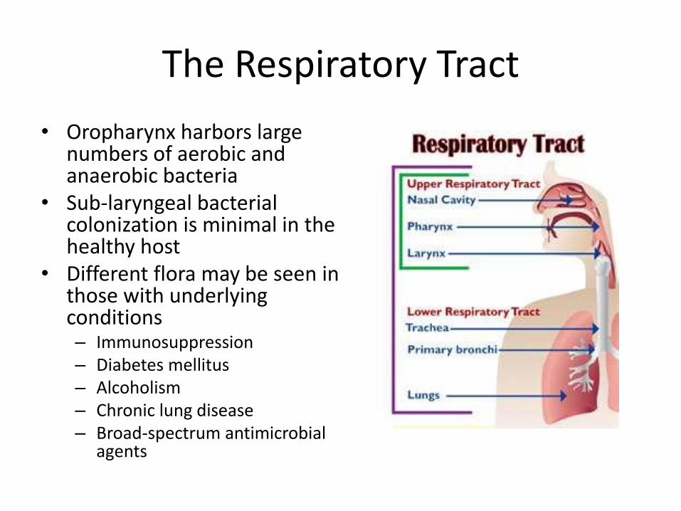
The Respiratory Tract
• Oropharynx harbors large numbers of aerobic and anaerobic bacteria
• Sub-laryngeal bacterial colonization is minimal in the healthy host
• Different flora may be seen in those with underlying conditions – Immunosuppression – Diabetes mellitus – Alcoholism – Chronic lung disease – Broad-spectrum antimicrobial
agents

Infection in the Lung
• Aspiration of bacterial into the alveoli is the most common mechanism initiating a pneumonic infection
• Asymptomatic aspiration commonly occurs, but organisms are usually cleared by the mucociliary apparatus
• Aerosol inhalation is a second, less frequent, mechanism for organisms to gain access to the LRT
• Hematogenous seeding of the lung from a distant focus of infection

Bacteria in the Respiratory Tract
Normal Respiratory Flora • Corynebacterium spp. • Coagulase negative
staphylococci • Staphylococcus aureus • Neisseria spp. • Haemophilus influenzae • Streptococcus pneumoniae • Moraxella catarrhalis • Gram negative bacilli
Oral flora –1010-1012CFU/mL
Potential Pathogens
• Staphylococcus aureus
• Haemophilus influenzae
• Streptococcus pneumoniae
• Moraxella catarrhalis
• Gram negative bacilli

Bacterial Agents of Acute Pneumonia Common Uncommon
Streptococcus pneumoniae Acinetobacter baumannii
Staphylococcus aureus Actinomyces species
Haemophilus influenzae Bacillus species
Mixed anaerobic bacteria from
aspiration (Bacteroides spp.,
Fusobacterium spp., anaerobic cocci,
Prevotella/Porphyromonas spp.)
Moraxella catarrhalis
Enterobacteriaceae (Escherichia coli,
Klebsiella pneumoniae, Enterobacter
spp., Serratia spp.)
Francisella tularensis
Pseudomonas aeruginosa Nocardia species
Legionella species (including L.
pneumophila and L. mcdadei)
Pasteurella multocida
Neisseria meningitidis

Common Viral Agents of Acute Pneumonia
Children Adults
Respiratory
Syncytial Virus
Influenza A
Parainfluenza 1-3 Influenza B
Influenza A Respiratory
Syncytial Virus
Human
Metapneumovirus
Adenovirus

Pneumonia
Community Acquired
• Diagnosis is based on the presence of specific symptoms and suggestive radiographic features, such as pulmonary infiltrates and/or pleural effusion.
• Carefully obtained microbiological data can support the diagnosis but often fails to provide an etiologic agent.
Common Pathogens
• Mycoplasma pneumoniae
• Respiratory viruses
• Streptococcal pneumoniae
• Chlamydophila pneumoniae
• Haemophilus influenzae
• Staphylococcus aureus

Pneumonia
Health Care/Ventilator Associated, Hospital Acquired • Viruses and fungi are rare causes
of HCAP, HA, and VAP in the immunocompetent patient.
• The clinical diagnosis is based imaging plus the presence of clinical features (fever, leukocytosis or leucopenia, purulent secretions)
• Determining the cause of the pneumonia relies diagnostic testing
• A smear lacking inflammatory cells and a culture absent of potential pathogens have a very high negative predictive value.
Common Pathogens in Immunocompromised Patients • Pseudomonas aeruginosa • Escherichia coli • Klebsiella pneumoniae • Enterobacter spp • Serratia marcescens • Acinetobacter spp • Stenotrophomonas maltophilia • Staphylococcus aureus and MRSA • Haemophilus influenzae • Streptococcus pneumoniae • Legionella • Aspergillus • Influenzae A,B • HPIV • Adenovirus • Rhinovirus • RSV • MDROs

Acute Bronchitis
• Inflammation of the epithelial lining of the bronchi – Obstructs airflow – Shortness of breath and coughing – Production of thick mucus
• >85% caused by viruses—rhinovirus, adenovirus, influenza A and B, and parainfluenza virus
• May occur as a secondary bacterial infection following a viral upper respiratory tract infection
• May result from primary infection with one of several specific agents – Bordetella pertussis – Mycoplasma pneumoniae – Chlamydophila pneumoniae
• May become chronic

Viral Bronchiolitis
• Lower respiratory tract infection seen in children less than 2 years old with peak occurrence seen in 2-8 month old children
• Symptoms – Starts with symptoms of the common cold and progresses to involve
bronchi and the bronchioles – Expiratory wheezing, tachypnea, retractions, irritability, and
dehydration – Complications include conjunctivitis, otitis media, pneumonia
• Viral agents
– Major agents: RSV (up to 80% of cases), parainfluenza virus type 3 – Minor agents: adenoviruses, influenza, and rhinoviruses

Specimens for Diagnosis of Lower Respiratory Tract Infections
• Non-invasive specimens – Sputum – Tracheal aspirates – Blood cultures – Urine – Serum
• Bronchoscopic specimens
– Bronchial wash/brush – Protected specimen
brushings – Bronchoalveolar lavage – Transbronchial biopsy – Transbronchial needle
aspirates
• Other invasive specimen types
– Pleural fluid
– Transthoracic-needle biopsy
– Open lung biopsy

Bronchoalveolar Lavage (BAL)
• Performed following general inspection of the tracheobronchial tree and before biopsy or brushing
• Performed during flexible bronchoscopy • Obtain specimens to rule out opportunistic infections in
immunocompromised hosts • Good general rule is to perform the lavage where the disease is most
prominent radiographically • In localized disease, lavage of the involved segment is most likely to yield
the best results. In diffuse disease, the right middle lobe or lingula is often chosen to optimize fluid recovery.
• Typically involves the delivery of a total of 100 to 240 mL of fluid in 20 to 60 mL aliquots
• A lavage volume of 100 mL samples approximately one million alveoli (1.5 to 3 percent of the lung).
• Lavage fluid should be pooled into a single container

Respiratory Testing in the Micro Lab
1. What should we be doing with these specimens in the lab?
2. Are there new pathogens we should be looking for?
Utility of Gram Stain
• Rapid, inexpensive, informational
• Evaluation of specimen quality – Identify superficially contaminated specimens
– Enhance discrimination between samples with potential pathogens vs. colonizing flora
• Presumptive organism ID
• Guide rational selection of preliminary antibiotic therapy
• Guides interpretation of culture results

Utility of Gram Stain
• Majority of the literature supports the clinical usefulness of gram stained sputum smears
• Wide range in reported sensitivity (35-96% and specificity (12-85%)
• Reference standard –sputum culture
• Multiple criteria for assessing Gram stain smears

Utility of Gram Stain
• Gram stain DOES NOT diagnose the presence of pneumonia • Once pneumonia diagnosed Gram stain is useful in
determining probable etiologic agent
A. sputum from a patient with pneumonia—Gram-positive, elongated cocci in pairs and short chains (Streptococcus pneumoniae)
B. a bronchoalveolar lavage specimen—Gram-negative intracellular rods (Klebsiella pneumoniae)
Utility of Gram Stain
Heineman et al. 1977. J Clin Microbiol 6:518-27
• 50% of the information gleaned from sputum cultures is clinically misleading in the absence of correlation with direct gram stain results
Gleckman et al. 1988. J Clin Microbiol 26:846-49
• Selection of appropriate monotherapy 94% of the time when guided by bacterial morphotypes from the gram stain

Sputum Culture in the Management of CAP
No
• Yield is variable and strongly influenced by the quality of the entire process
• Infrequent positive impact on clinical care
• Argue against the routine use of common tests, such as blood and sputum cultures.
• Optional in outpatients
Yes
• Cultures may have a major impact on the care of an individual patient and are important for epidemiologic reasons, including the antibiotic susceptibility patterns used to develop treatment guidelines
• Hospitalized patients with listed clinical indications
Mandell et al. 2007 CID44 (Suppl 2):S27-S72

Non-Invasive Specimens
Indication Blood culture Sputum culture Legionella urine antigen test
Pneumococcal urine antigen test Other
Admission to intensive care unit
✓ ✓ ✓ ✓ Endotracheal aspirate if intubated
Alcohol abuse ✓ ✓ ✓ ✓
Asplenia ✓ ✓
Cavitary infiltrates ✓ ✓ Fungal and tuberculosis cultures
Chronic severe liver disease ✓ ✓
Leukopenia ✓ ✓
Outpatient therapy ineffective
✓ ✓ ✓
Pleural effusion ✓ ✓ ✓ ✓ Thoracentesis and pleural fluid cultures
Positive Legionella urine antigen test result
✓
Positive pneumococcal urine antigen test result
✓ ✓
Recent travel (within past two weeks)
✓
Severe obstructive lung disease
✓
IDSA/ATS Consensus Guidelines on the Management of CAP
The benefit of a good quality sputum Gram stain
• Impact to therapy
– Broadens initial empirical coverage for less common etiologies (S. aureus or gram-negative bacilli)
– Early discontinuation of empirical treatment if results are negative
• Validates subsequent sputum culture results
Mandell et al. 2007 CID44 (Suppl 2):S27-S72

Work up of Respiratory Cultures
Specimen Quality Premise: • PMNs are an indication of infection or
inflammation • SEC indicate superficial contamination • If a specimen contains a large amount of SEC,
superficial contamination is likely the specimen should be recollected
• Extensive testing on heavily mixed cultures should not routinely be performed

Screening Sputum Specimens for Acceptability
Methods Minimum Criteria for Specimen Acceptance
Sum of neutrophils/LPF (10-15 = +1; >25 = +2); Mucus (+1); SEC/LPF (10-25=-1; >25 =-2)
Score of >0
Enumerate SEC/LPF <10 SEC/LPF
Enumerate Neutrophils/LPF >25 neutrophils/LPF
Enumerate SEC/LPF <25 SEC/LPF
Sum of neutrophils/LPF (1-75=+1; 76-150=+2; >150=+3) and SEC/LPF (5-15=-1; 16-25=-2; >25=-3)
Positive summation score
Ratio, neutrophils to SEC >10 neutrophils/SEC
Ratio, neutrophils to SEC >5 neutrophils/SEC
Enumerate SEC/LPF and presence/absence of organisms/OIF
<10SEC/LPF and organisms present
Presence/absence of organisms/OIF Organisms present
Sharp, SE, et al. 2004. Cumitech 7B, Lower Respiratory Tract Infections. ASM Press, Washington, DC

Screening Sputum Specimens for Acceptability
> 25 epithelial cells/lpf
[lpf, x10])
Interpretation: Unsuitable for culture
4+ (>25/lpf) neutrophils, no epithelial cells seen, 3+ (11-50/oif) Gram negative diplococci,
2+ (1-10/oif) yeast cells
Interpretation: Suitable for culture
Yeast cells = Cryptococcus neoformans; Gram-negative diplococci=Moraxella catarrhalis

Screening Sputum Specimens for Acceptability
>25 epithelial cells/lpf
Multiple bacterial morphologies
suggesting oral contamination Interpretation: Unsuitable for culture
High power view.
3+ neutrophils, 3+ Gram-positive
diplococci
Interpretation: Suitable for culture;
Gram-positive diplococci=
Streptococcus pneumoniae

Mixed Flora
• Used only with respiratory specimens • Use of objective criteria (# of organisms present
per OIF) to distinguish resident flora or colonizers from potential pathogens:
Bartlett 1982 JAMAWright et al. 1990 Am J Med Normandin et al. 1997 ASM C-91
Morphology OK to Report if:
Gram negative bacilli ≥ 10 organisms/OIF
Moraxella ≥ 25 organisms/OIF
Staph ≥ 50 organisms/OIF
S. pneumoniae ≥25 pairs/OIF
Aspiration event >50 organisms/OIF* * intracellular gram-positive and gram-negative organisms in at least one field
Gram Stain Screening
• Use interpretive comments
DIRECT SMEAR SUGGESTS:
No neutrophils
Many squamous epithelial cells
Not representative of lower respiratory tract secretions. Culture not performed. Please consult Microbiology if clinical considerations warrant complete processing of this specimen. (Specimen will be held 5 days).

Work up of Respiratory Cultures
• No definitive guidelines for working up bacterial cultures
– Standardized methods
– Uniformity in work up and reporting of bacterial isolates
– When to perform AST

Culture Set Up
• 5% sheep blood agar, MacConkey agar, and chocolate agar
• Add on Hemophilus plate? – Quad plate
• Factor X (hemin) or V factor (Nicotinamide adenine dinucleotide (NAD)) along with the hemolytic reaction on horse blood
– Remel Haemophilus Isolation Agar • Bacitracin and horse blood
• BCYE for Legionella

Work up of Respiratory Cultures
• Standardized pathogen list • Basic correlation with Gram stain
– Gram stain results used to guide the selection of potential pathogens in the culture that merit further identification and susceptibility testing
• Q-Score System
• Q234 System
• PMN-association System Sharp, SE, et al. 2004. Cumitech 7B, Lower Respiratory Tract Infections. ASM Press,
Washington, DC

Work up of Respiratory Cultures
Q-Score System
• Up to 3 organisms can be considered potential pathogens (PP) and be worked up (ID/AST) if from a good quality specimen (Q3)
• The lower quality of the specimen (e.g., the more SEC present) the fewer the organisms worked up (Q2, Q1)

Q-SCORE = # of potential pathogens (PP) to work up Squamous cells (-)
Report value
Key:
0 = no cells
1 = 1-9/lpf
2 = 10-24/lpf
3 = >25/lpf
Q0 = no cult
Q1 = 1PP
Q2 = 2PP
Q3 = 3PP
Q-Score System
0 -1 -2 -3
0 3
+1 3
Neutrophils (+) +2 3
+3 3 2 1
Work up of Respiratory Cultures
0
0
0
0
0
0
1 0 0
0

Work up of Respiratory Cultures
Q-Score System # PP in culture < Q-score:
(2PP) (Q3)
work up PP with ID/AST
# PP in culture > Q-score:
(3PP) (Q2)
Look to Gram stain
Work up PP that were seen in Gram stain with ID/AST
If all PP in the culture are seen in Gram stain = do not work up; perform morphological identification
Work up of Respiratory Cultures
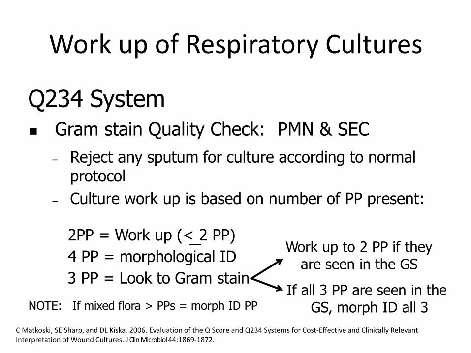
Q234 System Gram stain Quality Check: PMN & SEC
– Reject any sputum for culture according to normal protocol
– Culture work up is based on number of PP present:
2PP = Work up (< 2 PP)
4 PP = morphological ID
3 PP = Look to Gram stain
NOTE: If mixed flora > PPs = morph ID PP
Work up to 2 PP if they are seen in the GS
If all 3 PP are seen in the GS, morph ID all 3
C Matkoski, SE Sharp, and DL Kiska. 2006. Evaluation of the Q Score and Q234 Systems for Cost-Effective and Clinically Relevant Interpretation of Wound Cultures. J Clin Microbiol 44:1869-1872.

Work up of Respiratory Cultures
PMN-Association System
• Success in microscopic evaluation relies on strict cytological criteria – >25 PMN, < 10 SEC – >25 PMN, <25 SEC – >10 PMN per SEC (10:1 ratio)
• Evaluate the presence of predominant morphotypes associated with WBC • Work up organisms seen in association with PMN
Mixed flora • 1 morphotype <10 organisms/OIF • 1 morphotype >10 organisms/OIF
Helpful in predicting primary pathogen

Work up of Respiratory Cultures
PMN-Association System
• Quantitation of organisms in smears inconsistent and inaccurate – Technologist variability
– Do not report
• Do not rely on quantitation to determine relatedness to infection – PMN more predictive

Example 1: Sputum
GS: many PMN (+3), few SEC (-1), many enteric-like gram negative bacilli, moderate gram positive cocci suggestive of Staph, few Mixed flora (yeast)
CULT: moderate P. aeruginosa, moderate E.coli, moderate Staph aureus, few yeast
WORK UP:
Q Score (Q2=2PP):
Q234 (3PP):

Example 1: Sputum
GS: many PMN (+3), few SEC (-1), many enteric-like gram negative bacilli, moderate gram positive cocci suggestive of Staph, few Mixed flora (yeast)
CULT: moderate P. aeruginosa, moderate E.coli, moderate Staph aureus, few yeast
WORK UP:
Q Score (Q2=2PP): Work up E. coli and S. aureus MID P. aeruginosa; Report Mixed flora
Q234 (3PP): Work up E. coli and S. aureus MID P. aeruginosa; Report Mixed flora

Example 2: Sputum
GS: many PMN (+3), moderate SEC (-2), many nonenteric- like gram negative bacilli, moderate Mixed flora
CULT: many P. aeruginosa, moderate Staph aureus, few viridans Strep
WORK UP: Q Score (Q1=1PP):
Q234 (2PP):

Example 2: Sputum
GS: many PMN (+3), moderate SEC (-2), many nonenteric- like gram negative bacilli, moderate Mixed flora
CULT: many P. aeruginosa, moderate Staph aureus, few viridans Strep
WORK UP: Q Score (Q1=1PP): Work up P. aeruginosa
MID S. aureus; Report Mixed flora
Q234 (2PP):

Example 2: Sputum
GS: many PMN (+3), moderate SEC (-2), many nonenteric- like gram negative bacilli, moderate Mixed flora
CULT: many P. aeruginosa, moderate Staph aureus, few viridans Strep
WORK UP: Q Score (Q1=1PP): Work up P. aeruginosa
MID S. aureus; Report Mixed flora
Q234 (2PP): Work up P. aeruginosa and S. aureus Report Mixed flora

Example 3: Tracheal Aspirate
GS: many PMN (+3), few SEC (-1), many Mixed flora (few enteric-like GNB; moderate gram positive cocci suggestive of Staph)
CULT: moderate diphtheroids, moderate coag negative Staph, few E.coli, rare Staph aureus
WORK UP:
Q Score (Q2=2PP):
Q234 (2PP):
Example 3: Tracheal Aspirate
GS: many PMN (+3), few SEC (-1), many Mixed flora (few enteric-like GNB; moderate gram positive cocci suggestive of Staph)
CULT: moderate diphtheroids, moderate coag negative Staph, few E.coli, rare Staph aureus
WORK UP:
Q score (Q2=2PP): Work up E. coli and S. aureus Report Mixed flora
Q234 (2PP):

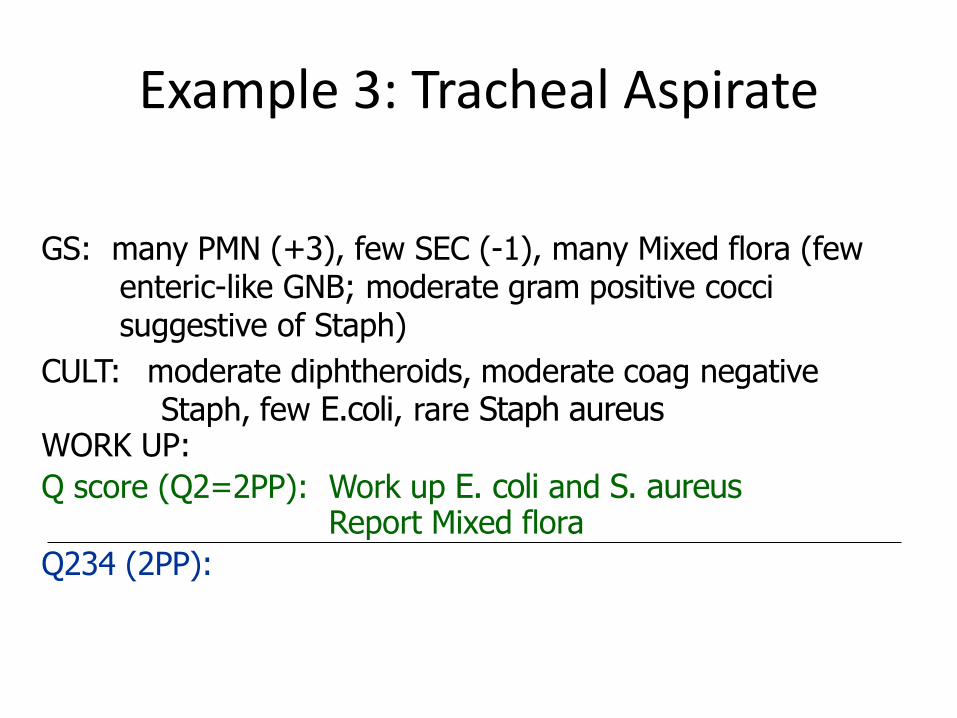
Example 3: Tracheal Aspirate
GS: many PMN (+3), few SEC (-1), many Mixed flora (few enteric-like GNB; moderate gram positive cocci suggestive of Staph)
CULT: moderate diphtheroids, moderate coag negative Staph, few E.coli, rare Staph aureus
WORK UP:
Q-Score (Q2=2PP): Work up E. coli and S. aureus Report Mixed flora
Q234 (2PP): Report Mixed flora MID E. coli and S. aureus **
** If mixed flora > PPs = MID PP
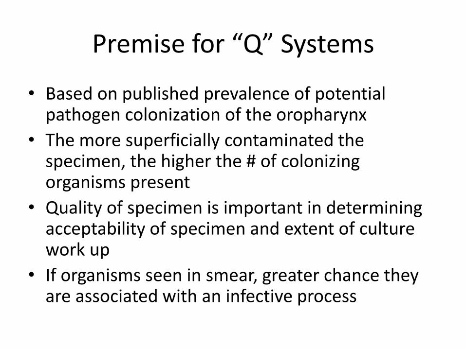
Premise for “Q” Systems
• Based on published prevalence of potential pathogen colonization of the oropharynx
• The more superficially contaminated the specimen, the higher the # of colonizing organisms present
• Quality of specimen is important in determining acceptability of specimen and extent of culture work up
• If organisms seen in smear, greater chance they are associated with an infective process

“Q” Systems Advantages
• Offers a consistent approach for interpreting cultures – Based on specimen quality – Based on organisms seen in Gram stain (organism
seen on smear should be in a significant number in the specimen, >105/mL)
– Limits number of organisms worked up from mixed cultures reporting of misleading information minimized
• All potential pathogens reported (may not perform full ID/AST)

Q System Caveats
• Gram stain sensitivity – Requires 104-105organisms per ml of fluid
• Standardization of Gram Stain – Specimen quality, type
– Smear preparation and staining
– Smear interpretation
• Culture and Gram Stain Correlation
• When not to apply the criteria – Legionella culture
Nagendra, S. et al. 2001. Sampling variability in the microbiological evaluation of expectorated sputa and endotracheal aspirates. J Clin Microbiol 39:2344–2347;

QUANTITATIVE CULTURE
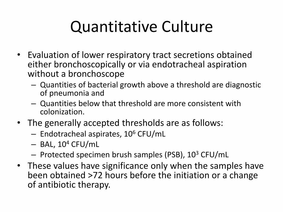
Quantitative Culture
• Evaluation of lower respiratory tract secretions obtained either bronchoscopically or via endotracheal aspiration without a bronchoscope – Quantities of bacterial growth above a threshold are diagnostic
of pneumonia and – Quantities below that threshold are more consistent with
colonization.
• The generally accepted thresholds are as follows: – Endotracheal aspirates, 106 CFU/mL – BAL, 104 CFU/mL – Protected specimen brush samples (PSB), 103 CFU/mL
• These values have significance only when the samples have been obtained >72 hours before the initiation or a change of antibiotic therapy.

Methods for Quantitative Culture
• Two approaches for quantitative culture: • Serial-dilution method
– Two 100-fold dilutions are made, and colony counts are obtained from 0.1-ml amounts of the diluted specimen inoculated onto media.
– Counts are made from the plate containing between 30 and 300 colonies. The results are expressed as CFU per milliliter.
• Calibrated-loop method, – 0.1 ml of PSB and 0.001 and 0.01 ml of BAL are inoculated onto agar
media. – The results are expressed as log10 ranges of bacteria.
• All morphotypes should be quantitated and reported. • Those organisms whose numbers approach or exceed the threshold
for significance should be identified and have susceptibility testing performed.
• Those bacteria present in smaller quantities should not be completely characterized.

• Cultures of respiratory secretions should be obtained from virtually all patients with suspected VAP
• Noninvasive sampling with semi-quantitative cultures to diagnose VAP, rather than invasive sampling with quantitative cultures and rather than noninvasive sampling with quantitative cultures
• Remain in favor of blood cultures for all patients with suspected VAP/HAP, although they don’t always correlate (25% positive from non-pumlonary source)
• When the 5 trials were pooled via meta-analysis, sampling technique did not affect any clinical outcome, including mean duration of mechanical ventilation, ICU length of stay, or mortality
• Strongly encourage diagnostic testing whenever the result is likely to change individual antibiotic management.
IDSA/ATS Consensus Guidelines on the Management of HA/VAP
Kalil, AC, et al. Management of Adults With Hospital-acquired and Ventilator-associated Pneumonia: 2016 Clinical Practice Guidelines by the Infectious Diseases Society of America and the American Thoracic Society. Clin Infect Dis. 2016 Sep 1;63(5):e61-e111.
• The guideline panel acknowledged that there is a potential that invasive sampling with quantitative cultures could lead to less antibiotic exposure if growth below defined thresholds is used as a trigger to stop antibiotics
Remarks: Clinical factors should also be considered because they may alter the decision of whether to withhold or continue antibiotics.
IDSA/ATS Consensus Guidelines on the Management of HA/VAP

EMERGING PATHOGENS

Corynebacterium sp.
• C. pseudodiphtheriticum, C. striatum
• Unlike C. diphtheriae and C. ulcerans, non–diphtheria corynebacteria do not produce toxins.
• Widely distributed in the environment
• Colonizers of the skin and mucosal membranes
• Patient-to-patient transmission in ICUs has been demonstrated for C. striatum
• Susceptible to vancomycin, linezolid
Nhan, TX et al. Microbiological investigation and clinical significance of Corynebacterium spp. In respiratory specimens. Diagnostic Microbiology and Infectious Disease 74 (2012) 236–241

Utility of Fungal Culture
• Histopathology alone is not sensitive enough to diagnose fungal infections
• Should be accompanied by immunostain, culture, and, when available, NAAT
• Has the ability to detect unsuspected fungi

Endemic Regions of the Systemic
Mycoses

Histoplasmosis
Histoplasmosis in a State Where It Is Not Known to Be Endemic — Montana, 2012–2013 MMWR Weekly October 25, 2013 / 62(42);834-837 • Diagnosed in four Montana residents by four different physicians • Three patients reported no recent travel outside of Montana and
likely were exposed in Montana, which is west of areas where H. capsulatum is recognized as endemic
• 4th patient likely acquired her infection in Montana before traveling out of state (could have been acquired during travel to California)
• Three patients experienced diagnostic delays, likely in part because none reported recent travel to areas where H. capsulatum is endemic

Direct Examination of Clinical Specimens
BAL: macrophages with yeasts, Wright-Giemsa

Phenotypic Morphology in Culture
• Colonies on BA and BHI are glabrous or wrinkled and cream-to-brown in color
• Subcultures grown on Sabouraud’s dextrose agar are white, tan, or light brown with abundant aerial hyphae
• Buff-to-brown colonies produce sparse aerial hyphae and abundant macroconidia at first, then turn white with dense aerial hyphae on subculture

Microscopic Morphology
• Produces two types of conidia at 30oC
• Both produced singly at the tips of short, narrow conidiophores. – Large (8-15 micron), thick-walled,
spherical/pear-shaped macroconidia with fingerlike projections (tuberculate macroconidia) • Tubercules are extension of the outer
wall, are not cellular, and do not bud
– Small (2-4 microns), oval microconidia with smooth to finely roughened walls
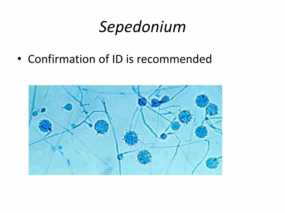
Sepedonium
• Confirmation of ID is recommended

Coccidioidomycosis
Notes from the Field: Coccidioides immitis Identified in Soil Outside of Its Known Range — Washington, 2013 MMWR Weekly May 23, 2014 / 63(20);450-450 • Three acute cases among residents of south central
Washington reported during 2010–2011 were suspicious for local acquisition;
• None of the three patients had traveled within 22 months of illness onset to an area where coccidioidomycosis is known to be endemic
• Novel PCR used to detect Coccidioides DNA in six of 22 soil samples
• Viable mold from 4 out of 6 samples

Coccidioides spp.
• C. immitis consists of two taxa – Non-CA → Arizona, Texas,
Mexico, and Argentina Group I
– CA → California Group II
• Two species within the genus Coccidioides – “Coccidioides immitis” is
restricted to isolates from California
– “Coccidioides posadasii” for all other isolates belonging to this genus

Phenotypic Morphology in Culture
• Growth within 3-5 days
• Cottony, velvety, powdery, granular, smooth, or wrinkled colonies
• Usually white, but may be gray, buff, lavender, cinnamon, yellow or brown
• Can be leathery on blood based agar

Arthroconidia of Coccidioides sp.
• Arthroconidia formed within 5-10 days – Barrel- or cask-shaped and measure 2.5-4 µ by 3-6 µ – Liberated arthroconidia carry a portion of the walls of the
intervening sterile segments (disjunctor cells) – Liberated arthroconidia survive desiccation and extreme
temperatures, and germinate to produce hyphae

Malbranchea
• Confirmation of ID is recommended

RESPIRATORY VIRUS TESTING

NAAT Based Respiratory Virus Testing
Yes • Infectious diseases present as a
constellation of symptoms
• Infectious causes are broad and diverse
• Empirical response is to treat for everything
• Knowledge of the etiologic agent allows informed decisions
• On-demand, rapid
• Ability to exclude known viruses
• Labs can serve as sentinels
• Cost
No
• Panels may be too broad
• Should be risk based not specimen based
• Test for uncommon pathogens
• Multiplex may impact sensitivity for some targets
• Cost
Schreckenberger P, McAdam A. (2015) Point-Counterpoint: Large multiplex PCR panels should be first line tests for detection of respiratory and intestinal pathogens. J Clin Microbiol. 53:3110-3115
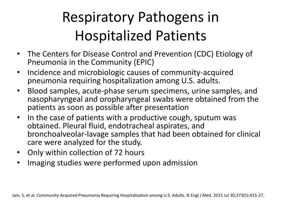
Respiratory Pathogens in Hospitalized Patients
• The Centers for Disease Control and Prevention (CDC) Etiology of Pneumonia in the Community (EPIC)
• Incidence and microbiologic causes of community-acquired pneumonia requiring hospitalization among U.S. adults.
• Blood samples, acute-phase serum specimens, urine samples, and nasopharyngeal and oropharyngeal swabs were obtained from the patients as soon as possible after presentation
• In the case of patients with a productive cough, sputum was obtained. Pleural fluid, endotracheal aspirates, and bronchoalveolar-lavage samples that had been obtained for clinical care were analyzed for the study.
• Only within collection of 72 hours • Imaging studies were performed upon admission
Jain, S, et al. Community-Acquired Pneumonia Requiring Hospitalization among U.S. Adults. N Engl J Med. 2015 Jul 30;373(5):415-27.

Respiratory Pathogens in Hospitalized Patients
• A PCR assay was performed on nasopharyngeal and oropharyngeal swabs with the use of CDC-developed methods for the detection of adenovirus; Chlamydophila pneumoniae; coronaviruses 229E, HKU1, NL63, and OC43; human metapneumovirus (HMPV); human rhinovirus; influenza A and B viruses; Mycoplasma pneumoniae; parainfluenza virus types 1, 2, and 3; and respiratory syncytial virus (RSV).
• A real-time polymerase-chain-reaction (PCR) assay for legionella was performed on sputum regardless of the quality of the sample
• PCR for bacterial targets
Jain, S, et al. Community-Acquired Pneumonia Requiring Hospitalization among U.S. Adults. N Engl J Med. 2015 Jul 30;373(5):415-27.

Jain S et al. N Engl J Med 2015;373:415-427.
Pathogen Detection among U.S. Adults with Community-Acquired Pneumonia Requiring Hospitalization, 2010–
2012.

Jain S et al. N Engl J Med 2015;373:415-427.
Pathogen Detection among U.S. Adults with Community-Acquired Pneumonia Requiring Hospitalization, 2010–
2012.
viruses detected in 27% and bacteria in 14%
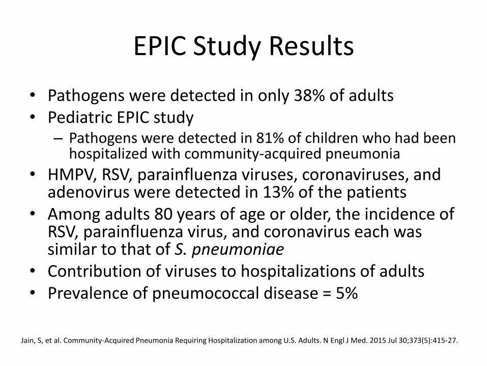
EPIC Study Results
• Pathogens were detected in only 38% of adults • Pediatric EPIC study
– Pathogens were detected in 81% of children who had been hospitalized with community-acquired pneumonia
• HMPV, RSV, parainfluenza viruses, coronaviruses, and adenovirus were detected in 13% of the patients
• Among adults 80 years of age or older, the incidence of RSV, parainfluenza virus, and coronavirus each was similar to that of S. pneumoniae
• Contribution of viruses to hospitalizations of adults • Prevalence of pneumococcal disease = 5%
Jain, S, et al. Community-Acquired Pneumonia Requiring Hospitalization among U.S. Adults. N Engl J Med. 2015 Jul 30;373(5):415-27.

EPIC Study Results
• Three pathogens were detected more commonly in patients in the ICU than in patients not in the ICU: – S. pneumoniae (8% vs. 4%) – S. aureus (5% vs. 1%) – Enterobacteriaceae (3% vs. 1%)
• Pathogens were detected less frequently in nasopharyngeal and oropharyngeal swabs obtained from asymptomatic controls than in swabs obtained from patients with pneumonia
• The incidences of influenza and of S. pneumoniae were almost 5 times as high among adults 65 years of age or older than among younger adults
• The incidence of human rhinovirus was almost 10 times as high among adults 65 years of age or older than among younger adults
Jain, S, et al. Community-Acquired Pneumonia Requiring Hospitalization among U.S. Adults. N Engl J Med. 2015 Jul 30;373(5):415-27.

Rhinovirus Seasonality and Transmission
• In temperate climates reported a peak in incidence in the early fall, with a smaller peak in the spring
• Most common cause of respiratory viral illness during the spring, summer, and fall months, while – influenza virus and RSV predominate in the winter
• Initiated by intranasal and conjunctival inoculation, but not oral
• Survive in an indoor environment for hours to days at an ambient temperature and on undisturbed skin for 2 h
• In one study, HRV was transmitted via an aerosolized route to 56% of volunteers who played cards for 12 h with experimentally infected subjects
Samantha E. Jacobs et al. Clin. Microbiol. Rev. 2013;26:135-162

Asymptomatic Rhinovirus
• Asymptomatic infection is relatively common
• In children <4 years old, rates of asymptomatic infection range from 12 to 32% and tend to be higher in the youngest age groups
• HRV in 0% and 2% of asymptomatic adults
Samantha E. Jacobs et al. Clin. Microbiol. Rev. 2013;26:135-162

Rhinovirus
• Detected in 49% of children admitted to ICUs with lower respiratory tract infection – in approximately one-half of cases, no other respiratory
pathogen was identified
• Can lead to an influenza-like illness, lower respiratory tract infections, chronic infections, and secondary bacterial infections, especially in immunocompromised patients, children with asthma, and adults with COPD
• Increases susceptibility to bacterial infection – Disrupts cell barrier function at tight junctions
– Stimulating the immune system
Samantha E. Jacobs et al. Clin. Microbiol. Rev. 2013;26:135-162

Middle East Respiratory Syndrome MERS-CoV
• A coronavirus
• Not detected by RVP panels
• Reported in Saudi Arabia in 2012
• Severe acute respiratory illness, including fever, cough, and shortness of breath
• 2 patients in US in May 2014 (IN and FL), both had traveled to Saudi Arabia
• Carried by camels

Case Definition

Specimens for MERS-CoV Testing
Collection of all three specimen types (not just one or two of the three), lower respiratory, upper respiratory and serum specimens for testing using the CDC MERS rRT-PCR assay is recommended.
Lower respiratory specimens are preferred, but collecting nasopharyngeal and oropharyngeal (NP/OP) specimens, and serum, are strongly recommended depending upon the length of time between symptom onset and specimen collection. Respiratory specimens should be collected as soon as possible after symptoms begin – ideally within 7 days. However, if more than a week has passed since symptom onset and the patient is still symptomatic, respiratory samples should still be collected, especially lower respiratory specimens since respiratory viruses can still be detected by rRT-PCR.
http://www.cdc.gov/coronavirus/mers/guidelines-clinical-specimens.html

What’s Next?
• Laboratory diagnosis of infectious diseases increasingly relies on pathogen-specific tests = a priori knowledge of likely etiologic agents
• Many different pathogens can cause clinically indistinguishable symptoms
• PCR amplification of marker genes strategy introduces bias and ignores effects of the relevant viral and phage flora for which no marker gene exists
• Next-generation sequencing (NGS) allows for unbiased, hypothesis-free detection
• High-throughput DNA and RNA-seq possible, computational analysis lacking
Flygare, S, et al. Taxonomer: an interactive metagenomics analysis portal for universal pathogen detection and host mRNA expression profiling. Genome Biology. 2016. 17:111

Metagenomics-Based Pathogen Detection Tools
Taxonomer
Flygare, S, et al. Taxonomer: an interactive metagenomics analysis portal for universal pathogen detection and host mRNA expression profiling. Genome Biology. 2016. 17:111

Summary
• A good Gram stain is useful for interpreting culture results
• Standardization may be a useful strategy
• Quantitative culture may not be required
• S. pneumoniae and Rhinovirus in hospitalized patients
• NAAT testing is required
• New pathogens coming to a location near you