Embed Size (px)
Citation preview
Vol. 55, No. 3INFECTION AND IMMUNITY, Mar. 1987, p. 572-5780019-9567/87/030572-07$02.00/0Copyright © 1987, American Society for Microbiology
Resistance to Pesticin, Storage of Iron, and Invasion of HeLa Cellsby Yersiniaet
DANIEL J. SIKKEMA AND ROBERT R. BRUBAKER*Department of Microbiology and Public Health, Michigan State University, East Lansing, Michigan 48824-1101
Received 21 August 1986/Accepted 17 November 1986
The independent abilities of Yersinia pestis to absorb exogenous pigments including hemin and Congo red(Pgm+) and to produce the bacteriocin pesticin with genetically linked invasive enzymes (Pst+) are establishedvirulence factors of the species. Pst- Pgm+ strains of Y. pestis are sensitive to pesticin (Psts), and mutation ofthese isolates to pesticin resistance (PStr) is known to result in concomitant conversion to Pgm-. Wild-type cellsof Yersinia pseudotuberculosis and Yersinia enterocolitica are Pgm- but may be Psts; mutation of the latter toPstr also results in avirulence. In this study, typical Pgm- mutants of Y. pestis exhibited a dramatic nutritionalrequirement at 37°C but not 260C for iron which could be fulfilled by either Fe3+ or hemin. Iron privation ofPgm- yersiniae resulted in formation of osmotically stable spheroplasts similar to those previously observedafter exposure of PstS bacteria to pesticin. At 370C, Pgm+ organisms rapidly overgrew initially predominantPgm- populations in iron-deficient medium. However, Pgm- isolates could undergo a second mutation thatpermitted successful competition with Pgm+ cells in this environment. The mutation to Pstr in Y.pseudotuberculosis and Y. enterocolitica did not promote a similar requirement for iron but rather preventedthese organisms from penetrating HeLa cells. The ability to invade these nonprofessional phagocytes was notshared by Pgm+ or Pgm- cells of Y. pestis.
Wild-type cells of Yersinia pestis, the causative agent ofbubonic plague, absorbed exogenous hemin (26) or the dyeCongo red (43) during growth at 26°C on solid media therebyforming intensely colored or pigmented colonies (Pgm+).Mutational loss of this evident chromosomal function (17,42) resulted in comparable growth as white colonies (26, 43),disappearance of a major outer membrane peptide (42), andmarked reduction of virulence in mice by intraperitoneal (27)or subcutaneous (47) routes of injection. Typical cells ofclosely related Yersinia pseudotuberculosis and Yersiniaenterocolitica were Pgm- as judged by their growth as whitecolonies under conditions used to define pigmentation in Y.pestis (43; R. R. Brubaker, unpublished observations). Sub-sequent work showed that other pathogenic species includ-ing neisseriae, shigellae, Vibrio, and certain isolates ofEscherichia coli were Pgm+ as judged by their ability toabsorb Congo red in environments distinct from that estab-lished as optimal for Y. pestis (5, 13, 33, 35); this determinantwas correlated with the ability of shigellae to penetratenonprofessional phagocytes (13).
Ability to produce the bacteriocin pesticin (2, 23, 24) andgenetically linked (11, 31) coagulase and fibrinolysin (1)serves as an additional virulence factor (9) of Y. pestis(Pst+). Loss of these functions, which are mediated by anapproximate 6 megadalton plasmid (3, 17, 42), also promotedavirulence in mice by intraperitoneal and subcutaneousroutes of injection (9). Y. pseudotuberculosis and Y. entero-colitica cells are Pst-, but isolates of certain serotypes ofthese species (12, 36) and a few strains of E. coli (10, 18, 24)were also sensitive to the antibacterial action of pesticin(Psts). This lethal effect was catalyzed by the N-acetylglu-cosaminidase activity of the bacteriocin (16) which promotedconversion of Pst' bacteria to osmotically stable sphero-plasts (20). Expression of Psts in Y. pestis required loss of
* Corresponding author.t Article no. 12071 from the Michigan Agricultural Experiment
Station.
immunity to the bacteriocin by mutation to Pst- (21) withretention of the Pgm + determinant (8). Subsequent mutationof these Pst- Pgm+ organisms to pesticin resistance (Pstr)resulted in selection for Pgm- isolates thereby permittingdetermination of a mutation rate from Pgm+ to Pgm- of 10-5(8). Analogous Pstr mutants of Y. pseudotuberculosis and Y.enterocolitica were subsequently isolated and, like Pgm-and Pst- cells of Y. pestis, were of reduced virulence in micevia intraperitoneal and subcutaneous routes of injection (47).Pgm+ and Psts yersiniae were maintained in mouse liver,
spleen, and lung, whereas Pgm- and Pstr mutants wererapidly cleared from these organs, suggesting loss of acommon mechanism of virulence (47). The Pgm+ phenotypewas assumed from the outset (7) to favor accumulation ofiron in vivo (26, 27). However, attempts to demonstrate aselective advantage of Pgm+ organisms in iron-deficientenvironments including serum (28) or 8-hydroxyquinoline-extracted medium (37) were not successful. Cells of Y.pseudotuberculosis, Y. enterocolitica, and both Pgm+ andPgm- Y. pestis utilized hemin as a sole source of iron andalso accumulated Fe3+ by an inducible, siderophore-independent, cell-bound, high-affinity transport system (37).This finding suggested that all these organisms acquire ironby the same mechanism and that the Pgm+ determinantmerely serves as a mechanism to store the cation. Thepurpose of this report is to provide evidence indicating thatavirulence associated with mutation to Pgm- in Y. pestis isindeed correlated with a significant lesion in iron accumula-tion. In contrast, avirulence caused by the analogous muta-tion to Pstr in Y. pseudotuberculosis and Y. enterocoliticareflected loss of ability to invade nonprofessional phago-cytes.
MATERIALS AND METHODS
Bacteria. All yersiniae used in this study were avirulentowing to loss of the -45-megadalton plasmid that mediatesthe low-calcium response (3, 17). Unless stated otherwise,
572
on June 23, 2018 by guesthttp://iai.asm
.org/D
ownloaded from
PESTICIN RESISTANCE IN YERSINIAE 573
TABLE 1. Properties of yersinia strains
SpeciesStrain Pigmen- Production Sensitivity Plasmids BiotyperSpeciesSrain taton of pesicin to esticin' (mega- or sero- Source or originSpecies ~~~~~~~tation of pesticin to pesticin"' atn) tpdaltons) type
Y. pestis Kuma + + 1 6, 65 A ManchuriaKIM + + I 6, 65 M IranG32 + - S 65 0 United StatesTS + + I 6, 65 0 JavaM23 + + I 6, 65 0 Nonencapsulated mutant
of MP6A12 + - S 65 0 From strain A1122
(United States)Salazar + + I 6, 65 0 BoliviaYokohama + + I 6, 65 A JapanKimberley + + 1 6, 65 0 AfricaDodson + - S 65 0 UnknownK10 + + I 6, 65 M IranMP6 + + I 6, 65 0 Unknown
Y. pseudotuberculosis PB1 - - S None I England1 - - S None I UnknownMD67 - - S None I United StatesHale - - S None I EnglandGalligue - - S None I EnglandParkin - - S None I England
Y. enterocolitica Wa - - S None 0:8 United StatesP76 - - S None 0:8 United StatesE701 - - S None 0:9 CanadaE736 - - S None 0:21 Canada
a S, Sensitive; I, immune.b Biotypes are antiqua (A), mediaevalis (M), and orientalis (0).
organisms used in experiments were Y. pestis Kuma (37), Y.pseudotuberculosis PB1 (12), and Y. enterocolitica WA (36).Salient properties of these strains and other yersiniae areshown in Table 1. Pgm- mutants of Y. pestis were obtainedon Congo red agar (43) incubated at 26°C, and Pstr mutantsof Y. pseudotuberculosis and Y. enterocolitica were isolatedon solid medium containing homogeneous pesticin (23) bythe method previously described for determining the muta-tion rate of Y. pestis to Pgm- (8). Streptomycin-resistantmutants were selected at 26°C for the ability to form colonieson tryptose-blood agar base (Difco Laboratories, Detroit,Mich.) containing the antibiotic (100 U/ml).
Cultivation. Slopes of tryptose-blood agar base were incu-bated for 1 day (Y. pseudotuberculosis or Y. enterocolitica)or 2 days (Y. pestis) at 26°C after inoculation from stockcultures of buffered glycerol maintained at -20°C as previ-ously described (1). Resulting organisms were suspendedand appropriately diluted in 0.033 M potassium phosphatebuffer (pH 7.0) (phosphate buffer) for use as inocula ofchemically defined medium (53), yielding an initial opticaldensity at 620 nm of 0.1 (corresponding to about 108 viablebacteria per ml). A single transfer was made in this mediumcontaining 50 ,uM Fe3+ or 20 to 50 ,uM hemin in Erlenmeyerflasks (10% [vol/vol]) aerated at 200 rpm on a model G76gyratory water bath shaker (New Brunswick Scientific Co.,Inc., Edison, N.J.). Organisms in late logarithmic growthwere harvested by centrifugation (17,000 x g for 10 min at4°C), washed once in phosphate buffer, and then suspendedat an optical density of 0.1 in control medium containing ironas described above or in iron-deficient medium. Thesecultures were aerated as already defined and, in someexperiments, were used to inoculate subsequent transferswhen the optical density had increased to 1.0. Iron-deficient
medium was prepared by extraction with 8-hydroxyquin-oline (50) and contained <0.3 ,uM iron as determined byflame absorption spectroscopy.
Host-cell invasion. HeLa cells were prepared in suspensionand used in experiments with yersiniae essentially as previ-ously described (15). The cells were routinely cultivated at37°C under 5% CO2 in Eagle minimal essential medium(Difco) with 5% fetal calf serum containing streptomycin (75,ug/ml) and potassium penicillin G (100 U/ml). After growthto a density of 5 x 10⁵ cells per ml, antibiotics were removedby three washes in minimal essential medium with fetal calfserum, and 2.0 ml of the final suspension was placed in rollertubes. The latter then received 108 yersiniae washed once inphosphate buffer in 0.1 to 0.2 ml of the same buffer and wereplaced in a model TC-5 roller apparatus (New Brunswick) at37°C. After infection for 2 h, extracellular organisms werekilled by further incubation in the presence of kanamycin (75p.g/ml) for 4 h. Intracellular bacteria were then released bydisruption of the HeLa cells with a Dounce homogenizer anddetermined by plating appropriate dilutions on tryptose-blood agar base. Results of preliminary studies showed thatsignificant uptake, when it occurred, commenced after 1 hand ceased after 2 h of incubation (15).
RESULTS
The degree of adaptation to the culture medium and theextent of prior exposure to iron markedly influenced bacte-rial growth during subsequent iron privation. To controlthese variables, the organisms were cultivated for a singletransfer with Fe3+ or hemin before washing and dilution foruse as inocula. Pgm+ cells of Y. pestis were capable ofevident unlimited growth thereafter at 37°C in iron-deficient
VOL. 55, 1987
on June 23, 2018 by guesthttp://iai.asm
.org/D
ownloaded from
574 SIKKEMA AND BRUBAKER
10.0
1.0
cnzw
-J
a-0
0.1
B C D
E F H
0 18 360 18 360 18 360 18 36
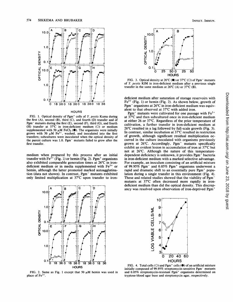
HOURSFIG. 1. Optical density of Pgm+ cells of Y. pestis Kuma during
the first (A), second (B), third (C), and fourth (D) transfer and ofPgm- mutants during the first (E), second (F), third (G), and fourth(H) transfer at 37°C in iron-deficient medium (0) or mediumsupplemented with 50 ,uM FeCl3 (0). The organisms were initiallygrown with 50 ,uM Fe3+, washed, and inoculated into the firsttransfers; subcultures were inoculated when the optical density ofthe parent culture was 1.0. Pgm- mutants failed to grow after thefirst transfer.
medium when prepared by this process after an initialtransfer with Fe3+ (Fig. 1) or hemin (Fig. 2). Pgm+ organismsalso exhibited comparable generation times at 26°C in iron-deficient medium or in media supplemented with Fe3+ orhemin, although the latter promoted marked autoagglutina-tion (data not shown). In contrast, Pgm- mutants exhibitedonly limited multiplication at 37°C upon transfer to iron-
A B DC10.0
1.0:
zw
0n 0.1
0 18 360 18 360 18 360 18 36HOURS
FIG. 2. Same as Fig. 1 except that 50 ,uM hemin was used inplace of Fe3".
A B
z
LU ~ HOR
a-J 1.0
0~0. 1
0 25 50 0 25 50HOURS
FIG. 3. Optical density at 26°C (X) or 37°C (0) of Pgm- mutantsof Y. pestis KIM in iron-deficient medium after a previous singletransfer in the same medium at 26°C (A) or 37°C (B).
deficient medium after saturation of storage reservoirs withFe3+ (Fig. 1) or hemin (Fig. 2). As shown below, growth ofPgm- organisms at 26°C in iron-deficient medium was equiv-alent to that observed at 37°C with added iron.Pgm- mutants were cultivated for one passage with Fe3+
at 37°C and then subcultured once in iron-deficient mediumat either 26 or 37°C. Regardless of the prior temperature ofcultivation, a further transfer in iron-deficient medium at26°C resulted in a lag followed by full-scale growth (Fig. 3).In contrast, similar incubation at 37°C resulted in restrictionof growth, although significant residual multiplication oc-curred in the culture inoculated with organisms previouslygrown at 26°C. Accordingly, Pgm- mutants specificallyexhibit an evident lesion in accumulation of iron at 37°C butnot at 26°C. Although the nature of this temperature-dependent deficiency is unknown, it provides Pgm+ bacteriain iron-deficient medium with a marked selective advantage.For example, an inoculum consisting of an artificial mixtureof 99.95% Pgm- and 0.05% Pgm+ organisms underwent arapid and dramatic shift to an essentially pure Pgm+ popu-lation during a single transfer in this environment (Fig. 4).These and related studies showed that the viability of Pgm-mutants at 37°C often decreased more rapidly in iron-deficient medium than did the optical density. This discrep-ancy was resolved upon observation of iron-deprived Pgm+
10
19
0l) 8
6--J
>5
0 20 40 60HOURS
FIG. 4. Total cells (0) and Pgm+ cells (0) of an artificial mixtureinitially composed of 99.95% streptomycin-sensitive Pgm- mutantsand 0.05% streptomycin-resistant Pgm+ organisms determined ontryptose-blood agar base and streptomycin agar, respectively.
INFECT. IMMUN.
on June 23, 2018 by guesthttp://iai.asm
.org/D
ownloaded from
PESTICIN RESISTANCE IN YERSINIAE 575
A' B
I
JN,7
4.141
1*7
FIG. 5. Pgm+ cells of Y. pestis Kuma exhibiting normal mor-phology (A) and osmotically stable spheroplasts of Pgm- mutants(B) after a single transfer in iron-deficient medium.
and Pgm- organisms by light microscopy. The former ex-hibited normal morphology, whereas the latter underwentconversion to nonviable osmotically stable spheroplasts thatnevertheless contributed to optical density (Fig. 5).Other strains of Y. pestis were examined to determine
whether the Pgm--specific lesion in iron metabolism was ageneral property of the species. Of the 11 additional isolatestested, 8 resembled strain Kuma in that only Pgm+ organ-isms were capable of growth at 37°C without added iron(Table 2). Pgm- mutants of the remaining three strains grewas well in this environment as did their Pgm+ parents. Thisfinding suggested that Pgm- bacteria typically exhibit thetemperature-dependent defect since the exceptions pos-
TABLE 2. Doubling times and maximum optical density of Pgm+and Pgm- strains of Y. pestis at 37°C in chemically defined iron-
deficient mediuma
Doubling MaximumStrain Pgm time (min) opticaltime(mm) ~density
Kuma + 110 8.160NGb 0.125
KIM + 110 6.120NG 0.122
G32 + 150 7.960NG 0.137
TS + 240 5.950NG 0.129
M23 + 170 6.340180 6.200
A12 + 220 7.000NG 0.119
Salazar + 170 7.740NG 0.141
Yokohama + 220 6.300NG 0.126
Kimberley + 240 4.500240 5.550
Dodson + 210 8.070230 7.080
K10 + 150 6.800NG 0.095
MP6 + 270 2.570NG 0.084
a Organisms were inoculated at an optical density of 0.1 after a singletransfer in iron-deficient medium.
b NG, No growth (doubling time >24 h).
sessed various atypical characteristics and were of uncertainbackground (Table 1). However, the existence of theseexceptions demonstrated that the ability to express thepigmentation reaction was not necessarily essential forgrowth at 37°C in iron-deficient medium. To further illustratethis point, Pgm- cells of strain Kuma were cultivated at 37°Cfor 4 days without added iron at which time a subpopulationof otherwise typical Pgm- organisms emerged that werecapable of evident indefinite growth in this environment.These observations indicate that a suppressor mutation canpromote multiplication ofPgm- cells at 37°C in iron-deficientmedium. Thus, acquisition of this ability does not reflect truereversion to Pgm+.
Since Pgm- mutants are analogous to Pstr mutants of Y.pseudotuberculosis and Y. enterocolitica, the latter werecompared for ability to grow at 37°C in iron-deficient me-dium. Control Pgm- cells of Y. pestis typically ceaseddivision in this environment (Fig. 6A), whereas resultsobtained with Y. pseudotuberculosis were equivocal in thatthe Pstr mutant grew more slowly than did the Psts parent buteventually achieved the same maximum optical density (Fig.6B). No difference was detected between Psts and Pstr cellsof Y. enterocolitica (Fig. 6C). These findings indicated thatmutation to Pstr in Y. enterocolitica and probably Y.pseudotuberculosis did not result in significant loss of abilityto accumulate iron as occurred with Pgm- mutants of Y.pestis.
Accordingly, a search was initiated for other putativevirulence functions present in Psts but not Pstr mutants of Y.pseudotuberculosis and Y. enterocolitica. Attempts to dem-onstrate differences in adherence or ability to damage hostcells were not successful, but the capability to penetratenonprofessional phagocytes was significantly reduced in Pstrmutants. This relationship is shown in Table 3 which showsthat mutation of Y. pseudotuberculosis and Y. enterocoliticato Pstr resulted in significant decreases in the ability toinvade HeLa cells. Neither Pgm+ nor Pgm- isolates of Y.pestis were capable of promoting detectable invasion ofHeLa cells.
DISCUSSIONIt is known that macrophages serve as target host cells for
Y. pestis, Y. pseudotuberculosis, and Y. enterocolitica (29,44, 46). This relationship is emphasized by the observationthat organisms of all three species are of highest virulence
A 'B C
10.0
a-Z 1.0w.0
0.
000 18 360 18 360 18 36HOURS
FIG. 6. Growth of Pgm+ (0) and Pgm- (0) cells of Y. pestisKuma (A), Psts (0) and PStr (0) cells of Y. pseudotuberculosis PBI(B), and Psts (0) and Pstr (0) cells of Y. enterocolitica WA (C)during a second transfer in iron-deficient medium.
VOL. 55, 1987
on June 23, 2018 by guesthttp://iai.asm
.org/D
ownloaded from
576 SIKKEMA AND BRUBAKER
via intravenous injection (9, 47), in which immediate inter-action occurs with fixed macrophages lining the blood ves-sels of the liver and spleen. Further evidence underscoringthe importance to yersiniae of obtaining access to macro-phages was the observation that virulence of Pgm- and Pst-mutants of Y. pestis (9, 47) and Pstr mutants of Y.pseudotuberculosis and Y. enterocolitica (47) could be phe-notypically restored via intravenous injection. This findingsuggests that these mutants are rapidly eliminated from thehost by nonspecific mechanisms of defense after injection byperipheral routes but are able to cause acute disease afteradministration by a route that permits immediate uptake bymacrophages (47). An important clue to the nature of thesenonspecific mechanisms of host defense was the discoverythat virulence of Pgm-, Pst-, and Pstr mutants could be fullyrestored via peripheral routes of infection by saturation ofserum transferrin by injected iron (9, 27; unpublished obser-
TABLE 3. Infectivity for HeLa cells of Pgm+ and Pgm- isolatesof Y. pestis, Pst' and Pstr isolates of Y. pseudotuberculosis, and
Psts and Pstr isolates of Y. enterocolitica
Species Strain
Y. pestis Kuma
KIM
G32
TS
M23
A12
Salazar
Yokohama
Kimberley
Dodson
K10
MP6
Y. pseudotuberculosis PB1
1
MD67
Hale
Galligue
Parkin
Y. enterocolitica WA
P76
E701
E736
Phenotype
Pgm+Pgm-Pgm+Pgm-Pgm+Pgm-Pgm+Pgm-Pgm+Pgm-Pgm+Pgm-Pgm+Pgm-Pgm+Pgm-Pgm+Pgm-Pgm+Pgm-Pgm+Pgm-Pgm+Pgm-
PstspstrPstspstrPstspstrPstspstrPstspstrPstspstr
PstspstrPstspstrPstspstrPstspStr
Irnfectivity'
<1<1<1<1<1<1<1<1<1<1<1<1<1<1<1<1<1<1<1<1<1<1<1<1
14.51.0
18.01.37.5
<18.0
<1
15.01.4
14.01.0
8.42.79.11.57.1
<18.31.0
a Number of viable bacteria per HeLa cell.
vations). This phenomenon, originally made with Pgm-mutants (27), first defined the debilitative effect of exogenousiron on the course of infectious diseases and has now beenextended to include a variety of other pathogenic microor-ganisms (48, 52). Initially, the explanation of this effect wasgenerally attributed to fulfilling a nutritional requirement forthe cation which is tightly sequestered in vivo (51, 52).However, it is now established (48) that exogenous iron canalso enhance virulence by interfering with a variety ofnonspecific mechanisms of host defense including blockingsynthesis of transferrin (34), preventing chemotaxis of pro-fessional phagocytes (49), and inhibiting intracellular killingby both oxidative (30, 40) and nonoxidative (19) processes.Accordingly, correct assessment of the process by whichinjected iron reverses the lesions in virulence present inPgm-, Pst-, and Pstr isolates may be requisite to defining thenature of the virulence factors lost by these mutations.
Injected iron was assumed to enhance the virulence ofPst- mutants of Y. pestis by inhibiting nonspecific mecha-nisms of host defense (7). Evidence favoring this possibilitywas the discovery that Pst- mutants lacked coagulase andfibrinolysin and thus, after typical infection of peripheraltissues via fleabite, would be unable to initiate systemicinfection (9, 11). Furthermore, it seems unlikely that thesmall 6-megadalton plasmid, known to encode pesticin,coagulase, fibrinolysin, and putative immunity plus mainte-nance functions, could also mediate iron transport activities.Accordingly, we suspect that the Pst+ determinant serves toenhance virulence by enabling the organisms to disseminaterapidly from peripheral foci of infection to favored nicheswithin fixed macrophages of lymphatics and internal organs.This capability would enable the organisms to avoid lethalencounters with professional phagocytes other than macro-phages. In contrast, the Pgm+ factor was predicted toincrease virulence by promoting acquisition of iron in vivo(7, 26, 27). Nevertheless, the first direct evidence linking thePgm+ determinant with transport of this cation is the obser-vation reported here that typical Pgm- cells do indeedexhibit an increased nutritional requirement for iron. Thisfinding was not anticipated because a different medium,rendered iron deficient by the same process, did not reveal adifference in growth between Pgm+ and Pgm- organisms(37). That medium, however, was prepared by avoidingincorporation of organic compounds capable of chelatingsignificant Fe3+. In contrast, the medium used in this studycontained high levels of a number of biologically significantchelators, including 0.01 M citric acid (53). The presence ofthese ligands may account for the inability of Pgm- mutantsto multiply after removal of extractable Fe3+ since over 100,uM FeCl2 was required for full-scale growth in the initialversion (22) of the medium used.An unexpected observation was that iron-deprived Pgm-
organisms underwent conversion to osmotically stablespheroplasts. Further study will be required to determinewhether this phenomenon reflects the enzymatic activity ofpesticin. If so, the dramatic loss of viability of Pgm- mutantsin iron-deficient medium may represent an inability to main-tain immunity to the bacteriocin in the absence of iron.Alternatively, normally compartmentalized pesticin may bereleased after death in iron-deficient medium thereby pro-moting formation of spheroplasts. Evidence supporting thissecond possibility was the finding that the Pst+ determinantwas not required for lethality of all Pgm- mutants in iron-deficient medium. Indeed, the observation that three Pgm-isolates and suppressor mutants of Pgm- cells of strainKuma grew normally in iron-deficient medium at 37°C also
INFECT. IMMUN.
on June 23, 2018 by guesthttp://iai.asm
.org/D
ownloaded from
PESTICIN RESISTANCE IN YERSINIAE 577
indicates that the Pgm+ determinant is not essential fortransport of the cation in this environment. We are presentlyinvestigating the nature of this suppressor mutation andintend to determine whether it restores virulence of Pgm-organisms via peripheral routes of injection.Attempts to demonstrate a similar requirement for iron in
Pstr mutants of Y. pseudotuberculosis and Y. enterocoliticawere not successful. Accordingly, a comparative study wasinitiated to identify other deficiencies that might account foravirulence in these isolates. No significant differences werenoted except in the ability to penetrate nonprofessionalphagocytes as typified by HeLa cells. This capability waspreviously defined in Y. pseudotuberculosis (6) and Y. en-terocolitica (13, 32, 45) and probably reflects that alsoreported for invasion of other types of nonprofessionalphagocytes (25, 38). Further study will be required todetermine whether this activity is identical to that trans-ferred to E. coli K-12 as a single genetic locus from apesticin-insensitive strain of Y. pseudotuberculosis (25).Similar penetration of host cells was not observed with Y.pestis, suggesting that this capability is only required bythose yersiniae normally transmitted via the oral route ofinfection. Nevertheless, it is difficult to reconcile that selec-tion for Pstr in Y. pestis results in recovery of Pgm- mutants,whereas similar selection with Psts isolates of Y. pseudotu-berculosis or Y. enterocolitica permitted recovery of mu-tants unable to invade HeLa cells. Before this finding, wesuspected that the pesticin receptor per se served as thehemin- and Congo red-binding component unique to Pgm+cells of Y. pestis. This notion became untenable with thediscovery that mutational loss of the pesticin receptor in theother yersiniae resulted in inability to invade HeLa cells.However, a strong possibility remains that the structuralgene for the pesticin receptor is linked to genes encodinghost-cell invasiveness in Y. pseudotuberculosis and Y. en-terocolitica and linked to distinct genes in Y. pestis thatmediate storage of iron. Development of methods permittingprecise genetic analysis of chromosomal functions may benecessary to resolve this relationship.The ability of Y. pseudotuberculosis and Y. enterocolitica
to invade host cells would, like expression of the Pst+ factor,clearly provide protection against a variety of nonspecificmechanisims of host defense including destruction by pro-fessional phagocytes other than macrophages. However,this activity could also facilitate accumulation of iron as doesthe Pgm+ determinant. Evidence supporting this possibilityis tenuous and dependent on the observations that otherfacultative intracellular parasites including neisseriae (41),listeriae (14), legionellae (39), and salmonellae (4) also utilizesiderophore-independent mechanisms of iron transport invivo possibly analogous to that first described for yersiniae(37). As initially suggested for legionellae (39), these findingsare consistent with the hypothesis that available iron isextremely scarce in extracellular spaces but easily obtainedin intracellular environments. If this assumption is correct,cells of Y. pestis may obtain and store needed iron via use ofthe Pgm+ factor, whereas the other yersiniae obtain thecation from intracellular reservoirs. In any event, we previ-ously established that Pstr mutants of Y. pseudotuberculosisand Y. enterocolitica were of reduced virulence (47) andhave now shown that this effect reflects the inability toinvade nonprofessional phagocytes.
ACKNOWLEDGMENTSThis work was supported by Public Health Service grant Al 13590
from the National Institutes of Health.
Preliminary experiments concerned with formation of osmoticallystable spheroplasts were performed by Robert D. Perry and SusanC. Straley. The excellent technical assistance of Janet M. Fowler isgratefully acknowledged.
LITERATURE CITED1. Beesley, E. D., R. R. Brubaker, W. A. Janssen, and M. J.
Surgalla. 1%7. Pesticins. III. Expression of coagulase andmechanism of fibrinolysis. J. Bacteriol. 94:19-26.
2. Ben-Gurion, R., and I. Hertman. 1958. Bacteriocin-like materialproduced by Pasteurella pestis. J. Gen. Microbiol. 19:289-297.
3. Ben-Gurion, R., and A. Shafferman. 1981. Essential virulencedeterminants of different Yersinia species are carried on acommon plasmid. Plasmid 5:183-187.
4. Benjamin, W. H., Jr., C. L. Turnbough, Jr., B. S. Posey, andD. E. Briles. 1985. The ability of Salmonella typhimurium toproduce the siderophore enterobactin is not a virulence factor inmouse typhoid. Infect. Immun. 50:392-397.
5. Berkhoff, H. A., and A. C. Venial. 1985. Congo red medium todistinguish between invasive and non-invasive Escherichia colipathogenic for poultry. Avian Dis. 30:117-121.
6. Bovallius, A., and G. Nilsson. 1975. Ingestion and survival ofYersinia pseudotuberculosis in HeLa cells. Can. J. Microbiol.21:1977-2007.
7. Brubaker, R. R. 1979. Expression of virulence in yersiniae, p.168-171. In D. Schlessinger (ed.), Microbiology-1979. Ameri-can Society for Microbiology, Washington, D.C.
8. Brubaker, R. R. 1970. Mutation rate to nonpigmentation inPasteurella pestis. J. Bacteriol. 98:1404-1406.
9. Brubaker, R. R., E. D. Beesley, and M. J. Surgalla. 1965.Pasteurella pestis: role of pesticin I and iron in experimentalplaque. Science 149:422-424.
10. Brubaker, R. R., and M. J. Surgalla. Pesticins. I. Pesticin-bacterium interrelationships, and environmental factors influ-encing activity. J. Bacteriol. 82:940-949.
11. Brubaker, R. R., M. J. Surgalla, and E. D. Beesley. 1965.Pesticinogeny and bacterial virulence. Zentralbl. Bakteriol.Parasitenkd. Infektionskr. Hyg. Abt. 1 Orig. 196:302-315.
12. Burrows, T. W., and G. A. Bacon. 1960. V and W antigens instrains of Pasteurella pseudotuberculosis. Br. J. Exp. Pathol.39:278-291.
13. Chambers, C. E., S. L. Stockman, and D. W. Niesel. 1985.Thermoregulated expression of a cloned Congo red bindingactivity gene of Shigella flexneri. FEMS Microbiol. Lett. 28:281-286.
14. Cowart, R. E., and B. G. Foster. 1985. Differential effects of ironon the growth of Listeria monocytogenes: minimum require-ments and mechanisms of acquisition. J. Infect. Dis. 151:721-730.
15. Devenish, J. A., and D. A. Schiemann. 1981. HeLa cell infectionby Yersinia enterocolitica: evidence for lack of intracellularmultiplication and development of a new procedure for quanti-tative expression of infectivity. Infect. Immun. 32:48-55.
16. Ferber, D. M., and R. R. Brubaker. 1979. Mode of action ofpesticin: N-acetylglucosaminidase activity. J. Bacteriol. 139:495-501.
17. Ferber, D. M., and R. R. Brubaker. 1981. Plasmids in Yersiniapestis. Infect. Immun. 31:839-841.
18. Ferber, D. M., J. M. Fowler, and R. R. Brubaker. 1981.Mutations to tolerance and resistance to pesticin and colicins inEscherichia coli 4). J. Bacteriol. 146:506-511.
19. Gladstone, G. P., and E. Walton. 1971. The effect of iron andhaematin on the killing of staphylococci by rabbit polymorphs.Br. J. Exp. Pathol. 152:452-464.
20. Hall, P. J., and R. R. Brubaker. 1978. Pesticin-dependentgeneration of osmotically stable spheroplast-like structures. J.Bacteriol. 136:786-789.
21. Hertman, I., and R. Ben-Gurion. 1959. A study of pesticinbiosynthesis. J. Gen. Microbiol. 21:135-143.
22. Higuchi, K., and C. E. Carlin. 1957. Studies on the nutrition andphysiology of Pasteurella pestis. I. A casein hydrolyzate me-dium for the growth of Pasteurella pestis. J. Bacteriol.73:122-129.
VOL. 55, 1987
on June 23, 2018 by guesthttp://iai.asm
.org/D
ownloaded from
578 SIKKEMA AND BRUBAKER
23. Hu, P. C., and R. R. Brubaker. 1974. Characterization ofpesticin: separation of antibacterial activities. J. Biol. Chem.249:4749-4753.
24. Hu, P. C., G. C. H. Yang, and R. R. Brubaker. 1972. Specificity,induction, and absorption of pesticin. J. Bacteriol. 112:212-219.
25. Isberg, R. R., and S. Falkow. 1985. A single genetic locusencoded by Yersinia pseudotuberculosis permits invasion ofcultured animal cells by Escherichia coli K-12. Nature (London)317:262-264.
26. Jackson, S., and T. W. Burrows. 1956. The pigmentation ofPasteurella pestis on a defined medium containing haemin. Br.J. Exp. Pathol. 37:570-576.
27. Jackson, S., and T. W. Burrows. 1956. The virulence enhancingeffect of iron on non-pigmented mutants of virulent strains ofPasteurella pestis. Br. J. Exp. Pathol. 37:577-583.
28. Jackson, S., and B. C. Morris. 1961. Enhancement of growth ofPasteurella pestis and other bacteria in serum by the addition ofiron. Br. J. Exp. Pathol. 37:570-576.
29. Janssen, W. A., and M. J. Surgalla. 1969. Plague bacillus:survival within host phagocytes. Science 163:950-952.
30. Kaplan, S. S., P. G. Quie, and R. E. Basford. 1975. Effect of ironon leukocyte function: inactivation of H202 by iron. Infect.Immun. 12:303-308.
31. Kol'tsova, E. G., Y. G. Suchkov, and S. A. Legedeva. 1973.Transmission of a bacteriocinogenic factor in Pasteurella pestis.Sov. Genet. 7:507-510.
32. Lee, W. H., P. P. McGrath, P. H. Carter, and E. L. Eide. 1977.The ability of some Yersinia enterocolitica strains to invadeHeLa cells. Can. J. Microbiol. 23:1714-1722.
33. Maurelli, A. T., B. Blackman, and R. C. Curtiss III. 1984. Lossof pigmentation in Shigella flexneri 2a is correlated with loss ofvirulence and virulence-associated plasmid. Infect. Immun.43:397-401.
34. McFarlane, H., M. Okubadejo, and S. Reddy. 1972. Transferrinand Staphylococcus aureus in kwashiokor. Am. J. Clin. Pathol.57:587-591.
35. Payne, S. M., and R. A. Finklestein. 1977. Detection anddifferentiation of iron-responsive avirulent mutants on Congored agar. Infect. Immun. 18:94-98.
36. Perry, R. D., and R. R. Brubaker. 1983. Vwa+ phenotype ofYersinia enterocolitica. Infect. Immun. 40:166-171.
37. Perry, R. D., and R. R. Brubaker. 1979. Accumulation of ironby yersiniae. Infect. Immun. 137:1290-1298.
38. Portnoy, D. A., S. L. Moseley, and S. Falkow. 1981. Character-
ization of plasmids and plasmid-associated determinants ofYersinia enterocolitica pathogenesis. Infect. Immun. 31:775-782.
39. Reeves, M. W., L. Pine, J. B. Neilands, and A. Balows. i983.Absence of siderophore activity in Legionella species grown iniron-deficient media. J. Bacteriol. 154:324-329.
40. Schultz, J., and S. Rosenthal. 1959. Iron. II. Inactivation ofmyeloperoxidase. J. Biol. Chem. 234:2486-2490.
41. Simonson, C., D. Brener, and I. W. DeVoe. 1982. Expression ofa high-affinity mechanism for acquisition of transferrin iron byNeisseria meningitidis. Infect. Immun. 36:107-113.
42. Straley, S. C., and R. R. Brubaker. 1982. Localization inYersinia pestis of peptides associated with virulence. Infect.Immun. 36:129-135.
43. Surgalla, M. J., and E. D. Beesley. 1969. Congo-red-agar platingmedium for detecting pigmentation in Pasteurella pestis. Appl.Microbiol. 18:834-837.
44. Une, T. 1977. Studies on the pathogenicity of Yersinia entero-colitica. I. Experimental infection in rabbits. Microbiol. Immu-nol. 21:349-363.
45. Une, T. 1977. Studies on the pathogenicity of Yersinia entero-colitica. II. Interaction with cultured cells in vitro. Microbiol.Immunol. 21:365-377.
46. Une, T. 1977. Studies on the pathogenicity of Yersinia entero-colitica. III. Comparative studies between Y. enterocolitica andY. pseudotuberculosis. Microbiol. Immunol. 21:505-516.
47. Une, T., and R. R. Brubaker. 1984. In vivo comparison ofavirulent Vwa- and Pgm- or Pstr phenotypes of yersiniae.Infect. Immun. 43:895-900.
48. van Asbeck, B. S., and J. Verhoef. 1983. Iron and host defense.Eur. J. Clin. Microbiol. 2:6-10.
49. Ward, P. A., P. Goldschmidt, and N. D. Greene. 1975. Suppres-sive effects of metal salts on leukocyte and fibroblast function.RES J. Reticuloendothel. Soc. 18:313-321.
50. Waring, W. S., and C. H. Werkman. 1942. Growth of bacteria inan iron-free medium. Arch. Biochem. 1:303-310.
51. Weinberg, E. D. 1974. Iron and susceptibility to infectiousdisease. Science 184:952-956.
52. Weinberg, E. D. 1978. Iron and infection. Microbiol. Rev.42:45-66.
53. Zahorchak, R. J., and R. R. Brubaker. 1982. Effect ofexogenous nucleotides on Ca2' dependence and V antigensynthesis in Yersinia pestis. Infect. Immun. 38:953-959.
INFECT. IMMUN.
on June 23, 2018 by guesthttp://iai.asm
.org/D
ownloaded from


![CY-'Proc. Nat. Acad. Sci. USA Vol. 70, No.8, pp. 2429-2433, August 1973 Synthetic Analogs ofthe Active Sites ofIron-Sulfur Proteins. Structure and Properties ofBis[o-xylyldithiolato-MA2-sulfidoferrate(III)],](https://img.pdfslide.us/doc/110x75/5e9dfa63d66a0c175d562bb1/cy-proc-nat-acad-sci-usa-vol-70-no8-pp-2429-2433-august-1973-synthetic.jpg)

![Vol. in U.S.A. Effect ofIron and Salt Prodigiosin Synthesis · culture on TS slants (1.0% ion agar no. 2 [Colab], 3.0% Trypticase soy broth [BBL]) or on slants of Brain Heart Infusion](https://img.pdfslide.us/doc/110x75/5f6410a6530e2f494935985b/vol-in-usa-effect-ofiron-and-salt-prodigiosin-synthesis-culture-on-ts-slants.jpg)