Embed Size (px)
Citation preview

Hu et al. Journal of Experimental & Clinical Cancer Research 2010, 29:43http://www.jeccr.com/content/29/1/43
Open AccessR E S E A R C H
ResearchThe expression and significance of IDH1 and p53 in osteosarcomaXiang Hu1, Ai-Xi Yu*1, Bai-Wen Qi1, Tao Fu1, Gang Wu1, Min Zhou1, Jun Luo2 and Jun-Hua Xu1
AbstractBackground: To detect the expression of isocitrate dehydrogenase 1 (IDH1) and transformation-related protein 53 (p53) in osteosarcoma and analyze the correlation between them and the clinico-pathological features.
Methods: The expressions of IDH1 and p53 were detected in human osteosarcoma cell lines (MG-63 and U2OS) by immunocytochemistry, Real-time PCR and Western Blotting. The expressions of IDH1 and p53 in formalin-fixed paraffin-embedded tissue sections from 44 osteosarcoma patients were determined by immunohistochemistry, and the correlation between them and clinicopagthological features were analyzed. None of these patients received chemotherapy prior to surgery.
Results: IDH1 is detected in osteosarcoma cell lines and biopsies. IDH1 expresses higher in U2OS cells with wild type p53 than in MG-63 cells with mutation p53. IDH1 correlates with histological Rosen grade and metastasis negatively. P53 correlates with histological Rosen grade, metastasis and overall survival in clinical osteosarcoma biopsies. Osteosarcoma patients with High IDH1 expression have a very high p53 expression.
Conclusion: IDH1 may correlate with p53 and be a candidate biomarker for osteosarcoma correlate with histological Rosen grade and metastasis.
BackgroundOsteosarcoma (OS) is the most current primary malig-nant bone tumor in children and adolescents. Presently,60% of the affected patients are cured by wide resectionof the tumor and aggressive adjuvant chemotherapy [1,2].However, around 40% of the individuals with metastasesstill emerge which normally exhibit resistance to cytostat-ics and acquire "second malignancies" [3]. The identifica-tion of biomarkers linked to clinicopagthological featuresand development of this disease is crucial for the diagno-sis and treatment of these patients [4,5].
Genetic alterations caused either by lost of heterozy-gosity or by mutations have been reported in osteosar-coma. Such alterations can occur in tumor suppressorgenes, such as tumor protein 53(p53) and phosphates andtensin homolog (PTEN). The p53 mutations occurs com-monly in primary osteosarcoma [6]. It is implicated in thepathogenesis of various human malignancies through lossof function mutations [7,8]. P53 contributes to the devel-
opment, life expectancy, and overall fitness of an organ-ism except for its role in protecting against cancerdevelopment [9]. PTEN is known to be the most highlymutated tumor suppressor gene after p53 [10]. It plays animportant role in regulating proliferation, migration, sur-vival, cell invasion and tumor angiogenesis [11,12]. Free-man et al. [13] reported that loss of PTEN was a commonoccurrence in osteosarcoma. It was further demonstratedthat PTEN can control p53 half-life independent via acurrently unknown mechanism [14]. In addition, muta-tions of tumor-suppressor retinoblastoma gene (Rb) inosteosarcoma are associated with a poor prognosis [15].However, none of these alterations can characteristicallyreflect the biologic nature or clinical features of all osteo-sarcomas.
IDH1 is a cytosolic NADP-dependent isocitrate dehy-drogenase. It catalyzes decarboxylation of isocitrate intoalpha-ketoglutarate [16]. Shechter et al. [17] describedthat the activity of IDH1 is coordinately regulatedthrough the cholesterol and fatty acid biosynthetic path-ways, suggesting that IDH1 provides the cytosolicNADPH required by these pathways. Memon et al. [18]found that expression of IDH1 was downregulated in a
* Correspondence: [email protected] Department of Orthopedics, Zhongnan Hospital of Wuhan University, No 169 Donghu Road, Wuchang District, 430071, Wuhan, ChinaFull list of author information is available at the end of the article
BioMed Central© 2010 Hu et al; licensee BioMed Central Ltd. This is an Open Access article distributed under the terms of the Creative Commons Attri-bution License (http://creativecommons.org/licenses/by/2.0), which permits unrestricted use, distribution, and reproduction in anymedium, provided the original work is properly cited.

Hu et al. Journal of Experimental & Clinical Cancer Research 2010, 29:43http://www.jeccr.com/content/29/1/43
Page 2 of 10
poorly differentiated bladder cancer cell line comparedwith a well-differentiated bladder cancer cell line. Tissuebiopsies of late-stage bladder cancers also showed IDH1downregulation compared with early-stage bladder can-cers. Yan et al. [19] described that mutations of NADP(+)-dependent isocitrate dehydrogenases encoded byIDH1 and IDH2 occur in a majority of several types ofmalignant gliomas.
Interestingly, Parsons et al. [20] found that IDH1 muta-tions in human glioblastoma had a very high frequency ofp53 mutation. Mutation of the IDH1 gene was alsostrongly correlated with a normal cytogenetic status [21].IDH1 appears to function as a tumor suppressor that,when mutationally inactivated, contributes to tumorigen-esis [21,22]. But, there is no study on the expression ofIDH1 in osteosarcoma. As to the previous study on IDH1and p53, we are also curious intensively about the correla-tion between IDH1 and p53. So, we developed a study tocharacterize the expression and significance of IDH1 andp53 in osteosarcoma cell lines (MG63 and U2OS) as wellas in clinical patient biopsies.
MethodsCell lines and cell cultureThe human osteosarcoma (OS) cell lines MG63 andU2OS (obtained from ATCC through LGC Promochem,Wesel, Germany) were cultured in RPMI 1640 media(Sigma, USA) with 10% fetal bovine serum (Amresco,USA) and antibiotics. Cells were cultured according tostandard techniques in cell culture flasks in a humidifiedincubator in 5% CO2 atmosphere.
ImmunocytochemistryCell lines were grown on coverslips treated with theappropriate growth media in 24 well cluster plates. Cellswere fixed in 2% formaldehyde in 0.1 mol/L phosphate-buffered saline (PBS, pH 7.4) for 20 min at room temper-ature and subsequently washed three times in PBS. Cov-erslips were permeabilized with 0.1% Triton X-100 for 15min and blocked in 3% H2O2-methyl alcohol for 15 min.The coverslips were incubated with anti-IDH1 rabbitpolyclonal antibody (protein technology group, USA) inblocking buffer overnight at 4°C. Coverslips were thenincubated with an anti-rabbit secondary antibody andperoxidase-conjugated strepavidin-biotin complex (SantaCruz, CA, USA) at 37°C for 45 min at room temperaturein the dark [23]. Immunoreactivity was visualized withdiaminobenzidine (DAB) (Zymed, South San Francisco,CA). Negative controls were obtained by omitting theprimary antibody. Slides were scanned using a micros-copy (Carl Zeiss AG, Germany), images were recordedusing a digital camera (DC 500, Leica) and the Leica FW4000 software and images were processed using AdobePhotoshop.
Real-time PCRCellular total RNA from OS cells was extracted with TRI-ZOL Reagent (Invitrogen, Carlsbad, CA, USA). The con-centration of RNA was determined by the absorbance at260 nm and the purity was determined by the 260/280ratio with a BioPhotometer(Eppendorf, Hamburg, Ger-many). For each reaction, 1 μg RNA was reverse-tran-scribed with random primer by ReverTra Ace (Toyobo,Osaka, Japan). RNA quality and efficiency of reverse tran-scription were examined by PCRs from each 1 μl cDNAaccording to the manufacturer's recommendations [24].The mRNA expression of IDH-1, p53 and internal controlgeneβ-actin was quantified by Real-time PCR DetectionSystem (SLAN, HONGSHI) with SYBR Green I (Toyobo,Osaka, Japan). As PCR was performed according to stan-dard procedures [24,25] after optimization, PCR-reac-tions were within the exponential range of amplification.The gene-specific exon-spanning PCR primer pair forIDH1 was 5'-TCAGTGGCGGTTCTGTGGTA-3',5'-CTTGGTGACTTGGTCGTTGGT-3', and for p537-8 was5'-CAGCCAAGTCTGTGACTTGCACGTA C-3',5'-CTATGTCGAAAAGTGTTTCTGTCATC-3', and for β-actin was 5'-GTCCACCGCAAATGCTTCTA-3',5'-TGCTGTCACCTTCACCGTTC-3'. The sequences ofthe primers were checked by Nucleotide BLAST for spe-cific gene amplification. Omission of cDNA template wasused as a negative control. Triplicate measurements weremade of all genes in each patient and data of mean wereused. For relative quantification of genes expression level,standard curves were built by considering at least threepoints of a ten-fold dilution series of cDNA in water. Rel-ative gene expression data are given as the n-fold changein transcription of the target genes normalized to theendogenous control in the same sample.
Protein extraction and Western blotLysates of cells were prepared using lysis buffer from theDual-Luciferase assay kit (Promega) according to themanufacturer's recommendations. The lysates were col-lected and centrifuged at 12,000 g for 10 min at 4°C. Theprotein in the supernatants were pooled together andstored at -80°C until concentration analyzed by the BCAProtein Assay Kit (Sangon, Shanghai, China). After beingheated at 99°C for 5 min in loading buffer, equal volumeof tissue lysates (40 μg of protein) were then loaded forsodium dodecyl sulphate-polyacrylamide gel electopho-resis (SDS-PAGE) analysis and subsequently electrotrans-ferred from the gels onto a polyvinylidene difluoride(PVDF) membranes (Millipore, MA, USA). The trans-ferred membranes were blocked with 5% skim milk inTris-buffered saline with 0.05% Tween (TBST) andwashed six times in TBST. IDH1 and p53 proteins weredetected by the rabbit polyclonal antibody for IDH1 (pro-tein technology group, USA) or p53 (Santa Cruz, CA,

Hu et al. Journal of Experimental & Clinical Cancer Research 2010, 29:43http://www.jeccr.com/content/29/1/43
Page 3 of 10
USA). β-actin proteins were recognized by the β-actin-specific monoclonal mouse IgG (Santa Cruz, CA, USA).Antibodies were diluted according to the manufacturedirection and were incubated overnight at 4°C followedby incubating with peroxidase-conjugated goat anti-rab-bit immunoglobulin (Santa Cruz, CA, USA, 1:2000) inTBST for 1 h. Signals were developed using enhancedchemiluminescent reagent (Pierce Biotechnology, Rock-ford, IL, USA). β-actin is used as the internal loading con-trol. The band intensity was analyzed using Quantity Onesoftware (Bio-Rad, Hercules, and CA). Relative expres-sion was calculated as the intensity ratio of target proteinto that of β-actin.
Tissue specimens and clinical dataFifty-one formalin-fixed, paraffin-embedded osteosar-coma biopsies (before the administration of neo-adjuvantchemotherapy) were collected according to the Chinesenational ethical guidelines ('Code for Proper SecondaryUse of Human Tissue', Chinese Federation of MedicalScientific Societies). Because of limitations in availabletumor material and following up information, only 44 ofthese osteosarcoma tumor samples including 32(72.7%)males and 12(27.3%) females with mean age(M ± SD) of25.25 ± 13.61 years (range 9-61) were amenable for use inthis study. Patients were followed until death from dis-ease, or until the latest clinical therapy at the end of thisstudy. The mean following-up time(M ± SD) were 4.26 ±1.99 years (range 0.5-9.0). All patients consisted with thediagnostic criteria of osteosarcoma defined in the WorldHealth Organization classification. Written informedconsent was obtained from each patient before enteringinto this study. Clinical information was available in Table1.
Immunohistochemistry for biopsiesSections were cut from formalin-fixed, paraffin-embed-ded granulation tissue. They were hydrated throughgraded alcohols. For antigen unmasking, sections weretreated in trypsin solution for 10 min at 37°C. Sectionswere then washed with deionized water and incubatedwith 3% H2O2 for 5 min. They were incubated in anti-IDH1 mAb (protein technology group, USA) or anti-p53mAb (Santa Cruz, CA, USA) for 1 h at room temperature,followed by secondary antibody and peroxidase-conju-gated strepavidin-biotin complex (Santa Cruz, CA, USA)at 37°C for 30 min. Immunoreactivity was visualized withdiaminobenzidine (DAB) (Zymed, South San Francisco,CA). Negative controls were obtained by omitting theprimary antibody.
Evaluation of immunohistochemistryThe slides were evaluated under the microscope. Thepercentage of cells showing positive nuclear staining for
p53 was calculated by reviewing the entire spot. ForIDH1, cytoplasmic immunostaining was considered to bepositive. The staining patterns were classified into scaleson the percentage of cells with positive staining [26,27]: 0,absence of nuclear (or cytoplasmic) stained cell; 1, <10%positive cells; 2, 10-25% positive cells; 3, 26-50% positivecells; 4, 51-75% positive cells; 5, >75% positive cells. Forstatistical analysis, osteosarcoma patients were alsogrouped as either low-staining group (scale 0-3: positivestaining ≤ 50%) or high-staining group (scale 4, 5: positivestaining >50%). Biopsy Stained less than 10% was consid-ered as a negative result, while stained more than 10%was considered as a positive one. At least 5 separated fociof neoplastic infiltration in each biopsy were analyzed.Assessment of Immunostaining intensity was completedby three independent observers. Slides were scannedusing a microscopy (Carl Zeiss AG, Germany), imageswere recorded using a digital camera (DC 500, Leica) andthe Leica FW 4000 software and images were processedusing Adobe Photoshop.
Statistical analysisAll statistical analyses were performed using the SPSS13.0 software package for Windows (SPSS Inc., Chicago,IL, USA). The values for the description of the statisticalsignificance of IDH1 or p53 expression in different osteo-sarcoma cell lines were calculated by independent, two-tailed Student's t-tests after the Levine's Test for Equalityof Variances. Mann-Whitney U was used for unnormalcontinuous variables. Categorical variables were analyzedby the Pearson Chis-square test and Fisher's exact test.Associations were assessed by Pearson correlation coeffi-cient for normal data or Spearman's correlation coeffi-cient for nonnormal data. Kaplan-Meier test was used foranalysis of survival versus IDH1 and survival versus p53expression. P < 0.05 was considered as statistically signifi-cant. P < 0.01 was considered as statistically highly signif-icant.
ResultsIDH1 expresses higher in U2OS compared with in MG63Expression of IDH1 is specifically detected in the cyto-plasm of both osteosarcoma cell lines U2OS and MG63(Fig. 1). The expression of IDH1 mRNA is higher inU2OS than in MG63, and P < 0.01(Fig. 2). The westernblotting result(Fig. 3A, Fig. 3C) shows that IDH1 is highlyexpressed in U2OS(P < 0.01), and these results corrobo-rate the immunocytochemistry(Fig. 1).
Expression of p53 in U2OS and MG63Consistent with data published previously [28,29]; ourMG63 demonstrates no detectable p53 while U2OS dem-onstrates high expressed p53. The result is shown in Fig.3B.

Hu et al. Journal of Experimental & Clinical Cancer Research 2010, 29:43http://www.jeccr.com/content/29/1/43
Page 4 of 10
IDH1 correlates with histological Rosen grade and metastasis in clinical osteosarcoma biopsiesIDH1 mainly locates on the cytoplasm (Such as Fig. 1A,Fig. 4A, and Fig. 5A). It's positive expression was identi-fied using immunohistochemistry in 40 of 44 (90.9%)osteosarcoma tumors, of which 23 of 44 (52.2%) exhibits
high staining (Table 2). The average IDH1 immunostain-ing percentage is 53.57%(SD: 28.99%, range from 8% to100%). The average score is 3.59 (SD: 1.22, range from 1to 5). IDH1 expresses higher in low Rosen grade osteosar-coma vs. high Rosen grade osteosarcoma [30-32] (Fig. 4,Fig. 5, Fig. 6, and Fig. 7). IDH1 correlates with metastasis
Table 1: Clinical Features
Features Total(N) Percentage
Age(year)
<12 3 6.8%
13--20 14 31.8%
21--30 8 18.2%
31--40 14 31.8%
41- 5 11.4%
Sex
Male 32 72.7%
Female 12 27.3%
Localization of primary tumor
Distal femur 13 29.5%
Proximal tibia 11 25.0%
Humerus 3 6.8%
Tibia diaphysis 5 11.4%
Femur diaphysis 7 15.9%
Other 5 11.4%
Histological type
Osteoblastic 29 65.9%
Small cell 1 2.3%
Chondroblastic 6 13.6%
Teleangetatic 1 2.3%
Round cell 2 4.5%
Fibroblastic 4 9.1%
Mixed 1 2.3%
Histological Rosen grade*
1 5 11.3%
2 16 36.4%
3 16 36.4%
4 7 15.9%
1+2 21 47.7%
3+4 23 52.3%
Metastasis
no 23 53.3%
lung 17 38.6%
other 4 9.1%
*As described previously [30-32], Grade 1: <50% tumor necrosis; Grade2: 50% to 90% tumor necrosis; Grade 3: > 90% tumor necrosis;Grade 4: 100% tumor necrosis.1+2: low grade; 3+4: high grade.
Hu et al. Journal of Experimental & Clinical Cancer Research 2010, 29:43http://www.jeccr.com/content/29/1/43
Page 5 of 10
negatively (P = 0.016, r = -0.361). There is no significantcorrelation between IDH1 expression and overall survival(P = 0.342) (Fig. 8).
P53 correlates with histological Rosen grade, metastasis and overall survival in clinical osteosarcoma biopsiesP53 mainly locates on the nuclear (Such as Fig 4B, Fig4D), Its positive expression is identified using immuno-histochemistry in 37 of 44 (84.1%) osteosarcoma tumors,of which 19 of 44 (43.2%) exhibits high staining (Table 2).The average p53 immunostaining percentage is
47.25%(SD: 28.82%, range from 4.5% to 100%). The aver-age score is 3.18 (SD: 1.35, range from 1 to 5). P53expresses higher in low Rosen grade osteosarcoma (Fig. 4,Fig. 5, Fig. 6, Fig. 7). P53 correlates with metastasis nega-tively (P = 0.001, r = -0.473). High-expression p53patients have better survival than low-expression p53patients do (P = 0.019) (Fig. 9).
IDH1 correlates with p53 in clinical osteosarcoma biopsiesThere is no significant difference between IDH1 and p53in clinical osteosarcoma biopsies. Positive correlationbetween IDH1 and p53 expression is demonstrated in ourstudy (Table 2, Fig. 4, and Fig. 5).
DiscussionIDH1 catalyzes decarboxylation of isocitrate into alpha-ketoglutarate 16. Shechter et al. [17] described that activ-ity of IDH1 is coordinately regulated with the cholesteroland fatty acid biosynthetic pathways, suggesting thatIDH1 provides NADPH required by these pathways. Itwas described IDH1 appears to function as a tumor sup-pressor that, when mutationally inactivated, contributesto tumorigenesis [22]. IDH1 is likely to function as atumor suppressor gene rather than as an oncogene [22].IDH1, encoding two TCA enzymes, fumarate hydratase(FH) and succinate dehydrogenase (SDH), has beenfound to sustain loss-of-function mutations in certainhuman tumors, which likewise contribute to tumorgrowth via stimulating the HIF-1a pathway and muta-tionally altering metabolic enzymes [33,34]. As IDH1 alsocatalyzes the production of NADPH, it is possible that adecrease in NADPH levels resulting from IDH1 mutationcontributes to tumorigenesis through effects on cellmetabolism and growth [17]. Zhao et al. [22] showed thatmutation of IDH1 impairs the enzyme's affinity for itssubstrate and dominantly inhibits wild type IDH1 activity
Figure 1 The immunocytochemistry of IDH1 in MG63 and U2OS. IDH1 is specifically detected in the cytoplasm of both osteosarcoma cell lines MG63 and U2OS.(A) Expression of IDH1 in U2OS, × 200; (B) Ex-pression of IDH1 in MG63,× 200; (C) Expression of IDH1 in U2OS,× 400; (D) Expression of IDH1 in MG63,× 400.
Figure 2 The mRNA levels of IDH1 in MG63 and U2OS (on fold). The mRNA levels of IDH1 is higher in U2OS than in MG63(P < 0.01).
Figure 3 The protein expression levels of IDH1 and p53 in U2OS and MG63. MG63 demonstrates no detectable p53 while U2OS cells demonstrates a high expressed p53. IDH1 expresses higher in U2OS than in MG63 at the protein level(P < 0.01).

Hu et al. Journal of Experimental & Clinical Cancer Research 2010, 29:43http://www.jeccr.com/content/29/1/43
Page 6 of 10
with the formation of catalytically inactive heterodimers.Mutation of the IDH1 gene was strongly correlated with anormal cytogenetic status [21].
In this study, we firstly demonstrate that IDH1 isdetected in U2OS with wild type p53 and MG63 with
mutation p53 by immnohistochemistry, Realtime-PCRand Western Blotting. Intriguingly, our study demon-strates that IDH1 markedly increases in U2OS comparewith MG63 not only in mRNA level but also in protein
Table 2: The expression of IDH1 and P53 in osteosarcoma biopsies
Proteins* Expression** Positive N***
1 2 3 4 5 Low High
N (%) N (%) N (%) N (%) N (%) N (%) N (%) N (%)
IDH1 4 (9.1) 2 (4.5) 15 (34.1) 10 (22.7) 13 (29.5) 21 (47.7) 23 (52.2) 40 (90.9)
P53 7 (15.9) 6 (13.6) 12 (27.3) 10 (22.7) 9 (20.5) 25 (56.8) 19 (43.2) 37 (84.1)
* P < 0.01(p = 0.000) r = 0.620, IDH1 correlates with P53 positively; Spearman's rho.** P > 3/40.05(P = 0.316), IDH1 vs. P53; Mann-Whitney U.*** P > 3/40.05(0.334), IDH1 vs. P53; Pearson Chis-square test;
Figure 4 The expression of IDH1 and p53 in low histological Rosen grade biopsy. IDH1 expresses at high level accompanying with high ex-pressed p53 in Low histological Rosen grade biopsy.(A) Expression of IDH1 in low histological Rosen grade biopsy, × 100;(B) Expression of p53 in low histological Rosen grade biopsy, × 100; (C) Expression of IDH1 in low histological Rosen grade biopsy, × 200;(D) Expression of p53 in low histological Rosen grade biopsy, × 200.
Hu et al. Journal of Experimental & Clinical Cancer Research 2010, 29:43http://www.jeccr.com/content/29/1/43
Page 7 of 10
level. It is conceivable that the expression of IDH1 mayrelate to p53.
Human osteosarcoma cell line MG63 was found withDeletion and rearrangement of the p53 gene [35-37]. NoWild type p53 expression could be detected in this cellline. Our results are in accordance with the results ofMasuda et al. [6] and Mulligan et al. [36] and indicate thatinactivation of p53 is a common event in osteosarcomadevelopment. In addition, we authenticate the wild typep53 in human osteosarcoma cell line U2OS in our study.
P53 is described as a tumor suppressor in many tumors.Culotta and Koshland [38] and Harris et al [39] gave anextensive account of its discovery and function as well asthe use of p53 in cancer risk assessment. Activity of p53ubiquitously lost in osteosarcoma either by mutation ofthe p53 gene itself or by loss of cell signaling upstream ordownstream of p53 [40]. Xue et al. [41] reported that p53inactive may be required for maintenance of aggressive
tumors. Marion et al. [42] showed that p53 is critical inpreventing the generation of human pluripotent cellsfrom suboptimal parental cells. Harris and Hollstein [39]highlighted the clinical implications of changes in the p53gene in the pathogenesis, diagnosis, prognosis, and ther-apy of human cancer. But, little is known about the com-binatory role of p53 and IDH1 in OS cells. We are curiousabout the role of p53 and IDH1 in osteosarcoma.
Most studies addressing the immunohistochemicalexpression of IDH1 or p53 in biopsies have used a semi-quantitative scoring approach of the staining results [43-47], often with a 10% threshold for scoring a tumor aspositive and with a 50% threshold for scoring a tumor ashigh expression level [48]. Using this approach, theimmunoreactivity for IDH1 or p53 has been used toinvestigate its correlation with clinical features [47]. Thestaining pattern, and thus the difference in IDH1 reactiv-ity, is highly different among individual tumors, showing
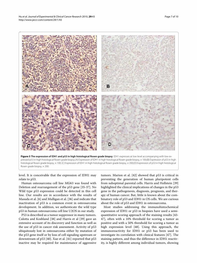
Figure 5 The expression of IDH1 and p53 in high histological Rosen grade biopsy. IDH1 expresses at low level accompanying with low ex-pressed p53 in high histological Rosen grade biopsy.(A) Expression of IDH1 in high histological Rosen grade biopsy, × 100;(B) Expression of p53 in high histological Rosen grade biopsy, × 100; (C) Expression of IDH1 in high histological Rosen grade biopsy, × 200;(D) Expression of p53 in high histological Rosen grade biopsy, × 200.

Hu et al. Journal of Experimental & Clinical Cancer Research 2010, 29:43http://www.jeccr.com/content/29/1/43
Page 8 of 10
a range from 8% through 100% IDH1-positive tumorcells, while the P53, ranging from 5% to 100%. In addi-tion, the positive rate of IDH1 is 90.9%, while the p53 is84.1%. The high staining rate of IDH1 is 52.2%, while thep53 is 43.2%. Furthermore, IDH1 expresses higher inpatients with low histological Rosen grade. IDH1 corre-lates with metastasis negatively. There is no significantcorrelation between IDH1 expression and overall sur-vival. In our study, lower IDH1 expression in higherRosen grade may not convey mutation in the gene. Tosubstitute, genetic studies of IDH1 gene alteration may beof value. The study is limited by the fact that there wereonly 44 patients and without intimate following up infor-
mation. However, it may, from the theoretical point ofview, still be valuable to study the role of IDH1 in osteo-sarcoma. In accordance with former results, p53 in ourosteosarcoma patients correlates with histological Rosengrade, metastasis and overall survival. In our study, theexpression of IDH1 does not correlate some other clinical
Figure 6 The immunostaining percentages of IDH1 and p53 in low Rosen grade vs. high Rosen grade. IDH1 expresses higher in Low histological Rosen grade compare with high histological Rosen grade at the level of the immunostaining percentages (P < 0.01), so does p53 (P < 0.01).
Figure 7 The immunostaining scores of IDH1 and p53 in low Ros-en grade vs. high Rosen grade. IDH1 expresses higher in Low histo-logical Rosen grade compare with high histological Rosen grade at the level of the immunostaining scores (P < 0.05), so does p53 (P < 0.01).
Figure 8 The relationship between IDH1 and survival. The IDH1 high expression group represents the osteosarcoma patients with >50% IDH1 positive staining. Patients with ≤ 50% IDH1 positive stain-ing are recorded as low-expression group. The survival time in the χ -axis was given as years. There is no significant correlation between IDH1 expression and overall survival (P = 0.342).
Figure 9 The relationship between p53 and survival. The p53 high expression group represents the osteosarcoma patients with >50% p53 positive staining. Patients with ≤ 50% p53 positive staining are re-corded as low-expression group. The survival time in the χ-axis was giv-en as years. High-expression p53 patients have better survival than low-expression p53 patients do (P = 0.019).

Hu et al. Journal of Experimental & Clinical Cancer Research 2010, 29:43http://www.jeccr.com/content/29/1/43
Page 9 of 10
features such as age, localization of primary tumor andhistological type.
Interestingly, patients in our study with High expres-sion of IDH1 had a very high p53 expression in osteosar-coma biopsies, which is accordance with our result inosteosarcoma cell lines MG63 and U2OS. A recent studyhas shown IDH1 appears to function as a tumor suppres-sor contributes to tumorigenesis in part through induc-tion of the HIF-1 pathway [22]. Parsons et al. [20] foundthat IDH1 mutations had a very high frequency of p53mutation in human glioblastoma. Accumulation of func-tional p53 protein followed by p53-dependent apoptosishas been described in cultured cells exposed to hypoxia[49]. P53 inhibits HIF-1 dependent transcription anddecrease the chances of normal cells surviving underhypoxia since the expression of most glycolytic enzymesis HIF-1 dependent [50]. It is conceivable that IDH1 mayrelate to p53 with the function of HIF-1.
ConclusionsIDH1 may correlate with p53 and be a biomarker forosteosarcoma correlate with histological Rosen grade andmetastasis.
List of abbreviationsIDH1: isocitrate dehydrogenase 1; p53: transformation-related protein 53; OS: osteosarcoma; PTEN: phosphatesand tensin homolog; Rb: retinoblastoma gene; TCA: Thecitric acid cycle; SD: Std. Deviation.
Competing interestsThe authors declare that they have no competing interests.
Authors' contributionsHu X carried out most parts of the experiment; Qi BW, Fu T, Wu G, Zhou M, LuoJ and Xu JH participated in the experiment; Yu AX conceives the study project,organizes the whole study process, provides financial support, and finalizes themanuscript. All authors have read and approved the final manuscript.
AcknowledgementsWe thank guorong Yu, zhenyu Pan, kai Deng, Shengxiang Tao for technical assistance. This work is supported by the grants from the Natural Science Foun-dation of China (No. 303131304), the health department Scientific Research Project of Hubei Province of China (No. 303121208).
Author Details1Department of Orthopedics, Zhongnan Hospital of Wuhan University, No 169 Donghu Road, Wuchang District, 430071, Wuhan, China and 2Department of pathology, Zhongnan Hospital of Wuhan University, Wuhan, China
References1. Kager L, Zoubek A, Pötschger U, Kastner U, Flege S, Kempf-Bielack B,
Branscheid D, Kotz R, Salzer-Kuntschik M, Winkelmann W, Jundt G, Kabisch H, Reichardt P, Jürgens H, Gadner H, Bielack SS: Primary metastatic osteosarcoma: presentation and outcome of patients treated on neoadjuvant Cooperative Osteosarcoma Study Group protocols. J Clin Oncol 2003, 21:2011-2018.
2. Ferguson WS, Goorin AM: Current treatment of osteosarcoma. Cancer Invest 2001, 19:292-315.
3. Overholtzer M, Rao PH, Favis R, Lu XY, Elowitz MB, Barany F, Ladanyi M, Gorlick R, Levine AJ: The presence of p53 mutations in human osteosarcomas correlates with high levels of genomic instability. Proceedings of the National Academy of Sciences of the United States of America 2003, 100:11547-11552.
4. Zheng Shui-er, Yso Yang, Dong Yang, Lin Feng, Zhao Hui, Shen Zan, Sun Yuan-jue, Tang Li-na: Down-regulation of ribosomal protein L7A in human osteosarcoma. J Cancer Res Clin Oncol 2009, 135:1025-1031.
5. Saleh HA, Jin B, Barnwell J, Alzohaili O: Utility of immunohistochemical markers in differentiating benign from malignant follicular-derived thyroid nodules. Diagn Pathol 2010, 26:5-9.
6. Masuda H, Miller C, Koeffler HP, Battifora H, Cline MJ: Rearrangement of the p53 gene in human osteogenic sarcoma. Proc Natl Acad Sci USA 1987, 84:7716-9.
7. Baker SJ, Fearon ER, Nigro JM, Hamilton SR, Preisinger AC, Jessup JM, vanTuinen P, Ledbetter DH, Barker DF, Nakamura Y, White R, Vogelstein B: Chromosome 17 deletions and p53 gene mutations in colorectal carcinomas. Science 1989, 244:217-21.
8. Miller G, Socci ND, Dhall D, D'Angelica M, DeMatteo RP, Allen PJ, Singh B, Fong Y, Blumgart LH, Klimstra DS, Jarnagin WR: Genome wide analysis and clinical correlation of chromosomal and transcriptional mutations in cancers of the biliary tract. Journal of Experimental & Clinical Cancer Research 2009, 28:62.
9. Vousden KH, Lane DP: P53 in health and disease. Nat Rev Mol Cell Biol 2007, 8:275-83.
10. Di Cristofano A, Pandolfi PP: The multiple roles of PTEN in tumor suppression. Cell 2000, 100:387-390.
11. Cantley LC, Neel BG, New insights into tumor suppression: PTEN suppresses tumor formation by restraining the phosphoinositide 3-kinase/AKT pathway. Proc Natl Acad Sci USA 1999, 96:4240-4245.
12. Hamada K, Sasaki T, Koni PA, Natsui M, Kishimoto H, Sasaki J, Yajima N, Horie Y, Hasegawa G, Naito M, Miyazaki J, Suda T, Itoh H, Nakao K, Mak TW, Nakano T, Suzuki A: The PTEN/PI3K pathway governs normal vascular development and tumor angiogenesis. Genes Dev 2005, 19:2054-2065.
13. Freeman SS, Allen SW, Ganti R, Wu J, Ma J, Su X, Neale G, Dome JS, Daw NC, Khoury JD: Copy number gains in EGFR and copy number losses in PTEN are common events in osteosarcoma tumors. Cancer 2008, 113:1453-61.
14. Ternovoi1 Vladimir V, Curiel David T, Smith Bruce F, Siegal Gene P: Adenovirus-mediated p53 tumor suppressor gene therapy of osteosarcoma. Laboratory Investigation 2006, 86:748-766.
15. Wadayama B, Toguchida J, Shimizu T, Ishizaki K, Sasaki MS, Kotoura Y, Yamamuro T: Mutation spectrum of the retinoblastoma gene in osteosarcoma. Cancer Res 1994, 54:3042-8.
16. Nekrutenko A, Hillis DM, Patton JC, Bradley RD, Baker RJ: Cytosolic isocitrate dehydrogenase in humans, mice, and voles and phylogenetic analysis of the enzyme family. Molec Biol Evol 1998, 15:1674-1684.
17. Shechter I, Dai P, Huo L, Guan G: IDH1 gene transcription is sterol regulated and activated by SREBP-1a and SREBP-2 in human hepatoma HepG2 cells: evidence that IDH1 may regulate lipogenesis in hepatic cells. J Lipid Res 2003, 44:2169-2180.
18. Memon AA, Chang JW, Oh BR, Yoo YJ: Identification of differentially expressed proteins during human urinary bladder cancer progression. Cancer Detect Prev 2005, 29:249-255.
19. Yan H, Parsons DW, Jin G, McLendon R, Rasheed BA, Yuan W, Kos I, Batinic-Haberle I, Jones S, Riggins GJ, Friedman H, Friedman A, Reardon D, Herndon J, Kinzler KW, Velculescu VE, Vogelstein B, Bigner DD: IDH1 and IDH2 mutations in gliomas. New Eng J Med 2009, 360:765-773.
20. Parsons DW, Jones S, Zhang X, Lin JC-H, Leary RJ, Angenendt P, Mankoo P, Carter H, Siu I-M, Gallia GL, Olivi A, McLendon R, 21 others: An integrated genomic analysis of human glioblastoma multiforme. Science 2008, 321:1807-1812.
21. Mardis ER, Ding L, Dooling DJ, Larson DE, McLellan MD, Chen K, Koboldt DC, Fulton RS, Delehaunty KD, McGrath SD, Fulton LA, Locke DP, 46 others: Recurring mutations found by sequencing an acute myeloid leukemia genome. New Eng J Med 2009, 361:1058-1066.
22. Zhao S, Lin Y, Xu W, Jiang W, Zha Z, Wang P, Yu W, Li Z, Gong L, Peng Y, Ding J, Lei Q, Guan K-L, Xiong Y: Glioma-derived mutations in IDH1 dominantly inhibit IDH1 catalytic activity and induce HIF-1-alpha. Science 2009, 324:261-265.
Received: 29 March 2010 Accepted: 7 May 2010 Published: 7 May 2010This article is available from: http://www.jeccr.com/content/29/1/43© 2010 Hu et al; licensee BioMed Central Ltd. This is an Open Access article distributed under the terms of the Creative Commons Attribution License (http://creativecommons.org/licenses/by/2.0), which permits unrestricted use, distribution, and reproduction in any medium, provided the original work is properly cited.Journal of Experimental & Clinical Cancer Research 2010, 29:43
Hu et al. Journal of Experimental & Clinical Cancer Research 2010, 29:43http://www.jeccr.com/content/29/1/43
Page 10 of 10
23. Jeong Ji-Hak, Nakajima Hiroo, Magae Junji, Furukawa Chiharu, Taki Keiko, Otsuka Kensuke, Tomita Masanori, Lee In-Seon, Kim Cheorl-Ho, Chang Hyeun-Wook, Min Kwan-Sik, Park Kwang-Kyun, Park Kwan-Kyu, Chang Young-Chae: Ascochlorin activates p53 in a manner distinct from DNA damaging agents. Int J Cancer 2009, 124:2797-2803.
24. Saiki RK, Gelfand DH, Stoffel S, Scharf SJ, Higuchi R, Horn GT, Mullis KB, Erlich HA: Primer-directed enzymatic amplification of DNA with a thermostable DNA polymerase. Science 1988, 239:487-491.
25. Hellwinkel OJ, Müller A, Struve D, Hiort O: Influence of androgens and age on androgen receptor and 5 alpha-reductase II transcription. Eur J Endocrinol 2000, 143:217-225.
26. Ryu K, Choy E, Yang C, Susa M, Hornicek FJ, Mankin H, Duan Z: Activation of Signal Transducer and Activator of Transcription 3 (Stat3) Pathway in Osteosarcoma Cells and Overexpression of Phosphorylated-Stat3 Correlates with Poor Prognosis. J Orthop Res 2010 in press.
27. Wu L, Peng CW, Hou JX, Zhang YH, Chen C, Chen LD, Li Y: Coronin-1C is a novel biomarker for hepatocellular carcinoma invasive progression identified by proteomics analysis and clinical validation. J Exp Clin Cancer Res 2010 in press.
28. Ponten J, Saksela E: Two established in vitro cell lines from human mesenchymal tumors. Int J Cancer 1967, 2:434-47.
29. Heremans H, Billiau A, Cassiman JJ, Mulier JC, de Somer P: In vitro cultivation of human tumor tissues. II. Morphological and virological characterization of three cell lines. Oncology 1978, 35:246-52.
30. Huvos AG, Rosen G, Marcove RC: Primary osteogenic sarcoma: pathologic aspects in 20 patients after treatment with chemotherapy en bloc resection, and prosthetic bone replacement. Arch Pathol Lab Med 1977, 101:14-18.
31. Rosen G, Marcove RC, Caparros B, Nirenberg A, Kosloff C, Huvos AG: Primary osteogenic sarcoma: the rationale for preoperative chemotherapy and delayed surgery. Cancer 1979, 43:2163-2177.
32. Rosen G, Murphy ML, Huvos AG, Gutierrez M, Marcove RC: Chemotherapy, en bloc resection, and prosthetic bone replacement in the treatment of osteogenic sarcoma. Cancer 1976, 37:1-11.
33. MacKenzie ED, Selak MA, Tennant DA, Payne LJ, Crosby S, Frederiksen CM, Watson DG, Gottlieb E: Cell-permeating alpha-ketoglutarate derivatives alleviate pseudohypoxia in succinate dehydrogenase-deficient cells. Mol Cell Biol 2007, 27:3282-9.
34. Ingebretsen OC: Mechanism of the inhibitory effect of glyoxylate plus oxaloacetate and oxalomalate on the NADP-specific isocitrate dehydrogenase. Biochim Biophys Acta 1976, 452:302-9.
35. Lindström MS, Nistér M: Silencing of ribosomal protein S9 elicits a multitude of cellular responses inhibiting the growth of cancer cells subsequent to p53 activation. PLoS One 2010, 5:e9578.
36. Mulligan LM, Matlashewski GJ, Scrable HJ, Cavenee WK: Mechanisms of p53 loss in human sarcomas. Proc Natl Acad Sci USA 1990, 87:5863-7.
37. Chandar N, Billig B, McMaster J, Novak J: Inactivation of p53 gene in human and murine osteosarcoma cells. Br J Cancer 1992, 65:208-14.
38. Culotta E, Koshland DE Jr: P53 sweeps through cancer research. Science 1993, 262:1958-61.
39. Harris CC, Hollstein M: Clinical implications of the p53 tumor-suppressor gene. N Engl J Med 1993, 329:1318-27.
40. Bourdon JC, Fernandes K, Murray-Zmijewski F, Liu G, Diot A, Xirodimas DP, Saville MK, Lane DP: P53 isoforms can regulate p53 transcriptional activity Genes. Dev 2005, 19:2122-37.
41. Xue C, Haber M, Flemming C, Marshall GM, Lock RB, MacKenzie KL, Gurova KV, Norris MD, Gudkov AV: P53 determines multidrug sensitivity of childhood neuroblastoma. Cancer Res 2007, 67:10351-60.
42. Marion RM, Strati K, Li H, Murga M, Blanco R, Ortega S, Fernandez-Capetillo O, Serrano M, Blasco MA: A p53-mediated DNA damage response limits reprogramming to ensure iPS cell genomic integrity. Nature 2009, 460:1149-1153.
43. Wadayama B, Toguchida J, Yamaguchi T, Sasaki MS, Yamamuro T: P53 expression and its relationship to DNA alterations in bone and soft tissue sarcomas. British Journal of Cancer 1993, 68:1134-1139.
44. Stefanou DG, Nonni AV, Agnantis NJ, Athanassiadou SE, Briassoulis E, Pavlidis N: p53/MDM-2 immunohistochemical expression correlated with proliferative activity in different subtypes of human sarcomas: a ten-year followup study. Anticancer Research 1998, 18:4673-4681.
45. Lonardo F, Ueda T, Huvos AG, Healey J, Ladanyi M: P53 and MDM2 alterations in osteosarcomas. Correlation with clinicopathologic features and proliferative rate. Cancer 1997, 79:1541-1547.
46. Matsuo T, Sugita T, Shimose S, Kubo T, Ishikawa M, Yasunaga Y, Ochi M: Immunohistochemical expression of promyelocytic leukemia body in soft tissue sarcomas. Journal of Experimental & Clinical Cancer Research 2008, 27:73.
47. Ueda Y, Dockhorn-Dworniczak B, Blasius S, Mellin W, Wuisman P, Böcker W, Roessner A: Analysis of mutant P53 protein in osteosarcomas and other malignant and benign lesions of bone. Journal of Cancer Research and Clinical Oncology 1993, 119:172-178.
48. Naka T, Fukuda T, Shinohara N, Iwamoto Y, Sugioka Y, Tsuneyoshi M: Osteosarcoma versus malignant fibrous histiocytoma of bone in patients older than 40 years. A clinicopathologic and immunohistochemical analysis with special reference to malignant fibrous histiocytoma-like osteosarcoma. Cancer 1995, 76:972-984.
49. Graeber TG, Osmanian C, Jacks T, Houseman DE, Koch CJ, Lowe SW, Giaccia AJ: Hypoxia-mediated selection of cells with diminished apoptotic potential in solid tumors. Nature 1996, 379:88-91.
50. Salnikow K, An WG, Melillo G, Blagosklonny MV, Costa M: Nickel-induced transformation shifts the balance between HIF-1 and p53 transcription factors. Carcinogenesis 1999, 20:1819-23.
doi: 10.1186/1756-9966-29-43Cite this article as: Hu et al., The expression and significance of IDH1 and p53 in osteosarcoma Journal of Experimental & Clinical Cancer Research 2010, 29:43