Embed Size (px)
Citation preview
RESEARCH ARTICLE
Regulation of Neuronal Cav3.1 Channels byCyclin-Dependent Kinase 5 (Cdk5)Aida Calderón-Rivera1☯, Alejandro Sandoval1☯, Ricardo González-Ramírez2,Christian González-Billault3, Ricardo Felix4*
1 School of Medicine FES Iztacala, National Autonomous University of Mexico (UNAM), Tlalnepantla,Mexico, 2 Department of Molecular Biology and Histocompatibility, “Dr. Manuel Gea González”GeneralHospital, Ministry of Health, Mexico City, Mexico, 3 Department of Biology, Faculty of Sciences, University ofChile, Santiago, Chile, 4 Department of Cell Biology, Center for Research and Advanced Studies of theNational Polytechnic Institute (Cinvestav-IPN), Mexico City, Mexico
☯ These authors contributed equally to this work.* [email protected]
AbstractLow voltage-activated (LVA) T-type Ca2+ channels activate in response to subthreshold
membrane depolarizations and therefore represent an important source of Ca2+ influx near
the resting membrane potential. In neurons, these proteins significantly contribute to control
relevant physiological processes including neuronal excitability, pacemaking and post-in-
hibitory rebound burst firing. Three subtypes of T-type channels (Cav3.1 to Cav3.3) have
been identified, and using functional expression of recombinant channels diverse studies
have validated the notion that T-type Ca2+ channels can be modulated by various endoge-
nous ligands as well as by second messenger pathways. In this context, the present study
reveals a previously unrecognized role for cyclin-dependent kinase 5 (Cdk5) in the regula-
tion of native T-type channels in N1E-115 neuroblastoma cells, as well as recombinant
Cav3.1channels heterologously expressed in HEK-293 cells. Cdk5 and its co-activators
play critical roles in the regulation of neuronal differentiation, cortical lamination, neuronal
cell migration and axon outgrowth. Our results show that overexpression of Cdk5 causes a
significant increase in whole cell patch clamp currents through T-type channels in N1E-115
cells, while siRNA knockdown of Cdk5 greatly reduced these currents. Consistent with this,
overexpression of Cdk5 in HEK-293 cells stably expressing Cav3.1channels upregulates
macroscopic currents. Furthermore, using site-directed mutagenesis we identified a major
phosphorylation site at serine 2234 within the C-terminal region of the Cav3.1subunit.
These results highlight a novel role for Cdk5 in the regulation of T-type Ca2+ channels.
IntroductionThe family of voltage-gated Ca2+ (CaV) channels are transmembrane proteins that serves astransducers of cell surface membrane potential changes into local intracellular Ca2+ transientsthat initiate a myriad of physiological events. CaV channels have been traditionally classified
PLOSONE | DOI:10.1371/journal.pone.0119134 March 11, 2015 1 / 19
OPEN ACCESS
Citation: Calderón-Rivera A, Sandoval A, González-Ramírez R, González-Billault C, Felix R (2015)Regulation of Neuronal Cav3.1 Channels by Cyclin-Dependent Kinase 5 (Cdk5). PLoS ONE 10(3):e0119134. doi:10.1371/journal.pone.0119134
Academic Editor: Alexander G. Obukhov, IndianaUniversity School of Medicine, UNITED STATES
Received: April 24, 2014
Accepted: January 19, 2015
Published: March 11, 2015
Copyright: © 2015 Calderón-Rivera et al. This is anopen access article distributed under the terms of theCreative Commons Attribution License, which permitsunrestricted use, distribution, and reproduction in anymedium, provided the original author and source arecredited.
Data Availability Statement: All relevant data arewithin the paper
Funding: This work received funding from thefollowing sources: 128707-Q, Conacyt Mexico, http://www.conacyt.mx/; IN-221011, PAPIIT;UNAM, http://dgapa.unam.mx/html/papiit/papit.html; and ACT114,CONICYT Chile, http://www.conicyt.cl/. The fundershad no role in study design, data collection andanalysis, decision to publish, or preparation of themanuscript.
Competing Interests: The authors have declaredthat no competing interests exist.
into high voltage-activated (HVA) and low voltage-activated (LVA) subtypes [1]. HVA chan-nels activate at relatively depolarized potentials and comprise L-, P/Q-, N-, and R-types. LVAchannels, also known as T-type, are critically important for regulating neuronal excitability,pacemaking and post-inhibitory rebound burst firing [2],[3]. Therefore, it should not come asa surprise that T-type channel hyperactivity has been associated to human neurological disor-ders such as absence epilepsy and neuropathic pain [4],[5],[6],[7].
Three different T-type channels, CaV3.1, CaV3.2 and CaV3.3, have been cloned and express-ed from mammals [1],[2]. Using recombinant channels diverse studies have validated the ideathat Cav3 channels can be modulated by various endogenous ligands as well as by second mes-senger pathways. Hence, it has been reported that Ca2+/CaM-dependent protein kinase II(CaMKII) differentially regulates the activation of CaV3 channels [8], and that protein kinaseA (PKA) and PKC increase CaV3 current density [9],[10],[11]. However, it remains unknownwhether other kinases play a role in modulating CaV3 channel function.
Interestingly, it has been shown that the inhibition of the cyclin-dependent kinase 5 (Cdk5)favors neurotransmitter release via enhancement of P/Q-type channel activity [12]. Cdk5seems to phosphorylate the intracellular loop that connects the second and third repeated do-mains in the CaV2.1α1 pore-forming subunit of the channels, affecting its interaction withSNAP-25 and synaptotagmin [12]. Likewise, recent evidence suggests that the N-type channel,the other major presynaptic Ca2+ channel, is also a substrate of Cdk5. In this case, phosphory-lation of the CaV2.2α1 pore-forming subunit by Cdk5 facilitates neurotransmitter release in-creasing Ca2+ influx by enhancing channel open probability [13].
Cdk5 is a neuron-specific, proline-directed serine/threonine kinase that forms a complexwith its activators p35 or p39. Diverse studies have shown that the complex of Cdk5 and its ac-tivators has multiple functions in immature neurons including migration, differentiation andsynaptogenesis [14],[15]. Although the physiological role of Cdk5 in mature neurons is lessclear, it has been suggested that several proteins of the soluble N-ethylmaleimide-sensitive fac-tor attachment protein (SNAP) receptor (SNARE) required for efficient neurotransmitter re-lease may act as physiological substrates of Cdk5. Likewise, it has been documented thatproteolytic cleavage of p35 may produce p25, which accumulates in the brain of patients withAlzheimer's disease [13],[16]. Furthermore, increased proteolysis of p35 is linked to abnormaltau phosphorylation and promotes neuronal apoptosis [17].
In the present study we analyzed CaV3.1 channels for potential phosphorylation by Cdk5.We report that Cdk5 can directly phosphorylate CaV3.1 channels at serine 2234 and that thisin turn modulates depolarization-dependent Ca2+ entry.
Materials and Methods
Cell culturesMouse neuroblastoma-derived N1E-115 cells (American Type Culture Collection; ATCCNum-ber CRL-2263) were grown in culture using Dulbecco’s modified Eagle’s medium plus 25 mMglucose (DMEM-HG) culture medium supplemented with 10% fetal bovine serum, 2 mML-glutamine, and penicillin-streptomycin (100 U/mL). Cells were incubated in a humid atmo-sphere of 5% CO2-95% air at 37°C. The incubation medium was changed every 2 days. Cellswere harvested once per week by treatment with a trypsin-EDTA solution, and reseeding wascarried out at 20% of the original density. Human embryonic kidney (HEK) 293 cells stably ex-pressing the Cav3.1a channel [18],[19] were grown as described elsewhere [20]. In brief, cellswere kept in culture in DMEM supplemented with 1 mg/ml G418 (Gibco/BRL Life Technolo-gies), 10% fetal bovine serum, and penicillin-streptomycin (100 U/mL) at 37°C in a 5%CO2-95% air humidified atmosphere and sub-cultured by mechanical dispersion every week.
Cav3.1 Channel Regulation by Cdk5
PLOS ONE | DOI:10.1371/journal.pone.0119134 March 11, 2015 2 / 19
ElectrophysiologyN1E-115 and HEK-293 cells were subjected to the standard whole cell patch-clamp techniqueusing an Axopatch 200B amplifier as described previously [20]. Current signals were filtered at2 kHz, digitized at 5.71 kHz and analyzed with pClamp software. Data were leak subtracted online by a P/4 protocol. The bath recording solution contained (in mM) either 5 CaCl2 (HEK-293 stable transfected), 2 BaCl2 (HEK-293 cells transiently transfected), or 10 BaCl2 (N1E-115cells), 125 TEA-Cl, 10 HEPES and 10 glucose (pH 7.3). The internal solution consisted of (in mM)110 CsCl, 5 MgCl2, 10 EGTA, 10 HEPES, 4 Mg-ATP and 0.1 GTP (pH 7.3). Pipettes with resis-tance values in the range 2–3 MO were used. Linear leak and parasitic capacitance componentswere subtracted on-line using a P/4 protocol. Membrane capacitance (Cm) was determined aspreviously described [21] and used to normalize currents.
Data analysisActivation of the currents was well described by a Boltzmann relation of the form G/Gmax = 1/{1+exp[(V-V½)/k]}, where G is peak conductance, Gmax is fitted maximal G, V½ is half-activa-tion voltage, and k is the slope factor. Steady-state current inactivation curves were fitted withthe following equation: I/Imax = 1/(1+exp(−(Vp−V½inact)/k)), where I/Imax is the normalizedpeak current, Vp is the conditioning prepulse, V½inact is the voltage for half-inactivation and kis the slope factor. Time constants of activation (τact) and inactivation (τinact) were obtained,using 140 ms test pulses, from single exponential fits to the raising and decaying phases ofthe currents.
Cdk5 gene silencing by specific siRNAsN1E-115 cells were seeded in 35-mm dishes (at*80% confluence) and transfected with a 100-pmol mixture of predesigned synthetic small interfering RNAs (siRNAs) that target Cdk5 orwith a non-targeting siRNA as negative control (Sigma-Aldrich) using Lipofectamine 2000transfection reagent (Invitrogen). Twenty-four hours after transfection, cells were harvested toanalyze the knockdown effect of siRNAs on endogenous Cdk5 protein levels by Western blotusing specific antibodies or re-seeded for electrophysiological recording.
Western blotTotal protein extracts were obtained using 200 μl of lysis buffer (50 mMTris-HCl pH 8.0, 150 mMNaCl, 0.5 mM PMSF, 1% Triton X-100, 1X protease inhibitor mix (Roche Applied Science).After centrifugation, supernatants were recovered for protein quantification by the bicinchoni-nic acid reaction. Aliquots of 50 μg of total protein extracts were boiled in SDS sample buffer(50 mM Tris-HCl pH 6.8, 2% SDS, 10% glycerol, 0.1% 2-mercaptoethanol, 0.001% bromophe-nol blue), electrophoresed on 10% SDS-polyacrylamide gels and transferred to nitrocellulosemembranes (Amersham Pharmacia GE Healthcare). After blocking with non-fat milk (5%)supplemented with 0.2% Tween 20, membranes were incubated 1 h with the primary antibodyanti-Cdk5 or anti-p35 (C-8 and C-19, respectively; Santa Cruz Biotechnology) at 1:1000 dilu-tion in TBS-T with 5% non-fat milk (Santa Cruz) washed with TBS-T (10 mM Tris-HCl, 0.15M NaCl, 0.05% Tween 20), and finally incubated with goat anti-rabbit secondary antibody cou-pled to horseradish peroxidase for 1 h. Immunoblots were developed by using the ECLWest-ern blotting analysis system (Amersham Pharmacia GE Healthcare).
Cav3.1 Channel Regulation by Cdk5
PLOS ONE | DOI:10.1371/journal.pone.0119134 March 11, 2015 3 / 19
Total RNA isolation and RT-PCRTotal RNA was isolated from N1E-115 cells using TRIzol reagent (Invitrogen). Five micro-grams of total RNA was reverse transcribed using random hexanucleotides and the M-MLV re-verse transcriptase (Invitrogen), according to the manufacturer’s instructions. Afterinactivation of reverse transcriptase cDNAs were processed immediately for amplification. ThePCR reaction was carried out in a total volume of 50 μl containing 5 μl of cDNA solution, 1XPCR buffer (20 mM Tris-HCl pH 8.4, 50 mM KCl), 0.2 mM of each deoxynucleotide triphos-phate, 1.5 mMMgCl2, 30 pmol of each primer, and 2.5 U of Taq DNA polymerase (Invitrogen)on a PCRsprint thermal cycler (Thermo Scientific). The PCR conditions included an initial de-naturation step at 94°C for 5 min followed by a cyclic reaction consisted of denaturation at 94°Cfor 30 s, annealing at 55°C for 30 s and extension for 1 min at 72°C. PCR products were thensubjected to 1% agarose gel electrophoresis and nucleic acid bands were visualized by ethidiumbromide staining. The amplified products were purified and sequenced. Primers used in thiswork are as follows (forward and reverse): 5’-TGCAGAAATACGAGAAACTGGA-3’ and 5’-ACATGTCGATGGACGTGGAGTAC-3’ for CDK5; 5’-AAGAAGAAGAACTCCAAGAA-3’and 5’-ACATCCCTGCAGAGCATGTA-3’ for p35; 5’-CACTTGTGCACCAGCCACTA-3’and 5’-AGGTCTCCAAAGAGCTCCAC-3’ for CaV3.1; 5’-TTCTTCAAGGACAGGTGGAA-3’ and 5’-GCCTCCTTGTTGCTCTCCTC-3’ for CaV3.2; 5’-TGTGCCTTCTTCATCATCTT-3’and 5’-AAGGTGATGAAGATGTCCAG-3’ for CaV3.3; and 5’-AAGATGACCCAGAT-CATGTT-3’ and 5’-GAGTACTTGCGCTCAGGAGG-3’ for actin.
Transient transfection of HEK-293 cellsHEK-293 cells (ATCC Number CRL-1573) were maintained in DMEM-HG medium supple-mented with 10% horse serum, 1% L-glutamine, 110 mg/L sodium pyruvate and antibiotics, at37°C in a 5% CO2-95% air humidified atmosphere. Gene transfer was performed using Lipofec-tamine Plus reagent (Invitrogen). Briefly, for a 35-mm Petri dish of HEK293 cells, 1.2 μg of theplasmid cDNA encoding the rat brain T-type Ca2+ channel CaV3.1 subunit (GenBank acces-sion number AF027984.1; Perez-Reyes et al., 1998), a generous gift of Dr. JH Lee (Sogang Uni-versity, Korea), or its S2234A mutant, as well as 1.5 μg Cdk5 and 0.5 μg p35 plasmid constructs(NM_007668.3 and U89527.1, respectively) were premixed with 6 μL of Lipofectamine in 100 μLserum-free medium according to the manufacturer’s instructions.
ImmunofluorescenceHEK-293 cell line stably expressing CaV3.1 channels were grown on poly-L lysine coated slides,fixed with 4% formaldehyde in PBS for 15 min at room temperature (RT) and washed withPBS. Cells were permeabilized with 0.2% Triton X-100 and blocked with 1% gelatin and 10%FBS for 30 min and incubated overnight (at 4°C) with the primary anti CaV3.1 antibody (Milli-pore at 1:20 dilution in 0.1% Triton X-100, 2% BSA and 5% FBS). Cells were then incubatedfor 1 h at RT with the secondary antibody (goat anti-rabbit IgG coupled to Cy5; Zymed). Afterwashing slides were mounted with VectaShield (Vector Laboratories) and fluorescence was vi-sualized with a confocal laser scanning microscope (TCS-SP2, Leica) using a 63x oil-immersionobjective.
Plasma membrane protein extractionHEK-293 cells were transfected with the plasmids codifying for CaV3.1-YFP in the absence(control) or the presence of Cdk5/p35. After 48 h the extraction of membrane proteins was car-ried out using the Membrane Protein Extraction Kit (Cat # K268-50; BioVision) according to
Cav3.1 Channel Regulation by Cdk5
PLOS ONE | DOI:10.1371/journal.pone.0119134 March 11, 2015 4 / 19
the manufacturer’s instruction. Briefly, cells were grown in p100 culture dishes and collectedby centrifugation (700 x g, 5 min), washed with cold PBS, resuspended and homogenized. Thehomogenate was centrifuged at 700 x g for 10 min. The pellets containing the cell membraneproteins were mixed both with 200 μl of the upper and lower phase solutions and centrifugedat 1000 x g for 5 min. The upper phase was carefully transferred to a new tube, diluted in 5 vol-umes of water and centrifuged for 30 min. The supernatant was removed, and the pellet con-taining the membrane proteins was dissolved in 0.5% Triton X-100 in PBS. Proteins were thenquantified using the BSA method and separated by SDS-PAGE electrophoresis gel, and trans-ferred to a nitrocellulose membrane. For CaV3.1 channel detection, the membrane was incu-bated with anti-GFP antibody (1:5000 dilution; Aves Labs) and the loading control protein wasdetected with an anti-pan-Cadherin antibody (1:50 dilution; Life Technologies). The quantifi-cation of the band intensity of the CaV3.1 signal was normalized to Cadherin and expressed asarbitrary units.
Site-directed mutagenesisMultiple nucleotide sequences were aligned using the modified Clustal W algorithm of VectorNTI 8 software package (Invitrogen). Phosphorylation site(s) prediction was performed usingthe GPS 2.0 software (Group-based Prediction System, ver 2.0), a state-of-the-art web-basedsoftware for prediction of phosphorylation available at http://gps.biocuckoo.org/. The CaV3.1cDNA was inserted into the expression plasmid pcDNA3 and expressed under the control ofthe cytomegalovirus promoter. A point mutation (S2234A) was introduced with*40-mersynthetic oligonucleotides using the Quik-Change XL-mutagenesis kit (Stratagene). Mutantchannel cDNA was sequenced (PerkinElmer Applied Biosystems).
Plasmid constructspGEX expression constructs encoding rat CaV3.1 C-terminus were generated by amplifying a frag-ment of*300 bp comprising amino acids 2154 to 2254 of the protein using the following primers:forward 5´ GAATTCAAGCGGCTCCCAACCCCGCCT 3´ and reverse 5´ GAATTCGGGGTCCATGTCTGTTGGGT 3´. Amplification was carried out in a PCR Sprint Thermal Cycler (ThermoScientific) with a preliminary denaturation step at 94°C for 5 min, followed by 30 cycles at 94°C for45 s, primer annealing at 55°C for 15 s and primer extension at 72°C for 60 s, followed by a 10 minfinal extension at 72°C. The fragment was then digested with EcoRI and cloned into the EcoRI siteof the pGEX-3X vector (pGEX-COOH). Twomutagenic (S2234A) oligonucleotides were designedas follows: forward 5’ CGGCTGCCTCACCCGCCCCAAAGAAAGAC 3’; reverse 5’GTCTTTCTTTGGGGCGGGTGAGGCAGCCG 3’. One hundred twenty five ng of mutagenic oligonucleo-tides were used in each synthesis reaction (full-length CaV3.1 or GST-carboxyl terminal) usingQuik-Change II XL-mutagenesis kit (Agilent Technologies). Digestion of the amplified productswith DpnI preceded transformation of XL10-Gold ultracompetent cells. All cDNAswere sequenced.
Expression and purification of the CaV3.1 C-terminal constructspGEX-COOH vector was transformed into BL21 cells for expression and purification of theGST-COOH proteins. Protein expression was induced with 1.0 mM IPTG after cells reachedan absorbance of 0.5 at 595 nm for 5 h. Cells were spun at 4°C for 20 min and resuspended inlysis buffer [10 mM Tris pH 8.0, 150 mMNaCl, 1 mM EDTA pH 8.0, 3% Sarcosyl and 1X pro-tease inhibitor mix (Roche Applied Science)] and sonicated (Vibra-Cell VCX 130) on ice andthen centrifuged at>12,000 g for 30 min to pellet the insoluble material. Supernatants were re-moved and mixed with 1 mL of glutathione agarose (Invitrogen) to purify the proteins. After
Cav3.1 Channel Regulation by Cdk5
PLOS ONE | DOI:10.1371/journal.pone.0119134 March 11, 2015 5 / 19
releasing the carboxyl-terminal from GST with Xa factor, proteins were used inphosphorylation assays.
In vitro phosphorylationThe CaV3.1 C-terminal WT or S2234A constructs were incubated with 100 ng of recombinanthuman CDK5-p35 complex (Invitrogen) and 2 mM ATP in kinase reaction buffer (20 mMTris-HCl, pH 7.5, 1 mMMgCl2, 1 mM DTT) in a final volume of 20 μL for 1h at 30°C. The re-action was stopped by adding 20 μL of 2X RSB (20 mM Tris HCl pH7.4, 20 mM NaCl, 6 mMMgCl2) and 8 μL (6X) SDS sample buffer, and subsequently boiled for 5 min at 90°C. Sampleswere separated by 15% SDS-PAGE and gel bands revealed by colloidal Coomassie blue stainingor treated with Pro-Q Diamond phosphoprotein gel stain (Invitrogen) according the manufac-turer's instructions. Gels were fixed with 50% methanol and 10% acetic acid overnight, washedwith ddH2O and stained with Pro-Q Diamond for 90 min. Gel was destained (20% acetonitrile,50 mM sodium acetate, pH4) and the phosphoprotein bands detected using a Typhoon instru-ment (excitation/emission: 532/560 nm; Molecular Dynamics).
ResultsTo investigate whether T-type (CaV3) channels are substrates of Cdk5, we initially conductedwhole-cell patch clamp recordings in the clonal neuroblastoma cell line N1E-115 which expressprominent Ca2+ currents through CaV3 channels [22],[23]. These transient inward currents(Fig. 1A) were evoked by a step depolarization from a holding potential (Vh) of −80mV to poten-tials more positive than −50 mV, and the current amplitude became maximum around −25 mV(Fig. 1B). Likewise, when preceded by a 1 s conditioning pulse to −30 mV to inactivate theT-type component, step depolarizations failed to evoke the long-lasting component of the cur-rent previously observed in N1E-115 cells induced to differentiate into mature neurons [22],[23], suggesting that the contribution of L-type channels to the whole-cell current in non-dif-ferentiated N1E-115 cells is negligible (Fig. 1A).
The potential effects of Cdk5 on the capacity of T-type CaV channels to conduct currentwere then examined using also whole-cell recordings in N1E-115 cells transfected with theCdk5 plus its activator p35 cDNAs. Using Ba2+ as the charge carrier, we found that followingtransfection with Cdk5/p35, the peak current amplitude (Fig. 1A) and current density were sig-nificantly increased (>2-fold) when compared to the control condition (Fig. 1B). The possiblerole of Cdk5/p35 on T-type current regulation was supported by using the inhibitor olomou-cine (50 μM), which fully prevented the effect of the kinase on the macroscopic currents. In-ward currents were measured and expressed as peak current density (pA/pF) to account forvariations in cell size and the current density-voltage relationships were normalized (Fig. 1B).
Given that changes in current amplitude could result from changes in channel gating, thevoltage-dependent properties of the currents were assessed in these cells. Activation of thechannels was well described by a Boltzmann relation of the form G/Gmax = 1/{1+exp[(V-V½)/k]}, where G is peak conductance, Gmax is fitted maximal G, V½ is half-activation voltage, and kis the slope factor. As shown in Fig. 1C, for control (GFP-transfected) cells, V½ was −37.5 mV(n = 13), which was not significantly different from that of Cdk5/p35-transfected cells (-38.6 mV,n = 19; P>0.05, Student's t-test). The slope factors for the Boltzmann fits were also similar.Likewise, the voltage-dependent properties of inactivation were determined by applying 1 sconditioning pre-pulses that ranged successively from −80 to −20 mV in 10 mV voltage steps,followed by a 140 ms step depolarization to −30 mV. The relationship of normalized test pulsevoltage to peak current amplitude was plotted against its corresponding Vh and fitted with theBoltzmann equation (Fig. 1C). The voltage dependence of inactivation was similar for currents
Cav3.1 Channel Regulation by Cdk5
PLOS ONE | DOI:10.1371/journal.pone.0119134 March 11, 2015 6 / 19
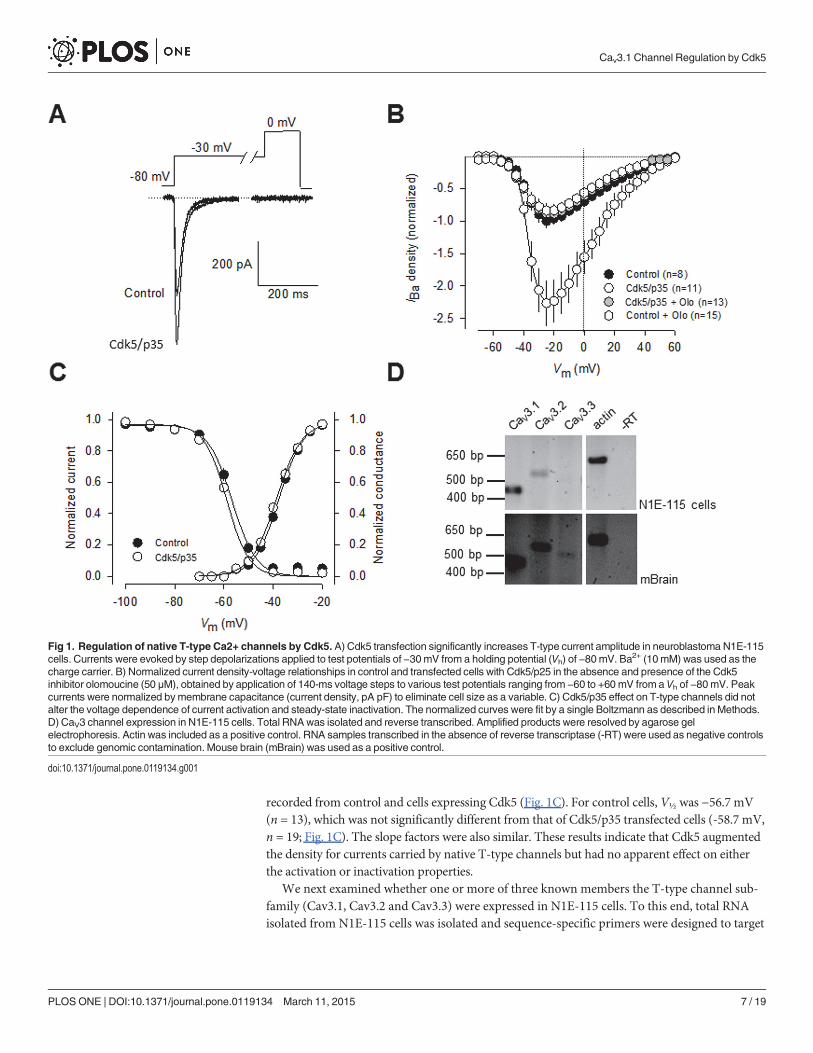
recorded from control and cells expressing Cdk5 (Fig. 1C). For control cells, V½ was −56.7 mV(n = 13), which was not significantly different from that of Cdk5/p35 transfected cells (-58.7 mV,n = 19; Fig. 1C). The slope factors were also similar. These results indicate that Cdk5 augmentedthe density for currents carried by native T-type channels but had no apparent effect on eitherthe activation or inactivation properties.
We next examined whether one or more of three known members the T-type channel sub-family (Cav3.1, Cav3.2 and Cav3.3) were expressed in N1E-115 cells. To this end, total RNAisolated from N1E-115 cells was isolated and sequence-specific primers were designed to target
Fig 1. Regulation of native T-type Ca2+ channels by Cdk5. A) Cdk5 transfection significantly increases T-type current amplitude in neuroblastoma N1E-115cells. Currents were evoked by step depolarizations applied to test potentials of −30mV from a holding potential (Vh) of −80mV. Ba2+ (10mM) was used as thecharge carrier. B) Normalized current density-voltage relationships in control and transfected cells with Cdk5/p25 in the absence and presence of the Cdk5inhibitor olomoucine (50 μM), obtained by application of 140-ms voltage steps to various test potentials ranging from −60 to +60 mV from a Vh of −80mV. Peakcurrents were normalized bymembrane capacitance (current density, pA∙pF) to eliminate cell size as a variable. C) Cdk5/p35 effect on T-type channels did notalter the voltage dependence of current activation and steady-state inactivation. The normalized curves were fit by a single Boltzmann as described in Methods.D) CaV3 channel expression in N1E-115 cells. Total RNAwas isolated and reverse transcribed. Amplified products were resolved by agarose gelelectrophoresis. Actin was included as a positive control. RNA samples transcribed in the absence of reverse transcriptase (-RT) were used as negative controlsto exclude genomic contamination. Mouse brain (mBrain) was used as a positive control.
doi:10.1371/journal.pone.0119134.g001
Cav3.1 Channel Regulation by Cdk5
PLOS ONE | DOI:10.1371/journal.pone.0119134 March 11, 2015 7 / 19
CaV3 channels in RT-PCR experiments. As previously reported [24] and corroborated here,single CaV3.1 and CaV3.2 products of the expected sizes can be selectively amplified from N1E-115 cells using RT-PCR (Fig. 1D, upper panel). In addition, no significant expression of CaV3.3mRNA could be detected. In contrast, evidence for the expression of the three CaV3 channelisotypes were found in the total RNA obtained from the mouse brain used as control (Fig. 1D,lower panel).
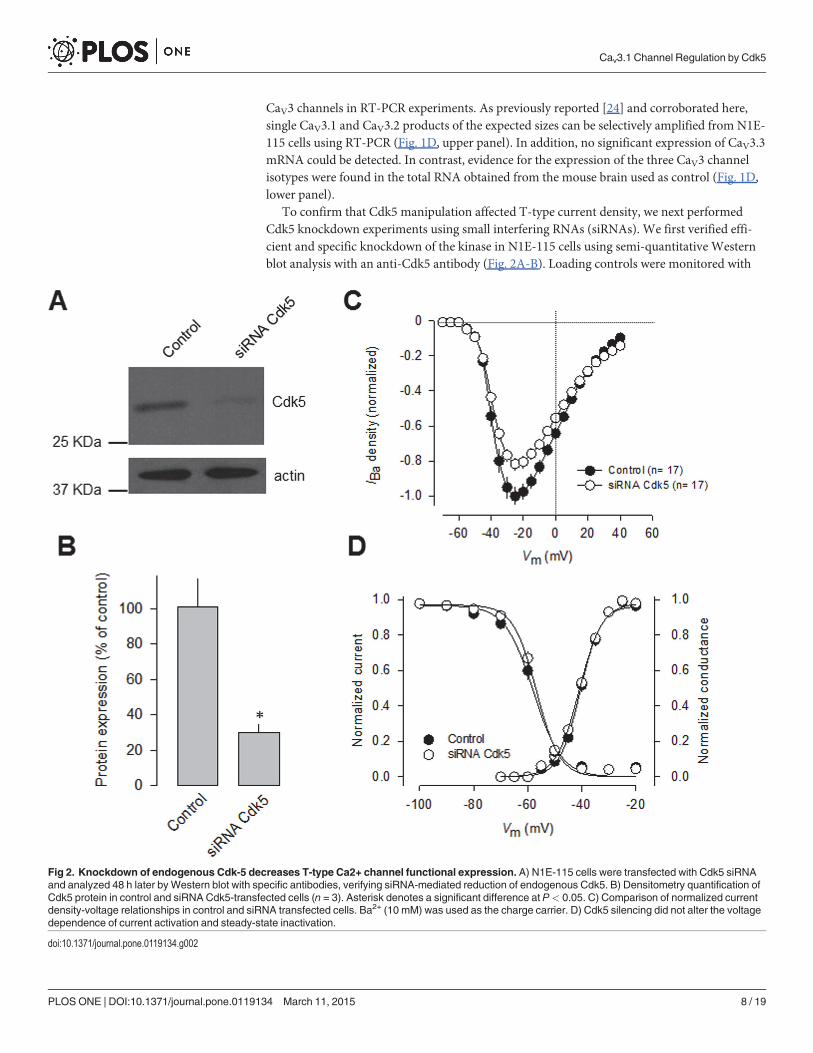
To confirm that Cdk5 manipulation affected T-type current density, we next performedCdk5 knockdown experiments using small interfering RNAs (siRNAs). We first verified effi-cient and specific knockdown of the kinase in N1E-115 cells using semi-quantitative Westernblot analysis with an anti-Cdk5 antibody (Fig. 2A-B). Loading controls were monitored with
Fig 2. Knockdown of endogenous Cdk-5 decreases T-type Ca2+ channel functional expression. A) N1E-115 cells were transfected with Cdk5 siRNAand analyzed 48 h later by Western blot with specific antibodies, verifying siRNA-mediated reduction of endogenous Cdk5. B) Densitometry quantification ofCdk5 protein in control and siRNA Cdk5-transfected cells (n = 3). Asterisk denotes a significant difference at P< 0.05. C) Comparison of normalized currentdensity-voltage relationships in control and siRNA transfected cells. Ba2+ (10 mM) was used as the charge carrier. D) Cdk5 silencing did not alter the voltagedependence of current activation and steady-state inactivation.
doi:10.1371/journal.pone.0119134.g002
Cav3.1 Channel Regulation by Cdk5
PLOS ONE | DOI:10.1371/journal.pone.0119134 March 11, 2015 8 / 19
an anti-actin antibody. The anti-Cdk5 antibody recognized a 32-kDa protein in N1E-115 cellline, as well as in mouse brain lysates and in HEK-293 cells (Fig. 2A). Our analysis of proteinexpression levels confirmed a significant decrease (*70%) in the levels of Cdk5 proteinafter knockdown.
Interestingly, whole-cell recordings of N1E-115 cells transfected with Cdk5 siRNAs for 48 hshowed a significant decrease (*20%) in T-type current density compared with scrambledsiRNA transfected cells. Scaled current density-voltage relationships confirmed that Cdk5knockdown has an inhibitory effect on T-type current density in N1E-115 cells (Fig. 2C). Thediscrepancy between the efficiency of the Cdk5 knockdown (protein decrease of*70%) andits effect on the T-type channels (current reduction of*20%) suggests that there might not bea linear relationship between the silencing of Cdk5 and its effect on T-current density. This isnot unexpected given that posttranslational modifications such as phosphorylation work in anon-stoichiometric manner. In this particular case, the amount of Cdk5 remaining after knock-down still could be phosphorylating a substantial proportion of channels. In addition, theCaV3.1 channels conduct current even in the absence of Cdk5-dependent phosphorylation, ascould be found for the mutant CaV3.1 channels (see below). Last, voltage-dependent propertiesof activation and inactivation of the T-type currents were examined in transfected N1E-115cells, and the results showed that there were no differences in the half-maximal activation (V½)and slope factors (k) of steady-state activation or inactivation between Cdk5 siRNA-transfectedand control cells (Fig. 2D).
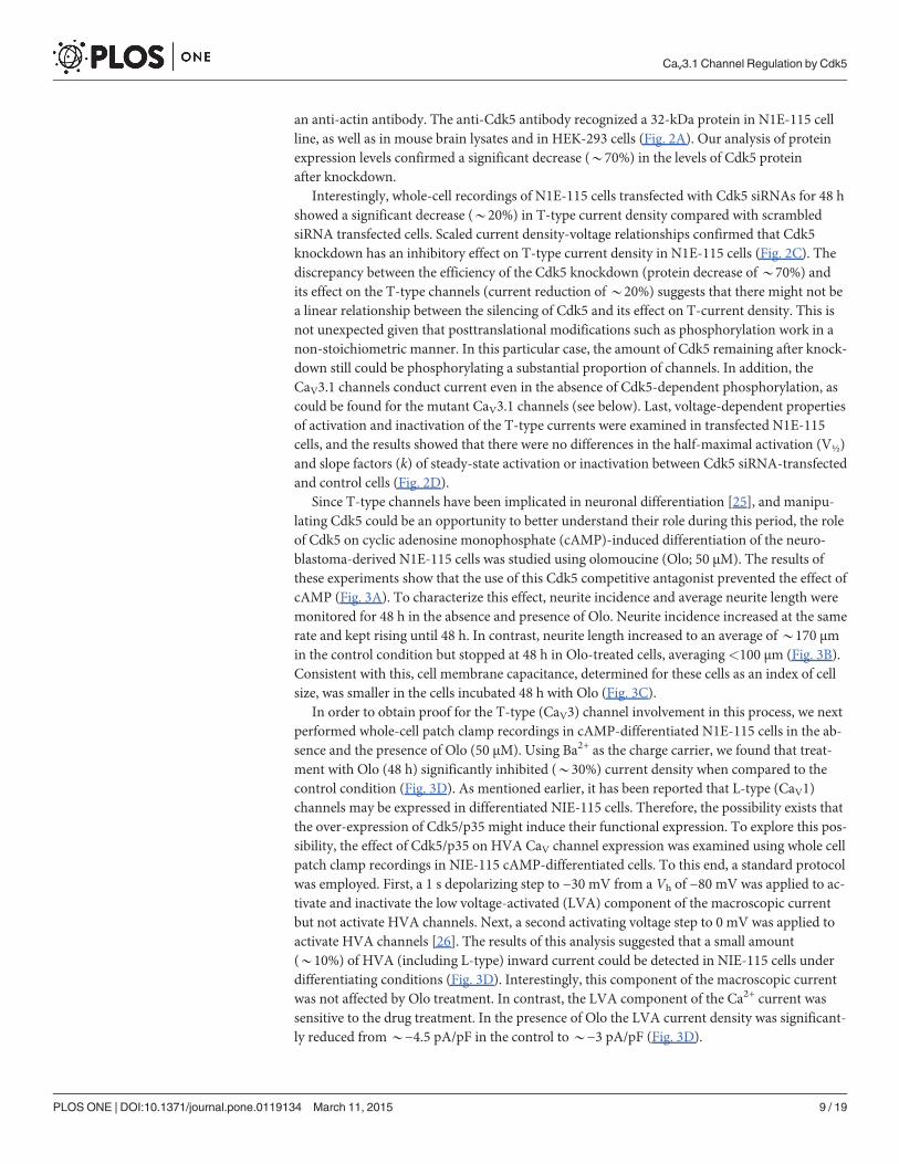
Since T-type channels have been implicated in neuronal differentiation [25], and manipu-lating Cdk5 could be an opportunity to better understand their role during this period, the roleof Cdk5 on cyclic adenosine monophosphate (cAMP)-induced differentiation of the neuro-blastoma-derived N1E-115 cells was studied using olomoucine (Olo; 50 μM). The results ofthese experiments show that the use of this Cdk5 competitive antagonist prevented the effect ofcAMP (Fig. 3A). To characterize this effect, neurite incidence and average neurite length weremonitored for 48 h in the absence and presence of Olo. Neurite incidence increased at the samerate and kept rising until 48 h. In contrast, neurite length increased to an average of*170 μmin the control condition but stopped at 48 h in Olo-treated cells, averaging<100 μm (Fig. 3B).Consistent with this, cell membrane capacitance, determined for these cells as an index of cellsize, was smaller in the cells incubated 48 h with Olo (Fig. 3C).
In order to obtain proof for the T-type (CaV3) channel involvement in this process, we nextperformed whole-cell patch clamp recordings in cAMP-differentiated N1E-115 cells in the ab-sence and the presence of Olo (50 μM). Using Ba2+ as the charge carrier, we found that treat-ment with Olo (48 h) significantly inhibited (*30%) current density when compared to thecontrol condition (Fig. 3D). As mentioned earlier, it has been reported that L-type (CaV1)channels may be expressed in differentiated NIE-115 cells. Therefore, the possibility exists thatthe over-expression of Cdk5/p35 might induce their functional expression. To explore this pos-sibility, the effect of Cdk5/p35 on HVA CaV channel expression was examined using whole cellpatch clamp recordings in NIE-115 cAMP-differentiated cells. To this end, a standard protocolwas employed. First, a 1 s depolarizing step to −30 mV from a Vh of −80 mV was applied to ac-tivate and inactivate the low voltage-activated (LVA) component of the macroscopic currentbut not activate HVA channels. Next, a second activating voltage step to 0 mV was applied toactivate HVA channels [26]. The results of this analysis suggested that a small amount(*10%) of HVA (including L-type) inward current could be detected in NIE-115 cells underdifferentiating conditions (Fig. 3D). Interestingly, this component of the macroscopic currentwas not affected by Olo treatment. In contrast, the LVA component of the Ca2+ current wassensitive to the drug treatment. In the presence of Olo the LVA current density was significant-ly reduced from*−4.5 pA/pF in the control to*−3 pA/pF (Fig. 3D).
Cav3.1 Channel Regulation by Cdk5
PLOS ONE | DOI:10.1371/journal.pone.0119134 March 11, 2015 9 / 19
It should be noted here, that the Cdk5 inhibitor roscovitine (Ro) has also shown to affectCaV3.1 channel activity. Specifically, the drug seems to inhibit these channels in part by stabi-lizing the closed-inactivated state [27]. In addition, it has been reported that Ro affects CaV2.2(N-type) current kinetics [28]. However, this effect appears to be specific for Ro since it is notpresent after Olo treatment [28], which points to distinct mechanisms of action. The
Fig 3. Cdk5 inhibits T-type Ca2+ channel functional expression and affect cAMP-mediated N1E-115 cell differentiation. A) Inhibition of neuriteoutgrowth by the specific Cdk5 inhibitor olomoucine (Olo) in N1E-115 differentiated with cyclic adenosine monophosphate (cAMP, 2 mM) for 48 h. Phasecontrast micrographs of cells grown in the absence or presence of Olo (50 μM). B) Comparison of neurite outgrowth from N1E-115 cells kept in culture in theabsence (control) and presence of Olo. Neurite analysis was carried out with ImageJ software (NIH). C) Comparison of theCm values in cAMP-differentiatedN1E-115 cells kept in culture in the presence or the absence of Olo. D) Representative superimposed trace currents recorded in response to 1 s depolarizingpulses to −30 mV from a Vh of −80 mV (to evoke LVA channel activity), and to +10 mV at the end of the 1 s LVA current inactivating pulses (to evoke the HVAcomponent of the current) in cAMP-differentiated N1E-115 cells in the presence or the absence of Olo (left panel). Comparison of the percentage of peakcurrent densities through HVA and LVA channels (right panel). Data are given as mean ± S.E.M. E) Comparison of the time constant of current andinactivation (τinact) at −30 mV in cAMP-differentiated N1E-115 cells in the presence or the absence of Olo as in D.
doi:10.1371/journal.pone.0119134.g003
Cav3.1 Channel Regulation by Cdk5
PLOS ONE | DOI:10.1371/journal.pone.0119134 March 11, 2015 10 / 19
differential effects of these two closely related Cdk5 inhibitors may also be true for the CaV3.1channels, where we found that current inactivation kinetics in control cells and cells treatedwith Olo did not differ significantly (Fig. 3E).
Although it is likely that the current recorded in N1E-115 cells may be mediated by CaV3.1,CaV3.2, or both channels, in a previous report we showed that the transcription factor Sp1 canregulate CaV3.1 promoter activity and that siRNA-mediated Sp1 silencing significantly de-creased the level of CaV3.1 protein and reduced the amplitude of whole-cell T-type currents ex-pressed in the N1E-115 cells [29]. These results indicated that CaV3.1 channels greatlycontribute to determine Ca2+ macroscopic currents in these cells.
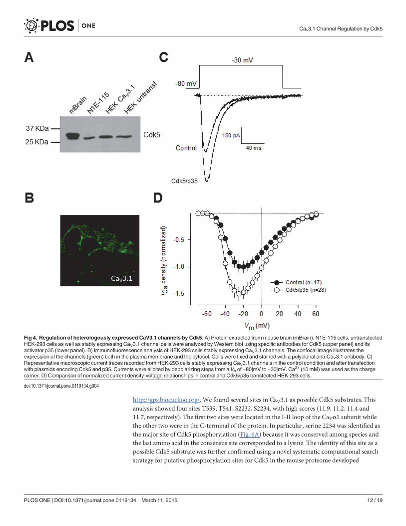
Consequently, we next investigated the functional significance of Cdk5-mediated phosphor-ylation on whole-cell currents recorded in HEK-293 cells stably expressing CaV3.1 channelsand transiently transfected with the cDNAs encoding for Cdk5 and p35. However, before ex-ploring this point, in an initial series of experiments, cell lysates from mouse brain, N1E-115,and HE-293 cells were subjected to Western blot analysis using anti-Cdk5 and anti-p35 to de-tect the expression of endogenous Cdk5 and p35 proteins. The results of these experiments re-vealed bands corroborating the expression of endogenous Cdk5 (Fig. 4A) and p35 in allsamples analyzed. However, given that the expression of p35 has not been detected previouslyin the HEK-293 cell line [30],[31], we decided to verify its expression at the level of mRNA inRT-PCR experiments using the same set of specific oligonucleotides as in Fig. 1A. Unexpected-ly, our results showed no specific p35 mRNA amplification in HEK-293 cells. Although thereare some possible explanations for the discrepancy between the data obtained by Western blotand RT-PCR, the actual reasons for these conflicting results remain presently unknown. How-ever, given that in all experiments examining the effect of Cdk5 phosphorylation on CaV3.1channels performed in HEK-293 cells, p35 was co-transfected with the kinase and the channels,whether or not p35 is endogenously expressed in this cell line does not affect the results of thisstudy. Additional studies are needed to unambiguously demonstrate the expression of p35 inthe HEK-293 cell line.
We further confirmed the expression of CaV3.1 in the surface of transfected HEK-293 cellsby immunofluorescence. As indicated by the green fluorescence signal in Fig. 4B, we foundthat the CaV3.1 channels show a distribution pattern consistent with predominant plasmamembrane expression, though there was also signal associated to cytoplasmic organelles. Like-wise, representative current traces elicited near the half-maximal (-30 mV) channel activationare shown in Fig. 4C. Remarkably, a*1.5-fold increase in current density was observed incells transfected with Cdk5/p35 for 48 h (n = 28) in comparison with control cells. Scaled cur-rent density-voltage relationships confirmed that co-expression of the Cdk5/p35 complex has astimulatory effect on current density in these cells (Fig. 4D).
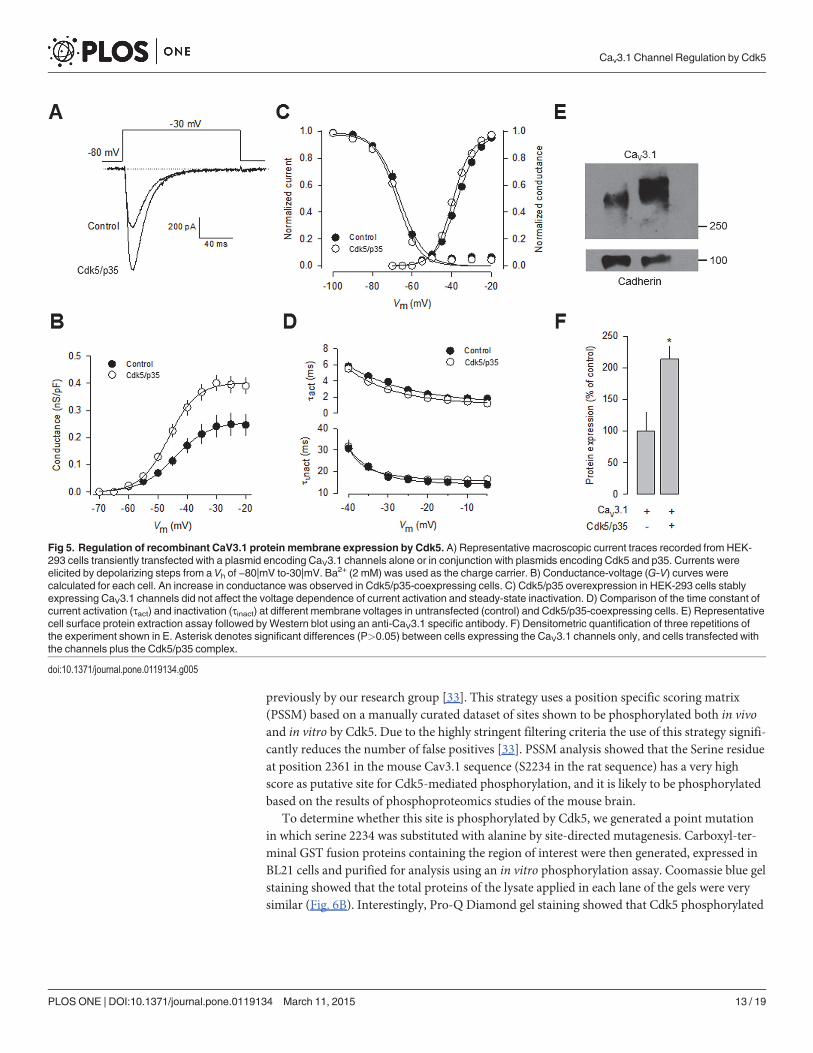
In contrast to the significant effect of Cdk5/p35 on current density and in conductance(Fig. 5A-B), the voltage dependence of channel activation and inactivation was not significantlyaltered (Fig. 5C). In addition, the time constants for current activation and inactivation werenot also significantly modified (Fig. 5D). These data are consistent with the results from the re-cordings obtained in N1E-115 cells (Figs. 1C and 3E). With a view to gaining further insightinto the mechanisms by which Cdk5 is affecting CaV3.1 channel currents, we examined wheth-er the surface expression of the CaV3.1 channel protein was altered. To this end, we measuredthe expression of the channels in plasma membrane protein extracts. By using the membrane-bound adhesion molecule E-cadherin as a control, we found a significant increase in CaV3.1subunit in HEK-293 cells transiently co-transfected with Cdk5/p35 compared with the control(Fig. 5E-F).
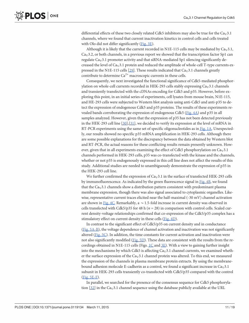
In parallel, we searched for the presence of the consensus sequence for Cdk5 phosphoryla-tion [32] in the CaV3.1 channel sequence using the database publicly available at the URL
Cav3.1 Channel Regulation by Cdk5
PLOS ONE | DOI:10.1371/journal.pone.0119134 March 11, 2015 11 / 19
http://gps.biocuckoo.org/. We found several sites in CaV3.1 as possible Cdk5 substrates. Thisanalysis showed four sites T539, T541, S2232, S2234, with high scores (11.9, 11.2, 11.4 and11.7, respectively). The first two sites were located in the I-II loop of the CaVα1 subunit whilethe other two were in the C-terminal of the protein. In particular, serine 2234 was identified asthe major site of Cdk5 phosphorylation (Fig. 6A) because it was conserved among species andthe last amino acid in the consensus site corresponded to a lysine. The identity of this site as apossible Cdk5 substrate was further confirmed using a novel systematic computational searchstrategy for putative phosphorylation sites for Cdk5 in the mouse proteome developed
Fig 4. Regulation of heterologously expressed CaV3.1 channels by Cdk5. A) Protein extracted frommouse brain (mBrain), N1E-115 cells, untransfectedHEK-293 cells as well as stably expressing CaV3.1 channel cells were analyzed byWestern blot using specific antibodies for Cdk5 (upper panel) and itsactivator p35 (lower panel). B) Immunofluorescence analysis of HEK-293 cells stably expressing CaV3.1 channels. The confocal image illustrates theexpression of the channels (green) both in the plasmamembrane and the cytosol. Cells were fixed and stained with a polyclonal anti-CaV3.1 antibody. C)Representative macroscopic current traces recorded from HEK-293 cells stably expressing CaV3.1 channels in the control condition and after transfectionwith plasmids encoding Cdk5 and p35. Currents were elicited by depolarizing steps from a Vh of −80|mV to −30|mV. Ca2+ (10 mM) was used as the chargecarrier. D) Comparison of normalized current density-voltage relationships in control and Cdk5/p35 transfected HEK-293 cells.
doi:10.1371/journal.pone.0119134.g004
Cav3.1 Channel Regulation by Cdk5
PLOS ONE | DOI:10.1371/journal.pone.0119134 March 11, 2015 12 / 19
previously by our research group [33]. This strategy uses a position specific scoring matrix(PSSM) based on a manually curated dataset of sites shown to be phosphorylated both in vivoand in vitro by Cdk5. Due to the highly stringent filtering criteria the use of this strategy signifi-cantly reduces the number of false positives [33]. PSSM analysis showed that the Serine residueat position 2361 in the mouse Cav3.1 sequence (S2234 in the rat sequence) has a very highscore as putative site for Cdk5-mediated phosphorylation, and it is likely to be phosphorylatedbased on the results of phosphoproteomics studies of the mouse brain.
To determine whether this site is phosphorylated by Cdk5, we generated a point mutationin which serine 2234 was substituted with alanine by site-directed mutagenesis. Carboxyl-ter-minal GST fusion proteins containing the region of interest were then generated, expressed inBL21 cells and purified for analysis using an in vitro phosphorylation assay. Coomassie blue gelstaining showed that the total proteins of the lysate applied in each lane of the gels were verysimilar (Fig. 6B). Interestingly, Pro-Q Diamond gel staining showed that Cdk5 phosphorylated
Fig 5. Regulation of recombinant CaV3.1 protein membrane expression by Cdk5. A) Representative macroscopic current traces recorded from HEK-293 cells transiently transfected with a plasmid encoding CaV3.1 channels alone or in conjunction with plasmids encoding Cdk5 and p35. Currents wereelicited by depolarizing steps from a Vh of −80|mV to-30|mV. Ba2+ (2 mM) was used as the charge carrier. B) Conductance-voltage (G-V) curves werecalculated for each cell. An increase in conductance was observed in Cdk5/p35-coexpressing cells. C) Cdk5/p35 overexpression in HEK-293 cells stablyexpressing CaV3.1 channels did not affect the voltage dependence of current activation and steady-state inactivation. D) Comparison of the time constant ofcurrent activation (τact) and inactivation (τinact) at different membrane voltages in untransfected (control) and Cdk5/p35-coexpressing cells. E) Representativecell surface protein extraction assay followed byWestern blot using an anti-CaV3.1 specific antibody. F) Densitometric quantification of three repetitions ofthe experiment shown in E. Asterisk denotes significant differences (P>0.05) between cells expressing the CaV3.1 channels only, and cells transfected withthe channels plus the Cdk5/p35 complex.
doi:10.1371/journal.pone.0119134.g005
Cav3.1 Channel Regulation by Cdk5
PLOS ONE | DOI:10.1371/journal.pone.0119134 March 11, 2015 13 / 19
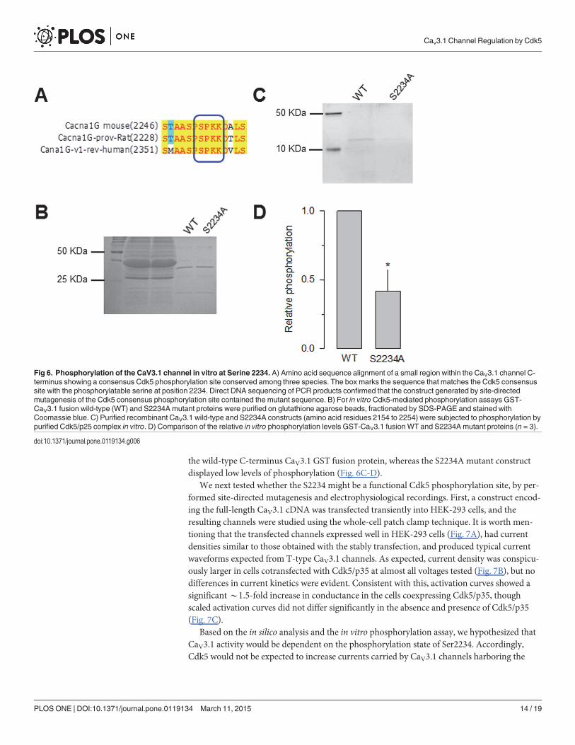
the wild-type C-terminus CaV3.1 GST fusion protein, whereas the S2234A mutant constructdisplayed low levels of phosphorylation (Fig. 6C-D).
We next tested whether the S2234 might be a functional Cdk5 phosphorylation site, by per-formed site-directed mutagenesis and electrophysiological recordings. First, a construct encod-ing the full-length CaV3.1 cDNA was transfected transiently into HEK-293 cells, and theresulting channels were studied using the whole-cell patch clamp technique. It is worth men-tioning that the transfected channels expressed well in HEK-293 cells (Fig. 7A), had currentdensities similar to those obtained with the stably transfection, and produced typical currentwaveforms expected from T-type CaV3.1 channels. As expected, current density was conspicu-ously larger in cells cotransfected with Cdk5/p35 at almost all voltages tested (Fig. 7B), but nodifferences in current kinetics were evident. Consistent with this, activation curves showed asignificant*1.5-fold increase in conductance in the cells coexpressing Cdk5/p35, thoughscaled activation curves did not differ significantly in the absence and presence of Cdk5/p35(Fig. 7C).
Based on the in silico analysis and the in vitro phosphorylation assay, we hypothesized thatCaV3.1 activity would be dependent on the phosphorylation state of Ser2234. Accordingly,Cdk5 would not be expected to increase currents carried by CaV3.1 channels harboring the
Fig 6. Phosphorylation of the CaV3.1 channel in vitro at Serine 2234. A) Amino acid sequence alignment of a small region within the CaV3.1 channel C-terminus showing a consensus Cdk5 phosphorylation site conserved among three species. The box marks the sequence that matches the Cdk5 consensussite with the phosphorylatable serine at position 2234. Direct DNA sequencing of PCR products confirmed that the construct generated by site-directedmutagenesis of the Cdk5 consensus phosphorylation site contained the mutant sequence. B) For in vitro Cdk5-mediated phosphorylation assays GST-CaV3.1 fusion wild-type (WT) and S2234Amutant proteins were purified on glutathione agarose beads, fractionated by SDS-PAGE and stained withCoomassie blue. C) Purified recombinant CaV3.1 wild-type and S2234A constructs (amino acid residues 2154 to 2254) were subjected to phosphorylation bypurified Cdk5/p25 complex in vitro. D) Comparison of the relative in vitro phosphorylation levels GST-CaV3.1 fusionWT and S2234Amutant proteins (n = 3).
doi:10.1371/journal.pone.0119134.g006
Cav3.1 Channel Regulation by Cdk5
PLOS ONE | DOI:10.1371/journal.pone.0119134 March 11, 2015 14 / 19
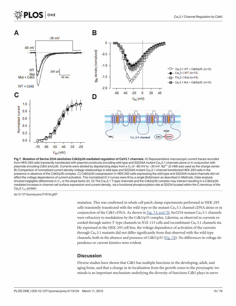
mutation. This was confirmed in whole cell patch clamp experiments performed in HEK-293cells transiently transfected with the wild-type or the mutant CaV3.1 channel cDNA alone or inconjunction of the Cdk5 cDNA. As shown in Fig. 7A and 7B, Ser2234 mutant CaV3.1 channelswere refractory to modulation by the Cdk5/p35 complex. Likewise, as observed in currents re-corded through native T-type channels in N1E-115 cells and recombinant CaV3.1 channels sta-bly expressed in the HEK-293 cell line, the voltage dependence of activation of the currentsthrough CaV3.1 mutants did not differ significantly from that observed with the wild typechannels, both in the absence and presence of Cdk5/p35 (Fig. 7B). No differences in voltage de-pendence or current kinetics were evident.
DiscussionDiverse studies have shown that Cdk5 has multiple functions in the developing, adult, andaging brain, and that a change in its localization from the growth cones to the presynaptic ter-minals is an important mechanism underlying the diversity of functions Cdk5 plays in nerve
Fig 7. Mutation of Serine 2234 abolishes Cdk5/p35-mediated regulation of CaV3.1 channels. A) Representative macroscopic current traces recordedfrom HEK-293 cells transiently transfected with plasmid constructs encoding wild-type and S2234Amutant CaV3.1 channels alone or in conjunction withplasmids encoding Cdk5 and p35. Currents were elicited by depolarizing steps from a Vh of −80 mV to −30 mV. Ba2+ (2 mM) was used as the charge carrier.B) Comparison of normalized current density-voltage relationships in wild-type and S2234Amutant CaV3.1 channel transfected HEK-293 cells in thepresence or absence of the Cdk5/p35 complex. C) Cdk5/p35 coexpression in HEK-293 cells expressing the wild-type and S2234Amutant channels did notaffect the voltage dependence of current activation. The normalizedG-V curves were fit by a single Boltzmann as described in Methods. Data analysisshowed negligible differences in V½ or the slope factor (k). D) The CaV3.1 T-type channels and the Cdk5/p35 complex may interact resulting in a Cdk5/p35-mediated increase in channel cell surface expression and current density, via a functional phosphorylation site at S2234 located within the C-terminus of theCaV3.1α1 protein.
doi:10.1371/journal.pone.0119134.g007
Cav3.1 Channel Regulation by Cdk5
PLOS ONE | DOI:10.1371/journal.pone.0119134 March 11, 2015 15 / 19
cells [34],[35],[36]. However, little is known concerning the regulatory effects of this kinase onCa2+ channels, essential regulators of neuronal activity.
Previous reports have shown that Cdk5 inhibitors favor neurotransmitter release by increas-ing the activity of P/Q-type channels. In this regard, initial work by Tomizawa and colleaguesshowed that Cdk5 may phosphorylate the II-III loop in the pore-forming (CaV2.1α1) subunit,inhibiting the interaction of the channels with SNARE proteins such as SNAP-25 and synapto-tagmin [12]. These results suggested that Cdk5 plays a key role in neurotransmitter release atthe presynaptic terminals of the adult brain.
More recently, Su and colleagues showed that the N-type (CaV2.2) channel, the other majorpresynaptic Ca2+ channel, is also a substrate of Cdk5. Phosphorylation of the pore-formingCaV2.2α 1 subunit by Cdk5 increased Ca
2+ influx and facilitated neurotransmitter release byenhancing channel open probability (Po). These events seemed to be mediated by an interac-tion between CaV2.2α1 and RIM1, which controls vesicle docking at the active zones. These re-sults outlined a mechanism by which Cdk5 may regulate N-type channels and consequentlymight determine presynaptic function [37]. Likewise, these studies corroborated the pivotalrole for Cdk5-mediated phosphorylation of CaV channels in regulating presynaptic function,and highlighted the close interaction between kinases and these channels in neurons.
Here, studying native T-type channels expressed in N1E-115 cells, as well as recombinantchannels transiently and stably expressed in HEK-293 cells, we show that CaV3.1 channels,which play a crucial role in determining neuronal excitability, are also regulated by Cdk5-medi-ated phosphorylation. The functional relevance of the Cdk5-mediated phosphorylation onCaV3.1 channel activity was firstly assessed by whole-cell recordings in Cdk5 transfected N1E-115 cells. Remarkably, following coexpression of Cdk5 peak current amplitude and currentdensity were significantly increased compared to control (GFP transfected) cells. Likewise, in acell line stably expressing CaV3.1, Cdk5 transfection also increased current density, providingindependent support for the notion that the increase in native neuronal T-type current densityobserved in N1E-115 cells may be mediated by Cdk5-mediated phosphorylation. In addition,we conducted whole-cell recordings in HEK-293 cells from transiently transfected with eitherthe wild-type or a phosphorylation mutant of CaV3.1 channels, in which a Cdk5 phosphoryla-tion site (S2234A) in the C-terminal region was abolished. In these experiments, we found thatthe phosphorylation mutant expressed a current density profile similar to that of wild-typeCaV3.1; however, cells in the presence of Cdk5 did not display an increase in current density.
Distinct mechanisms may underlie the increase in CaV3.1 current density following Cdk5-mediated phosphorylation. Previous studies have shown a role for diverse post-translationalmodifications in the regulation of T-type channels and enhanced Ca2+ influx due to increasedchannel surface expression [38],[39],[40]. Indeed, this could be the most plausible explanationfor our results, considering particularly that there were no significant differences in activationkinetics or voltage dependence of activation and inactivation between currents through CaV3.1channels in the presence or absence of Cdk5. In this regard, the role of Cdk5 in channel for-ward trafficking or in endocytosis is an interesting issue that remains unexplored.
In addition, it is worth mentioning that Cdk5-mediated phosphorylation increases Po ofCaV2.2 (N-type) channels, and that this regulation is abolished in a mutant channel in whichall Cdk5 phosphorylation sites in the CaV2.2α1 protein were eliminated [37]. Furthermore, ithas been shown that protein kinase C (PKC) can modulate Cav3 channels activity but pharma-cological and fluorescence studies revealed that the surface density of Cav3.1 T-type channelswas not significantly changed by activation of PKC, suggesting that CaV3.1 channel Po mightbe affected by phosphorylation [41]. It would be of great interest, therefore, to investigatewhether phosphorylation of the CaV3.1 channel by Cdk5 also involves changes in channel Po.
Cav3.1 Channel Regulation by Cdk5
PLOS ONE | DOI:10.1371/journal.pone.0119134 March 11, 2015 16 / 19
The physiological relevance of phosphorylation of native T-type channels and their contri-bution to the function of the cells expressing these channels has not been totally understood. Itis well recognized that CaV channels modulate the function of different neuronal types by influ-encing synaptic transmission and neuronal excitability. While HVA CaV channels participatein fast synaptic transmission, T-type channels have a unique function in neuronal excitability[2]. Neuronal T-type channels have been shown to promote burst firing and low-amplitude in-trinsic neuronal oscillations, as well as Ca2+ entry and amplification of dendritic synaptic sig-nals. Interestingly, evidence obtained in the last few years show that protein kinase activity mayimpact T-type channel function in different manners [42]. In this scenario, Cdk5 can pro-foundly affect cellular excitability by increasing the amplitude of the T-type currents.
Last, as mentioned earlier, Cdk5 is a serine/threonine kinase that is activated upon associa-tion with its activators p35 and p39 (non-cyclin proteins). It is predominantly neuronal,though Cdk5 can be active also in several non-neuronal tissues. Cdk5 plays a pivotal role in theCNS development, and Cdk5 dysregulation has been implicated in different pathologies in-cluding Alzheimer's and Parkinson's disease [43]. Interestingly, it has been reported thatnerve-growth factor (NGF) induces strong, sustained expression of p35 through activation ofthe ERK pathway [44]. The induced kinase activity of Cdk5 may be required for NGF-inducedneurite outgrowth. These results suggest the possibility of Cdk5 activation directly via theNGF receptor expressed heterologously in conjunction with recombinant CaV3.1 channelswhile performing electrophysiological recordings. This is a very interesting topic for futureexperiments.
In summary, here we demonstrated a previously uncharacterized interaction betweenCaV3.1 and the Cdk5/p35 complex that results in a Cdk5/p35-mediated increase in CaV3.1 cur-rent density, and identified a major phosphorylation site at serine 2234 within the C-terminusof the CaV3.1α1 pore forming subunit (Fig. 7D). These findings provide a framework to exam-ine how CaV3.1 channels are regulated in the context of Cdk5 physiological activity.
AcknowledgmentsACR was supported by fellowships from Conacyt and DGAPA-UNAM (Mexico). The excel-lent technical assistance of Mercedes Urbán is also gratefully acknowledged. The authorswould like to thank Dr. Antony A. Boucard (Cinvestav) for kindly revising the style and En-glish grammar, as well as the three anonymous reviewers who gave us helpful feedback to im-prove our earlier version of the manuscript.
Author ContributionsConceived and designed the experiments: ACR AS RGR CGB RF. Performed the experiments:ACR AS RGR. Analyzed the data: ACR AS RGR CGB RF. Contributed reagents/materials/anal-ysis tools: CGB RF. Wrote the paper: ACR AS RGR CGB RF.
References1. Catterall WA. Voltage-gated calcium channels. Cold Spring Harb Perspect Biol 2011; 3: a003947. doi:
10.1101/cshperspect.a003947 PMID: 21746798
2. Perez-Reyes E. Molecular physiology of low-voltage-activated t-type calcium channels. Physiol Rev2003; 83: 117–161. PMID: 12506128
3. Iftinca MC, Zamponi GW. Regulation of neuronal T-type calcium channels. Trends Pharmacol Sci2009; 30: 32–40. doi: 10.1016/j.tips.2008.10.004 PMID: 19042038
4. Nelson MT, Todorovic SM, Perez-Reyes E. The role of T-type calcium channels in epilepsy and pain.Curr Pharm Des 2006; 12: 2189–2197. PMID: 16787249
Cav3.1 Channel Regulation by Cdk5
PLOS ONE | DOI:10.1371/journal.pone.0119134 March 11, 2015 17 / 19
5. Zamponi GW, Lory P, Perez-Reyes E. Role of voltage-gated calcium channels in epilepsy. PflugersArch 2010; 460: 395–403. doi: 10.1007/s00424-009-0772-x PMID: 20091047
6. Bourinet E, Altier C, Hildebrand ME, Trang T, Salter MW, Zamponi GW. Calcium-permeable ion chan-nels in pain signaling. Physiol Rev 2014; 94: 81–140. doi: 10.1152/physrev.00023.2013 PMID:24382884
7. François A, Laffray S, Pizzoccaro A, Eschalier A, Bourinet E. T-type calcium channels in chronic pain:mouse models and specific blockers. Pflugers Arch. 2014; 466: 707–717. doi: 10.1007/s00424-014-1484-4 PMID: 24590509
8. Welsby PJ, Wang H, Wolfe JT, Colbran RJ, Johnson ML, Barrett PQ. A mechanism for the direct regu-lation of T-type calcium channels by Ca2+/calmodulin-dependent kinase II. J Neurosci 2003; 23:10116–10121. PMID: 14602827
9. Kim JA, Park JY, Kang HW, Huh SU, Jeong SW, Lee JH. Augmentation of CaV3.2 T-type calciumchannel activity by cAMP-dependent protein kinase A. J Pharmacol Exp Ther 2006; 318: 230–237.PMID: 16569752
10. Chemin J, Mezghrani A, Bidaud I, Dupasquier S, Marger F, Barrère C, et al. Temperature-dependentmodulation of CaV3 T-type calcium channels by protein kinases C and A in mammalian cells. J BiolChem 2007; 282: 32710–32718. PMID: 17855364
11. Hu C, Depuy SD, Yao J, McIntire WE, Barrett PQ. Protein kinase A activity controls the regulation of T-type CaV3.2 channels by Gβγ dimers. J Biol Chem 2009; 284: 7465–7473. doi: 10.1074/jbc.M808049200 PMID: 19131331
12. Tomizawa K, Ohta J, Matsushita M, Moriwaki A, Li ST, Takei K, et al. Cdk5/p35 regulates neurotrans-mitter release through phosphorylation and downregulation of P/Q-type voltage-dependent calciumchannel activity. J Neurosci. 2002; 22: 2590–2297. PMID: 11923424
13. Su SC, Tsai LH. Cyclin-dependent kinases in brain development and disease. Annu Rev Cell Dev Biol2011; 27: 465–491. doi: 10.1146/annurev-cellbio-092910-154023 PMID: 21740229
14. Jessberger S, Gage FH, Eisch AJ, Lagace DC. Making a neuron: Cdk5 in embryonic and adult neuro-genesis. Trends Neurosci 2009; 32: 575–582. doi: 10.1016/j.tins.2009.07.002 PMID: 19782409
15. Lai KO, Ip NY. Recent advances in understanding the roles of Cdk5 in synaptic plasticity. Biochim Bio-phys Acta 2009; 1792: 741–745. doi: 10.1016/j.bbadis.2009.05.001 PMID: 19442718
16. Cheung ZH, Fu AK, Ip NY. Synaptic roles of Cdk5: implications in higher cognitive functions and neuro-degenerative diseases. Neuron 2006; 50: 13–18. PMID: 16600851
17. Utreras E, Maccioni R, Gonzalez-Billault C. Cyclin-dependent kinase 5 activator p35 over-expressionand amyloid beta synergism increase apoptosis in cultured neuronal cells. Neuroscience 2009; 161:978–987. doi: 10.1016/j.neuroscience.2009.04.002 PMID: 19362124
18. Lee JH, Daud AN, Cribbs LL, Lacerda AE, Pereverzev A, Klöckner U, et al. Cloning and expression of anovel member of the low voltage-activated T-type calcium channel family. J Neurosci 1999; 19: 1912–1921. PMID: 10066244
19. Cribbs LL, Gomora JC, Daud AN, Lee JH, Perez-Reyes E. Molecular cloning and functional expressionof CaV3.1c, a T-type calcium channel from human brain, FEBS Letters 2000; 466: 54–58. PMID:10648811
20. Avila T, Andrade A, Felix R. Transforming growth factor-β1 and bone morphogenetic protein-2 down-regulate CaV3.1 channel expression in mouse C2C12 myoblasts. J Cell Physiol 2006; 209: 448–456.PMID: 16883604
21. Avila G, Sandoval A, Felix R. Intramembrane charge movement associated with endogenous K+ chan-nel activity in HEK-293 cells. Cell Mol Neurobiol 2004; 24: 317–330. PMID: 15206817
22. Narahashi T, Tsunoo A, Yoshii M. Characterization of two types of calcium channels in mouse neuro-blastoma cells. J Physiol 1987; 383: 231–249. PMID: 2443646
23. Yoshii M, Tsunoo A, Narahashi T. Gating and permeation properties of two types of calcium channelsin neuroblastoma cells. Biophys J 1988; 54: 885–895. PMID: 2853978
24. Panner A, Cribbs LL, Zainelli GM, Origitano TC, Singh S, Wurster D. Variation of T-type calcium chan-nel protein expression affects cell division of cultured tumor cells. Cell Calcium 2005; 37: 105–119.PMID: 15589991
25. Lory P, Bidaud I, Chemin J. T-type calcium channels in differentiation and proliferation. Cell Calcium2006; 40: 135–146. PMID: 16797068
26. Gandini MA, Sandoval A, Felix R. Whole-cell patch-clamp recordings of Ca2+ currents from isolatedneonatal mouse dorsal root ganglion (DRG) neurons. Cold Spring Harb Protoc 2014; 4: 389–395. doi:10.1101/pdb.prot073205 PMID: 24692487
Cav3.1 Channel Regulation by Cdk5
PLOS ONE | DOI:10.1371/journal.pone.0119134 March 11, 2015 18 / 19
27. Yarotskyy V, Gao G, Peterson BZ, Elmslie KS. Domain III regulates N-type (CaV2.2) calcium channelclosing kinetics. J Neurophysiol 2012; 107: 1942–1951. doi: 10.1152/jn.00993.2011 PMID: 22205645
28. Buraei Z, Anghelescu M, Elmslie KS. Slowed N-type calcium channel (CaV2.2) deactivation by the cy-clin-dependent kinase inhibitor roscovitine. Biophys J 2005; 89: 1681–1691. PMID: 15951378
29. González-Ramírez R, Martínez-Hernández E, Sandoval A, Felix R. Transcription factor Sp1 regulatesT-type Ca2+ channel CaV3.1 gene expression. J Cell Physiol 2014; 229: 551–560. doi: 10.1002/jcp.24432 PMID: 23868804
30. Kesavapany S, Lau KF, McLoughlin DM, Brownlees J, Ackerley S, Leigh PN, et al. p35/cdk5 binds andphosphorylates β-catenin and regulates β-catenin/presenilin-1 interaction. Eur J Neurosci 2001; 13:241–247. PMID: 11168528
31. Kamei H, Saito T, OzawaM, Fujita Y, Asada A, Bibb JA, et al. Suppression of calpain-dependent cleav-age of the CDK5 activator p35 to p25 by site-specific phosphorylation. J Biol Chem 2007; 282: 1687–1694. PMID: 17121855
32. Songyang Z, Lu KP, Kwon YT, Tsai LH, Filhol O, Cochet C, et al. A structural basis for substrate speci-ficities of protein Ser/Thr kinases: primary sequence preference of casein kinases I and II, NIMA, phos-phorylase kinase, calmodulin-dependent kinase II, CDK5, and Erk1. Mol Cell Biol 1996; 16: 6486–6493. PMID: 8887677
33. Bórquez DA, Olmos C, Álvarez S, Di Genova A, Maass A, González-Billault C. Bioinformatic survey fornew physiological substrates of Cyclin-dependent kinase 5. Genomics. 2013; 101: 221–228. doi: 10.1016/j.ygeno.2013.01.003 PMID: 23384938
34. Nikolic M, Dudek H, Kwon YT, Ramos YF, Tsai LH. The cdk5/p35 kinase is essential for neurite out-growth during neuronal differentiation. Genes Dev 1996; 10: 816–825. PMID: 8846918
35. Chae T, Kwon YT, Bronson R, Dikkes P, Li E, Tsai LH. Mice lacking p35, a neuronal specific activatorof Cdk5, display cortical lamination defects, seizures, and adult lethality. Neuron 1997; 18: 29–42.PMID: 9010203
36. Matsushita M, Tomizawa K, Lu YF, Moriwaki A, Tokuda M, Itano T, et al. Distinct cellular compartmentof cyclin-dependent kinase 5 (Cdk5) and neuron-specific Cdk5 activator protein (p35nck5a) in the de-veloping rat cerebellum. Brain Res 1996; 734: 319–322. PMID: 8896840
37. Su SC, Seo J, Pan JQ, Samuels BA, Rudenko A, Ericsson M, et al. Regulation of N-type voltage-gatedcalcium channels and presynaptic function by cyclin-dependent kinase 5. Neuron 2012; 75: 675–687.doi: 10.1016/j.neuron.2012.06.023 PMID: 22920258
38. Mor M, Beharier O, Levy S, Kahn J, Dror S, Blumenthal D, et al. ZnT-1 enhances the activity and sur-face expression of T-type calcium channels through activation of Ras-ERK signaling. Am J Physiol CellPhysiol 2012; 303: C192–C203. doi: 10.1152/ajpcell.00427.2011 PMID: 22572848
39. Toledo A, Sandoval A, González-Ramírez R, Ávila T, Almanza A, Monjaraz E, et al. Insulin-mediatedupregulation of T-type Ca2+ currents in GH3 cells is mediated by increased endosomal recycling andincorporation of surface membrane Cav3.1 channels. Cell Calcium 2012; 52: 377–387. doi: 10.1016/j.ceca.2012.06.005 PMID: 22770883
40. Weiss N, Black SA, Bladen C, Chen L, Zamponi GW. Surface expression and function of Cav3.2 T-typecalcium channels are controlled by asparagine-linked glycosylation. Pflugers Arch 2013; 465: 1159–1170. doi: 10.1007/s00424-013-1259-3 PMID: 23503728
41. Park JY, Kang HW, Moon HJ, Huh SU, Jeong SW, Soldatov NM, et al. Activation of protein kinase Caugments T-type Ca2+ channel activity without changing channel surface density. J Physiol 2006; 577(Pt 2): 513–523. PMID: 17008378
42. Todorovic SM, Jevtovic-Todorovic V. T-type voltage-gated calcium channels as targets for the develop-ment of novel pain therapies. Br J Pharmacol 2011; 163: 484–495. doi: 10.1111/j.1476-5381.2011.01256.x PMID: 21306582
43. Lopes JP, Agostinho P. Cdk5: multitasking between physiological and pathological conditions. ProgNeurobiol 2011; 94: 49–63. doi: 10.1016/j.pneurobio.2011.03.006 PMID: 21473899
44. Harada T, Morooka T, Ogawa S, Nishida E. ERK induces p35, a neuron-specific activator of Cdk5,through induction of Egr1. Nat Cell Biol 2001; 3: 453–459. PMID: 11331872
Cav3.1 Channel Regulation by Cdk5
PLOS ONE | DOI:10.1371/journal.pone.0119134 March 11, 2015 19 / 19