Embed Size (px)
Citation preview
RESEARCH ARTICLE
Catheter Ablation of Right-Sided AccessoryPathways in Adults Using the Three-Dimensional Mapping System: A RandomizedComparison to the Conventional ApproachYuedong Ma1☯, Jia Qiu1☯, Yang Yang2☯, Anli Tang1*
1 Department of Cardiology, the First Affiliated Hospital of Sun Yat-Sen University, Guangzhou, Guangdong,China, 2 Department of Pathology, the First Affiliated Hospital of Sun Yat-Sen University, Guangzhou,Guangdong, China
☯ These authors contributed equally to this work.* [email protected]
AbstractThree-dimensional (3D) mapping and navigation systems have been widely used for the ab-
lation of atrial fibrillation and ventricular tachycardia, but the applicability of these systems
for the ablation of supraventricular tachycardia (SVT) due to right-sided accessory path-
ways (RAPs) remains unknown. The goal of this prospective randomized study was to com-
pare the safety, efficiency, and efficacy of nonfluoroscopic and conventional fluoroscopic
mapping techniques in guiding catheter ablation of SVT due to RAPs. Of the 393 consecu-
tive patients with SVT who were randomized to receive either conventional fluoroscopic or
Ensite NavX mapping-guided ablation, 64 patients with RAPs were included for analysis.
Endpoints for ablation were no evidence of RAP conduction and no inducible atrioventricu-
lar reentrant tachycardia (AVRT). The 3D group showed fewer ablation pulses and a shorter
total ablation time compared to the conventional group, although the acute procedural suc-
cess did not differ significantly between the two groups. Total procedure time, electrophysio-
logical study time, total fluoroscopy time, and cumulative radiation doses were also
significantly reduced in the 3D group. Patients in the conventional group with a right atrium
diameter (RAD)� 47 mm required a longer fluoroscopy time. There was no significant dif-
ference in the recurrence rates between the two groups over a follow-up period of 3 to 29
months. There were no permanent complications. The 3D mapping systemmay be a pre-
ferred alternative for patients with AVRT due to RAPs, especially for patients with a large
RAD (� 47 mm).
IntroductionRadiofrequency catheter ablation (RFCA) is an effective curative therapy for atrioventricularreentrant tachycardia (AVRT) supported by accessory pathways (APs). Reported success rates
PLOSONE | DOI:10.1371/journal.pone.0128760 June 17, 2015 1 / 12
OPEN ACCESS
Citation: Ma Y, Qiu J, Yang Y, Tang A (2015)Catheter Ablation of Right-Sided AccessoryPathways in Adults Using the Three-DimensionalMapping System: A Randomized Comparison to theConventional Approach. PLoS ONE 10(6): e0128760.doi:10.1371/journal.pone.0128760
Academic Editor: Elena Cavarretta, SapienzaUniversity of Rome, ITALY
Received: January 26, 2015
Accepted: April 28, 2015
Published: June 17, 2015
Copyright: © 2015 Ma et al. This is an open accessarticle distributed under the terms of the CreativeCommons Attribution License, which permitsunrestricted use, distribution, and reproduction in anymedium, provided the original author and source arecredited.
Data Availability Statement: All relevant data arewithin the paper and its Supporting Information files.
Funding: This study was supported by the Sun Yat-Sen University Clinical Research 5010 Program(Grant No. 2007011), the National Natural ScienceFoundation of China (Grant No. 81200173), and theSpecialized Research Fund for the Doctoral Programof Higher Education (Grant No. 20120171120078).The funding provided resources to assist with thecollection, management, analysis and interpretationof the data, and preparation and review of themanuscript. These academic institutions provided
of left-sided AP ablation exceed 92%, and recurrence rates range from approximately 2% to5%. In contrast, patients receiving right-sided accessory pathway (RAP) ablation have shownlower success rates of 67% to 100% and higher recurrence rates of approximately 9% to 16.7%[1].
The challenge of RAPs ablation lies in the unique anatomical characteristics such as the ab-sence of a venous structure paralleling the tricuspid annulus (TA), greater circumference thanthe mitral valve, and the difference in angle with which the valve attaches to the TA [1]. Theseanatomical characteristics lead to difficulty in defining the ablation target and positioning cath-eters. In addition to low success rates, these properties lead to remarkably prolonged exposureto radiation for patients and laboratory staff, which could result in skin injury, radiation-in-duced cancer, and genetic malformations [2–4].
Ensite NavX is a three-dimensional (3D) electronic navigation system that has been success-fully used to track the precise position of the tip of the mapping and ablation catheter tips. Thesystem generates spatially accurate activation maps, enhancing the safety, efficiency, and effica-cy of catheter ablation [5]. It has been widely used to ablate complicated arrhythmias, such asatrial fibrillation and ventricular tachycardia [2], and its safety and efficiency have been investi-gated in the ablation of uncomplicated arrhythmias, including atrioventricular nodal reentranttachycardia (AVNRT) and AVRT [3, 4]. However, little is known about the utility of 3D map-ping systems for specifically ablating AVRT due to RAPs. Although retrospective studies havesuggested that NavX can reduce radiation exposure during supraventricular tachycardia (SVT)ablation in both pediatric and adult patients [1, 6], no direct, prospective, and randomizedstudies have compared 3D mapping system and conventional fluoroscopic mapping to investi-gate potential differences in the efficiency, efficacy, and safety of the two approaches for RAPscatheter ablation. Therefore, this small, prospective, and randomized study sought to comparethe effectiveness of traditional fluoroscopic and 3D approaches for this clinical problem.
MethodsThe protocol for this trial and supporting CONSORT checklist are available as supporting in-formation; see S1 CONSORT Checklist, S1 Protocol (English) and S2 Protocol (Chinese).
This study was conducted according to the Helsinki Declaration and approved by the EthicsCommittee of Sun Yat-Sen University, Guangzhou, China (January 07, 2008) (S1 Ethical Ap-proval Document). The study was registered with the Chinese Clinical Trial Registry (http://www.chictr.org, ChiCTR-TRC-08000268). The authors confirm that all ongoing and relatedtrials for this intervention have been registered. Randomization was performed according to acomputer-generated randomization scheme in blocks of four (S1 Protocol). Assignments wereconcealed in opaque, sealed envelopes that were numbered consecutively.
Study populationThe study included consecutive patients with documented paroxysmal SVT who were admittedto our institutions from June 2011 to November 2013. Thereafter, 393 patients who had nostructural heart disease were randomized to receive either conventional fluoroscopic (conven-tional group) or Ensite NavX (3D group) mapping to guide catheter ablation, which was per-formed by a single, experienced operator using a standard procedure [5]. All antiarrhythmicdrugs were discontinued for at least 5 half-lives before electrophysiological studies (EPS) wereperformed. Written informed consent for the procedure was provided by all patients. Patientswith AVNRT, left-sided APs, atrial ectopic tachycardia, intra-atrial reentry tachycardia orjunctional ectopic tachycardia were excluded. Finally, 64 patients with AVRT due to RAPswere analyzed in this study.
3DMapping System - A Preferred Alternative for the Ablation of RAPs
PLOSONE | DOI:10.1371/journal.pone.0128760 June 17, 2015 2 / 12
funding and oversight of funding, but were not directlyinvolved in collection or cleaning of data, analysis ofresults, or drafting of the manuscript.
Competing Interests: The authors have declaredthat no competing interests exist.
Echocardiographic studyTransthoracic echocardiography was performed on an echocardiograph (Vivid 7, GE Health-care, Milwaukee, MI, USA) equipped with a 2–4 MHz linear transducer with patients in theleft lateral decubitus position before and after the RFCA procedure. The right atrium diameter(RAD) was measured at end systole in the apical four-chamber view. The rest of the procedurewas performed through standard protocols and procedures, according to the guidelines of theAmerican Society of Echocardiography [7]. A post-procedural echocardiogram was performedon all patients to exclude pericardial effusion or other acute complications.
EP procedureEach patient underwent the EP procedure while fasting and without sedation.
Conventional fluoroscopic mapping. Under local anesthesia, venous access was obtainedfrom the femoral, right internal jugular, or subclavian veins to introduce diagnostic electrodecatheters into the high right atrium, right ventricle, bundle of His, and coronary sinus. Ablationcatheter access to the heart was achieved via the femoral vein. Fluoroscopy was used through-out all phases of the procedure, including confirmation of the guidewire position, EPS, map-ping, and ablation.
3D electronic navigation system. Seven skin patches were applied to guide the non-fluo-roscopic Ensite NavX navigation system (version 8.0; St. Jude Medical, St Paul, MN, USA), aspreviously described [5]. The point clouds feature of the Ensite NavX system was used to posi-tion the diagnostic and ablation catheters through the right femoral vein and, if necessary,through the right internal jugular vein and/or left subclavian vein as in the fluoroscopic map-ping procedure [5]. Briefly, the right atrium was reached and confirmed by the presence of atri-al electrograms. The catheter was advanced and pulled back to mark the superior vena cava,followed by the inferior vena cava. After the right ventricle was reached, the catheter was pulledback with clockwise rotation to the right atrioventricular annulus (A and V wave amplitudes atroughly equal levels) and then delivered to the coronary sinus. Other diagnostic (high rightatrium, right ventricle, and His bundle) or ablation catheters were placed with the point cloudstechnique. Thus, a rough sketch of the right atrial geometry was constructed from the placedcatheters. The coronary sinus catheter served as a positional reference for the remainder of theprocedure. An adequate, 3D image of the right atrium was completed by moving the ablationcatheter for several minutes. Technical details of the electroanatomical mapping system(NavX) have been previously described [1, 5–7].
The EP study and ablation were performed in accordance with standard protocols and pro-cedures [5]. Briefly, during sinus rhythm or atrial pacing, NavX showed a breakout of activa-tion around the tricuspid annulus simultaneously with or just before onset of the delta wave.Radiofrequency energy was applied at the breakouts, with power and temperature limits of 40W and 55°C, respectively. Radiofrequency energy was applied via a 7-F, 4-mm electrode-tippedsteerable ablation catheter (Stinger, Bard EP, Lowell, MA, USA). The endpoint of the proce-dure was persistent absence of both retrograde and antegrade pathway conduction. When nec-essary for orienting or confirming the catheter location, fluoroscopy was performed with aradiographic/fluoroscopic unit (Innova 2100, GE Healthcare, Waukesha, WI, USA). The mini-mum fluoroscopy dose compatible with adequate imaging was used.
Procedure and fluoroscopy timesThe preparation time was calculated from the time the patient entered the procedure roomuntil the time of the beginning of needle puncture. The geometry time was measured from theinsertion of the first catheter until the beginning of the EP study. The EP study time was defined
3DMapping System - A Preferred Alternative for the Ablation of RAPs
PLOSONE | DOI:10.1371/journal.pone.0128760 June 17, 2015 3 / 12
as the interval from the beginning of initial premature extrastimuli until the definition of theablation target. The total procedure time was defined as the interval from the placement of thefirst venous sheath until the removal of the last sheath from the patient. The numbers of abla-tion pulses and total ablation time (i.e., total time that ablation energy was on) were recorded.The total fluoroscopy time was defined as the cumulative duration of fluoroscopy during theentire procedure. The cumulative radiation dose was calculated as the total dose received bythe patient.
Acute success and procedural complicationsAcute success was defined as follows: 1) no evidence of RAP conduction, and 2) no relatedAVRT could be induced for more than 30 minutes after the last radiofrequency energy applica-tion, under basal conditions or with intravenous isoprenaline, accompanied by documentationof transient atrioventricular block with adenosine. Complications were defined as permanentsecond- or third-degree atrioventricular block, vascular or cardiac injury, and pericardialeffusion.
Postablation assessment and follow-upA 12-lead electrocardiogram (ECG) was performed for all patients when the patient returnedto the ward from the catheter room. Patients received a physical examination and ECG prior todischarge. Arrhythmia recurrence was documented by ECG and patient diary records. In de-tail, patients were assessed at 1, 6, 12, 18, 24, and 30 months after RFCA by clinical evaluationwith standard ECG and ECG Holter monitoring. An exercise stress test was performed on con-senting patients. Telephone contact was maintained with patients throughout the entire studyto assess the long-term recurrences of symptoms. Recurrence was defined as a relapse of pre-excitation (delta-waves) due to RAPs, ECG-documented tachycardia, or return of clinicalsymptoms that were identical to those before ablation and eventually proven to be AVRT dueto RAPs by the subsequent EP study.
Statistical analysesData are reported as means ± standard deviations (SDs). Continuous variables were comparedby using an independent sample t-test. Pearson’s chi-square (χ2) and Fisher’s exact tests wereused as appropriate to compare categorical values. Correlations between variables were as-sessed with Pearson’s correlation coefficient. Receiver operator characteristic (ROC) curveswere constructed for RAD as a predictor of total fluoroscopy time, and the area under the ROCcurve (AUC) was calculated. The Youden index was used to calculate optimal cutoff points forRAD. A P-value< 0.05 was considered statistically significant. Statistical analyses were per-formed with SPSS 10.0 software (SPSS Inc., Chicago, IL, USA).
Results
Patient characteristicsA total of 393 ablation procedures were performed during the study period (Fig 1). In total, 64patients (40 males, and 24 females; age range: 18 to 52 years; mean age: 36.1 ± 8.1 years) withRAPs met the inclusion criteria and were analyzed in this study, including 31 patients in theconventional group and 33 patients in the 3D group. Distributions of gender, age, weight, leftventricular ejection fraction, RAD, underlying heart disease, substrate location, AVRT type,and AP location were not significantly different between the two groups (Table 1).
3DMapping System - A Preferred Alternative for the Ablation of RAPs
PLOSONE | DOI:10.1371/journal.pone.0128760 June 17, 2015 4 / 12
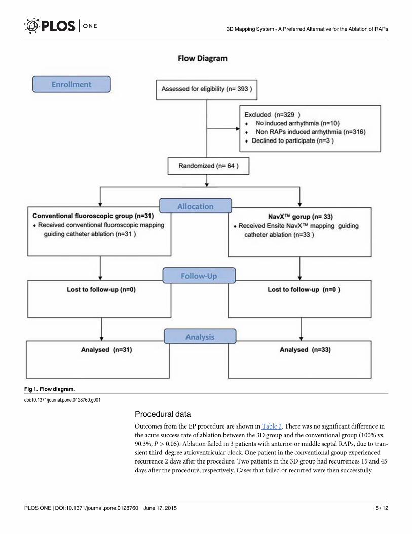
Procedural dataOutcomes from the EP procedure are shown in Table 2. There was no significant difference inthe acute success rate of ablation between the 3D group and the conventional group (100% vs.90.3%, P> 0.05). Ablation failed in 3 patients with anterior or middle septal RAPs, due to tran-sient third-degree atrioventricular block. One patient in the conventional group experiencedrecurrence 2 days after the procedure. Two patients in the 3D group had recurrences 15 and 45days after the procedure, respectively. Cases that failed or recurred were then successfully
Fig 1. Flow diagram.
doi:10.1371/journal.pone.0128760.g001
3DMapping System - A Preferred Alternative for the Ablation of RAPs
PLOSONE | DOI:10.1371/journal.pone.0128760 June 17, 2015 5 / 12
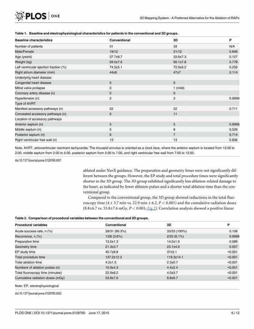
ablated under NavX guidance. The preparation and geometry times were not significantly dif-ferent between the groups. However, the EP study and total procedure times were significantlyshorter in the 3D group. The 3D group exhibited significantly less ablation-related damage tothe heart, as indicated by fewer ablation pulses and a shorter total ablation time than the con-ventional group.
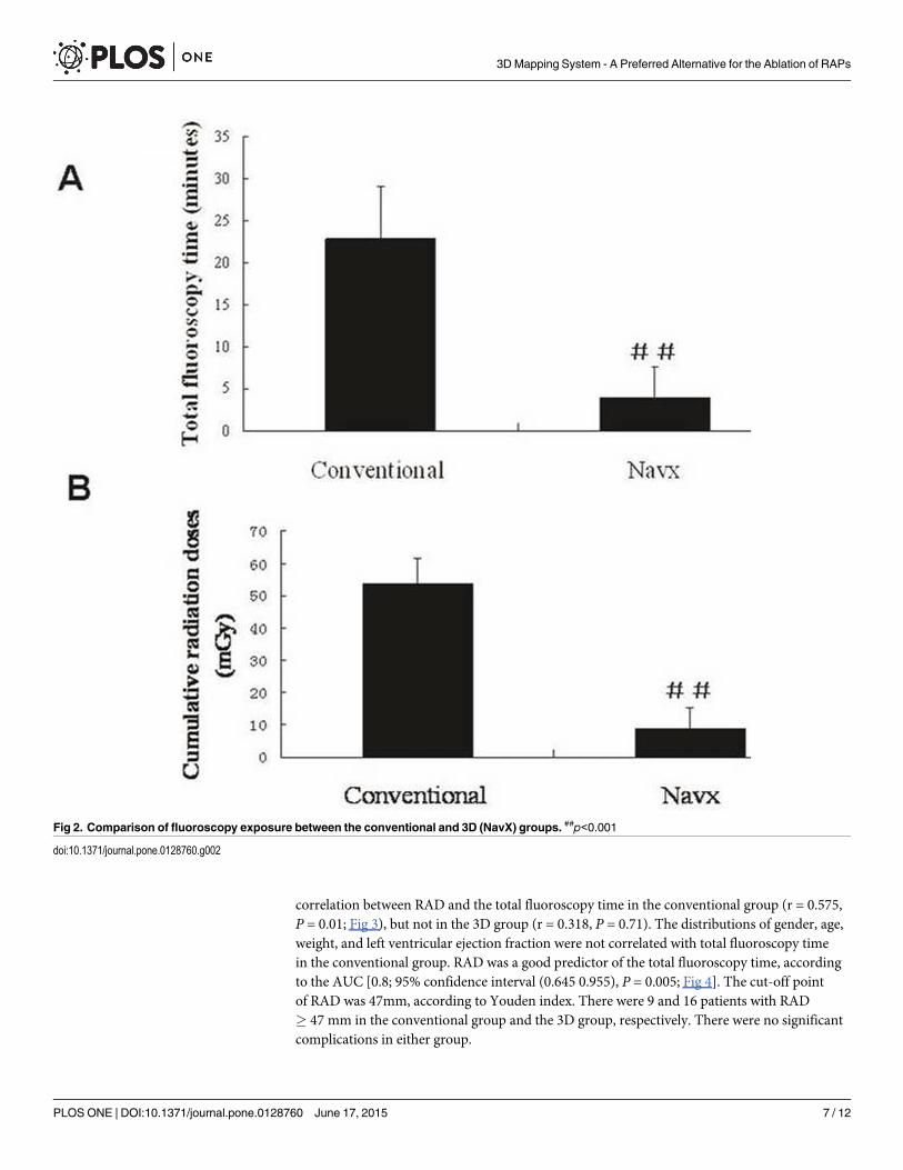
Compared to the conventional group, the 3D group showed reductions in the total fluo-roscopy time (4 ± 3.7 min vs. 22.9 min ± 6.2, P< 0.001) and the cumulative radiation doses(8.8±6.7 vs. 53.8±7.6 mGy, P< 0.001; Fig 2). Correlation analysis showed a positive linear
Table 1. Baseline and electrophysiological characteristics for patients in the conventional and 3D groups.
Baseline characteristics Conventional 3D P
Number of patients 31 33 N/A
Male/Female 19/12 21/12 0.846
Age (years) 37.7±8.7 33.6±7.3 0.127
Weight (kg) 59.5±7.6 60.1±7.8 0.778
Left ventricular ejection fraction (%) 74.5±5.1 72.9±6.2 0.259
Right atrium diameter (mm) 44±6 47±7 0.114
Underlying heart disease
Congenital heart disease 0 0
Mitral valve prolapse 0 1 (mild)
Coronary artery disease (n) 0 0
Hypertension (n) 2 3 0.9999
Type of AVRT
Manifest accessory pathways (n) 22 22 0.711
Concealed accessory pathways (n) 9 11
Location of accessory pathways
Anterior septum (n) 5 5 0.9999
Middle septum (n) 5 8 0.529
Posterior septum (n) 8 7 0.714
Right ventricular free wall (n) 13 13 0.836
Note: AVRT, atrioventricular reentrant tachycardia; The tricuspid annulus is oriented as a clock face, where the anterior septum is located from 12:00 to
2:00, middle septum from 2:00 to 5:00, posterior septum from 5:00 to 7:00, and right ventricular free wall from 7:00 to 12:00.
doi:10.1371/journal.pone.0128760.t001
Table 2. Comparison of procedural variables between the conventional and 3D groups.
Procedural variables Conventional 3D P
Acute success rate, n (%) 28/31 (90.3%) 33/33 (100%) 0.106
Recurrence, n (%) 1/28 (3.6%) 2/33 (6.1%) 0.9999
Preparation time 13.5±1.3 14.2±1.9 0.089
Geometry time 21.3±3.7 23.1±4.0 0.057
EP study time 45.7±8.8 37±5.1 <0.001
Total procedure time 137.2±12.3 119.3±14.1 <0.001
Total ablation time 4.2±1.5 2.3±0.7 <0.001
Numbers of ablation pulses (n) 10.9±4.3 4.4±2.4 <0.001
Total fluoroscopy time (minutes) 22.9±6.2 4.0±3.7 <0.001
Cumulative radiation doses (mGy) 53.8±7.6 8.8±6.7 <0.001
Note: EP, electrophysiological
doi:10.1371/journal.pone.0128760.t002
3DMapping System - A Preferred Alternative for the Ablation of RAPs
PLOSONE | DOI:10.1371/journal.pone.0128760 June 17, 2015 6 / 12
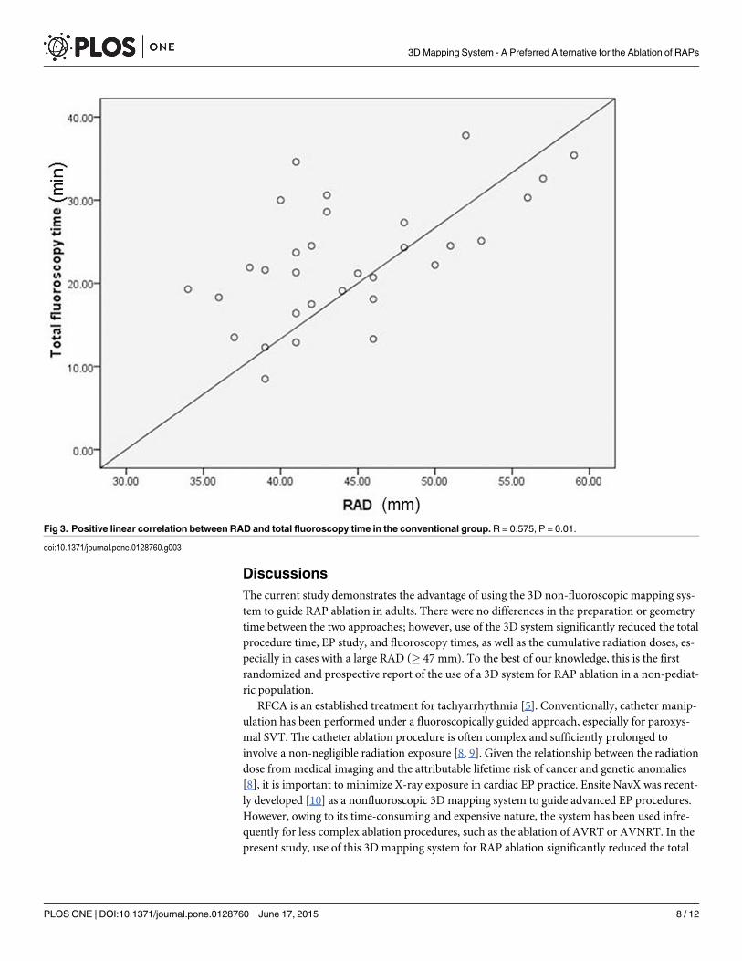
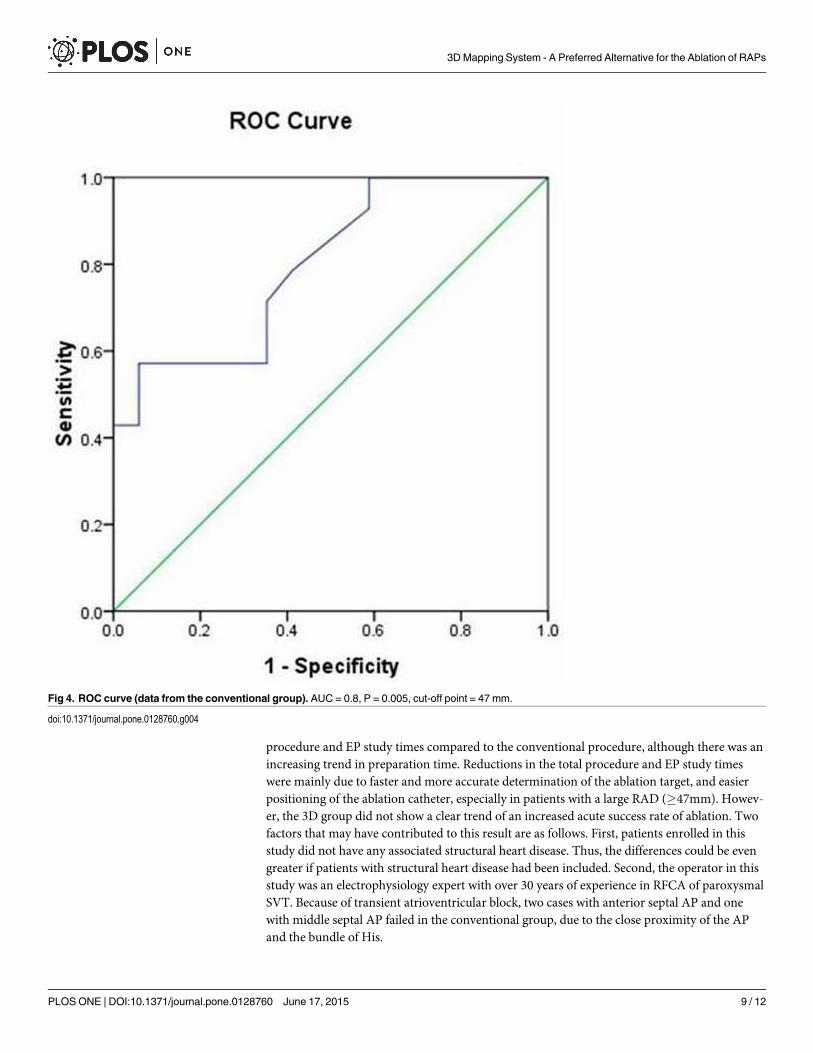
correlation between RAD and the total fluoroscopy time in the conventional group (r = 0.575,P = 0.01; Fig 3), but not in the 3D group (r = 0.318, P = 0.71). The distributions of gender, age,weight, and left ventricular ejection fraction were not correlated with total fluoroscopy timein the conventional group. RAD was a good predictor of the total fluoroscopy time, accordingto the AUC [0.8; 95% confidence interval (0.645 0.955), P = 0.005; Fig 4]. The cut-off pointof RAD was 47mm, according to Youden index. There were 9 and 16 patients with RAD� 47 mm in the conventional group and the 3D group, respectively. There were no significantcomplications in either group.
Fig 2. Comparison of fluoroscopy exposure between the conventional and 3D (NavX) groups. ##p<0.001
doi:10.1371/journal.pone.0128760.g002
3DMapping System - A Preferred Alternative for the Ablation of RAPs
PLOSONE | DOI:10.1371/journal.pone.0128760 June 17, 2015 7 / 12
DiscussionsThe current study demonstrates the advantage of using the 3D non-fluoroscopic mapping sys-tem to guide RAP ablation in adults. There were no differences in the preparation or geometrytime between the two approaches; however, use of the 3D system significantly reduced the totalprocedure time, EP study, and fluoroscopy times, as well as the cumulative radiation doses, es-pecially in cases with a large RAD (� 47 mm). To the best of our knowledge, this is the firstrandomized and prospective report of the use of a 3D system for RAP ablation in a non-pediat-ric population.
RFCA is an established treatment for tachyarrhythmia [5]. Conventionally, catheter manip-ulation has been performed under a fluoroscopically guided approach, especially for paroxys-mal SVT. The catheter ablation procedure is often complex and sufficiently prolonged toinvolve a non-negligible radiation exposure [8, 9]. Given the relationship between the radiationdose from medical imaging and the attributable lifetime risk of cancer and genetic anomalies[8], it is important to minimize X-ray exposure in cardiac EP practice. Ensite NavX was recent-ly developed [10] as a nonfluoroscopic 3D mapping system to guide advanced EP procedures.However, owing to its time-consuming and expensive nature, the system has been used infre-quently for less complex ablation procedures, such as the ablation of AVRT or AVNRT. In thepresent study, use of this 3D mapping system for RAP ablation significantly reduced the total
Fig 3. Positive linear correlation between RAD and total fluoroscopy time in the conventional group. R = 0.575, P = 0.01.
doi:10.1371/journal.pone.0128760.g003
3DMapping System - A Preferred Alternative for the Ablation of RAPs
PLOSONE | DOI:10.1371/journal.pone.0128760 June 17, 2015 8 / 12
procedure and EP study times compared to the conventional procedure, although there was anincreasing trend in preparation time. Reductions in the total procedure and EP study timeswere mainly due to faster and more accurate determination of the ablation target, and easierpositioning of the ablation catheter, especially in patients with a large RAD (�47mm). Howev-er, the 3D group did not show a clear trend of an increased acute success rate of ablation. Twofactors that may have contributed to this result are as follows. First, patients enrolled in thisstudy did not have any associated structural heart disease. Thus, the differences could be evengreater if patients with structural heart disease had been included. Second, the operator in thisstudy was an electrophysiology expert with over 30 years of experience in RFCA of paroxysmalSVT. Because of transient atrioventricular block, two cases with anterior septal AP and onewith middle septal AP failed in the conventional group, due to the close proximity of the APand the bundle of His.
Fig 4. ROC curve (data from the conventional group). AUC = 0.8, P = 0.005, cut-off point = 47 mm.
doi:10.1371/journal.pone.0128760.g004
3DMapping System - A Preferred Alternative for the Ablation of RAPs
PLOSONE | DOI:10.1371/journal.pone.0128760 June 17, 2015 9 / 12
RAPs are thought to be distributed mainly in the free wall and posterior septum, whereasAPs located in anterior and middle septal are rare [11]. In our study, anterior septal RAPs, nearthe bundle of His, were fairly common. The 3D system had more advantages for these patientswith para-His RAPs, since the three failed cases with anterior or middle septal RAPs were thensuccessfully treated guided by 3D system with no subsequent recurrence and complication.
Interestingly, we found that the RAD had a linear correlation with the total fluoroscopytime in the conventional group. The RAD can also be used as a basis for choosing the operationapproach, because it was a good predictor of fluoroscopy time according to the ROC analysisand had an optional cutoff point of 47 mm.
Statistical limitations make it difficult to evaluate the relationship between long-term cancerrisk and low doses of radiation, but the Biological Effects of Ionizing Radiation VII (BEIR VII)of the US National Academies concluded that current evidence supports a “linear-no-thresh-old”model. According to this model, a simple linear relationship exists between cancer riskand radiation dose [5, 10], and there is no threshold dose below which radiation carries no risk.Even low doses of radiation may have appreciable noxious effects, as evidenced by the observa-tion of both acute and long-term DNA damage in circulating lymphocytes of children under-going cardiac catheterization [12]. It also bears particular importance to note that radiationrisks are not distributed homogeneously among the population, as women and younger indi-viduals are at relatively higher risk, due to their greater vulnerability to radiation effects andlonger life expectancy [10, 13, and 14]. Therefore, the greatest benefits of applying the 3Dsystem in RAP ablation are the significant reduction in fluoroscopy time and radiation dose(Fig 2).
In addition to reduced radiation exposure, 3D system-guided RAP ablation has other ad-vantages over the conventional procedure. It allows for elucidation of detailed individual varia-tions in anatomy and electrogram distributions, and has the ability to “tag” important sites,such as catheter locations and lesion sites, which can be revisited with great accuracy. The sys-tem can create “shadows” of the catheters, which can be used for the repositioning of the cathe-ters in the case of dislodgment. Thus, compared to the conventional fluoroscopy-guidedprocedure, 3D system can be used to ablate RAPs more effectively, with fewer radiofrequencyapplications, lower radiofrequency energy, shorter procedure time, and reduced risk of inad-vertent atrioventricular block.
Although 3D mapping costs more than conventional mapping due to the electrodes used,the extra cost is balanced by the added benefits, such as reduction in total procedure time andradiation exposure. Moreover, the cost may go down in the future if electrodes can be recycled.
Study LimitationsThis study involved a relatively small number of subjects. Therefore, the low complication ratecan not be statistically evaluated. A much larger sample size will be necessary to determinewhether or not this approach will have any impact on the complication rates. The follow-upperiod was also short in some cases (range: 3–29 months). However, the follow-up period waslong enough to judge the outcome of ablation because most recurrences reportedly occur with-in 3 months after ablation [15]. In the present study, all recurrences in both groups occurredwithin the first 2 months after ablation.
ConclusionsThis prospective, randomized, single-center study demonstrated that the 3D mapping ap-proach significantly reduced fluoroscopy exposure, although it did not show a significant trendof an increased acute success rate of RAP ablation in an adult population. Cases with anterior
3DMapping System - A Preferred Alternative for the Ablation of RAPs
PLOSONE | DOI:10.1371/journal.pone.0128760 June 17, 2015 10 / 12
or middle septal RAPs are not rare. The 3D mapping system may be more beneficial for thesepatients, especially those with a large RAD (�47 mm).
Supporting InformationS1 CONSORT Checklist.(DOC)
S1 Ethical Approval Document.(PDF)
S1 Protocol. Study protocol in English.(DOCX)
S2 Protocol. Study protocol in Chinese.(DOC)
Author ContributionsConceived and designed the experiments: YDM YY JQ ALT. Performed the experiments:YDM YY JQ ALT. Analyzed the data: YDM YY. Contributed reagents/materials/analysis tools:YDM YY. Wrote the paper: YDM YY JQ ALT. Revised the mauscript and translated the proto-col: YDM JQ ALT.
References1. Nishida T, Nakajima T, Kaitani K, Takitsume A, Soeda T, Okayama S, et al. (2012) Non-contact map-
ping system accurately localizes right-sided accessory pathways in type BWolff-Parkinson-White syn-drome. Europace. 14:752–60. doi: 10.1093/europace/eur369 PMID: 22135318
2. Lickfett L, Mahesh M, Vasamreddy C, Bradley D, Jayam V, Eldadah Z, et al. (2004) Radiation exposureduring catheter ablation of atrial fibrillation. Circulation. 110:3003–10. PMID: 15505084
3. Rosenthal LS, Beck TJ, Williams J, Mahesh M, Herman MG, Dinerman JL, et al. (1997) Acute radiationdermatitis following radiofrequency catheter ablation of atrioventricular nodal reentrant tachycardia.Pacing Clin Electrophysiol. 20:1834–9. PMID: 9249839
4. Nahass GT. (1995) Fluoroscopy and the skin: implications for radiofrequency catheter ablation. Am JCardiol. 76:174–6. PMID: 7611155
5. Casella M, Pelargonio G, Dello Russo A, Riva S, Bartoletti S, Santangeli P, et al. (2011) "Near-zero"fluoroscopic exposure in supraventricular arrhythmia ablation using the EnSite NavX mapping system:personal experience and review of the literature. J Interv Card Electrophysiol. 31:109–18. doi: 10.1007/s10840-011-9553-5 PMID: 21365263
6. Papagiannis J, Tsoutsinos A, Kirvassilis G, Sofianidou I, Koussi T, Laskari C, et al. (2006) Nonfluoro-scopic catheter navigation for radiofrequency catheter ablation of supraventricular tachycardia in chil-dren. Pacing Clin Electrophysiol. 29:971–8. PMID: 16981921
7. Schilling RJ, Peters NS, Davies DW. (1998) Simultaneous endocardial mapping in the human left ven-tricle using a noncontact catheter: comparison of contact and reconstructed electrograms during sinusrhythm. Circulation. 98:887–98. PMID: 9738644
8. Perisinakis K, Damilakis J, Theocharopoulos N, Manios E, Vardas P, Gourtsoyiannis N. (2001) Accu-rate assessment of patient effective radiation dose and associated detriment risk from radiofrequencycatheter ablation procedures. Circulation. 104:58–62. PMID: 11435338
9. Smith IR, Rivers JT, Hayes J, Stafford W, Codd C. (2009) Reassessment of radiation risks from electro-physiology procedures compared to coronary angiography. Heart Lung Circ. 18:191–9. doi: 10.1016/j.hlc.2008.10.006 PMID: 19119073
10. Packer DL. (2005) Three-dimensional mapping in interventional electrophysiology: techniques andtechnology. J Cardiovasc Electrophysiol. 16:1110–6. PMID: 16191123
11. Moorman AF, Christoffels VM, Anderson RH. (2005) Anatomic substrates for cardiac conduction. HeartRhythm. 2:875–86. PMID: 16051128
3DMapping System - A Preferred Alternative for the Ablation of RAPs
PLOSONE | DOI:10.1371/journal.pone.0128760 June 17, 2015 11 / 12
12. Ait-Ali L, Andreassi MG, Foffa I, Spadoni I, Vano E, Picano E. (2010) Cumulative patient effective doseand acute radiation-induced chromosomal DNA damage in children with congenital heart disease.Heart. 96:269–74. doi: 10.1136/hrt.2008.160309 PMID: 19687017
13. Beels L, Bacher K, DeWolf D, Werbrouck J, Thierens H. (2009) gamma-H2AX foci as a biomarker forpatient X-ray exposure in pediatric cardiac catheterization: are we underestimating radiation risks? Cir-culation. 120:1903–9. doi: 10.1161/CIRCULATIONAHA.109.880385 PMID: 19858412
14. Fazel R, Krumholz HM, Wang Y, Ross JS, Chen J, Ting HH, et al. (2009) Exposure to low-dose ionizingradiation frommedical imaging procedures. N Engl J Med. 361:849–57. doi: 10.1056/NEJMoa0901249 PMID: 19710483
15. Calkins H, Yong P, Miller JM, Olshansky B, Carlson M, Saul JP, et al. (1999) Catheter ablation of acces-sory pathways, atrioventricular nodal reentrant tachycardia, and the atrioventricular junction: final re-sults of a prospective, multicenter clinical trial. The Atakr Multicenter Investigators Group. Circulation.99:262–70. PMID: 9892593
3DMapping System - A Preferred Alternative for the Ablation of RAPs
PLOSONE | DOI:10.1371/journal.pone.0128760 June 17, 2015 12 / 12