Embed Size (px)
Citation preview
Cheng et al. Molecular Cancer 2010, 9:126http://www.molecular-cancer.com/content/9/1/126
Open AccessR E S E A R C H
ResearchArecoline induces HA22T/VGH hepatoma cells to undergo anoikis - involvement of STAT3 and RhoA activationHsiao-Ling Cheng1, Shu-Jem Su2, Li-Wen Huang3, Bau-Shan Hsieh1, Yu-Chen Hu1, Thu-Ching Hung1 and Kee-Lung Chang*4
AbstractBackground: Our previous study showed that, in basal cell carcinoma cells, arecoline reduces levels of the tumor cell survival factor interleukin-6 (IL-6), increases levels of tumor suppressor factor p53, and elicits cell cycle arrest, followed by apoptosis. In preliminarily studies, we observed that arecoline induces detachment of the human-derived hepatoma cell line HA22T/VGH from the extracellular matrix. In the present study, we explored the fate of the detached HA22T/VGH cells and investigated the underlying mechanism.
Methods: HA22T/VGH cells or primary cultured rat hepatocytes were treated with arecoline, then changes in morphology, viability, apoptosis, and the expression of surface β1-integrin, apoptosis-related proteins, and IL-6 were examined. Furthermore, activation of the signal transducer and activator of transcription 3 (STAT3) pathway and the RhoA/Rock signaling pathway, including p190RhoGAP and Src homology-2 domain-containing phosphatase SHP2, was examined.
Results: A low concentration of arecoline (≤ 100 μg/ml) caused cytoskeletal changes in HA22T/VGH cells, but not hepatocytes, and this was accompanied by decreased β1-integrin expression and followed by apoptosis, indicating that HA22T/VGH cells undergo anoikis after arecoline treatment. IL-6 expression and phosphorylation of STAT3, which provides protection against anoikis, were inhibited and levels of downstream signaling proteins, including Bcl-XL and Bcl-2, were decreased, while Bax expression, mitochondrial cytochrome c release, and caspase-3 activity were increased. In addition, phosphorylation/activation of p190RhoGAP, a RhoA inhibitor, and of its upstream regulator, SHP2, was inhibited by arecoline treatment, while Rho/Rock activation was increased. Addition of the RhoA inhibitor attenuated the effects of arecoline.
Conclusions: This study demonstrated that arecoline induces anoikis of HA22T/VGH cells involving inhibition of STAT3 and increased RhoA/Rock activation and that the STAT3 and RhoA/Rock signaling pathways are connected.
BackgroundArecoline has been suggested as a possible cognitionenhancer in Alzheimer's type dementia [1,2]. Recentstudies have shown that it decreases interleukin-6 (IL-6)production in keratinocytes and KB cancer cells [3,4]. Inaddition, Chang et al. [3] reported that arecoline elicitscell cycle deregulation in KB cancer cells. Moreover, ourprevious study [Chang et al.: Arecoline decreases inter-
leukin-6 production and induces apoptosis and cell cyclearrest in human basal cell carcinoma cells (BCC/KMC),submitted] showed that, in basal cell carcinoma cells, are-coline reduces levels of the tumor cell survival factor IL-6,increases levels of the tumor suppressor factor p53, andelicits cell cycle arrest, followed by apoptosis, showingthat arecoline interferes with cancer cell cycle progres-sion. Our preliminary data showed that arecoline inducesdetachment of the hepatoma cell line HA22T/VGH fromthe extracellular matrix (ECM).
Adherence of epithelial cells to the ECM is importantfor cell growth and survival and detachment from the
* Correspondence: [email protected] Department of Biochemistry, Faculty of Medicine, College of Medicine, Kaohsiung Medical University, Kaohsiung 80708, TaiwanFull list of author information is available at the end of the article
© 2010 Cheng et al; licensee BioMed Central Ltd. This is an Open Access article distributed under the terms of the Creative CommonsAttribution License (http://creativecommons.org/licenses/by/2.0), which permits unrestricted use, distribution, and reproduction inany medium, provided the original work is properly cited.
Cheng et al. Molecular Cancer 2010, 9:126http://www.molecular-cancer.com/content/9/1/126
Page 2 of 12
ECM induces cell apoptosis, known as anoikis [5,6]. Theexpression of certain oncogenes, such as activation of sig-nal transducer and activator of transcription 3 (STAT3)[7], phosphatidylinositol 3-kinase (PI3K)/Akt [8], and Src[8], provides anchorage-independent growth ability andprotection against anoikis, and this protection is thoughtto be critical during tumorigenesis.
The small GTPase RhoA has emerged as a pivotal con-trol point through which cells sense changes in ECMmechanics and cytoskeletal organization and translatethe 'cell shape signal' to downstream effectors that medi-ate these behaviors [8]. RhoA activity can be suppressedby any one of a variety of different RhoGAP proteins.p190RhoGAP has been shown to be phosphorylated bySrc tyrosine kinase when cells first attach to the ECMsubstrate and integrin receptors become ligated, allowingp190RhoGAP to exert its RhoGAP activity and leading toinactivation of RhoA [9,10]. Cell detachment and round-ing in mitosis have also been reported to inhibitp190RhoGAP activity and increase RhoA activity [11].
Src homology-2 domain-containing phosphatases(SHPs) are a small, highly conserved subfamily of protein-tyrosine phosphatases, members of which are present inboth vertebrates and invertebrates. In most receptortyrosine kinase signaling pathways, SHP2 is required forfull activation [12]. SHP2 has been reported to play anessential role in integrin signaling, and dominant-nega-tive mutants of SHP2 inhibit integrin-stimulated focaladhesion and stress fiber turnover, cell spreading, andproliferation [12].
In the present study, we explored the fate of theHA22T/VGH cells detached by the action of arecolineand investigated the underlying mechanisms of thisdetachment. Cytokine IL-6 expression and activation ofits downstream effector STAT3 and expression and acti-vation of RhoA/Rock, p190RhoGAP, and SHP2 were alsoexamined. Our results showed that arecoline inducesanoikis in HA22T/VGH cells by inhibiting the activationof STAT3, SHP2 and p190RhoGAP and enhancing theactivation of RhoA/Rock.
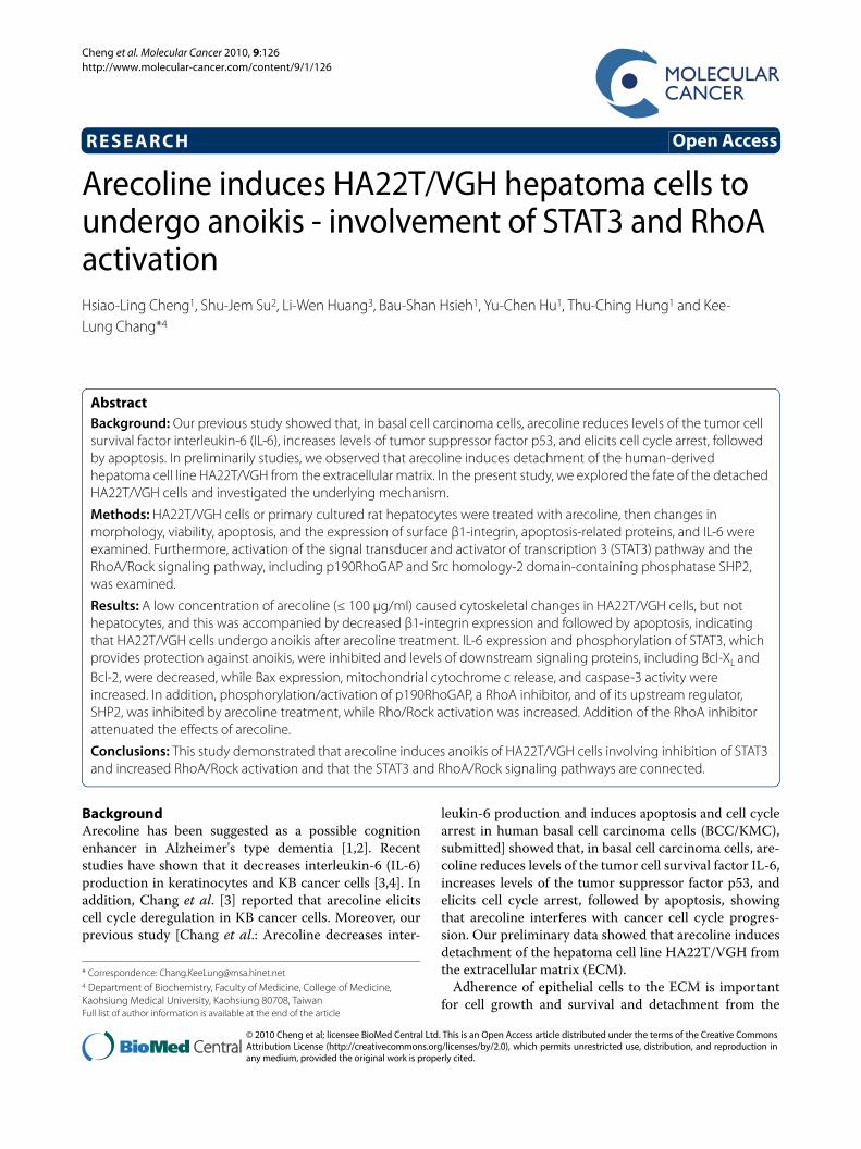
ResultsArecoline induces cell detachment, followed by apoptosisAs in our preliminary study, some HA22T/VGH cellsbecame detached after 24 h of treatment with 30 or 100μg/ml of arecoline, and more became detached after 48 hof treatment (Fig. 1A). This arecoline-induced celldetachment was accompanied by decreased expression ofthe cell surface adhesion molecule β1-integrin (Fig. 1B).To clarify whether this detachment was due to cell cycleprogression, we examined the distribution of cell cyclephases and found there was no difference with or withoutarecoline treatment (data not shown). Having excludedcell cycle progression, we explored the fate of these
detached cells and examined the effects of arecoline onnormal rat hepatocytes. Interestingly, no detachment ofnormal hepatocytes was seen with arecoline treatment.After 72 h of arecoline treatment, the viability of normalhepatocytes was not significantly changed, whereas thatof HA22T/VGH cells decreased in a dose-dependentmanner (Fig. 1C). In addition, DNA fragmentation wasseen in arecoline-treated HA22T/VGH cells and wasrestricted to the detached cells (Fig. 1D). As shown in Fig.1E, more than 90% of the detached cells were positive for
Figure 1 Arecoline induces detachment of HA22T/VGH cells, fol-lowed by apoptosis. (A) HA22T/VGH cells were treated with 0 (left), 30 (center), or 100 (right) μg/ml of arecoline for 24 h or 48 h, then cell mor-phology was observed under a phase-contrast microscopy at 200× magnification. The black arrows indicate detached cells. (B) After treat-ment of arecoline for 24 h, β1-integrin expression was measured by flow cytometric analysis using RPE-conjugated antibody. The histo-gram of red filled area is the untreated control and the black lines the treated groups. The values shown are the mean fluorescence intensity as a percentage of the untreated control value. (C) HA22T/VGH cells or primary normal rat hepatocytes were treated with the indicated con-centration of arecoline for 72 h, then viable cells were counted using Trypan blue and the results expressed as a percentage of the untreated control value. (D) After treatment of arecoline for 72 h, the cells were harvested all together or the detached and adherent cells separately and genomic DNA extracted and analyzed on an agarose gel for DNA fragmentation. The left panel shows the total cells and the right panel adherent and detached cells separately. (E) TUNEL staining of the de-tached cells detected by flow cytometric analysis. The green filled area is the untreated control and the black lines the treated groups. The val-ues shown are the percentage of TUNEL-positive cells in the detached cells. All data are the mean ± S.D. for three independent experiments. *: p < 0.05 as compared to the untreated control.
Cheng et al. Molecular Cancer 2010, 9:126http://www.molecular-cancer.com/content/9/1/126
Page 3 of 12
TUNEL staining over the concentration range of 10 μg/ml to 60 μg/ml arecoline, while only 74% were positive atthe concentration of 100 μg/ml, which could be explainedby the fact that some of the detached cells had died.These results demonstrate that arecoline inducesHA22T/VGH detachment, followed by apoptosis.
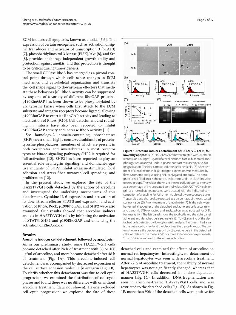
Expression of apoptosis-related proteins and caspase activityTo determine whether this arecoline-induced apoptosiswas associated with altered expression of apoptosis-regu-lated proteins, HA22T/VGH cells were treated for 24 hwith 30 or 100 μg/ml of arecoline. At 100 μg/ml of areco-line, Western blots showed a significant decrease in Bcl-2, Bcl-XL, and procaspase-9 levels and a significantincrease in Bax levels and cytochrome c release (Fig. 2A).Members of the caspase family are expressed in cells asinactive procaspases, which are activated during apopto-sis. As shown in Fig. 2B, treatment of HA22T/VGH cellsfor 24 h with 100 μg/ml of arecoline resulted in a markedincrease in active caspase-3, as shown by flow cytometryusing an antibody against active caspase-3. These results
show that arecoline induces apoptosis by caspase-3 acti-vation and a reduction in expression of anti-apoptoticproteins.
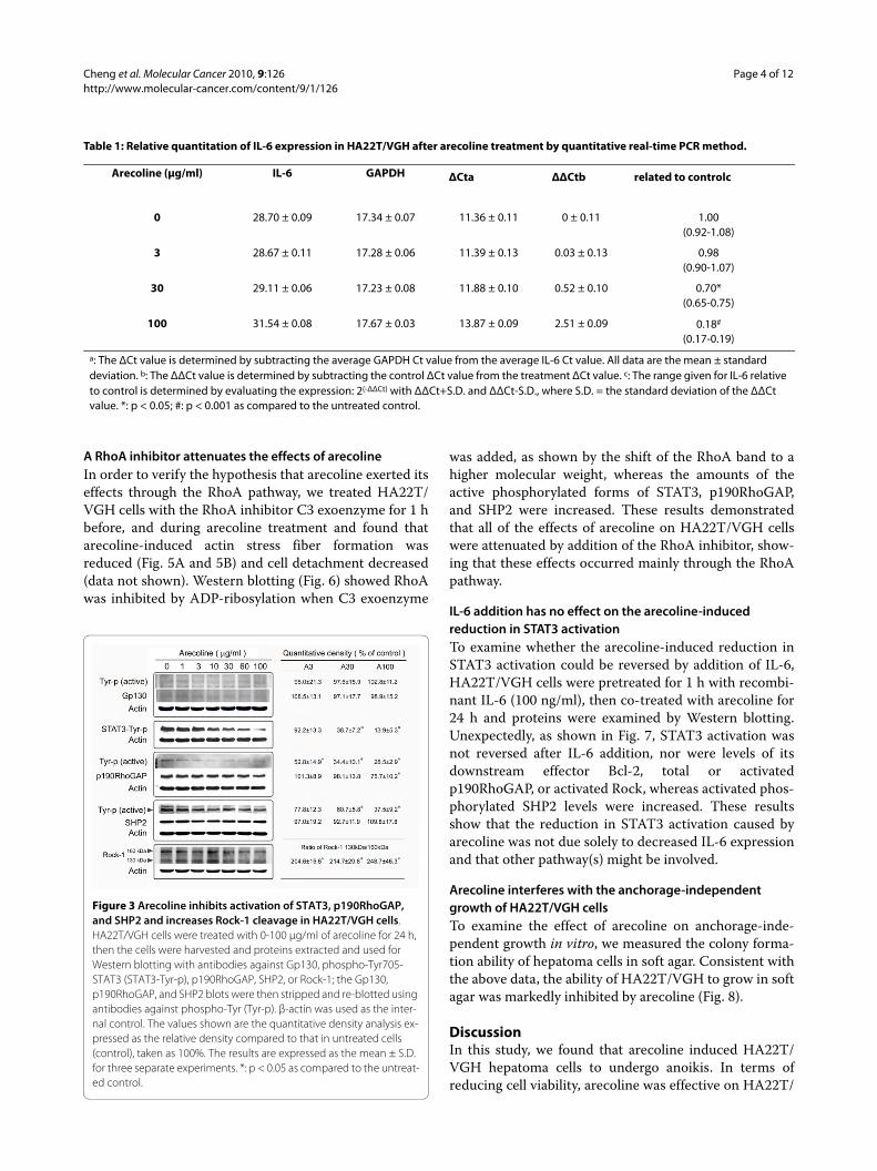
Arecoline decreases IL-6 expression and STAT3 activationSeveral studies have reported that anti-apoptotic genesare regulated by IL-6 and STAT3 [13,14]. Moreover, inthe liver, STAT3 is mainly activated by IL-6 and relatedcytokines and promotes anchorage-independent growth[15-17]. To examine whether arecoline had an effect onIL-6 and/or STAT3 levels, HA22T/VGH cells weretreated with 1-100 μg/ml of arecoline for 24 h, then IL-6mRNA levels were measured by quantitative real-timePCR and levels of gp130 (an IL-6 signal transducingreceptor component) and Tyr705 phosphorylated STAT3(the active form of STAT3) were assayed by Western blot-ting. Table 1 shows that arecoline at concentrations of 30-100 μg/ml markedly decreased IL-6 expression, while Fig.3 shows that levels of gp130 or activated gp130 were notaltered by arecoline, while Tyr705 phosphorylation ofSTAT3 was markedly inhibited.
Arecoline inhibits p190RhoGAP and SHP2 activation and increases Rho-associated kinasep190RhoGAP is phosphorylated by Src tyrosine kinase,allowing it to exert its RhoGAP activity, leading to inacti-vation of RhoA [9,10]. Fig. 3 shows that, after 24 h of are-coline treatment, p190RhoGAP levels were significantlydecreased at the concentration of 100 μg/ml of arecoline,while levels of the active phosphorylated p190RhoGAPwere decreased at arecoline concentrations higher than 3μg/ml. Src homology-2 domain-containing phosphatases(SHPs) are present in vertebrates and invertebrates. SHP2plays an essential role in integrin signaling [12] upstreamof RhoA to regulate its activity [18]. As shown in Fig. 3,after 24 h of arecoline treatment, the amount of SHP2was not changed, but the amount of active phosphory-lated SHP2 was decreased at arecoline concentrationshigher than 30 μg/ml. Furthermore, Rho-associatedkinase-1 (Rock-1) was cleaved to generate an activecleaved form at arecoline concentrations higher than 3μg/ml. The decreased activation of p190RhoGAP andSHP2 might result in enhanced RhoA activity and activityof its effector, Rock.
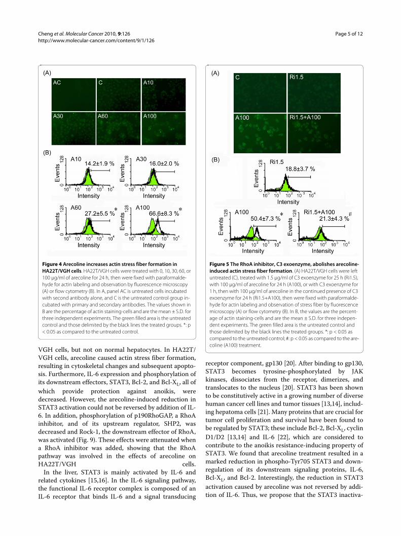
Arecoline stimulates actin stress fiber formationRhoA induces assembly of actin-myosin filamentsthrough activation of Rock kinase [19]. Since arecolinewas found to increase the amount of active Rock, weexamined whether it also stimulated actin stress fiber for-mation. After treatment with arecoline for 24 h, actinstress fiber formation was increased (Fig. 4A) and wasincreased by almost 6-fold at the concentration of 100 μg/ml arecoline compared to the control (Fig 4B).
Figure 2 Arecoline alters the expression of apoptosis-related proteins and caspase activity in HA22T/VGH cells. (A) HA22T/VGH cells were treated with 0, 30, or 100 μg/ml of arecoline for 24 h, then the cells were harvested and proteins extracted and used for Western blotting for Bcl-2, Bcl-XL, Bax, cytochrome c, or procaspase-9. β-actin was used as the internal control. The values shown are the quantitative density analysis expressed as the relative density compared to that in untreated cells (control), taken as 100%. The results are expressed as the mean ± S.D. for three separate experiments. (B) Caspase-3 activity was detected using RPE-conjugated anti-active caspase-3 antibody by flow cytometric analysis. The values shown are the percentage of cells with active caspase-3 and are the mean ± S.D. of three independent experiments. The red filled area is the untreated control and the black lines the treated groups. *: p < 0.05 as compared to the untreated con-trol.
Cheng et al. Molecular Cancer 2010, 9:126http://www.molecular-cancer.com/content/9/1/126
Page 4 of 12
A RhoA inhibitor attenuates the effects of arecolineIn order to verify the hypothesis that arecoline exerted itseffects through the RhoA pathway, we treated HA22T/VGH cells with the RhoA inhibitor C3 exoenzyme for 1 hbefore, and during arecoline treatment and found thatarecoline-induced actin stress fiber formation wasreduced (Fig. 5A and 5B) and cell detachment decreased(data not shown). Western blotting (Fig. 6) showed RhoAwas inhibited by ADP-ribosylation when C3 exoenzyme
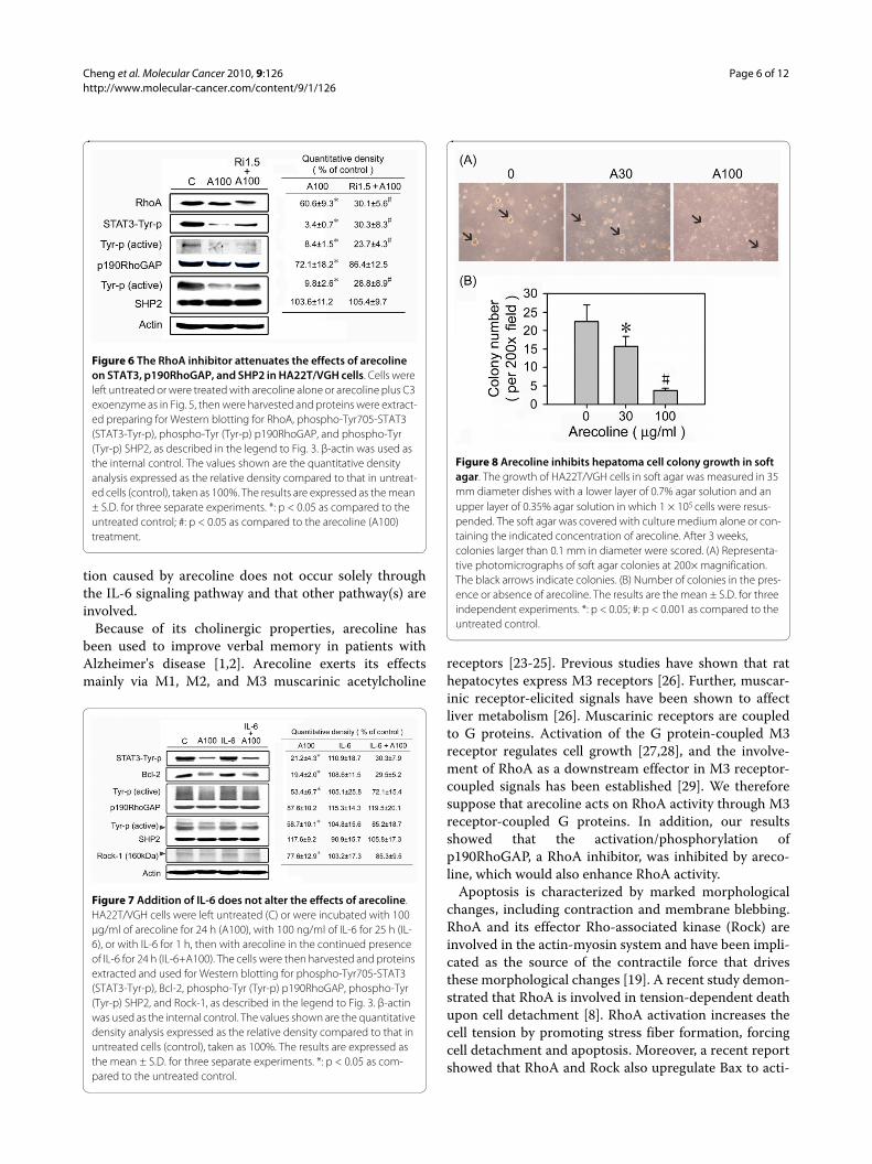
was added, as shown by the shift of the RhoA band to ahigher molecular weight, whereas the amounts of theactive phosphorylated forms of STAT3, p190RhoGAP,and SHP2 were increased. These results demonstratedthat all of the effects of arecoline on HA22T/VGH cellswere attenuated by addition of the RhoA inhibitor, show-ing that these effects occurred mainly through the RhoApathway.
IL-6 addition has no effect on the arecoline-induced reduction in STAT3 activationTo examine whether the arecoline-induced reduction inSTAT3 activation could be reversed by addition of IL-6,HA22T/VGH cells were pretreated for 1 h with recombi-nant IL-6 (100 ng/ml), then co-treated with arecoline for24 h and proteins were examined by Western blotting.Unexpectedly, as shown in Fig. 7, STAT3 activation wasnot reversed after IL-6 addition, nor were levels of itsdownstream effector Bcl-2, total or activatedp190RhoGAP, or activated Rock, whereas activated phos-phorylated SHP2 levels were increased. These resultsshow that the reduction in STAT3 activation caused byarecoline was not due solely to decreased IL-6 expressionand that other pathway(s) might be involved.
Arecoline interferes with the anchorage-independent growth of HA22T/VGH cellsTo examine the effect of arecoline on anchorage-inde-pendent growth in vitro, we measured the colony forma-tion ability of hepatoma cells in soft agar. Consistent withthe above data, the ability of HA22T/VGH to grow in softagar was markedly inhibited by arecoline (Fig. 8).
DiscussionIn this study, we found that arecoline induced HA22T/VGH hepatoma cells to undergo anoikis. In terms ofreducing cell viability, arecoline was effective on HA22T/
Table 1: Relative quantitation of IL-6 expression in HA22T/VGH after arecoline treatment by quantitative real-time PCR method.
Arecoline (μg/ml) IL-6 GAPDH ΔCta ΔΔCtb related to controlc
0 28.70 ± 0.09 17.34 ± 0.07 11.36 ± 0.11 0 ± 0.11 1.00(0.92-1.08)
3 28.67 ± 0.11 17.28 ± 0.06 11.39 ± 0.13 0.03 ± 0.13 0.98(0.90-1.07)
30 29.11 ± 0.06 17.23 ± 0.08 11.88 ± 0.10 0.52 ± 0.10 0.70*(0.65-0.75)
100 31.54 ± 0.08 17.67 ± 0.03 13.87 ± 0.09 2.51 ± 0.09 0.18#
(0.17-0.19)
a: The ΔCt value is determined by subtracting the average GAPDH Ct value from the average IL-6 Ct value. All data are the mean ± standard deviation. b: The ΔΔCt value is determined by subtracting the control ΔCt value from the treatment ΔCt value. c: The range given for IL-6 relative to control is determined by evaluating the expression: 2(-ΔΔCt) with ΔΔCt+S.D. and ΔΔCt-S.D., where S.D. = the standard deviation of the ΔΔCt value. *: p < 0.05; #: p < 0.001 as compared to the untreated control.
Figure 3 Arecoline inhibits activation of STAT3, p190RhoGAP, and SHP2 and increases Rock-1 cleavage in HA22T/VGH cells. HA22T/VGH cells were treated with 0-100 μg/ml of arecoline for 24 h, then the cells were harvested and proteins extracted and used for Western blotting with antibodies against Gp130, phospho-Tyr705-STAT3 (STAT3-Tyr-p), p190RhoGAP, SHP2, or Rock-1; the Gp130, p190RhoGAP, and SHP2 blots were then stripped and re-blotted using antibodies against phospho-Tyr (Tyr-p). β-actin was used as the inter-nal control. The values shown are the quantitative density analysis ex-pressed as the relative density compared to that in untreated cells (control), taken as 100%. The results are expressed as the mean ± S.D. for three separate experiments. *: p < 0.05 as compared to the untreat-ed control.
Cheng et al. Molecular Cancer 2010, 9:126http://www.molecular-cancer.com/content/9/1/126
Page 5 of 12
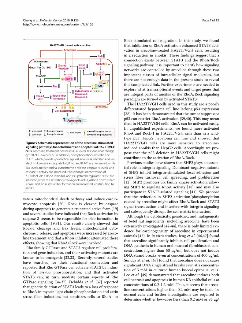
VGH cells, but not on normal hepatocytes. In HA22T/VGH cells, arecoline caused actin stress fiber formation,resulting in cytoskeletal changes and subsequent apopto-sis. Furthermore, IL-6 expression and phosphorylation ofits downstream effectors, STAT3, Bcl-2, and Bcl-XL, all ofwhich provide protection against anoikis, weredecreased. However, the arecoline-induced reduction inSTAT3 activation could not be reversed by addition of IL-6. In addition, phosphorylation of p190RhoGAP, a RhoAinhibitor, and of its upstream regulator, SHP2, wasdecreased and Rock-1, the downstream effector of RhoA,was activated (Fig. 9). These effects were attenuated whena RhoA inhibitor was added, showing that the RhoApathway was involved in the effects of arecoline onHA22T/VGH cells.
In the liver, STAT3 is mainly activated by IL-6 andrelated cytokines [15,16]. In the IL-6 signaling pathway,the functional IL-6 receptor complex is composed of anIL-6 receptor that binds IL-6 and a signal transducing
receptor component, gp130 [20]. After binding to gp130,STAT3 becomes tyrosine-phosphorylated by JAKkinases, dissociates from the receptor, dimerizes, andtranslocates to the nucleus [20]. STAT3 has been shownto be constitutively active in a growing number of diversehuman cancer cell lines and tumor tissues [13,14], includ-ing hepatoma cells [21]. Many proteins that are crucial fortumor cell proliferation and survival have been found tobe regulated by STAT3; these include Bcl-2, Bcl-XL, cyclinD1/D2 [13,14] and IL-6 [22], which are considered tocontribute to the anoikis resistance-inducing property ofSTAT3. We found that arecoline treatment resulted in amarked reduction in phospho-Tyr705 STAT3 and down-regulation of its downstream signaling proteins, IL-6,Bcl-XL, and Bcl-2. Interestingly, the reduction in STAT3activation caused by arecoline was not reversed by addi-tion of IL-6. Thus, we propose that the STAT3 inactiva-
Figure 4 Arecoline increases actin stress fiber formation in HA22T/VGH cells. HA22T/VGH cells were treated with 0, 10, 30, 60, or 100 μg/ml of arecoline for 24 h, then were fixed with paraformalde-hyde for actin labeling and observation by fluorescence microscopy (A) or flow cytometry (B). In A, panel AC is untreated cells incubated with second antibody alone, and C is the untreated control group in-cubated with primary and secondary antibodies. The values shown in B are the percentage of actin staining-cells and are the mean ± S.D. for three independent experiments. The green filled area is the untreated control and those delimited by the black lines the treated groups. *: p < 0.05 as compared to the untreated control.
Figure 5 The RhoA inhibitor, C3 exoenzyme, abolishes arecoline-induced actin stress fiber formation. (A) HA22T/VGH cells were left untreated (C), treated with 1.5 μg/ml of C3 exoenzyme for 25 h (Ri1.5), with 100 μg/ml of arecoline for 24 h (A100), or with C3 exoenzyme for 1 h, then with 100 μg/ml of arecoline in the continued presence of C3 exoenzyme for 24 h (Ri1.5+A100), then were fixed with paraformalde-hyde for actin labeling and observation of stress fiber by fluorescence microscopy (A) or flow cytometry (B). In B, the values are the percent-age of actin staining-cells and are the mean ± S.D. for three indepen-dent experiments. The green filled area is the untreated control and those delimited by the black lines the treated groups. *: p < 0.05 as compared to the untreated control; #: p < 0.05 as compared to the are-coline (A100) treatment.
Cheng et al. Molecular Cancer 2010, 9:126http://www.molecular-cancer.com/content/9/1/126
Page 6 of 12
tion caused by arecoline does not occur solely throughthe IL-6 signaling pathway and that other pathway(s) areinvolved.
Because of its cholinergic properties, arecoline hasbeen used to improve verbal memory in patients withAlzheimer's disease [1,2]. Arecoline exerts its effectsmainly via M1, M2, and M3 muscarinic acetylcholine
receptors [23-25]. Previous studies have shown that rathepatocytes express M3 receptors [26]. Further, muscar-inic receptor-elicited signals have been shown to affectliver metabolism [26]. Muscarinic receptors are coupledto G proteins. Activation of the G protein-coupled M3receptor regulates cell growth [27,28], and the involve-ment of RhoA as a downstream effector in M3 receptor-coupled signals has been established [29]. We thereforesuppose that arecoline acts on RhoA activity through M3receptor-coupled G proteins. In addition, our resultsshowed that the activation/phosphorylation ofp190RhoGAP, a RhoA inhibitor, was inhibited by areco-line, which would also enhance RhoA activity.
Apoptosis is characterized by marked morphologicalchanges, including contraction and membrane blebbing.RhoA and its effector Rho-associated kinase (Rock) areinvolved in the actin-myosin system and have been impli-cated as the source of the contractile force that drivesthese morphological changes [19]. A recent study demon-strated that RhoA is involved in tension-dependent deathupon cell detachment [8]. RhoA activation increases thecell tension by promoting stress fiber formation, forcingcell detachment and apoptosis. Moreover, a recent reportshowed that RhoA and Rock also upregulate Bax to acti-
Figure 6 The RhoA inhibitor attenuates the effects of arecoline on STAT3, p190RhoGAP, and SHP2 in HA22T/VGH cells. Cells were left untreated or were treated with arecoline alone or arecoline plus C3 exoenzyme as in Fig. 5, then were harvested and proteins were extract-ed preparing for Western blotting for RhoA, phospho-Tyr705-STAT3 (STAT3-Tyr-p), phospho-Tyr (Tyr-p) p190RhoGAP, and phospho-Tyr (Tyr-p) SHP2, as described in the legend to Fig. 3. β-actin was used as the internal control. The values shown are the quantitative density analysis expressed as the relative density compared to that in untreat-ed cells (control), taken as 100%. The results are expressed as the mean ± S.D. for three separate experiments. *: p < 0.05 as compared to the untreated control; #: p < 0.05 as compared to the arecoline (A100) treatment.
Figure 7 Addition of IL-6 does not alter the effects of arecoline. HA22T/VGH cells were left untreated (C) or were incubated with 100 μg/ml of arecoline for 24 h (A100), with 100 ng/ml of IL-6 for 25 h (IL-6), or with IL-6 for 1 h, then with arecoline in the continued presence of IL-6 for 24 h (IL-6+A100). The cells were then harvested and proteins extracted and used for Western blotting for phospho-Tyr705-STAT3 (STAT3-Tyr-p), Bcl-2, phospho-Tyr (Tyr-p) p190RhoGAP, phospho-Tyr (Tyr-p) SHP2, and Rock-1, as described in the legend to Fig. 3. β-actin was used as the internal control. The values shown are the quantitative density analysis expressed as the relative density compared to that in untreated cells (control), taken as 100%. The results are expressed as the mean ± S.D. for three separate experiments. *: p < 0.05 as com-pared to the untreated control.
Figure 8 Arecoline inhibits hepatoma cell colony growth in soft agar. The growth of HA22T/VGH cells in soft agar was measured in 35 mm diameter dishes with a lower layer of 0.7% agar solution and an upper layer of 0.35% agar solution in which 1 × 10⁵ cells were resus-pended. The soft agar was covered with culture medium alone or con-taining the indicated concentration of arecoline. After 3 weeks, colonies larger than 0.1 mm in diameter were scored. (A) Representa-tive photomicrographs of soft agar colonies at 200× magnification. The black arrows indicate colonies. (B) Number of colonies in the pres-ence or absence of arecoline. The results are the mean ± S.D. for three independent experiments. *: p < 0.05; #: p < 0.001 as compared to the untreated control.
Cheng et al. Molecular Cancer 2010, 9:126http://www.molecular-cancer.com/content/9/1/126
Page 7 of 12
vate a mitochondrial death pathway and induce cardio-myocyte apoptosis [30]. Rock is cleaved by caspaseduring apoptosis to generate a truncated active form [19]and several studies have indicated that Rock activation bycaspase-3 seems to be responsible for bleb formation inapoptotic cells [19,31]. Our results clearly showed thatRock-1 cleavage and Bax levels, mitochondrial cyto-chrome c release, and apoptosis were increased by areco-line treatment and that a RhoA inhibitor attenuated theseeffects, showing that RhoA/Rock were involved.
Rho family GTPases and STAT3 regulate cell prolifera-tion and gene induction, and their activating mutants areknown to be oncogenic [32,33]. Recently, several studieshave searched for their functional connection andreported that Rho GTPase can activate STAT3 by induc-tion of Tyr705 phosphorylation, and that activatedSTAT3 can, in turn, mediate certain aspects of RhoGTPase signaling [34-37]. Debidda et al. [37] reportedthat genetic deletion of STAT3 leads to a loss of responseto RhoA in myosin light chain phosphorylation and actinstress fiber induction, but sensitizes cells to RhoA- or
Rock-stimulated cell migration. In this study, we foundthat inhibition of RhoA activation enhanced STAT3 acti-vation in arecoline-treated HA22T/VGH cells, resultingin a reduction in anoikis. These findings suggest that aconnection exists between STAT3 and the RhoA/Rocksignaling pathway. It is important to clarify how signalingnetworks are controlled by arecoline through these twoimportant classes of intracellular signal molecules, butthere are not enough data in the present study to revealthis complicated link. Further experiments are needed toexplore what transcriptional events and target genes thatare integral parts of anoikis of the RhoA/Rock signalingparadigm are turned on by activated STAT3.
The HA22T/VGH cells used in this study are a poorlydifferentiated hepatoma cell line lacking p53 expression[38]. It has been demonstrated that the tumor suppressorp53 can restrict RhoA activation [39,40]. This may meanthat, in HA22T/VGH cells, RhoA can be activated easily.In unpublished experiments, we found more activatedRhoA and Rock-1 in HA22T/VGH cells than in a wild-type p53 HepG2 hepatoma cell line and showed thatHA22T/VGH cells are more sensitive to arecoline-induced anoikis than HepG2 cells. Accordingly, we pro-pose that the p53 deletion in HA22T/VGH cells mightcontribute to the activation of RhoA/Rock.
Previous studies have shown that SHP2 plays an essen-tial role in integrin signaling. Dominant-negative mutantsof SHP2 inhibit integrin-stimulated focal adhesion andstress fiber turnover, cell spreading, and proliferation[12]. SHP2 promotes Src family kinase activation, allow-ing SHP2 to regulate RhoA activity [18], and may alsoparticipate in STAT3-related signaling [41]. We proposethat the reduction in SHP2 activation/phosphorylationcaused by arecoline might affect RhoA/Rock and STAT3signal transduction and interfere with integrin signalingand subsequently disrupt the cell-matrix interaction.
Although the cytotoxicity, genotoxic, and mutagenicityof betel nut ingredients, including arecoline, have beenextensively investigated [42-44], there is only limited evi-dence for carcinogenicity of arecoline in experimentalanimals [45]. In in vitro studies, Jeng et al. [46,47] foundthat arecoline significantly inhibits cell proliferation andDNA synthesis in human oral mucosal fibroblasts at con-centrations higher than 50 μg/ml, but does not induceDNA strand breaks, even at concentrations of 400 μg/ml.Sundqvist et al. [48] found that arecoline does not causesignificant DNA single strand breaks even at a concentra-tion of 5 mM in cultured human buccal epithelial cells.Lee et al. [49] demonstrated that arecoline induces bothcell necrosis and apoptosis in human KB epithelial cells atconcentrations of 0.2-1.2 mM. Thus, it seems that areco-line concentrations higher than 0.2 mM may be toxic fornormal cells and further investigations are required todetermine whether low-dose (less than 0.2 mM or 85 μg/
Figure 9 Schematic representation of the arecoline-stimulated signaling pathways for detachment and apoptosis of HA22T/VGH cells. Arecoline treatment decreases IL-6 levels, but does not change gp130 of IL-6 receptor. In addition, phosphorylation/activation of STAT3, which provides protection against anoikis, is inhibited and lev-els of its downstream signals IL-6, Bcl-2, and Bcl-XL are decreased, while Bax levels, mitochondrial cytochrome c release, caspase-9 levels, and caspase-3 activity are increased. Phosphorylation/activation of p190RhoGAP, a RhoA inhibitor, and its upstream regulator, SHP2, are inhibited, while the activation/cleavage of Rock-1, a RhoA downstream kinase, and actin stress fiber formation are increased, contributing to anoikis.
Cheng et al. Molecular Cancer 2010, 9:126http://www.molecular-cancer.com/content/9/1/126
Page 8 of 12
ml) arecoline is toxic. However, this study highlights thepossibility that low-dose arecoline might be useful in thetreatment of hepatoma and provides clues for studyingthe arecoline-induced detachment of hepatoma cells.
ConclusionsWe propose that arecoline induces anoikis of HA22T/VGH cells that involves inhibition of STAT3 andincreased RhoA/Rock activation and that the STAT3 andRhoA/Rock signaling pathways are connected. Impor-tantly, this study shows that arecoline induces the deathof HA22T/VGH hepatoma cells, but not normal hepato-cytes. Since arecoline has diverse biological functions,additional studies are needed to evaluate its cytotoxicityfor other cells.
MethodsReagents and antibodiesArecoline hydrobromide (methyl 1-methyl-1,2,5,6-tetra-hydronicotinate hydrobromide) was obtained fromSigma-Aldrich (St. Louis, MO, USA); its purity wasgreater than 99.0%. Collagenase type II was from Wor-thington Biochemical Corporation (Lakewood, NJ, USA).Percoll was from Amersham Pharmacia Biotech (Upp-sala, Sweden). RNase A and the protease inhibitor cock-tail were from Sigma-Aldrich (St. Louis, MO, USA).Protein assay reagents were from Bio-Rad Laboratories(Hercules, CA, USA). TRIzol reagent was from Invitro-gen Life Technologies (Carlsbad, CA, USA). All otherchemicals were of analytical grade and purchased fromSigma-Aldrich (St. Louis, MO, USA). The cell-permeableRhoA inhibitor, C3 exoenzyme, was from CytoskeletonInc. (Denver, CO, USA). Recombinant human IL-6 wasfrom Peprotech (Rocky Hill, NJ, USA). Mouse monoclo-nal antibodies against phospho-tyrosine (phospho-Tyr),phospho-Tyr705-STAT3, SHP2, p190RhoGAP, RhoA,Rock-1, Bcl-2, Bax, procaspase-9, or cytochrome c, rabbitpolyclonal antibodies against Bcl-XL or gp130, and goatpolyclonal antibodies against β-actin were purchasedfrom Santa Cruz Biotechnology (Santa Cruz, CA, USA).Horseradish peroxidase-conjugated anti-mouse, goat,and rabbit IgG antibodies were purchased from BDPharmingen Inc. (San Diego, CA, USA). R-phycoerythrin(RPE)-conjugated rabbit anti-active caspase-3 polyclonalantibodies, RPE-conjugated mouse anti-human β1-integ-rin monoclonal antibody and the RPE-conjugated mouseIgG isotype control were purchased from BD Pharmin-gen Inc. (San Diego, CA, USA). The RPE-conjugated rab-bit IgG isotype control was from R&D Systems(Minneapolis, MN, USA). FITC-conjugated swine anti-goat IgG antibodies were from Invitrogen Corporation(Camarillo, CA, USA).
Cell line, cell culture, and arecoline treatmentHA22T/VGH, a poorly differentiated human hepatomacell line, was obtained from the Bioresource Collectionand Research Center (BCRC) in the Food IndustryResearch and Development Institute (Hsinchu, Taiwan)and was cultured in Dulbecco's Modified Eagle's Medium(DMEM) (Gibco BRL, Grand Island, NY, USA) contain-ing 10% fetal bovine serum (FBS) (Hyclone, Auckland,NZ), 2 mM L-glutamine (Gibco BRL, Grand Island, NY,USA), 0.1 mM non-essential amino acids (Gibco BRL,Grand Island, NY, USA), 100 units/ml of penicillin, and100 μg/ml of streptomycin (Gibco BRL, Grand Island,NY, USA) at 37°C in a humidified chamber with 5% CO2.To investigate the effects of arecoline, various concentra-tions of arecoline were added to the culture medium forthe indicated time period, then the cells were harvestedand analyzed.
Primary hepatocyte isolationHepatocytes were isolated using a previously describedmethod [50]. Sprague-Dawley rats were purchased fromBioLASCO Taiwan Co., Ltd. (Charles River Technology,Taipei, Taiwan). This study was performed in accordancewith the Guide for the Care and Use of Laboratory Ani-mals of the United States National Institutes of Health.The protocol for animal use was reviewed and approvedby the Institutional Animal Care and Use Committee(IACUC) of Kaohsiung Medical University (Approval No.96137). Livers from newborn Sprague-Dawley rats weremechanically dissociated using a scalpel and the liverpieces incubated in Hank's balanced salt solution (pH 7.4)(HBSS) containing 0.6 mM EGTA and 2% bovine serumalbumin (BSA) (Ca2+-free) (Sigma-Aldrich, St. Louis,MO, USA) by shaking for 10 min at 37°C followed by abrief wash with HBSS. Hepatocytes were further dissoci-ated from the tissue by shaking for 15 min at 37°C inHBSS (pH 7.4) containing 5 mM CaCl2 and 1.5 mg/ml ofcollagenase type II, then were filtered through a 70 μmnylon cell strainer (BD Falcon, Bedford, MA, USA) toremove cellular aggregates and tissue debris. The cell fil-trate was centrifuged at 50 ×g for 10 min and the cell pel-let resuspended in DMEM and centrifuged at 800 ×g for30 min on a discontinuous Percoll gradient comprised of3 ml of 70% and 6 ml of 30% Percoll. The dissociated cellswere stratified and viable hepatocytes were found at theinterface between the two Percoll layers. The hepatocytefraction was collected and the cells plated in flasks inDMEM containing 10% FBS and allowed to attach for 2-3h at 37°C in a humidified chamber with 5% CO2, thenwere washed with DMEM to remove non-adherenthematopoietic cells. The cells were fed with freshmedium every other day and were split at 80-90% conflu-
Cheng et al. Molecular Cancer 2010, 9:126http://www.molecular-cancer.com/content/9/1/126
Page 9 of 12
ence. Experiments were performed on day 7-10 post-iso-lation.
Cell morphologyMorphological changes were observed under an invertedphase-contrast microscope (Olympus, Tokyo, Japan).Photographs were taken at 200× magnification.
Cell viability assayAfter arecoline treatment, the cells were harvested andviable cells counted using a dye exclusion technique. Thecell suspension was centrifuged at 5 000 ×g, the superna-tant discarded and the cell pellet resuspended in serum-free medium. One volume of 0.4% Trypan blue (GibcoBRL, Grand Island, NY, USA) was added to one volume ofcell suspension, then, after incubation at room tempera-ture for 3 min, cells were counted in a hemocytometer.All counts were done in triplicate.
Detection of cell surface adhesion moleculesHA22T/VGH cells were harvested and washed withserum-free DMEM, then were suspended in DMEM con-taining 1% BSA and incubated in the dark at 4°C for 30min with RPE-conjugated mouse anti-human monoclo-nal antibody against β1-integrin. After two washed with1% BSA/phosphate-buffered saline (PBS), the cells werefixed by mixing the cells with 4% paraformaldehyde inPBS, then resuspended in 1% BSA/PBS for flow cytome-try analysis. Cell fluorescence was measured using aCoulter Epics XL cytometer (Beckman Coulter, Miami,FL, USA). A control sample incubated with RPE-conju-gated normal mouse IgG was run in parallel as a negativecontrol. The data were analyzed using WINMDI softwareversion 2.8 (Scripps Research Institute, La Jolla, CA,USA), a minimum of 1 × 10⁴ cells per sample being evalu-ated in each case.
Apoptosis assayDetection of active caspase-3Active caspase-3 was detected as described previously[51]. Briefly, cells were pelleted, resuspended in 1 ml of4% paraformaldehyde, and incubated for 30 min at roomtemperature. The suspension was then centrifuged, thepellet washed twice with PBS, the cells resuspended in 1ml of 0.1% Triton X-100 and incubated for 30 min atroom temperature, then washed as above. Labeling wasperformed by addition of 100 μl of PBS containing 5 μl ofpolyclonal RPE-conjugated rabbit anti-active caspase-3antibodies, incubation at 37°C for 1 h, washing with PBS,and analysis on a Coulter Epics XL cytometer (BeckmanCoulter, Miami, FL, USA). A control sample incubatedwith RPE-conjugated normal rabbit IgG was run in paral-lel. The data were analyzed using WINMDI software ver-sion 2.8 (Scripps Research Institute, La Jolla, CA, USA), a
minimum of 1 × 10⁴ cells per sample being evaluated ineach case.DNA fragmentation assayCells (5 × 10⁶) were treated with arecoline for 72 h, thenthe adherent or detached cells were harvested separatelyor pooled together for DNA fragmentation analysis asdescribed previously [52]. The cells were pelleted andresuspended in 200 μl of lysis solution [10 mM Tris (pH8.0), 100 mM NaCl, 25 mM EDTA, 0.5% sodium dodecylsulfate (SDS), 0.5 mg/ml proteinase K] and incubated for15 h at 50°C. Nucleic acids were extracted by addition ofan equal volume of phenol/chloroform/isoamyl alcohol,centrifugation for 20 min at 10 000 ×g 4°C, and harvestingthe aqueous (top) layer. DNA were precipitated by addi-tion of a 1/2 volume of 7.5 M ammonium acetate and 2volumes of 100% ethanol, and recovered by centrifuga-tion at 10 000 ×g 4°C for 5 min. After rinsing with 70%ethanol, the DNA was resuspended in TE buffer (10 mMTris, 1 mM EDTA, pH 8.0) and residual RNA removed byaddition of 10 μg/ml of RNase A and incubation at 60°Cfor 1 h. Samples were resolved on a 1.5% Tris-acetate-EDTA-agarose gel, which was stained with ethidium bro-mide, and the bands visualized and photographed undershort-wave UV.TUNEL assayTerminal deoxynucleotide transferase-mediated dUTPnick-end labeling (TUNEL) assays were performed usingan APO-BrdU™ TUNEL Assay Kit (Molecular Probes,Eugene, OR) according to the manufacturer's protocol.Briefly, the cells were incubated for the indicated timebefore being trypsinized, washed with PBS, and fixed in2% paraformaldehyde (pH 7.4) for 15 min. The fixed cellswere washed twice in PBS and stored at -20°C in 70% eth-anol for 12-18 h prior to performing the TUNEL assay.After removing the 70% ethanol by centrifugation, thecells were washed twice in wash buffer, then incubated at37°C for 60 min with DNA-labeling solution containingterminal deoxynucleotidyl transferase and BrdUTP. Afterwashing twice with rinse buffer, the cells were resus-pended for 30 min in the dark at room temperature inantibody solution containing Alexa Fluor® 488-labeledanti-BrdU antibody. Flow cytometric analysis was thenperformed using a Coulter Epics XL cytometer (BeckmanCoulter, Miami, FL, USA) to quantify apoptosis. The datawere analyzed using WINMDI software version 2.8(Scripps Research Institute, La Jolla, CA, USA), a mini-mum of 1 × 10⁴ cells per sample being evaluated in eachcase.
Protein lysate preparation and Western blottingSample preparation and Western blotting procedureswere performed as described previously [53]. Briefly, cellswere harvested and cytosolic extracts prepared using lysisbuffer [20 mM Tris-HCl (pH 7.2), 2 mM EGTA, 5 mM
Cheng et al. Molecular Cancer 2010, 9:126http://www.molecular-cancer.com/content/9/1/126
Page 10 of 12
EDTA, 500 μM sodium orthovanadate, 10 mM sodiumfluoride, 1% Triton X-100, 0.1% SDS and protease inhibi-tor cocktail]. Protein concentrations were determinedusing protein assay reagents. Forty to sixty micrograms ofprotein lysate was analyzed by SDS-polyacrylamide gelelectrophoresis. After transfer of the proteins from thegel to a nitrocellulose membrane (Amersham PharmaciaBiotech, Freiburg, Germany), the membranes wereblocked for 1 h at room temperature in PBS with 0.05%Tween 20 (PBS-T) containing 5% nonfat dry milk, thenincubated with specific primary antibodies and horserad-ish peroxidase-conjugated secondary antibodies. Forreblotting of other proteins on the same membrane, anti-bodies were stripped using heated 0.1 M glycine solution(pH 2.0) three times and the membrane washed twicewith PBS-T. The immunoreactive bands were visualizedusing an enhanced chemiluminescence kit (Perkin-ElmerLife Sciences, Boston, MA, USA).
Stress fiber formation assayImmunofluorescence stainingFor actin staining, HA22T/VGH cells were incubated forthe indicated time before being washed with PBS, fixedfor 10 min at room temperature in 2% paraformaldehydein PBS, and permeabilized for 10 min at room tempera-ture with 0.5% Triton X-100 in PBS. Filamentous actinwas stained for 1 h at 37°C with polyclonal goat anti-actinantibodies in PBS, then for 1 h at 37°C with FITC-conju-gated swine anti-goat IgG antibodies. Images wereobtained at 200× magnification using a Zeiss Axiovert200 fluorescence microscope.Flow cytometry analysisHA22T/VGH cells were incubated for the indicated timebefore being trypsinized, washed with PBS, fixed for 10min at room temperature in 2% paraformaldehyde inPBS, and permeabilized for 15 min on ice with 90% meth-anol. Filamentous actin was then stained as above, and,after washing with PBS, samples were analyzed on aCoulter Epics XL cytometer (Beckman Coulter, Miami,FL, USA). The data were analyzed using WINMDI soft-ware version 2.8 (Scripps Research Institute, La Jolla, CA,USA), a minimum of 1 × 10⁴ cells per sample being evalu-ated in each case.
Quantitative real-time PCR analysisTotal RNA was isolated using TRIzol reagent accordingto the manufacturer's instructions. RNA samples (2 μg)were reverse transcribed using random hexamer primersand M-MLV reverse transcriptase (Promega Corpora-tion, Madison, WI, USA) and the cDNA used for real-time PCR performed on a MiniOpticon™ Real-Time PCRDetection System (Bio-Rad Laboratories, Hercules, CA,USA) using iQ™ SYBR® Green Supermix (Bio-Rad Labora-tories, Hercules, CA, USA) following the manufacturer's
protocol. The PCR amplification reaction mixture (25 μl)contained 50 ng of cDNA, 12.5 μl of SYBR Green Super-mix, and 0.2 μM of the IL-6- or GAPDH-specific primerpair. The optimal primer concentrations were determinedin preliminary experiments. PCR primers were designedusing Beacon Designer software version 2.0 (Premier Bio-soft International, Palo Alto, CA, USA) and theirsequences were as follows: IL-6 forward, 5'-TCC TGGTGT TGC CTG CTG-3'; reverse, 5'-TCG TTC TGAAGA GGT GAG TGG-3' and GAPDH forward, 5'-GACATC AAG AAG GTG GTG AAG CAG-3'; reverse, 5'-GCG TCA AAG GTG GAG GAG TGG-3'. In order toconfirm amplification specificity, the PCR products fromeach primer pair were subjected to melting curve analy-sis. The reaction conditions were incubation at 50°C for 2min and initial denaturation at 95°C for 10 min, followedby 40 cycles of denaturation at 95°C for 20 s and anneal-ing at 60°C for 1 min. After real-time PCR, the tempera-ture was increased from 60 to 95°C at a rate of 0.5°C persecond to construct a melting curve. A negative controlwithout cDNA was run in parallel with each assay.Results were collected and analyzed using MJ OpticonMonitor Analysis software version 3.1 (Bio-Rad Labora-tories, Hercules, CA, USA). Each reaction mixture wasamplified in triplicate and the results calculated based onthe ΔΔCt method [54]. The cycle threshold (Ct) value forthe IL-6 gene was corrected using the mean Ct value forthe GAPDH gene. Relative gene expression was expressedas the fold change (2-ΔΔCt) relative to expression in theuntreated control.
Anchorage-independent growth in soft agarA soft agar assay was performed as described previously[33]. Briefly, growth in soft agar was measured in 35 mmdiameter dishes containing a lower layer of 0.7% agar(Bitek agar; Difco Laboratories, Detroit, MI, USA) solu-tion in DMEM containing 10% FBS and 0.1 mM non-essential amino acids overlaid with 0.35% agar solution,also in growth medium, in which 1 × 10⁵ cells were resus-pended. The soft agar was covered with culture mediumalone or containing the indicated concentration of areco-line. Colonies were scored 21 days after preparation (col-onies larger than ~0.1 mm in diameter were scored aspositive). Cells were maintained in DMEM with 10% FBSand 0.1 mM non-essential amino acids.
Statistical analysisAll data are presented as the mean ± standard deviation(S.D.) for the number of experiments indicated. Otherdifferences between treated and control groups were ana-lyzed using Student's t-test. Statistical analyses were per-formed using SAS version 6.011 (SAS Institute Inc, Cary,NC). A p value < 0.05 was considered statistically signifi-cant.
Cheng et al. Molecular Cancer 2010, 9:126http://www.molecular-cancer.com/content/9/1/126
Page 11 of 12
Competing interestsThe authors declare that they have no competing interests.
Authors' contributionsHLC performed the research, analyzed the data, and drafted the manuscript.SJS and LWH helped in drafting the manuscript. BSH helped with the Westernblotting techniques. YCH helped with the normal hepatocyte isolation tech-niques. TCH performed the flow cytometry study. KLC designed the research,interpreted the data, revised the manuscript, and gave final approval for theversion to be published. All authors approved the final version of the manu-script.
AcknowledgementsThis study was supported by the National Science Council, Executive Yuan, Tai-wan (Grant NSC 94-2320-B-037-039; NSC 95-2320-B-037-011; NSC 96-2320-B-037-003).
Author Details1Graduate Institute of Medicine, College of Medicine, Kaohsiung Medical University, Kaohsiung 80708, Taiwan, 2Bachelor Degree Program of Health Beauty, Department of Medical Technology, School of Medicine and Health Sciences, FooYin University, Kaohsiung 83101, Taiwan, 3Department of Medical Laboratory Science and Biotechnology, Kaohsiung Medical University, Kaohsiung 80708, Taiwan and 4Department of Biochemistry, Faculty of Medicine, College of Medicine, Kaohsiung Medical University, Kaohsiung 80708, Taiwan
References1. Gray JA, Enz A, Spiegel R: Muscarinic agonists for senile dementia: past
experience and future trends. Trends Pharmacol Sci 1989:85-88.2. Raffaele KC, Berardi A, Asthana S, Morris P, Haxby JV, Soncrant TT: Effects
of long-term continuous infusion of the muscarinic cholinergic agonist arecoline on verbal memory in dementia of the Alzheimer type. Psychopharmacol Bull 1991, 27:315-319.
3. Chang MC, Wu HL, Lee JJ, Lee PH, Chang HH, Hahn LJ, Lin BR, Chen YJ, Jeng JH: The induction of prostaglandin E2 production, interleukin-6 production, cell cycle arrest, and cytotoxicity in primary oral keratinocytes and KB cancer cells by areca nut ingredients is differentially regulated by MEK/ERK activation. J Biol Chem 2004, 279:50676-50683.
4. Jeng JH, Wang YJ, Chiang BL, Lee PH, Chan CP, Ho YS, Wang TM, Lee JJ, Hahn LJ, Chang MC: Roles of keratinocyte inflammation in oral cancer: regulating the prostaglandin E2, interleukin-6 and TNF-alpha production of oral epithelial cells by areca nut extract and arecoline. Carcinogenesis 2003, 24:1301-1315.
5. Frisch SM, Francis H: Disruption of epithelial cell-matrix interactions induces apoptosis. J Cell Biol 1994, 124:619-626.
6. Frisch SM, Ruoslahti E: Integrins and anoikis. Curr Opin Cell Biol 1997, 9:701-706.
7. Schlessinger K, Levy DE: Malignant transformation but not normal cell growth depends on signal transducer and activator of transcription 3. Cancer Res 2005, 65:5828-5834.
8. Ma Z, Myers DP, Wu RF, Nwariaku FE, Terada LS: p66Shc mediates anoikis through RhoA. J Cell Biol 2007, 179:23-31.
9. Arthur WT, Burridge K: RhoA inactivation by p190RhoGAP regulates cell spreading and migration by promoting membrane protrusion and polarity. Mol Biol Cell 2001, 12:2711-2720.
10. Brouns MR, Matheson SF, Settleman J: p190 RhoGAP is the principal Src substrate in brain and regulates axon outgrowth, guidance and fasciculation. Nat Cell Biol 2001, 3:361-367.
11. Maddox AS, Burridge K: RhoA is required for cortical retraction and rigidity during mitotic cell rounding. J Cell Biol 2003, 160:255-265.
12. Neel BG, Gu H, Pao L: The 'Shp'ing news: SH2 domain-containing tyrosine phosphatases in cell signaling. Trends Biochem Sci 2003, 28:284-293.
13. Aggarwal BB, Sethi G, Ahn KS, Sandur SK, Pandey MK, Kunnumakkara AB, Sung B, Ichikawa H: Targeting signal-transducer-and-activator-of-
transcription-3 for prevention and therapy of cancer: modern target but ancient solution. Ann N Y Acad Sci 2006, 1091:151-169.
14. Chen CL, Cen L, Kohout J, Hutzen B, Chan C, Hsieh FC, Loy A, Huang V, Cheng G, Lin J: Signal transducer and activator of transcription 3 activation is associated with bladder cancer cell growth and survival. Mol Cancer 2008, 7:78.
15. Berasain C, Castillo J, Perugorria MJ, Latasa MU, Prieto J, Avila MA: Inflammation and liver cancer: new molecular links. Ann N Y Acad Sci 2009, 1155:206-221.
16. Gao B: Cytokines, STATs and liver disease. Cell Mol Immunol 2005, 2:92-100.
17. Yoshikawa H, Matsubara K, Qian GS, Jackson P, Groopman JD, Manning JE, Harris CC, Herman JG: SOCS-1, a negative regulator of the JAK/STAT pathway, is silenced by methylation in human hepatocellular carcinoma and shows growth-suppression activity. Nat Genet 2001, 28:29-35.
18. Zhang SQ, Yang W, Kontaridis MI, Bivona TG, Wen G, Araki T, Luo J, Thompson JA, Schraven BL, Philips MR, Neel BG: Shp2 regulates SRC family kinase activity and Ras/Erk activation by controlling Csk recruitment. Mol Cell 2004, 13:341-355.
19. Coleman ML, Sahai EA, Yeo M, Bosch M, Dewar A, Olson MF: Membrane blebbing during apoptosis results from caspase-mediated activation of ROCK I. Nat Cell Biol 2001, 3:339-345.
20. Taga T, Kishimoto T: Gp130 and the interleukin-6 family of cytokines. Annu Rev Immunol 1997, 15:797-819.
21. Yang SF, Wang SN, Wu CF, Yeh YT, Chai CY, Chunag SC, Sheen MC, Lee KT: Altered p-STAT3 (tyr705) expression is associated with histological grading and intratumour microvessel density in hepatocellular carcinoma. J Clin Pathol 2007, 60:642-648.
22. Grivennikov S, Karin M: Autocrine IL-6 signaling: a key event in tumorigenesis? Cancer Cell 2008, 13:7-9.
23. Chandra JN, Malviya M, Sadashiva CT, Subhash MN, Rangappa KS: Effect of novel arecoline thiazolidinones as muscarinic receptor 1 agonist in Alzheimer's dementia models. Neurochem Int 2008, 52:376-383.
24. Xie DP, Chen LB, Liu CY, Zhang CL, Liu KJ, Wang PS: Arecoline excites the colonic smooth muscle motility via M3 receptor in rabbits. Chin J Physiol 2004, 47:89-94.
25. Yang YR, Chang KC, Chen CL, Chiu TH: Arecoline excites rat locus coeruleus neurons by activating the M2-muscarinic receptor. Chin J Physiol 2000, 43:23-28.
26. Vatamaniuk MZ, Horyn OV, Vatamaniuk OK, Doliba NM: Acetylcholine affects rat liver metabolism via type 3 muscarinic receptors in hepatocytes. Life Sci 2003, 72:1871-1882.
27. Burdon D, Patel R, Challiss RA, Blank JL: Growth inhibition by the muscarinic M(3) acetylcholine receptor: evidence for p21(Cip1/Waf1) involvement in G(1) arrest. Biochem J 2002, 367:549-559.
28. Nicke B, Detjen K, Logsdon CD: Muscarinic cholinergic receptors activate both inhibitory and stimulatory growth mechanisms in NIH3T3 cells. J Biol Chem 1999, 274:21701-21706.
29. Strassheim D, May LG, Varker KA, Puhl HL, Phelps SH, Porter RA, Aronstam RS, Noti JD, Williams CL: M3 muscarinic acetylcholine receptors regulate cytoplasmic myosin by a process involving RhoA and requiring conventional protein kinase C isoforms. J Biol Chem 1999, 274:18675-18685.
30. Del Re DP, Miyamoto S, Brown JH: RhoA/Rho kinase up-regulate Bax to activate a mitochondrial death pathway and induce cardiomyocyte apoptosis. J Biol Chem 2007, 282:8069-8078.
31. Sebbagh M, Renvoize C, Hamelin J, Riche N, Bertoglio J, Breard J: Caspase-3-mediated cleavage of ROCK I induces MLC phosphorylation and apoptotic membrane blebbing. Nat Cell Biol 2001, 3:346-352.
32. Van Aelst L, D'Souza-Schorey C: Rho GTPases and signaling networks. Genes Dev 1997, 11:2295-2322.
33. Bromberg JF, Wrzeszczynska MH, Devgan G, Zhao Y, Pestell RG, Albanese C, Darnell JE Jr: Stat3 as an oncogene. Cell 1999, 98:295-303.
34. Simon AR, Vikis HG, Stewart S, Fanburg BL, Cochran BH, Guan KL: Regulation of STAT3 by direct binding to the Rac1 GTPase. Science 2000, 290:144-147.
35. Faruqi TR, Gomez D, Bustelo XR, Bar-Sagi D, Reich NC: Rac1 mediates STAT3 activation by autocrine IL-6. Proc Natl Acad Sci USA 2001, 98:9014-9019.
36. Aznar S, Valeron PF, del Rincon SV, Perez LF, Perona R, Lacal JC: Simultaneous tyrosine and serine phosphorylation of STAT3
Received: 5 October 2009 Accepted: 28 May 2010 Published: 28 May 2010This article is available from: http://www.molecular-cancer.com/content/9/1/126© 2010 Cheng et al; licensee BioMed Central Ltd. This is an Open Access article distributed under the terms of the Creative Commons Attribution License (http://creativecommons.org/licenses/by/2.0), which permits unrestricted use, distribution, and reproduction in any medium, provided the original work is properly cited.Molecular Cancer 2010, 9:126
Cheng et al. Molecular Cancer 2010, 9:126http://www.molecular-cancer.com/content/9/1/126
Page 12 of 12
transcription factor is involved in Rho A GTPase oncogenic transformation. Mol Biol Cell 2001, 12:3282-3294.
37. Debidda M, Wang L, Zang H, Poli V, Zheng Y: A role of STAT3 in Rho GTPase-regulated cell migration and proliferation. J Biol Chem 2005, 280:17275-17285.
38. Puisieux A, Galvin K, Troalen F, Bressac B, Marcais C, Galun E, Ponchel F, Yakicier C, Ji J, Ozturk M: Retinoblastoma and p53 tumor suppressor genes in human hepatoma cell lines. FASEB J 1993, 7:1407-1413.
39. Gadea G, de Toledo M, Anguille C, Roux P: Loss of p53 promotes RhoA-ROCK-dependent cell migration and invasion in 3D matrices. J Cell Biol 2007, 178:23-30.
40. Xia M, Land H: Tumor suppressor p53 restricts Ras stimulation of RhoA and cancer cell motility. Nat Struct Mol Biol 2007, 14:215-223.
41. Karni R, Jove R, Levitzki A: Inhibition of pp60c-Src reduces Bcl-XL expression and reverses the transformed phenotype of cells overexpressing EGF and HER-2 receptors. Oncogene 1999, 18:4654-4662.
42. Dave BJ, Trivedi AH, Adhvaryu SG: In vitro genotoxic effects of areca nut extract and arecoline. J Cancer Res Clin Oncol 1992, 118:283-288.
43. Sharan RN, Wary KK: Study of unscheduled DNA synthesis following exposure of human cells to arecoline and extracts of betel nut in vitro. Mutat Res 1992, 278:271-276.
44. Shirname LP, Menon MM, Bhide SV: Mutagenicity of betel quid and its ingredients using mammalian test systems. Carcinogenesis 1984, 5:501-503.
45. Betel-quid and areca-nut chewing and some areca-nut derived nitrosamines. IARC Monogr Eval Carcinog Risks Hum 2004, 85:1-334.
46. Jeng JH, Kuo ML, Hahn LJ, Kuo MY: Genotoxic and non-genotoxic effects of betel quid ingredients on oral mucosal fibroblasts in vitro. J Dent Res 1994, 73:1043-1049.
47. Jeng JH, Tsai CL, Hahn LJ, Yang PJ, Kuo YS, Kuo MY: Arecoline cytotoxicity on human oral mucosal fibroblasts related to cellular thiol and esterase activities. Food Chem Toxicol 1999, 37:751-756.
48. Sundqvist K, Liu Y, Nair J, Bartsch H, Arvidson K, Grafstrom RC: Cytotoxic and genotoxic effects of areca nut-related compounds in cultured human buccal epithelial cells. Cancer Res 1989, 49:5294-5298.
49. Lee PH, Chang MC, Chang WH, Wang TM, Wang YJ, Hahn LJ, Ho YS, Lin CY, Jeng JH: Prolonged exposure to arecoline arrested human KB epithelial cell growth: regulatory mechanisms of cell cycle and apoptosis. Toxicology 2006, 220:81-89.
50. Devirgiliis LC, Dini L, Di Pierro A, Leoni S, Spagnuolo S, Stefanini S: An improved non-perfusion method for the isolation and purification of rat foetal and neonatal hepatocytes. Cell Mol Biol 1981, 27:687-694.
51. Belloc F, Belaud-Rotureau MA, Lavignolle V, Bascans E, Braz-Pereira E, Durrieu F, Lacombe F: Flow cytometry detection of caspase 3 activation in preapoptotic leukemic cells. Cytometry 2000, 40:151-160.
52. McGill G, Shimamura A, Bates RC, Savage RE, Fisher DE: Loss of matrix adhesion triggers rapid transformation-selective apoptosis in fibroblasts. J Cell Biol 1997, 138:901-911.
53. Su SJ, Chow NH, Kung ML, Hung TC, Chang KL: Effects of soy isoflavones on apoptosis induction and G2-M arrest in human hepatoma cells involvement of caspase-3 activation, Bcl-2 and Bcl-XL downregulation, and Cdc2 kinase activity. Nutr Cancer 2003, 45:113-123.
54. Livak KJ, Schmittgen TD: Analysis of relative gene expression data using real-time quantitative PCR and the 2(-Delta Delta C(T)) Method. Methods 2001, 25:402-408.
doi: 10.1186/1476-4598-9-126Cite this article as: Cheng et al., Arecoline induces HA22T/VGH hepatoma cells to undergo anoikis - involvement of STAT3 and RhoA activation Molecu-lar Cancer 2010, 9:126