Embed Size (px)
Citation preview

Yasmin et al. Nano Convergence 2014, 1:12http://www.nanoconvergencejournal.com/content/1/1/12
RESEARCH Open Access
Optimization and stabilization of goldnanoparticles by using herbal plant extract withmicrowave heatingAkbar Yasmin, Kumaraswamy Ramesh and Shanmugam Rajeshkumar*
Abstract
In this study, we have synthesized the gold nanoparticles by using Hibiscus rosa-sinensis, a medicinal plant. The goldnanoparticles were synthesized rapidly by the involvement of microwave heating. By changing of plant extractconcentration, gold solution concentration, microwave heating time and power of microwave heating theoptimized condition was identified. The surface Plasmon resonance found at 520 nm confirmed the goldnanoparticles synthesis. The spherical sized nanoparticles in the size range of 16–30 nm were confirmed byTransmission Electron Microscope (TEM). The stability of the nanoparticles is very well proved in the invitro stabilitytests. The biochemical like alkaloids and flavonoids play a vital role in the nanoparticles synthesis was identifiedusing the Fourier Transform Infrared Spectroscopy (FTIR). Combining the phytochemical and microwave heating,the rapid synthesis of gold nanoparticles is the novel process for the medically applicable gold nanoparticlesproduction.
Keywords: Gold nanoparticles; Green synthesis; Microwave; Stability; Optimization
1 BackgroundNanotechnology is mainly concerned with the synthesisof nanoparticles of variable sizes, shapes, chemical com-positions and controlled dispersity and their potentialuse for biomedical applications [1]. Although chemicaland physical methods may successfully produce pure,well defined nanoparticles, these are quite expensive andpotentially dangerous to the environment.As an alternative to toxic and expensive physical methods
for nanoparticles fabrication, using microorganisms, plantsand algae will help a lot to synthesize the materials in thenano range and in addition, the toxicity of the by-productwould be lesser than the other synthetic methods [2-4].Many of the scientists are strongly supporting that therewill not be any release of toxic substances during the nano-particles synthesis with the help of green materials. Thecause is chemicals which will be used in nanoparticles syn-thesis will get degraded by the enzymatic substances whichare produced by the microbes during the time of growth.Plants also by trapping the bio-chemical materials with in
* Correspondence: [email protected] of Biochemistry, Adhiparasakthi College of Arts and Science,Kalavai, 632506 Vellore District, TN, India
© 2014 Yasmin et al.; licensee Springer. This isAttribution License (http://creativecommons.orin any medium, provided the original work is p
their parts use the same as nutritive materials for metabolicprocesses of their own [5]. Using the biological organismssuch as micro organisms [6], plant extract or plant biomasscould be an alternative to chemical and physical methodsfor the production of nanoparticles [7,8].Stabilization of gold nanoparticles using phyto-synthesis
and microwave heating techniques is an emerging area inthe field of advanced nanoparticles synthesis. Severalplants and plant products have been successfully usedfor efficient and rapid extracellular synthesis of silverand gold nanoparticles. Leaf extracts of Coleus aromaticus[9], Garcinia mangostana [10], Barbated skullcup herb ex-tract [11], Magnolia kobus and Diopyros kaki leaf extracts[12], Rosa hybrid petal extract [13], Nyctanthes arbortristisethanolic flower extract [14], Cassia fistula leaf [15], Lippiacitriodora (Lemon verbena) leaves extract [16] have alsobeen used for gold nanoparticles synthesis. Using plantsand plant materials for synthesis of metal nanoparticlescould be advantageous over other environmentally ben-evolent biological processes by eliminating the elaborateprocess of maintaining bacterial and fungal cultures. Itcan also be easily scaled up for large scale production ofbeneficial nanoparticles.
an Open Access article distributed under the terms of the Creative Commonsg/licenses/by/2.0), which permits unrestricted use, distribution, and reproductionroperly cited.

Table 1 Preparation of gold nanoparticles by 2% (2 ml)leaf extracts concentration
Volume of goldsolution 1 mM (μl)
Microwave heatingtime (sec)
Power of microwaveheating (W)
300 30 140
300 60 140
300 90 140
300 30 280
300 60 280
300 90 280
300 30 420
300 60 420
300 90 420
Table 2 Preparation of gold nanoparticles by 4% (4 ml)leaf extracts concentration
Volume of goldsolution 0.1 M (μl)
Microwave heatingtime (sec)
Power of microwaveheating (W)
300 30 140
300 60 140
300 90 140
300 30 280
300 60 280
300 90 280
300 30 420
300 60 420
300 90 420
Yasmin et al. Nano Convergence 2014, 1:12 Page 2 of 7http://www.nanoconvergencejournal.com/content/1/1/12
A well known member of the family Malvaceae, Hibiscusrosa-sinensis grows as an important herbaceous plant.The bio-chemical constituents of the plants are Taraxeryacetate, β-sitosterol, campesterol, stigmasterol, cholesterol,erogosterol, lipids, critic, tartaric and oxalic acids, fructose,glucose, sucrose, falvonoids and flavonoid glycosides[17], Hibiscentin, cyaniding and cyanin glucosides are al-kanes. This plant has lot of applications like anti-infectious,anthelmintic, anti-inflammatory, diuretic, antipyretic [18].The young leaves and flowers are used in inducing abortionand as a cure for headache. The plant has many hiddenmedical benefits [19].In this study we synthesized the optimized gold
nanoparticles by using medicinal plant extract Hibiscusrosa-sinensis. For the optimized production of goldnanoparticles, five different concentrations of Hibiscusrosa-sinensis leaf extract was added with gold chloridesolution in different microwave heating voltage andtime intervals. After the synthesis, the nanoparticleswere confirmed by UV–vis spectrophotometry, Transmis-sion Electron Microscopy and the stability of nanoparticlesalso measured by UV–vis spectroscopy.
2 Methods2.1 MaterialsThe Hibiscus leaves for analysis were collected from VelloreDistrict, South India. The chlororauric acid (HAuCl4),cysteine, Sodium chloride and sodium phosphate werepurchased from Hi Media, Mumbai.
2.2 Green synthesis of optimized gold nanoparticles2, 4, 6, 8 and 10 g of Hibiscus rosa-sinensis leaves cutinto appropriate size was taken to in 100 ml wide neckborosil conical flask and washed several times with deio-nised water. 100 ml deionised water was added to the flaskcontaining freshly cut Hibiscus rosa-sinensis leaves exposedto microwave heating for 3 min. The resultant crude extractwas filtered with Whatman filter paper No. 40 and usedfor synthesis process. 1 mM of gold chloride solution wastaken for the gold nanoparticles synthesis.Optimization of gold nanoparticles synthesis was done
by changing the parameters like Plants extracts concen-tration, temperature (Power of microwave heating) andMicrowave heating time. The details of the optimizationparameters are given in Tables 1, 2, 3, 4 and 5.
2.3 Characterization of gold nanoparticlesPreliminary characterization of gold nanoparticles wascarried out using UV–vis spectroscopy (Tecan platereader-infinite m200 model). Gold nanoparticle powdersample was prepared by centrifuging the synthesized goldnanoparticle solution at 10,000 rpm for 15 min for Fouriertransformed infrared radiation spectroscopy measurement.The solid residue layer which contains gold nanoparticles
was dispersed in sterile deionised water three times to re-move the attached biological impurities. The pure residuewas then dried in an oven overnight at 65°C. The obtainedpowder was subjected to FT-IR measurement carried outon a Perkin Elmer spectrum-one instrument at a resolutionof 4 cm−1 in KBr pellets. Transmission electron microscopymeasurement of synthesized gold nanoparticles usingHibiscus rosa-sinensis leaf extract were carried out onTechni-20 Philips transmission electron microscopeoperated at 80 kev. TEM sample were prepared by thedispersion of 2–3 drops of Hibiscus rosa-sinensis stabi-lized gold nanoparticle solution on a copper grid anddried at room temperature after the removal of excesssolution with filter paper.
2.4 In-vitro stability studies of synthesized goldnanoparticle using Hibiscus rosa-sinensis leavesIn-vitro stability study of optimized Hibiscus rosa-sinensisstabilized gold nanoparticle was tested in the presenceof 10% NaCl, 0.2 M Cysteine, PBS 6, PBS 7.4 and PBS 8.Typically, 1 ml of gold nanoparticle solution was added
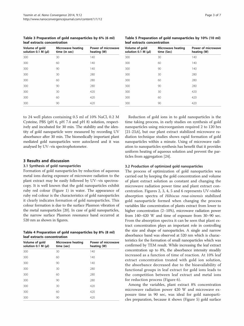
Table 3 Preparation of gold nanoparticles by 6% (6 ml)leaf extracts concentration
Volume of goldsolution 0.1 M (μl)
Microwave heatingtime (in sec)
Power of microwaveheating (W)
300 30 140
300 60 140
300 90 140
300 30 280
300 60 280
300 90 280
300 30 420
300 60 420
300 90 420
Table 5 Preparation of gold nanoparticles by 10% (10 ml)leaf extracts concentration
Volume of goldsolution 0.1 M (μl)
Microwave heatingtime (Sec)
Power of microwaveheating (W)
300 30 140
300 60 140
300 90 140
300 30 280
300 60 280
300 90 280
300 30 420
300 60 420
300 90 420
Yasmin et al. Nano Convergence 2014, 1:12 Page 3 of 7http://www.nanoconvergencejournal.com/content/1/1/12
to 24 well plates containing 0.5 ml of 10% NaCl, 0.2 MCysteine, PBS (pH 6, pH 7.4 and pH 8) solution, respect-ively and incubated for 30 min. The stability and the iden-tity of gold nanoparticle were measured by recording UVabsorbance after 30 min. The biomedically important plantmediated gold nanoparticles were autoclaved and it wasanalysed by UV–vis spectrophotometer.
3 Results and discussion3.1 Synthesis of gold nanoparticlesFormation of gold nanoparticles by reduction of aqueousmetal ions during exposure of microwave radiation to theplant extract may be easily followed by UV–vis spectros-copy. It is well known that the gold nanoparticles exhibitruby red colour (Figure 1) in water. The appearance ofruby red colour is the characteristics of gold nanoparticlesit clearly indicates formation of gold nanoparticles. Thiscolour formation is due to the surface Plasmon vibration ofthe metal nanoparticles [20]. In case of gold nanoparticles,the narrow surface Plasmon resonance band occurred at520 nm as shown in figures.
Table 4 Preparation of gold nanoparticles by 8% (8 ml)leaf extracts concentration
Volume of goldsolution 0.1 M (μl)
Microwave heatingtime (sec)
Power of microwaveheating (W)
300 30 140
300 60 140
300 90 140
300 30 280
300 60 280
300 90 280
300 30 420
300 60 420
300 90 420
Reduction of gold ions in to gold nanoparticles is thetime taking process, in early studies on synthesis of goldnanoparticles using microorganism required 2 to 120 hrs[21-23,6], but our plant extract stabilized microwave ra-diation technique studies shows rapid formation of goldnanoparticles within a minute. Using of microwave radi-ation to nanoparticles synthesis has benefit that it providesuniform heating of aqueous solution and prevent the par-ticles from aggregation [24].
3.2 Production of optimized gold nanoparticlesThe process of optimization of gold nanoparticles wascarried out by keeping the gold concentration and volumeof plant extract solution as constant and changing themicrowave radiation power time and plant extract con-centration. Figures 2, 3, 4, 5 and 6 represents UV-visibleabsorption spectra of Hibiscus rosa-sinensis stabilizedgold nanoparticle formed when changing the processvariables like concentration of plants extract from lower tohigher concentration (2–10%), microwave radiation powerfrom 140–420 W and time of exposure from 30–90 sec.From the absorption spectra it can be seen that plant ex-tract concentration plays an important role in controllingthe size and shape of nanoparticles. A single and narrowabsorbance band was observed at 520 nm which is charac-teristics for the formation of small nanoparticles which wasconfirmed by TEM result. While increasing the leaf extractconcentration up to 8%, the absorbance intensity steadilyincreased as a function of time of reaction. At 10% leafextract concentration treated with gold ion solution,the absorbance decreased due to the bioavailability offunctional groups in leaf extract for gold ions leads tothe competition between leaf extract and metal ionsfor reduction process (Figure 6).Among the variables, plant extract 8% concentration
microwave radiation power 420 W and microwave ex-posure time in 90 sec, was ideal for gold nanoparti-cles preparation, because it shows (Figure 5) gold surface

Figure 1 Visual observations (a) Plant leaf extract (b) gold chloride solution and (c) formation of gold nanoparticles.
Yasmin et al. Nano Convergence 2014, 1:12 Page 4 of 7http://www.nanoconvergencejournal.com/content/1/1/12
resonance occur at ~520 nm and steadily increased inintensity as a function microwave power and time ofreaction without any shift in peak wavelength. Thereis no change in peak wavelength suggest that particlesare monodispersed in the aqueous solution withoutaggregation [4].
3.3 Stability testingThe stability of Hibiscus rosa-sinensis stabilized gold na-noparticles was evaluated by monitoring the plasmonλmax in 0.2 M cysteine, 10% NaCl, phosphate buffersolution at pH 6, 7.4 and 8. The plasmon wavelengthin all above shows shift of ~1 – 8 nm. Our resultfrom the in-vitro stability studies has confirmed thatgold nanoparticles were stable in biological fluids atphysiological pH (Figure 7).
Figure 2 2% of Plant extracts and microwave heating at threedifferent temperature.
Gold nanoparticles used varies biomedical applica-tion before going to in-vivo models. Sterilization is veryimportant process to complete destruction of all organ-ism including bacteria, spores, virus.Several sterilization methods can be used, including
physical methods such as autoclaving and UV irradiation,which comprise moist heat and drug heat, respectively andchemical treatment such as using hydrogen peroxide gasplasma, ethylene oxide and chemical vapour, which includeboth gaseous and liquid solutions.Sterilization remains a critical step for the in-vivo
use of gold nanoparticles, and the effects of steriliza-tion on the integrity of the physiochemical propertiesof gold nanoparticles used to be investigated. Hence,gold nanoparticles sterilized by autoclave at 120°C for15 min before and after sterilization of gold nanoparticles
Figure 3 4% of Plant extracts and microwave heating at threedifferent temperatures.
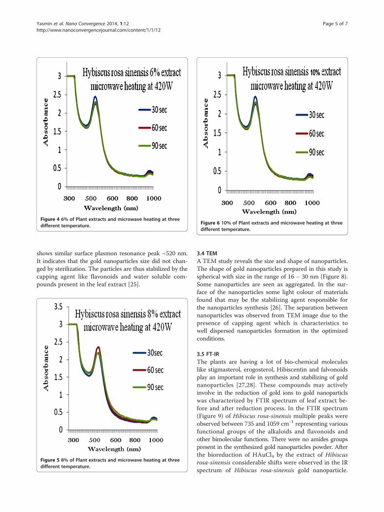
Figure 4 6% of Plant extracts and microwave heating at threedifferent temperature. Figure 6 10% of Plant extracts and microwave heating at three
different temperature.
Yasmin et al. Nano Convergence 2014, 1:12 Page 5 of 7http://www.nanoconvergencejournal.com/content/1/1/12
shows similar surface plasmon resonance peak ~520 nm.It indicates that the gold nanoparticles size did not chan-ged by sterilization. The particles are thus stabilized by thecapping agent like flavonoids and water soluble com-pounds present in the leaf extract [25].
Figure 5 8% of Plant extracts and microwave heating at threedifferent temperature.
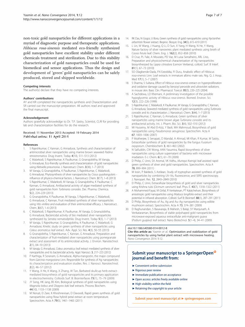
3.4 TEMA TEM study reveals the size and shape of nanoparticles.The shape of gold nanoparticles prepared in this study isspherical with size in the range of 16 – 30 nm (Figure 8).Some nanoparticles are seen as aggregated. In the sur-face of the nanoparticles some light colour of materialsfound that may be the stabilizing agent responsible forthe nanoparticles synthesis [26]. The separation betweennanoparticles was observed from TEM image due to thepresence of capping agent which is characteristics towell dispersed nanoparticles formation in the optimizedconditions.
3.5 FT-IRThe plants are having a lot of bio-chemical moleculeslike stigmasterol, erogosterol, Hibiscentin and falvonoidsplay an important role in synthesis and stabilizing of goldnanoparticles [27,28]. These compounds may activelyinvolve in the reduction of gold ions to gold nanoparticlswas characterized by FTIR spectrum of leaf extract be-fore and after reduction process. In the FTIR spectrum(Figure 9) of Hibiscus rosa-sinensis multiple peaks wereobserved between 735 and 1059 cmˉ1 representing variousfunctional groups of the alkaloids and flavonoids andother bimolecular functions. There were no amides groupspresent in the synthesized gold nanoparticles powder. Afterthe bioreduction of HAuCl4 by the extract of Hibiscusrosa-sinensis considerable shifts were observed in the IRspectrum of Hibiscus rosa-sinensis gold nanoparticle.
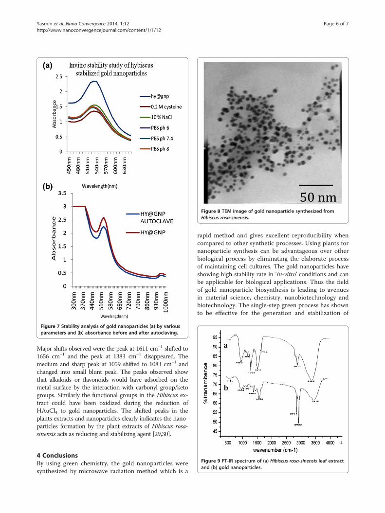
Figure 7 Stability analysis of gold nanoparticles (a) by variousparameters and (b) absorbance before and after autoclaving.
Figure 8 TEM image of gold nanoparticle synthesized fromHibiscus rosa-sinensis.
Yasmin et al. Nano Convergence 2014, 1:12 Page 6 of 7http://www.nanoconvergencejournal.com/content/1/1/12
Major shifts observed were the peak at 1611 cm−1 shifted to1656 cm−1 and the peak at 1383 cm−1 disappeared. Themedium and sharp peak at 1059 shifted to 1083 cm−1 andchanged into small blunt peak. The peaks observed showthat alkaloids or flavonoids would have adsorbed on themetal surface by the interaction with carbonyl group/ketogroups. Similarly the functional groups in the Hibiscus ex-tract could have been oxidized during the reduction ofHAuCl4 to gold nanoparticles. The shifted peaks in theplants extracts and nanoparticles clearly indicates the nano-particles formation by the plant extracts of Hibiscus rosa-sinensis acts as reducing and stabilizing agent [29,30].
Figure 9 FT-IR spectrum of (a) Hibiscus rosa-sinensis leaf extractand (b) gold nanoparticles.
4 ConclusionsBy using green chemistry, the gold nanoparticles weresynthesized by microwave radiation method which is a
rapid method and gives excellent reproducibility whencompared to other synthetic processes. Using plants fornanoparticle synthesis can be advantageous over otherbiological process by eliminating the elaborate processof maintaining cell cultures. The gold nanoparticles haveshowing high stability rate in ‘in-vitro’ conditions and canbe applicable for biological applications. Thus the fieldof gold nanoparticle biosynthesis is leading to avenuesin material science, chemistry, nanobiotechnology andbiotechnology. The single-step green process has shownto be effective for the generation and stabilization of
Yasmin et al. Nano Convergence 2014, 1:12 Page 7 of 7http://www.nanoconvergencejournal.com/content/1/1/12
non-toxic gold nanoparticles for different applications in amyriad of diagnostic purpose and therapeutic applications.Hibiscus rosa-sinensis mediated eco-friendly synthesizedgold nanoparticles have excellent stability under differentchemicals treatment and sterilization. Due to this stabilitycharacterization of gold nanoparticles could be used forbiomedical and sensor applications. Thus the design anddevelopment of ‘green’ gold nanoparticles can be safelyproduced, stored and shipped worldwide.
Competing interestsThe author(s) declare that they have no competing interests.
Authors’ contributionsAY and KR completed the nanoparticles synthesis and Characterization andSR carried out the manuscript preparation. All authors read and approvedthe final manuscript.
AcknowledgementAuthors gratefully acknowledge to Dr. T.P. Sastry, Scientist, CLRI for providinglab and characterization facilities for do the research.
Received: 11 November 2013 Accepted: 19 February 2014
References1. S Rajeshkumar, C Kannan, G Annadurai, Synthesis and characterization of
antimicrobial silver nanoparticles using marine brown seaweed Padinatetrastromatica. Drug Invent. Today 4(10), 511–513 (2012)
2. C Malarkodi, S Rajeshkumar, K Paulkumar, G Gnanajobitha, M Vanaja,G Annadurai, Eco-friendly synthesis and characterization of gold nanoparticlesusing Klebsiella pneumonia. J. Nanostruct. Chem. 3(30), 1–7 (2013)
3. M Vanaja, G Gnanajobitha, K Paulkumar, S Rajeshkumar, C Malarkodi,G Annadurai, Phytosynthesis of silver nanoparticles by Cissus quadrangularis -influence of physico-chemical factors. J. Nanostruct. Chem. 3(17), 1–8 (2013)
4. S Rajeshkumar, C Malarkodi, M Vanaja, G Gnanajobitha, K Paulkumar, CKannan, G Annadurai, Antibacterial activity of algae mediated synthesis ofgold nanoparticles from Turbinaria conoides. Der. Pharma. Chemica.5(2), 224–229 (2013)
5. G Gnanajobitha, K Paulkumar, M Vanaja, S Rajeshkumar, C Malarkodi,G Annadurai, C Kannan, Fruit mediated synthesis of silver nanoparticlesusing Vitis vinifera and evaluation of their antimicrobial efficacy. J. Nanostruct.Chem. 3(67), 1–6 (2013)
6. C Malarkodi, S Rajeshkumar, K Paulkumar, M Vanaja, G GnanaJobitha,G Annadurai, Bactericidal activity of bio mediated silver nanoparticlessynthesized by Serratia nematodiphila. Drug Invent. Today 5(3), 1–7 (2013)
7. M Vanaja, S Rajeshkumar, G Gnanajobitha, K Paulkumar, C Malarkodi, GAnnadurai, Kinetic study on green synthesis of silver nanoparticles usingColeus aromaticus leaf extract. Adv. Appl. Sci. Res. 4(3), 50–55 (2013)
8. G Gnanajobitha, S Rajeshkumar, C Kannan, G Annadurai, Preparation andcharacterization of fruit-mediated silver nanoparticles using pomegranateextract and assessment of its antimicrobial activity. J. Environ. Nanotechnol.2(1), 04–10 (2013)
9. M Vanaja, G Annadurai, Coleus aromaticus leaf extract mediated synthesis of silvernanoparticles and its bactericidal activity. Appl. Nanosci. 3, 217–223 (2012)
10. P Karthiga, R Soranam, G Annadurai, Alpha-mangostin, the major compoundfrom Garcinia mangostana Linn. Responsible for synthesis of Ag nanoparticles:its characterization and evaluation studies. Res. J. Nanosci. Nanotechnol.2(2), 46–57 (2012)
11. Y Wang, X He, K Wang, X Zhang, W Tan, Barbated skullcup herb extract-mediated biosynthesis of gold nanoparticles and its primary applicationin electrochemistry. Colloids Surf. B: Biointerfaces 73(1), 75–79 (2009)
12. JY Song, HK Jang, BS Kim, Biological synthesis of gold nanoparticles usingMagnolia kobus and Diopyros kaki leaf extracts. Process Biochem.44(10), 1133–1138 (2009)
13. M Noruzi, D Zare, K Khoshnevisan, D Davoodi, Rapid green synthesis of goldnanoparticles using Rosa hybrid petal extract at room temperature.Spectrochim. Acta A 79(5), 1461–1465 (2011)
14. RK Das, N Gogoi, U Bora, Green synthesis of gold nanoparticles using Nyctanthesarbortristis flower extract. Bioproc. Biosyst. Eng. 34(5), 615–619 (2011)
15. L Lin, W Wang, J Huang, Q Li, D Sun, X Yang, H Wang, N He, Y Wang,Nature factory of silver nanowires: plant mediated synthesis using broth ofCassia fistula leaf. Chem. Eng. J. 162(2), 852–858 (2010)
16. D Cruz, PL Fale, A Mourato, PD Vaz, M Luisa Serralheiro, ARL Lino,Preparation and physicochemical characterization of Ag nanoparticlesbiosynthesized by Lippia citriodora (Lemon Verbena), colloid. Surf. B Interf.81(1), 67–73 (2010)
17. SO Olagbende-Dada, FN Ezeobika, FI Duru, Anabolic effect of Hibiscusrosa-sinensis Linn. Leaf extracts in immature albino male rats. Nig. Q. J. Hosp.Med 17(1), 5–7 (2007)
18. S Sharma, S Sultana, Effect of Hibiscus rosa-sinensis extract on hyperproliferationand oxidative damage caused by benzoyl peroxide and ultraviolet radiationsin mouse skin. Basic Clin. Pharmacol. Toxicol. 95(5), 220–225 (2004)
19. A Sachdewa, LD Khemani, A preliminary investigation of the possiblehypoglycemic activity of Hibiscus rosa-sinensis. Biomed. Environ. Sci.12(3), 222–226 (1999)
20. S Rajeshkumar, C Malarkodi, K Paulkumar, M Vanaja, G Gnanajobitha, C Kannan,G Annadurai, Seaweed mediated synthesis of gold nanoparticles using Turbinariaconoides and its characterization. J. Nanostruct. Chem. 3(44), 1–7 (2013)
21. S Rajeshkumar, C Kannan, G Annadurai, Green synthesis of silvernanoparticles using marine brown algae Turbinaria conoides and itsantibacterial activity. Int. J. Pharm. Bio. Sci. 3(4), 502–510 (2012)
22. MI Husseiny, M Abd El-Aziz, Y Badr, MA Mahmoud, Biosynthesis of goldnanoparticles using Pseudomonas aeruginosa. Spectrochim. Acta A67, 1003–1006 (2007)
23. P Mukherjee, S Senapati, D Mandal, A Ahmad, MI Khan, R Kumar, M Sastry,Extracellular synthesis of gold nanoparticles by the fungus Fusariumoxysporum. Chembiochem 3, 461–463 (2002)
24. N Saifuddin, CW Wong, AAN Yasumira, Rapid biosynthesis of silvernanoparticles using culture supernatant of bacteria with microwaveirradiation. E-J. Chem. 6(1), 61–70 (2009)
25. D Philip, C Unni, SA Aromal, VK Vidhu, Murraya Koenigii leaf-assisted rapidgreen synthesis of silver and gold nanoparticles. Spectrochim. Acta A78, 899–904 (2011)
26. M Iosin, P Baldeck, S Astilean, Study of tryptophan assisted synthesis of goldnanoparticles by combining UV–Vis, fluorescence, and SERS spectroscopy.J. Nanopart. Res. 12, 2843–2849 (2010)
27. D Philip, C Unni, Extracellular biosynthesis of gold and silver nanoparticlesusing Krishna tulsi (Ocimum sanctum) leaf. Phys. E. 43(7), 1318–1322 (2011)
28. A Mohammed-Fayaz, M Girilal, R Venkatesan, PT Kalaichelvan, Biosynthesis ofanisotropic gold nanoparticles using Maduca longifolia extract and theirpotential in infrared absorption. Colloid. Surf B: Biointerf. 88(1), 287–291 (2011)
29. D Philip, Biosynthesis of Au, Ag and Au–Ag nanoparticles using ediblemushroom extract. Spectrochim. Acta A 73, 374–381 (2009)
30. D Raghunandan, S Basavaraja, B Mahesh, S Balaji, SY Manjunath, AVenkataraman, Biosynthesis of stable polyshaped gold nanoparticles frommicrowave-exposed aqueous extracellular anti-malignant guava(Psidium guajava) leaf extract. Nano Biotechnol. 5(1–4), 34–41 (2009)
doi:10.1186/s40580-014-0012-8Cite this article as: Yasmin et al.: Optimization and stabilization of goldnanoparticles by using herbal plant extract with microwave heating.Nano Convergence 2014 1:12.
Submit your manuscript to a journal and benefi t from:
7 Convenient online submission
7 Rigorous peer review
7 Immediate publication on acceptance
7 Open access: articles freely available online
7 High visibility within the fi eld
7 Retaining the copyright to your article
Submit your next manuscript at 7 springeropen.com