Embed Size (px)
Citation preview

RESEARCH Open Access
Developmental expression of p97/VCP (Valosin-containing protein) and Jab1/CSN5 in the rattestis and epididymisSevil Cayli1*, Seda Ocakli1, Fikret Erdemir2, Ufuk Tas3, Huseyin Aslan1, Tamer Yener4 and Zafer Karaca1
Abstract
Background: The ubiquitin proteasome system (UPS) is a key player in regulating many cellular processes viaproteasomal degradation of ubiquitinated proteins. Recently published data show that Jab1/CSN5 interacts withp97/VCP and controls the ubiquitination status of proteins bound to p97/VCP in mouse and human cells. However,coexpression of p97/VCP and Jab1/CSN5 in the developing rat testis and epididymis has not previously beenstudied.
Methods: Testicular and epididymal tissues from 5-, 15-, 30-, and 60-day-old rats were examined byimmunohistochemistry and Western blotting. Colocalisation of proteins was determined by immunofluorescencemicroscopy.
Results: In the 5-day-old rat testis, p97/VCP and Jab1/CSN5 were specifically expressed in gonocytes. Theexpression of p97/VCP and Jab1/CSN5 significantly increased at day 15 and was found in spermatogonia, Sertolicells and spermatocytes. In 30- and 60-day-old rat testes, p97/VCP indicated moderate to strong expression inSertoli cells, spermatogonia, round and elongating spermatids. However, moderate to weak expression wasobserved in spermatocytes. Jab1/CSN5 showed strong expression in spermatogonia and spermatocytes, whilerelatively moderate expression was observed in round and elongating spermatids in 30- and 60-day-old rat testes.In contrast, in the epididymis, the expression of both proteins gradually increased from 5 to 60 days of age. Afterrats reached 2 weeks of age, the expression of both proteins was mostly restricted to the basal and principal cellsof the caput epididymis.
Conclusions: Our study suggests that p97/VCP and Jab1/CSN5 could be an important part of the UPS in thedeveloping rat testis and epididymis and that both proteins may be involved in the regulation of spermatogenesisand epididymal epithelial functions.
BackgroundThe testis has the specific function of generating sperma-tozoa from precursors called spermatogonia after anintricate series of divisions [1]. This process takes placewithin the seminiferous epithelium, which is a complexstructure composed of germ cells with radially orientedsupporting cells termed Sertoli cells. In postnatal animals,spermatogenesis is initiated in the testes when gonocytesresume proliferation, migrate to the seminiferous tubulebasal membrane and differentiate into spermatogonial
stem cells [2]. The postnatal phase is divided into threemain stages: 1) mitotic proliferation of spermatogonialstem cells and premeiotic differentiation of spermatogo-nia into diploid primary spermatocytes; 2) meiotic differ-entiation of primary spermatocytes into haploid early,round spermatids; and 3) spermiogenesis, a cellular andnuclear reorganisation process that differentiates sperma-tids into spermatozoa [3].The epididymis provides an adequate environment for
the final maturation of sperm [4-6]. During embryonicand postnatal development of the testis and epididymis,regulated proteolysis and organelle degradation arerequired [7-9]. The ubiquitin proteasome system (UPS)is a developmentally regulated and highly substrate-
* Correspondence: [email protected] of Histology and Embryology, Faculty of Medicine,Gaziosmanpasa University, Tokat, TurkeyFull list of author information is available at the end of the article
Cayli et al. Reproductive Biology and Endocrinology 2011, 9:117http://www.rbej.com/content/9/1/117
© 2011 Cayli et al; licensee BioMed Central Ltd. This is an Open Access article distributed under the terms of the Creative CommonsAttribution License (http://creativecommons.org/licenses/by/2.0), which permits unrestricted use, distribution, and reproduction inany medium, provided the original work is properly cited.

specific pathway for the removal of damaged and aber-rant proteins. It is well known that the UPS fulfilsnecessary requirements for sperm cell differentiationinside the testicular seminiferous tubules and cell cyclecontrol throughout spermatogenesis and fertilisation inadult males. Moreover, the ubiquitin-activating enzymes(E1), ubiquitin-conjugating enzymes (E2), ubiquitinligase (E3) and some proteasomal subunits are expressedduring spermatogenesis and postnatal testicular develop-ment [10-13].In the male reproductive system, the UPS contributes
to gamete quality control mechanisms, carrying outselective spermatogonial removal at the haploid phase ofspermatogenesis [14], protein and organelle degradationduring spermiogenesis [8,15], and the tagging defectivespermatozoa with ubiquitin in the epididymis [7,16]. Forthese reasons, exploring the expression of proteins thatare the components of the UPS may lead to advances inunderstanding the biology of the testis and epididymis.In the ATP-dependent ubiquitin pathway, the attach-
ment of ubiquitin to a target protein, referred to as ubi-quitination, is carried out by E1, E2 and E3 [17]. Themain purpose of ubiquitination is to deliver the ubiquiti-nated proteins to a cellular trash bin, a lysosome, anautophagosomal vacuole, or a 26S proteasome. Ubiquiti-nated proteins can either be transferred directly to theproteasome or indirectly transferred via p97/Valosin-containing protein (VCP), a member of the ATPasesuper-family associated with diverse cellular activities(AAA-ATPase). p97/VCP has been associated with awide variety of essential cellular protein pathways,including nuclear envelope reconstruction, cell cycleregulation, Golgi reassembly, suppression of apoptosis,DNA damage responses, maturation of autophagosomeand sperm capacitation [18-26]. In addition, duringendoplasmic reticulum-associated degradation, p97/VCPdislodges ubiquitinated proteins from the endoplasmicreticulum (ER) and chaperones them to the cytosol forproteasomal degradation [27]. For ubiquitination of mis-folded proteins in the ER, interaction with p97/VCP isrequired [28]. Moreover, it has recently been shown thatthe COP9 signalosome (CSN) interacts in an ATP-dependent manner with p97/VCP and controls the ubi-quitination status of proteins bound to p97/VCP [29].The CSN, which is involved in the ubiquitin/protea-
some system, contains eight core subunits (CSN1-8),like the proteasome lid complex. CSN5 (also known asJab1) facilitates the 26S proteasome-dependent degrada-tion of several proteins, including p27Kip, luteinisinghormone receptor (LHR), p53, oestrogen receptor,Smad4, Smad7, Id1, Id3, and I�Ba [30-35]. The JAMM(JAB1/MPN/Mov34 metalloenzyme) domain present inJab1/CSN5 exhibits deubiquitinase activity regulatingubiquitinlated protein sorting when associated with the
CSN [36]. In addition to its role in the UPS, Jab1/CSN5regulates many signalling pathways, such as transform-ing growth factor (TGF)-b signalling, cell proliferation,apoptosis and DNA repair [37].Although it seems clear that the UPS is fundamentally
required for the male reproductive system, there cur-rently exists no information on the colocalisation pat-terns of p97/VCP and Jab1/CSN5 and their possibleroles in the developing rat testis and epididymis. There-fore, the goal of this study was to assess the develop-mental expression of p97/VCP and Jab1/CSN5 and toshow the cellular localisation of both complexes in rattestis and epididymis. For this reason, we analysed theexpression of p97/VCP and Jab1/CSN5 in rat testis andepididymis during postnatal development using immu-nohistochemistry, immunofluorescence and Westernblot techniques.
MethodsAnimals and tissue preparationAfter obtaining approval from the local ethics commit-tee (2010-HADYEK-012), twenty-four male Wistaralbino rats at postnatal ages of 5, 15, 30 and 60 days(six rats per group), i.e., corresponding to infantile (5day), prepubertal (15 day), pubertal (30 day), and adult(60 day) periods, were obtained from the GaziosmanpasaUniversity Experimental Animal Research Laboratory.The rats were cared in the laboratory according to insti-tutional guidelines and the Guide for Care and Use ofLaboratory Animals of the National Research Council.All rats were observed for several days to ascertain theirhealth status before sample collection. Pups were rearedwith their dams. They were maintained in a tempera-ture-controlled room (20-23 °C) on a 12 h light/darkcycle with food (commercial rat chow) and fresh wateravailable adlibitum. Six rats of the same age were killedby administration of an overdose of sodium pentobarbi-tal (150 mg/kg, i.p.) before removal of the testis and epi-didymis. Epididymes were dissected and subdivided intothree anatomical regions: the caput (CT), corpus (CS)and cauda (CA) epididymis. One testis and epididymisfrom each animal were fixed in Bouin’s fluid for 12 himmediately upon collection, then dehydrated andembedded in paraffin for histochemistry and immuno-histochemistry experiments (see below). The contralat-eral testis and epididymal regions from each animalwere snap frozen in liquid nitrogen and strored at -80°Cfor Western blotting.The definition of the stages and cell types with respect
to the cycle of the seminiferous epithelium in adult tes-tis was determined as previously published [38,39].Germ cells were distinguished as being spermatogonia,spermatocytes, round and elongating spermatids basedon their morphology and position in the seminiferous
Cayli et al. Reproductive Biology and Endocrinology 2011, 9:117http://www.rbej.com/content/9/1/117
Page 2 of 13

epithelium [40-42]. To spesificially identify mature Ser-toli cells, p27kip1 immunostaining was performed at days30 and 60 testis and cytokeratin 18 immunostainingwere used to determine immature Sertoli cells at days15 [43,44].
ImmunohistochemistryFor immunohistochemical analysis, 5 μm-thick serialsections were collected on poly-L-lysine-coated slides(Sigma-Aldrich, St. Louis, MO, USA) and incubatedovernight at 56 °C. Tissue sections were deparaffinisedin xylene and rehydrated in a graded series of ethanol.Sections were then treated in a microwave oven in 10mM citrate buffer, pH 6.0 and left to cool for 20 min.After three washes in phosphate buffered saline (PBS),endogenous peroxidase activity was quenched by 3%hydrogen peroxide in PBS for 20 min., and the sectionswere washed again three times in PBS. The sectionswere then incubated in a blocking serum (Ultra VBlock, TP-060-HL; NeoMarker, Fremont, CA, USA) for10 min. to block non-specific binding. Subsequently,sections were incubated overnight at 4°C with followingprimary antibodies: mouse monoclonal p97/VCP (MA3-004, 1: 500, Affinity BioReagent, USA), Jab1/CSN5 (sc-9074, 1: 200, Santa Cruz Tech, USA), p27kip1 (sc-1641,1: 100, Santa Cruz Tech, USA), p27kip1 (sc-776, 1: 100,Santa Cruz Tech, USA), cytokeratin 18 (sc-58729, 1:200, Santa Cruz Tech, USA), p97/VCP (sc-20799, 1:200, Santa Cruz Tech, USA) and Jab1/CSN5 (ab495, 1:250, Abcam, UK). The sections were then washed threetimes in PBS and incubated with biotinylated anti-mouse (BA-9200; 1:400 Dilution; Vector Laboratories,Burlingame, CA) and biotinylated anti-rabbit (BA-1000;1:400 Dilution; Vector Laboratories) secondary antibo-dies for 45 min. at room temperature. After threewashes with PBS, the antigen-antibody complexes weredetected using a streptavidin-peroxidase complex (TP-060-HL; LabVision, Fremont, CA, USA) for 15 min., fol-lowed by three rinses with PBS. Bound peroxidase wasdeveloped with 3-amino-9-ethylcarbazol (AEC) (ScyTekLaboratories, USA) chromogen, and sections were coun-terstained with Mayer’s hematoxylin (ScyTek Labora-tories, Utah, USA) and mounted with Permount (FisherChemicals, Springfield, NJ, USA) on glass slides. Forcontrols, sections were treated with the appropriate iso-type of mouse IgG or rabbit IgG, depending on the pri-mary antibody used, which was diluted to the same finalprotein concentration as the primary antibody. Photomi-crographs were collected with a Leica microscope (LeicaDM2500, Nussloch, Germany).
Double immunofluorescenceTo identify colocalisation of proteins, immunofluores-cence was performed. Both primary antibodies (rabbit
anti-Jab1/CSN5 and mouse anti-p97/VCP, mouse anti-p97/VCP and rabbit anti-p27kip1, mouse anti-Jab1/CSN5and rabbit anti-p27kip1) were applied simultaneously andincubated overnight at 4°C. After rinsing in PBS, sec-tions were incubated with FITC-conjugated goat anti-rabbit secondary antibody (at a 1:200 dilution, Millipore,USA) and rhodamine-conjugated goat anti-mouse sec-ondary antibody (at a 1:500 dilution, sc-2092, SantaCruz Biotechnology, USA) for 1 hour at room tempera-ture. Thereafter, all sections were rinsed in PBS andnuclei were stained with DAPI (Invitrogen, MolecularProbes, D-1306). Fluorescence images were taken with aNikon microscope (Nikon E600, Germany).
H-SCORE analysisEvaluation of the immunohistochemical labelling wasperformed using H-SCORE analyses as previouslydescribed [45]. The intensities of the p97/VCP andJab1/CSN5 immunoreactivities were evaluated semi-quantitatively using the following intensity categories: 0(no staining), 1+ (weak but detectable staining), 2+(moderate or distinct staining), and 3+ (intense stain-ing). For each tissue, an H-SCORE value was derived bycalculating the sum of the percentages of cells thatstained at each intensity category and multiplying thatvalue by the weighted intensity of the staining using theFormula H-SCORE: ∑Pi(i+ l), where ‘i’ represents theintensity scores and ‘Pi’ is the corresponding percentageof cells. For each slide, five randomly selected areaswere evaluated under a light microscope (40 x objec-tive), and the percentage of cells exhibiting each inten-sity within these areas was determined at different timesby two investigators who were not informed about thetype and source of the tissues. The average score ofboth observers was used.
SDS-PAGE and Western blot analysesTotal testicular and epididymal proteins were extractedusing modified RIPA buffer (1% NP-40; 0.25% sodiumdeoxycholate; 150 mM NaCl; 1 mM EDTA; 1 mMPMSF; 1 mg/ml each of aprotinin, leupeptin, and pep-statin; 1 mM Na3VO4; and 1 mM NaF in 50 mM Tris-Cl, pH 7.4) and quantitated using the Bradford proce-dure (Bio-Rad, Hercules, CA). Then, 40-μg sampleswere separated by 8% SDS-PAGE and electroblottedonto a nitrocellulose membrane (Bio-Rad Laboratories).The membrane was blocked with 5% non-fat dry milkin TBS containing 0.1% Tween 20 (TBS-T) for 1 h toreduce non-specific binding. Subsequently, the mem-brane was incubated for 1 h with primary antibodiesagainst p97/VCP (MA3-004, Affinity BioReagent, USA,1:10,000 in 5% non-fat dry milk), Jab1/CSN5 (sc-9074,Santa Cruz Biotechnology, 1:500 in 5% non-fat dry milk)and b-actin (sc-47778, C4; Santa Cruz Biotechnology
Cayli et al. Reproductive Biology and Endocrinology 2011, 9:117http://www.rbej.com/content/9/1/117
Page 3 of 13

Inc., 0.05 mg/ml in 5% non-fat dry milk). The mem-brane was washed with TBS-T for 1 h and incubatedwith horseradish peroxidase-conjugated anti-mouse andanti-rabbit secondary antibodies (Vector Laboratories)diluted in 5% non-fat dry milk in TBS-T. Bound second-ary antibodies were visualised using an enhanced chemi-luminescence substrate (GE Healthcare). Immunoblotbands for p97/VCP, Jab1/CSN5 and b-actin were quan-tified using an Alpha DigiDoc 1000 gel documentationunit (Alpha Innotech Corporation, CA, USA). The opti-cal density (OD) values for the p97/VCP and JAb1/CSN5 bands were divided by the OD values of cognateb-actin bands to normalise the OD values for loadingdifferences.
Statistical analysisThe Mann-Whitney U-test was employed for compari-son of independent groups of samples, and Kruskall-Wallis analysis with the Dunn posthoc test was per-formed for multiple comparisons of independent groupsof samples. A P-value of less than 0.05 was consideredto indicate a statistically significant difference. Statisticalcalculations were performed using SigmaStat for Win-dows, version 3.5 (Jandel Scientific Corp., San Rafael,CA).
ResultsCellular localisation of p97/VCP and Jab1/CSN5 in thedeveloping rat testisImmunohistochemical analysis of 5-day-old rat testesrevealed that p97/VCP was mainly localised to nuclearand cytoplasmic regions of gonocytes in the lumen ofthe seminiferous tubules (Figure 1A). No immunoreac-tivity was detected in somatic cells, though some inter-stitial cells showed immunopositivity for p97/VCP(Figure 1A). In the 15-day-old rat testis, spermatogoniaat the basal membrane, Sertoli cells and spermatocyteswere positive for p97/VCP (Figure 1B, Table 1). Addi-tionally, interstitial cells showed weak to moderateimmunostaining for p97/VCP at day 15 (Figure 1B). Thestaining intensity and the number of cells positivelystained for p97/VCP significantly increased by day 15,coinciding with the appearance of leptotene spermato-cytes [39] (Figure 1B and 1M). The other cell types pre-sent in the 30-day-old rat testis, specifically round andelongating spermatids, also expressed p97/VCP (Figure1C and 1D). Additionally, supporting Sertoli and peritu-buler myoid cells, interstitial cells showed relativelymoderate expression of p97/VCP, reaching the highestexpression level at day 30 (Figure 1C-D and 1M). Inadult rat (60 day) testis, p97/VCP indicated moderate tostrong expression in the Sertoli cells, spermatogonia,round and elongating spermatids; however, spermato-cytes showed moderate to weak expression of p97/VCP
(Figure 1E-F, Table 1). The staining intensity and thenumber of p97/VCP-positive cells were found to be thesame in all stages of seminiferous tubules in the adultrat testis. There was no immunoreactivity observed onnegative control slides that were treated with the isotypemouse antibody instead of the p97/VCP primary anti-body at the same final concentration (Figure 1A, 1C,inserts).In the 5-day-old rat testis, Jab1/CSN5 showed moder-
ate immunolabelling in gonocytes (Figure 1G). Thestaining intensity and the number of Jab1/CSN5-positi-vite cells were significantly increased in the 15-day-oldrat testis compared to the 5-day-old rat testis (Figure1H and 1M). In the 15-day-old rat testis, Jab1/CSN5localised strongly in spermatogonia and spermatocytes,but Sertoli cells presented only moderate to weakimmunostaining (Figure 1H, and Table 1). In the 30-day-old rat testis, spermatogonia, spermatocytes, roundand elongating spermatids, and Sertoli and interstitialcells were positively labelled with Jab1/CSN5, presentingthe highest expression level observed (Figure 1I, Table1).In the 60-day-old rat testis, Jab1/CSN5 showed stron-
ger nuclear staining in spermatogonia and moderatecytoplasmic staining in spermatocytes at stages 8-12(Figure 1J-L). However, the acrosomes of round sperma-tids at stages 6-8 and the heads of elongating spermatidsat stages 1-5 and 9-14 displayed relatively weak immu-nostaining for Jab1/CSN5 (Figure 1K, 1L). A few inter-stitial cells and Sertoli cells presented weak labelling forJab1/CSN5. No immunoreactivity was detected on con-trol slides (Figure 1G, 1H and 1L, inserts). According toH-SCORE analysis, the expression of both Jab1/CSN5and p97/VCP significantly increased from day 5 to day30 in the testis, whereas no significant differences werefound between day 30 and day 60 testes (Figure 1M).Immunoexpression of immature and mature Sertoli
cell markers such as cytokeratin 18 and p27kip1 wasused to confirm whether p97/VCP and Jab1/CSN5 wasexpressed in Sertoli cells at days 15, 30 and 60 days ofrat testis (Figure 2A-F). At days 15, Sertoli cells wereimmunopositive for cytokeratin 18 (Figure 2A). At days30 and 60, p27kip1 was found to be expressed in themature Sertoli cells (Figure 2B and 2C). Moreover,p27kip1 was colocalised with p97/VCP (Figure 2D-F) andJab1/CSN5 (Figure 2G-I) in the mature Sertoli cells.
Cellular localisation of p97/VCP and Jab1/CSN5 in thedeveloping rat epididymisIn the 5-day-old rat epididymis, p97/VCP immunostain-ing was found in the CT, CS and CA epididymis.Epithelial cells of epididymis were positively labelled forp97/VCP (Figure 3A-C). p97/VCP immunoreactivityshowed no uniformity in different regions of the
Cayli et al. Reproductive Biology and Endocrinology 2011, 9:117http://www.rbej.com/content/9/1/117
Page 4 of 13

epididymis, and high levels of expression were observedin CS at day 5 (Figure 3B). However, p97/VCP expres-sion was higher in the CT and CS epididymis comparedto the CA epididymis from day 15 to day 60 (Figure3D-L, Table 2). p97/VCP immunostaining was seen inboth the basal and principal cells of the adult rat epidi-dymis (Figure 3J-L, Table 2). Moreover, in the lumen of
the epididymis, spermatozoa presented weak staining forp97/VCP at day 60 (Figure 3L).In 5-day-old rat epididymes, Jab1/CSN5 immunoposi-
tivity was detected in the CT, CS and CA regions (Fig-ure 4A-C). Although some epithelial cells showed noimmunoreactivity, Jab1/CSN5 was expressed in both theprincipal and basal cells at day 15, 30 and 60 in the
Figure 1 Localisation of p97/VCP and Jab1/CSN5 in the developing rat testis. Immunohistochemistry was used to show the cellularlocalisation of p97/VCP and Jab1/CSN5 in the testis at days 5 (A, G), 15 (B, H), 30 (C, D, I), and 60 (E, F, J, K, L) after birth. The negative controls(A, C, G, H, L, inserts). A: Positive staining of p97/VCP expression was demonstrated in the nuclear and cytoplasmic regions of gonocytes (GC) atday 5. Interstitial cells (I) show weak immunoreactivity, and peritubuler cells (P) present no immunoreactivity. B: p97/VCP is highly expressed inspermatogonia (Sg), spermatocytes (Sc) and Sertoli cells (Se) at day 15. Some I show moderate immunostaining for p97/VCP. C, D: Elongating(eSt) and round (rSt) spermatids, Se and Sg show moderate to strong immunostaining for p97/VCP, while cytoplasm of Sc are moderatelylabelled with p97/VCP. E, F: In the adult testis, p97/VCP is weakly to moderately expressed in the cytoplasm of Sc, whereas Se, Sg, rSt and eSt aremoderate to strong immunopositive for p97/VCP. G: Moderate Jab1/CSN5 expression is seen at day 5 in GC. H: Jab1/CSN5 is strongly expressedin Sg and Sc while Se exhibit moderate expression at day 15. I: Sg, Sc, eSt and Se show moderate to strong immunostaining for Jab1/CSN5. J-L:In the adult testis, Jab1/CSN5 is strongly expressed in Sg and Sc, while rSt and eSt show weak to moderate immunostaining for Jab1/CSN5.Roman numerals indicate stages of the seminiferous epithelial cycle. M: H-SCORE of the p97/VCP and Jab1/CSN5 immunostaining intensities inthe developing rat testis. The data are represented as the means ± SEM. a: p < 0.05, day 5 vs. days 15, 30 and 60, b: day 15 vs. day 5 and day30, c: day 30 vs. days 5 and 15. Scale bars: 50 μm.
Cayli et al. Reproductive Biology and Endocrinology 2011, 9:117http://www.rbej.com/content/9/1/117
Page 5 of 13

epididymis (Figure 4D-L). On days 5 and 15, Jab1/CSN5expression was relatively higher in the CS epididymis(Figure 4B-E); however, the CT epididymis presentedincreased expression in day 30 and 60 testes (Figure 4G,4J). Spermatozoa showed weak immunostaining in thelumen of day 60 epididymes (Figure 4J-L).
Colocalisation of Jab1/CSN5 and p97/VCP in thedeveloping rat testis and epididymisTo determine Jab1/CSN5 and p97/VCP colocalisation,double immunofluorescence was performed in the testi-cular (Figure 5) and epididymal tissues (Figure 6). In the5-day-old rat testis, Jab1/CSN5 and p97/VCP werefound to be colocalised in gonocytes (Figure 5A). In the15-day-old rat testis, p97/VCP immunostaining wasoverlapped with Jab1/CSN5 in spermatogonia, sperma-tocytes and Sertoli cells (Figure 5B). Although p97/VCPand Jab1/CSN5 staining intensities indicated differencesin day 30, cytoplasm of spermatocytes, Sertoli cells,
spermatogonia, round (rSt) and elongating (eSt) sperma-tids were found to be immunopositive for Jab1/CSN5and p97/VCP (Figure 5C). In the 60-day-old rat testis,spermatogonia, spermatocytes, Sertoli cells and elongat-ing spermatids were found to be double immunopositivefor Jab1/CSN5 and p97/VCP, although some Sertolicells and spermatogonia showed only Jab1/CSN5 orp97/VCP immunpositivity (Figure 5D).In the 5-day-old rat epididymis, Jab1/CSN5 and p97/
VCP were colocalised in the cytoplasm of the epithelialcells of corpus epididymis (Figure 6A). In the 15, 30 and60-day-old rat epididymis, both proteins were found tocolocalise in the nuclear and cytoplasmic regions (Figure6B-D). Overall, the expression of Jab1/CSN5 and p97/VCPwas gradually increased from 5 to 60 days of epididymis.
Changes in protein expression levels of p97/VCP andJab1/CSN5 in the developing rat testis and epididymisIn agreement with immunohistochemistry, Western blotresults confirmed the presence of p97/VCP and Jab1/CSN5 in the developing rat testis and epididymis. Wes-tern blot analyses revealed a specific band at 97 kDa forp97/VCP and at 38 kDa for Jab1/CSN5. The intensity ofboth bands was quantified and normalised for the inten-sity of b-actin controls (Figure 7A-B, lower panels). Theexpression level of p97/VCP was low on day 5 of testisand epididymis but increased and reached a peak on day30. However, no significant changes were observedbetween 30 and 60-day-old rat testis for p97/VCPexpression (Figure 7A). Jab1/CSN5 expression was alsoincreased gradually but reached the peak on day 30 intestis and on day 60 in the epididymis (Figure 7A-B). Inaccordance with immunohistocehemical results, p97/VCP and Jab1/CSN5 expressions were significantlyhigher in the CT epididymis compared to the CS andCA epididymis at day 30 and day 60 epididymis (Figure7B, lower panel).
DiscussionTo our knowledge, this is the first study demonstratingcoexpression of the UPS components p97/VCP andJab1/CSN5 during postnatal development of the rat tes-tis and epididymis using immunohistochemistry,
Table 1 Localisation of Jab1/CSN5 and p97/VCP in rat testis
GC Sg Sc rSt eSt Se I
Postnatal Day Jab1 VCP Jab1 VCP Jab1 VCP Jab1 VCP Jab1 VCP Jab1 VCP Jab1 VCP
5 ++ ++ – – – – – – – – – – – +
15 – – +++ +++ +++ ++ – – – – ++ ++ + +
30 – – +++ +++ ++ ++ ++ ++ ++ ++ ++ ++ ++ ++
60 – – +++ +++ ++ + + ++ ++ ++ + +++ + +
Semiquantitive evaluation of Jab1/CSN5 and p97/VCP immunoreactive cells in the developing rat testis. GC: Gonocytes, Sg: Spermatogonia, Sc: Spermatocyte, rSt:Round spermatid, eSt: Elongating spermatid, Se: Sertoli cell, I: Interstitial cells.
weak immunoreactivity: +, moderate immunoreactivity: ++, strong immunoreactivity: +++
Figure 2 Immunohistochemical analysis of Sertoli cells at day 5(A), 15 (B), 60 (C) and colocalisation of p97/VCP - p27kip1 (D-F)and Jab1/CSN5 - p27kip1 (G-I) in rat testis. Cytokeratin 18 (CK) aredetected in immature Sertoli cells (Se) at day 15 (A) and p27kip1 isobserved in mature Sertoli cells (Se) of day 30 and 60 (B, C) in rattestis. Mature Sertoli cells (Se) are immunopositive for p27kip1 (green,D, G), p97/VCP (red, E) and Jab1/CSN5 (red, H), resulting in a yellowcolor (overlay, F, I). Scale bars: 25 μm.
Cayli et al. Reproductive Biology and Endocrinology 2011, 9:117http://www.rbej.com/content/9/1/117
Page 6 of 13

immunofluorescence and Western blotting. Our resultsindicate that p97/VCP expression overlapped with Jab1/CSN5 expression in gonocytes, spermatogonia, sperma-tocytes, Sertoli cells, spermatids and epididymal epithe-lial cells in the 5-, 15-, 30- and 60-day-old rat testis andepididymis.
To define specific sites where p97/VCP and Jab1/CSN5 expression might be important, we explored cell-specific expression of p97/VCP and Jab1/CSN5 in thedeveloping rat testis and epididymis. In the 5-day-oldrat testis, we observed that p97/VCP and Jab1/CSN5were specifically expressed in gonocytes and that the
Figure 3 Distribution of p97/VCP in different regions of the rat epididymis during postnatal development. p97/VCP immunopositivity isdetected in the CT, CS and CA of the epididymis at days 5 (A, B, C), 15 (D, E, F), 30 (G, H, I) and 60 (J, K, L). p97/VCP expression is seen in theepithelial cells (EC) of the CT, CS and CA epididymis at day 5 (A, B, C). No significant staining is observed in the negative controls (at the cornerof panels A-L). p97/VCP is localised both in the basal (BS) and principal (PC) cells of the epididymis at days 15, 30 and 60 (D-L). Of note, p97/VCPis also expressed in the apical layer of the cytoplasm in all epididymal regions at days 30 and 60 (G-L). Immunopositivity for p97/VCP is detectedboth in the principal cells and basal epithelial cells, while clear cells (C) show no immunoreactivity at day 60 (J-L). Spermatozoa (Sp) are weaklyimmunopositive for p97/VCP. CT: caput, CS: corpus, CA: cauda. Scale bars: 25 μm (A-C); scale bars: 50 μm (D-L).
Cayli et al. Reproductive Biology and Endocrinology 2011, 9:117http://www.rbej.com/content/9/1/117
Page 7 of 13

Table 2 Localisation of Jab1/CSN5 and p97/VCP in rat epididymis
Caput Corpus Cauda
PR BS PR BS PR BS
Postnatal Day Jab1 VCP Jab1 VCP Jab1 VCP Jab1 VCP Jab1 VCP Jab1 VCP
5 + + – + + ++ – + + + – +
15 + + + ++ ++ ++ + ++ + + + +
30 +++ +++ ++ ++ ++ +++ ++ ++ ++ ++ + +
60 +++ +++ +++ +++ +++ +++ +++ +++ ++ ++ ++ ++
Semiquantitive evaluation of Jab1/CSN5 and p97/VCP immunoreactive cells in the developing rat epididymis. PR: Principal cell, BS: Basal cell.
weak immunoreactivity: +, moderate immunoreactivity: ++, strong immunoreactivity: +++
Figure 4 Distribution of Jab1/CSN5 in different regions of the rat epididymis during postnatal development. Jab1/CSN5immunopositivity is detected in the CT, CS and CA at days 5 (A, B, C), 15 (D, E, F), 30 (G, H, I) and 60 (J, K, L). Jab1/CSN5 is found in thecytoplasm of epithelial cells (EC) of the epididymis at day 5 (A, B, C). Both the basal (BS) and principal (PC) cells of the CT, CS and CA epididymisare found to be immunopositive for Jab1/CSN5 at days 15, 30 and 60 (D-L). In addition to the nuclear region of epithelial cells (EC), Jab1/CSN5 ishighly expressed in the cytoplasm in the CT and CS epididymis at days 30 (G-I) and 60 (J-L). Spermatozoa (Sp) are weakly immunopositive forJab1/CSN5 at day 60 (J-L). No significant staining is observed in the negative controls (at the upper corner of panels A-L). CT: caput, CS: corpus,CA: cauda. Scale bars: 25 μm (A-C); scale bars: 50 μm (D-L).
Cayli et al. Reproductive Biology and Endocrinology 2011, 9:117http://www.rbej.com/content/9/1/117
Page 8 of 13
expression levels of these proteins were significantlylower compared to what was seen at other ages. Thelow levels of p97/VCP and Jab1/CSN5 expression in theneonatal testis suggest that neither protein has a majorrole during very early postnatal stages. However, fromday 5 after birth to day 30, the levels of the p97/VCPand Jab1/CSN5 expression significantly increased. It haspreviously been shown that the period from 4 days afterbirth to 6 weeks of age corresponds to a period of rapidcell proliferation and growth of the rat testis and epidi-dymis [3]. Therefore, the gradual increase in the expres-sion of p97/VCP and Jab1/CSN5 coincides withtesticular growth, suggesting that the p97/VCP andJab1/CSN5 proteins may play important roles in cellproliferation in the testis and epididymis. In fact, it iswell known that Jab1/CSN5 plays an essential role incell growth and that strong expression of Jab1/CSN5 isassociated with accelerated proliferation [46].During postnatal development, the rat testis is com-
posed of several developmental stages [39]. The rat tes-tis at postnatal days 0-5 only contains gonocytes andsomatic cells. By days 6-7, spermatogonia appear. By day13-23, spermatocytes are present, and round spermatids
are first observed by day 24-25. At postnatal day 30,elongating spermatids are seen, and by day 36, elongatedspermatozoa can be found. In the present study, p97/VCP and Jab1/CSN5 were found to exhibit notableexpression in the rat testis and to be present at virtuallyevery phase of germ cell development and maturation.Therefore, these proteins might have important roles inthe developing rat testis and epididymis. For example, asshown by the staining presented in Figure 1 and Figure5, p97/VCP and Jab1/CSN5 localised in round and elon-gating spermatids, where the proteins are included inthe developing acrosome and the sperm tail. Addition-ally, both proteins were expressed in maturing sperma-tocytes and spermatogonia, suggesting that theseproteins may also be critical for normal spermatocytedevelopment. Moreover, p97/VCP and Jab1/CSN5immunoreactivity appeared in the Sertoli cells of juve-nile animals (15- and 30-day-old rats) and mature rats(60-day-old rats).Ultrastructural investigations of Sertoli cell have
revealed that numerous phagosomes are located in thecytoplasm of these cells, which indicates the occurrenceof phagocytic activity [43]. These authors indicated that
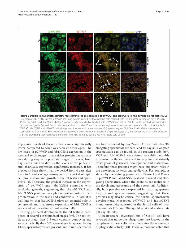
Figure 5 Double immunohistochemistry representing the colocalisation of p97/VCP and Jab1/CSN5 in the developing rat testis (A-D).Detection of Jab1/CSN5 (green), p97/VCP (red), and double-stained (yellow) positive cells merged with DAPI nuclear staining on day 5 (A), day15 (B), day 30 (C) and day 60 (D). A: Day 5 gonocytes (GC) are double labelled with p97/VCP and Jab1/CSN5. B: Double-labelled spermatocytes(Sc), spermatogonia (Sg) and Sertoli cells (Se) are shown on day 15, and the nuclear regions of some spermatocytes are only positive for Jab1/CSN5. C: Jab1/CSN5 and p97/VCP positivity (yellow) is observed in spermatocytes (Sc), spermatogonia (Sg), Sertoli cells (Se) and elongatingspermatids (eSt) on day 30. D: Double staining (yellow) is detected in the cytoplasm of spermatocytes (Sc), the nuclear region of spermatogonia(Sg) and elongating spermatids (eSt) and Sertoli cells (Se) in the 60-day-old rat testis. Scale bars: 25 μm.
Cayli et al. Reproductive Biology and Endocrinology 2011, 9:117http://www.rbej.com/content/9/1/117
Page 9 of 13

Sertoli cells are highly involved in the digestion of germcells that degenerate during spermatogenesis and oflobules of residual spermatid cytoplasm left during sper-miation. Some researchers have also reported that Ser-toli cells might be involved in the exchange andelimination of excessive sperm cell substances [47,48].Recently, p97/VCP was found to be essential for autop-hagosome maturation, suggesting that p97/VCP mightbe selectively required for autophagic degradation ofubiquitinated substrates [24,25]. Based on the localisa-tion of the p97/VCP in immature and mature Sertolicells, our results support previous observations, and wesuggest that p97/VCP may be necessary for the proces-sing of ubiqutinylated and misfolded proteins from lar-ger protein complexes or membranes in Sertoli cells.Further functional studies will be required to clarify thephysiological role of p97/VCP in Sertoli cells.In the present study, it was found that the expression
of p97/VCP and Jab1/CSN5 gradually increased duringpostnatal development of the rat epididymis. Moreover,both proteins were observed in the epididymal epithe-lium and produced by all regions of the epididymis but
were highly expressed in the CT and CS epididymis.These findings also support the idea that p97/VCP andJab1/CSN5 probably contribute to the sperm maturationprocess in the epididymis.There are number of pieces of evidence that removal
and degradation of defective spermatozoa occurs duringepididymal passage [8,16,49]. However, it is not clearhow the defective spermatozoa are removed or whatcomponents of the UPS contribute proteasomal proteo-lysis in the epididymis. One possibility that arises fromthe present study is that Jab1/CSN5 together with p97/VCP may act as an important mediator during the pro-teasomal degradation of defective spermatozoa. It is wellknown that the JAMM domain of Jab1/CSN5 has deubi-quitinase activity when associated with the CSN [36].This activity of Jab1/CSN5 might be critical for proteindegradation in the epididymis. Furthermore, it was pre-viously shown when the CSN was inactivated by knock-down of Jab1/CSN5, the amount of polyubiquitinatedproteins bound to p97/VCP increased, indicating thatthe CSN is required for proper processing of substrateproteins bound to p97/VCP [29]. Clearly, p97/VCP and
Figure 6 Colocalisations of Jab1/CSN5 and p97/VCP in the developing rat epididymis (A-D). Detection of Jab1/CSN5 (green), p97/VCP(red), and double (yellow) positive cells merged with DAPI nuclear staining on day 5 (A), 15 (B), 30 (C) and 60 (D) CS epididymis. Both the basal(BS) and principal cells (PC) of CS epididymis are immunopositive for Jab1/CSN5 (green) and for p97/VCP (red), resulting a yellow color.Spermatozoa (Sp) are weakly immunopositive for Jab1/CSN5 and p97/VCP on day 60. Scale bars: 25 μm (A), 50 μm (B-D).
Cayli et al. Reproductive Biology and Endocrinology 2011, 9:117http://www.rbej.com/content/9/1/117
Page 10 of 13

Figure 7 Western blot analysis of p97/VCP and Jab1/CSN5 in the developing rat testis (A) and epididymis (B). A: p97/VCP (97 kDa) andJab1/CSN5 (38 kDa) were detected by western blotting. b-actin (43 kDa) was used as loading control. Immunoblot bands were quantified by anoptical densitometer. The OD (optical density) values of the p97/VCP and Jab1/CSN5 bands were normalised to the OD values of b-actin bands.The data in the graphs are presented as the means ± SEM. The expression of p97/VCP and Jab1/CSN5 is significantly increased from days 5 to30 in the testis (Asterisks indicate, p < 0.05); however, no differences are observed between days 30 and 60. B: p97/VCP and Jab1/CSN5expression levels are significantly higher in the CT epididymis than the CS and CA epididymis at days 30 and 60. Asterisks indicate p < 0.05, days5 CS vs. days 5 CA and CT epididymis, days 30 and 60 CT vs. days 30 and 60 CA and CS epididymis. CT: caput, CS: corpus, CA: cauda.
Cayli et al. Reproductive Biology and Endocrinology 2011, 9:117http://www.rbej.com/content/9/1/117
Page 11 of 13

Jab1/CSN5 may also be involved in the proper proces-sing of polyubiquitinated substrates in the epididymis.Moreover, the colocalisation of p97/VCP and Jab1/CSN5 in 5-, 15-, and 30-day-old rat testes and epidi-dymes also supports the previous findings indicatingthat Jab1/CSN5 may regulate the ubiquitination statusof proteins bound to p97/VCP.
ConclusionsHere, we report the developmental expression and colo-calisation of the UPS components p97/VCP and Jab1/CSN5 in the rat testis and epididymis. Further researchmight contribute to the clarification of the exact func-tions of these proteins in the development of the rat tes-tis and epididymis.
AcknowledgementsThis work was partially supported by the Research Foundation ofGaziosmanpasa University (2011/08). We would like to thank Dr. AhmetEyibilen for generously providing AEC chromogen and Dr. Emin TurkayKorgun for providing p27kip1 antibody. This manuscript was edited byElsevier Language Editing Services.
Author details1Department of Histology and Embryology, Faculty of Medicine,Gaziosmanpasa University, Tokat, Turkey. 2Department of Urology, Faculty ofMedicine, Gaziosmanpasa University, Tokat, Turkey. 3Department of Anatomy,Faculty of Medicine, Gaziosmanpasa University, Tokat, Turkey. 4ExperimentalAnimal Center, Faculty of Medicine, Gaziosmanpasa University, Tokat, Turkey.
Authors’ contributionsSC and SO performed the major part of the histological analysis and wrotethe manuscript. FE, TY, ZK, UT and HA participated in the study design andthe analysis. All authors read and approved the final manuscript.
Competing interestsThe authors declare that they have no competing interests.
Received: 27 May 2011 Accepted: 19 August 2011Published: 19 August 2011
References1. de Kretser DM: Clinical male infertility. I. Prevalence of and progress in
understanding male infertility. Reprod Fertil Dev 1994, 6:3-8.2. Kierszenbaum AL: Mammalian spermatogenesis in vivo and in vitro: a
partnership of spermatogenic and somatic cell lineages. Endocr Rev 1994,15:116-134.
3. Clermont Y, Perey B: Quantitative study of the cell population of theseminiferous tubules in immature rats. Am J Anat 1957, 100:241-267.
4. Cooper TG: Interactions between epididymal secretions andspermatozoa. J Reprod Fertil Suppl 1998, 53:119-136.
5. Orgebin-Crist MC: Studies on the function of the epididymis. Biol Reprod1969, 1(Suppl 1):155-175.
6. Hinton BT, Palladino MA, Rudolph D, Labus JC: The epididymis asprotector of maturing spermatozoa. Reprod Fertil Dev 1995, 7:731-745.
7. Sutovsky P, Moreno R, Ramalho-Santos J, Dominko T, Thompson WE,Schatten G: A putative, ubiquitin-dependent mechanism for therecognition and elimination of defective spermatozoa in the mammalianepididymis. J Cell Sci 2001, 114:1665-1675.
8. Sutovsky P: Ubiquitin-dependent proteolysis in mammalianspermatogenesis, fertilization, and sperm quality control: killing threebirds with one stone. Microsc Res Tech 2003, 61:88-102.
9. Glickman MH, Ciechanover A: The ubiquitin-proteasome proteolyticpathway: destruction for the sake of construction. Physiol Rev 2002,82:373-428.
10. Bedard N, Hingamp P, Pang Z, Karaplis A, Morales C, Trasler J, Cyr D,Gagnon C, Wing SS: Mice lacking the UBC4-testis gene have a delay inpostnatal testis development but normal spermatogenesis and fertility.Mol Cell Biol 2005, 25:6346-6354.
11. Liu Z, Oughtred R, Wing SS: Characterization of E3Histone, a novel testisubiquitin protein ligase which ubiquitinates histones. Mol Cell Biol 2005,25:2819-2831.
12. Wing SS: Deubiquitinating enzymes–the importance of driving in reversealong the ubiquitin-proteasome pathway. Int J Biochem Cell Biol 2003,35:590-605.
13. Roest HP, van Klaveren J, de Wit J, van Gurp CG, Koken MH, Vermey M, vanRoijen JH, Hoogerbrugge JW, Vreeburg JT, Baarends WM, et al: Inactivationof the HR6B ubiquitin-conjugating DNA repair enzyme in mice causesmale sterility associated with chromatin modification. Cell 1996,86:799-810.
14. Kwon J, Mochida K, Wang YL, Sekiguchi S, Sankai T, Aoki S, Ogura A,Yoshikawa Y, Wada K: Ubiquitin C-terminal hydrolase L-1 is essential forthe early apoptotic wave of germinal cells and for sperm quality controlduring spermatogenesis. Biol Reprod 2005, 73:29-35.
15. Baarends WM, Roest HP, Grootegoed JA: The ubiquitin system ingametogenesis. Mol Cell Endocrinol 1999, 151:5-16.
16. Baska KM, Manandhar G, Feng D, Agca Y, Tengowski MW, Sutovsky M, Yi YJ,Sutovsky P: Mechanism of extracellular ubiquitination in the mammalianepididymis. J Cell Physiol 2008, 215:684-696.
17. Hochstrasser M: Ubiquitin, proteasomes, and the regulation ofintracellular protein degradation. Curr Opin Cell Biol 1995, 7:215-223.
18. Kondo H, Rabouille C, Newman R, Levine TP, Pappin D, Freemont P,Warren G: p47 is a cofactor for p97-mediated membrane fusion. Nature1997, 388:75-78.
19. Hirabayashi M, Inoue K, Tanaka K, Nakadate K, Ohsawa Y, Kamei Y,Popiel AH, Sinohara A, Iwamatsu A, Kimura Y, et al: VCP/p97 in abnormalprotein aggregates, cytoplasmic vacuoles, and cell death, phenotypesrelevant to neurodegeneration. Cell Death Differ 2001, 8:977-984.
20. Rabinovich E, Kerem A, Frohlich KU, Diamant N, Bar-Nun S: AAA-ATPasep97/Cdc48p, a cytosolic chaperone required for endoplasmic reticulum-associated protein degradation. Mol Cell Biol 2002, 22:626-634.
21. Meyer HH, Shorter JG, Seemann J, Pappin D, Warren G: A complex ofmammalian ufd1 and npl4 links the AAA-ATPase, p97, to ubiquitin andnuclear transport pathways. Embo J 2000, 19:2181-2192.
22. Hetzer M, Meyer HH, Walther TC, Bilbao-Cortes D, Warren G, Mattaj IW:Distinct AAA-ATPase p97 complexes function in discrete steps ofnuclear assembly. Nat Cell Biol 2001, 3:1086-1091.
23. Ficarro S, Chertihin O, Westbrook VA, White F, Jayes F, Kalab P, Marto JA,Shabanowitz J, Herr JC, Hunt DF, Visconti PE: Phosphoproteome analysisof capacitated human sperm. Evidence of tyrosine phosphorylation of akinase-anchoring protein 3 and valosin-containing protein/p97 duringcapacitation. J Biol Chem 2003, 278:11579-11589.
24. Krick R, Bremer S, Welter E, Schlotterhose P, Muehe Y, Eskelinen EL,Thumm M: Cdc48/p97 and Shp1/p47 regulate autophagosomebiogenesis in concert with ubiquitin-like Atg8. J Cell Biol 2010,190:965-973.
25. Tresse E, Salomons FA, Vesa J, Bott LC, Kimonis V, Yao TP, Dantuma NP,Taylor JP: VCP/p97 is essential for maturation of ubiquitin-containingautophagosomes and this function is impaired by mutations that causeIBMPFD. Autophagy 2010, 6:217-227.
26. Dai RM, Li CC: Valosin-containing protein is a multi-ubiquitin chain-targeting factor required in ubiquitin-proteasome degradation. Nat CellBiol 2001, 3:740-744.
27. Meusser B, Hirsch C, Jarosch E, Sommer T: ERAD: the long road todestruction. Nat Cell Biol 2005, 7:766-772.
28. Klein JB, Barati MT, Wu R, Gozal D, Sachleben LR Jr, Kausar H, Trent JO,Gozal E, Rane MJ: Akt-mediated valosin-containing protein 97phosphorylation regulates its association with ubiquitinated proteins. JBiol Chem 2005, 280:31870-31881.
29. Cayli S, Klug J, Chapiro J, Froehlich S, Krasteva G, Orel L, Meinhardt A: TheCOP9 signalosome interacts ATP-dependently with p97/VCP andcontrols the ubiquitination status of proteins bound to p97/VCP. J BiolChem 2009, 284:34944-34953.
30. Berse M, Bounpheng M, Huang X, Christy B, Pollmann C, Dubiel W:Ubiquitin-dependent degradation of Id1 and Id3 is mediated by theCOP9 signalosome. J Mol Biol 2004, 343:361-370.
Cayli et al. Reproductive Biology and Endocrinology 2011, 9:117http://www.rbej.com/content/9/1/117
Page 12 of 13

31. Yun J, Tomida A, Andoh T, Tsuruo T: Interaction between glucose-regulated destruction domain of DNA topoisomerase IIalpha and MPNdomain of Jab1/CSN5. J Biol Chem 2004, 279:31296-31303.
32. Callige M, Kieffer I, Richard-Foy H: CSN5/Jab1 is involved in ligand-dependent degradation of estrogen receptor {alpha} by the proteasome.Mol Cell Biol 2005, 25:4349-4358.
33. Li S, Liu X, Ascoli M: p38JAB1 binds to the intracellular precursor of thelutropin/choriogonadotropin receptor and promotes its degradation. JBiol Chem 2000, 275:13386-13393.
34. Kim BC, Lee HJ, Park SH, Lee SR, Karpova TS, McNally JG, Felici A, Lee DK,Kim SJ: Jab1/CSN5, a component of the COP9 signalosome, regulatestransforming growth factor beta signaling by binding to Smad7 andpromoting its degradation. Mol Cell Biol 2004, 24:2251-2262.
35. Wan M, Cao X, Wu Y, Bai S, Wu L, Shi X, Wang N: Jab1 antagonizes TGF-beta signaling by inducing Smad4 degradation. EMBO Rep 2002,3:171-176.
36. Cope GA, Suh GS, Aravind L, Schwarz SE, Zipursky SL, Koonin EV,Deshaies RJ: Role of predicted metalloprotease motif of Jab1/Csn5 incleavage of Nedd8 from Cul1. Science 2002, 298:608-611.
37. Shackleford TJ, Claret FX: JAB1/CSN5: a new player in cell cycle controland cancer. Cell Div 2010, 5:26.
38. Leblond CP, Clermont Y: Definition of the stages of the cycle of theseminiferous epithelium in the rat. Ann N Y Acad Sci 1952, 55:548-573.
39. Malkov M, Fisher Y, Don J: Developmental schedule of the postnatal rattestis determined by flow cytometry. Biol Reprod 1998, 59:84-92.
40. Parvinen M: Regulation of the seminiferous epithelium. Endocr Rev 1982,3:404-417.
41. Griswold MD: Interactions between germ cells and Sertoli cells in thetestis. Biol Reprod 1995, 52:211-216.
42. Cheng CY, Mruk DD: The biology of spermatogenesis: the past, presentand future. Philos Trans R Soc Lond B Biol Sci 2010, 365:1459-1463.
43. Hutchison GR, Scott HM, Walker M, McKinnell C, Ferrara D, Mahood IK,Sharpe RM: Sertoli cell development and function in an animal model oftesticular dysgenesis syndrome. Biol Reprod 2008, 78:352-360.
44. Sharpe RM, McKinnell C, Kivlin C, Fisher JS: Proliferation and functionalmaturation of Sertoli cells, and their relevance to disorders of testisfunction in adulthood. Reproduction 2003, 125:769-784.
45. Kayisli UA, Cayli S, Seval Y, Tertemiz F, Huppertz B, Demir R: Spatial andtemporal distribution of Tie-1 and Tie-2 during very early developmentof the human placenta. Placenta 2006, 27:648-659.
46. Bounpheng MA, Melnikova IN, Dodds SG, Chen H, Copeland NG, Gilbert DJ,Jenkins NA, Christy BA: Characterization of the mouse JAB1 cDNA andprotein. Gene 2000, 242:41-50.
47. Sakai Y, Nakamoto T, Yamashina S: Dynamic changes in Sertoli cellprocesses invading spermatid cytoplasm during mouse spermiogenesis.Anat Rec 1988, 220:51-57.
48. Russell LD, Tallon-Doran M, Weber JE, Wong V, Peterson RN: Three-dimensional reconstruction of a rat stage V Sertoli cell: III. A study ofspecific cellular relationships. Am J Anat 1983, 167:181-192.
49. Tengowski MW, Feng D, Sutovsky M, Sutovsky P: Differential expression ofgenes encoding constitutive and inducible 20S proteasomal coresubunits in the testis and epididymis of theophylline- or 1,3-dinitrobenzene-exposed rats. Biol Reprod 2007, 76:149-163.
doi:10.1186/1477-7827-9-117Cite this article as: Cayli et al.: Developmental expression of p97/VCP(Valosin-containing protein) and Jab1/CSN5 in the rat testis andepididymis. Reproductive Biology and Endocrinology 2011 9:117.
Submit your next manuscript to BioMed Centraland take full advantage of:
• Convenient online submission
• Thorough peer review
• No space constraints or color figure charges
• Immediate publication on acceptance
• Inclusion in PubMed, CAS, Scopus and Google Scholar
• Research which is freely available for redistribution
Submit your manuscript at www.biomedcentral.com/submit
Cayli et al. Reproductive Biology and Endocrinology 2011, 9:117http://www.rbej.com/content/9/1/117
Page 13 of 13