Embed Size (px)
Citation preview

JCB: ARTICLE
The Rockefeller University Press $30.00J. Cell Biol. Vol. 187 No. 2 201–217www.jcb.org/cgi/doi/10.1083/jcb.200903024 JCB 201
Correspondence to Mervyn J. Monteiro: [email protected]. Liang’s present address is Dept. of Biochemistry and Molecular Biology, Peking University Health Science Center, Beijing 100191, China.Abbreviations used in this paper: AD, Alzheimer’s disease; ERAD, ER-associated protein degradation; JAMP, JNK-associated membrane protein; UBA, ubiquitin-associated; UBL, ubiquitin-like; UPR, unfolded protein response; UBX, ubiquitin regulatory X; Y2H, yeast two-hybrid.
IntroductionThe ER is a fundamental eukaryotic organelle where proteins are made and where stringent quality-control systems operate to ensure that only correctly folded and properly assembled pro-tein complexes are allowed to exit the organelle for delivery to their intended sites of function (Ellgaard et al., 1999; Kostova and Wolf, 2003; Hebert and Molinari, 2007). The mechanisms by which the ER operates to monitor and dispose of misfolded and misassembled protein complexes are beginning to be under-stood. One mechanism is ER-associated protein degradation (ERAD), a regulated process by which proteins in the ER are exported to the cytoplasm for destruction by the proteasome (Kanehara et al., 2007; Vembar and Brodsky, 2008; Hirsch et al., 2009). Inefficient clearance of misfolded and misassembled proteins from the ER leads to ER stress, which results in the activation of a signaling cascade called the unfolded protein response (UPR; Schröder and Kaufman, 2005; Malhotra and Kaufman, 2007; Ron and Walter, 2007). In mammals, UPR is regulated by three main sensors called IRE1, PERK, and ATF6,
each of which triggers, via distinct mechanisms, a coordinated intracellular response designed to restore protein homeostasis in the ER. If, however, ER stress is not alleviated, a terminal cell death program is then executed.
There is growing evidence that disturbances in ERAD are involved in human disease. For example, several diseases ap-pear to result from mutations in proteins that cause them to mis-fold in the ER during synthesis, where they are retained and eliminated by ERAD (Schröder and Kaufman, 2005; Zhao and Ackerman, 2006; Brodsky, 2007). Although in this scenario, ERAD is detrimental and leads to disease, under normal cir-cumstances, it presumably functions as a safety mechanism to remove damaged and misassembled proteins before they accu-mulate to toxic levels. However, inefficient ERAD has also been linked to disease. For example, mutant proteins involved in Huntington’s disease and amyotrophic lateral sclerosis were recently shown to induce ER stress by interfering with ERAD (Nishitoh et al., 2008; Duennwald and Lindquist, 2008).
Our knowledge of proteins involved in ERAD is expand-ing (for reviews see Kanehara et al., 2007; Vembar and Brodsky, 2008; Hirsch et al., 2009). Derlin proteins are thought to form
Unwanted proteins in the endoplasmic reticu-lum (ER) are exported into the cytoplasm and degraded by the proteasome through the ER-
associated protein degradation pathway (ERAD). Distur-bances in ERAD are linked to ER stress, which has been implicated in the pathogenesis of several human diseases. However, the composition and organization of ERAD complexes in human cells is still poorly understood. In this paper, we describe a trimeric complex that we propose functions in ERAD. Knockdown of erasin, a platform for
p97/VCP and ubiquilin binding, or knockdown of ubiq-uilin in human cells slowed degradation of two classical ERAD substrates. In Caenorhabditis elegans, ubiquilin and erasin are ER stress-response genes that are regu-lated by the ire-1 branch of the unfolded protein response pathway. Loss of ubiquilin or erasin resulted in activation of ER stress, increased accumulation of polyubiquitinated proteins, and shortened lifespan in worms. Our results strongly support a role for this complex in ERAD and in the regulation of ER stress.
Ubiquilin and p97/VCP bind erasin, forming a complex involved in ERAD
Precious J. Lim,1 Rebecca Danner,1 Jing Liang,1 Howard Doong,1 Christine Harman,1 Deepa Srinivasan,1 Cara Rothenberg,1 Hongmin Wang,1 Yihong Ye,2 Shengyun Fang,1 and Mervyn J. Monteiro1
1Medical Biotechnology Center, University of Maryland Biotechnology Institute, Baltimore, MD 212012National Institute of Diabetes and Digestive and Kidney Disease, National Institutes of Health, Bethesda, MD 20892
© 2009 Lim et al. This article is distributed under the terms of an Attribution–Noncommercial–Share Alike–No Mirror Sites license for the first six months after the publication date (see http://www.jcb.org/misc/terms.shtml). After six months it is available under a Creative Commons License (Attribution–Noncommercial–Share Alike 3.0 Unported license, as de-scribed at http://creativecommons.org/licenses/by-nc-sa/3.0/).
TH
EJ
OU
RN
AL
OF
CE
LL
BIO
LO
GY
Dow
nloaded from http://rupress.org/jcb/article-pdf/187/2/201/1343756/jcb_200903024.pdf by guest on 04 D
ecember 2021

JCB • VOLUME 187 • NUMBER 2 • 2009 202
bind radiolabeled erasin protein (Fig. 1 C). Fusion proteins con-taining the central domain of ubiquilin-1 bound erasin effi-ciently, whereas those that lacked the domain failed to bind erasin (Fig. 1 C).
By additional pull-down experiments and systematic N- and C-terminal deletion of the erasin polypeptide, we mapped the ubiquilin-1–binding site to the N-terminal end of erasin (between amino acids 1 and 200; see Fig. S1), which is distinct from the p97/VCP-binding site located at the ubiquitin regulatory X (UBX) domain (amino acids 316–395) of the pro-tein (Liang et al., 2006).
Interaction between erasin and other ubiquitin–proteasome shuttle factorsWe next investigated, using similar pull-down assays, whether erasin binds hHR23A, another ubiquitin–proteasome shuttle protein, which like ubiquilin contains both UBL and UBA do-mains (Elsasser and Finley, 2005; Raasi and Wolf, 2007). As shown in Fig. 1 (D and E), erasin-His was efficiently pulled down by GST-ubiquilin but not by GST-hHR23A or GST-His. These data suggest that erasin binds selectively to ubiquilin-1 and not to all ubiquitin–proteasome shuttle factors.
Recombinant erasin, ubiquilin, and p97/VCP proteins bind together in a single complexGiven that erasin binds p97/VCP (Liang et al., 2006; Alexandru et al., 2008), we next examined whether recombinant erasin, ubiquilin, and p97/VCP proteins bind together in one complex. In these experiments, purified GST-ubiquilin or GST-His pro-tein alone, which served as a control for nonspecific binding, were mixed with purified p97/VCP-His, erasin-His, or both p97/VCP-His and erasin-His proteins, and then the GST proteins were affinity purified. The purified complexes were examined by immunoblotting for recovery of the added His-tagged pro-teins (Fig. 1 F). The blots show that erasin-His bound strongly to GST-ubiquilin but not to GST alone. Also, p97/VCP-His was isolated in a complex with GST-ubiquilin, but only after addi-tion of erasin-His. These results strongly suggest that p97/VCP binds ubiquilin through erasin and that all three proteins coexist in a single complex. Quantification of the stoichiometry of the complex suggests that erasin-His and p97/VCP-His bind GST-ubiquilin in an 1:1 ratio (see Fig. S2). Because p97/VCP self-assembles to form a homohexamer, it is likely that in these in vitro binding assays, erasin has saturated each of the six UBX-binding sites of the p97/VCP hexamer (Meyer et al., 2000; Dreveny et al., 2004).
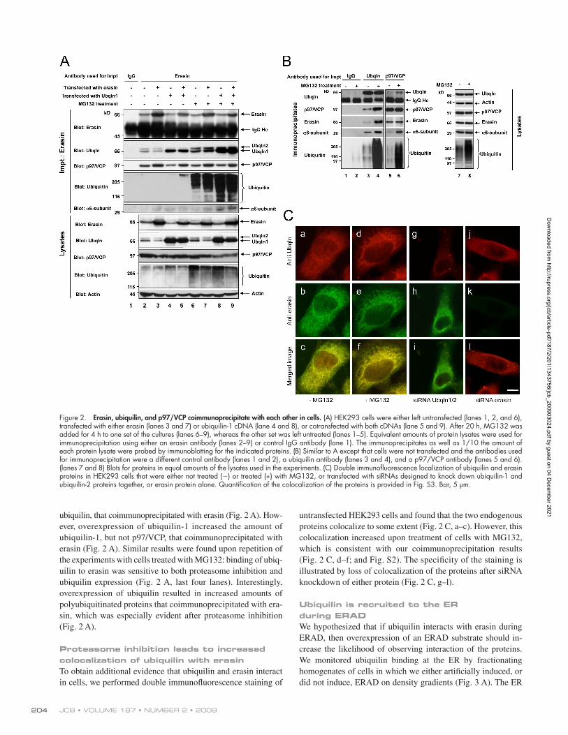
Erasin, ubiquilin, and p97/VCP coimmunoprecipitate with each other in cellsTo determine if erasin, ubiquilin, and p97/VCP form a complex in cells, we immunoprecipitated each of the proteins from HEK293 cells and found that the other two proteins were indeed coimmunoprecipitated (Fig. 2 A, lane 2; and Fig. 2 B, lanes 3 and 5). The specificity of these immunoprecipitations is illus-trated by the failure to detect any of the proteins in parallel
the portal in the ER through which misfolded and misassembled proteins are exported. p97 (valosin-containing protein: p97/VCP) provides the main driving force for extraction of the ERAD sub-strate (Lilley and Ploegh 2004; Ye et al., 2004). p97/VCP is re-cruited to the ER for ERAD by binding to ER-localized proteins, candidates of which are gp78, derlin, VIMP, ubx2, Hrd1, erasin, and UBXD8. During extraction, the misfolded protein is tagged with a polyubiquitin chain by ubiquitin ligases (Kostova et al., 2007); the polyubiquitin chain serves as a signal for the recogni-tion and destruction of the protein by the proteasome. At least five ER resident ubiquitin ligases—Hrd1/Der3, gp78, Doa10/Teb4, RMA1, and RFP2—conduct this modification, although oth-ers probably exist. The length of the ubiquitin chain that is tagged onto substrates is regulated by Ufd2, which is recruited to the ERAD complex by binding to p97/VCP (Richly et al., 2005). Ufd2, in turn, binds two additional factors, Rad23 and Dsk2, both of which contain ubiquitin-associated (UBA) and ubiquitin-like (UBL) domains, which appear to function in the transfer of the ubiquitinated substrate to the proteasome for deg-radation (Richly et al., 2005; Elsasser and Finley, 2005). The UBA domains of Rad23 and Dsk2 bind polyubiquitin chains, and their UBL domains bind the proteasome, thus positioning these ubiquitin–proteasome shuttle proteins as the terminal fac-tors that deliver the substrate to the proteasome for degradation (Verma et al., 2004; Raasi and Wolf, 2007).
Many of the ERAD components mentioned were first identified in yeast. Interestingly, mammals contain additional ERAD components for which no counterparts exist in yeast, which suggests that mammals use a more elaborate repertoire of proteins in ERAD (Carvalho et al., 2006; Jentsch and Rumpf, 2007; Kanehara et al., 2007).
In this paper, we demonstrate that human ubiquilin proteins (Wu et al., 1999; Mah et al., 2000; Kleijnen et al., 2000) that are found in the cytoplasm and nucleoplasm (Mah et al., 2000), and which are believed to function as ubiquitin–proteasome shuttle factors, interact with erasin, an ER localized protein (Liang et al., 2006), in a novel complex that functions in ERAD. Using a vari-ety of techniques, including in vitro binding assays, coimmuno-precipitation and biochemical fractionation of proteins from cells, and silencing of the proteins in cells and worms, we obtained evi-dence in support of this novel complex in ERAD.
ResultsIn vitro interaction between erasin and ubiquilinA yeast two-hybrid (Y2H) screen using the central domain of human ubiquilin-1 (UBQLN1UBLUBA; Fig. 1 A) netted four clones containing different portions of the N-terminal end of erasin, an ER-localized protein that promotes ERAD (Liang et al., 2006). The longest clone interacted 100-fold stronger with the same ubiquilin bait than with the UBA domain of ubiquilin or with unrelated baits, which indicates strong and specific interaction between erasin and the central domain of ubiquilin (Fig. 1 B).
To confirm binding, and to map the erasin-binding site in ubiquilin-1, we conducted GST pull-down experiments exam-ining whether GST fusions of different regions of ubiquilin-1
Dow
nloaded from http://rupress.org/jcb/article-pdf/187/2/201/1343756/jcb_200903024.pdf by guest on 04 D
ecember 2021

203INTERACTION OF ERASIN WITH UBIQUILIN AND P97/VCP • Lim et al.
Indeed, more ubiquilin was coimmunoprecipitated with erasin or p97/VCP proteins, or vice versa, in cells cultured in the presence of MG132 than in the absence of the inhibitor (Fig. 2, A and B). Furthermore, more 6 subunit components of the proteasome and ubiquitinated proteins were coimmunoprecipitated with ubiquilin, erasin, and p97/VCP proteins after proteasome inhibition (Fig. 2, A and B). Interestingly, the amount of p97/VCP or erasin proteins that coimmunoprecipitated with each other was not altered after proteasome inhibition (Fig. 2, A and B), which indicates that asso-ciation of ubiquilin, but not p97/VCP, with erasin is regulated by proteasome activity.
To explore the interaction between ubiquilin and erasin further, we examined whether overexpression of either pro-tein affects assembly of the complex. Overexpression of era-sin predominantly increased the amount of p97/VCP, but not
immunoprecipitations conducted with two different control anti-bodies (Fig. 2 A, lane 1; and Fig. 2 B, lane 1).
Because erasin promotes ERAD (Liang et al., 2006), we considered that the erasin–ubiquilin–p97/VCP complex might function for this purpose. We reasoned that erasin might recruit p97/VCP and ubiquilin to the ER during ERAD for separate pur-poses: p97/VCP to aid in the retrotranslocation of ERAD substrates from the ER, whereas ubiquilin might function to bridge binding of the extracted ubiquitinated substrate and the proteasome through interaction with its UBA and UBL domains, respectively. To test this idea, we made a comparison to see if assembly of the complex was altered in cells treated with or without the proteasome inhibitor MG132. We predicted that proteasome inhibition would lead to a buildup of the complex in cells because of the inability of ubiquilin to deliver the ERAD substrate to the proteasome for degradation.
Figure 1. Erasin interaction with ubiquilin-1, p97/VCP, and hHR23A. (A) Schematic drawing of ubiquilin-1. Ubiquilin-1UBLUBA (178–428 aa) was used as the bait in a Y2H screen. (B) Yeast interaction assay of an erasin prey clone with different baits measured in five independent colonies showing the mean and SD (error bars). (C) GST pull-down assays of ubiquilin-1 (UBQ) fusion proteins with 35S-radiolabeled erasin. (C, right) Schematic drawings of the GST constructs and a summary of their binding (+, binding; , no binding). (C, left) Autoradiogram (top panel) and Coomassie-stained gel (bottom panel) of the pull-down experiment. (D) Coomassie-stained gel of purified proteins used for pull-down assays. The black line indicates that intervening lanes have been spliced out. (E) GST pull-down assays showing that erasin-His binds to GST-ubiquilin but not to GST or GST-hHR23A proteins. The input erasin, the recovered erasin, and GST proteins are shown. (F) GST pull-down assays showing that erasin, p97/VCP, and ubiquilin bind together in a trimeric complex. The input p97/VCP and erasin proteins and the recovered GST complexes (ponceau) are also shown. Arrowheads indicate the position of full-length GST-fusion proteins.
Dow
nloaded from http://rupress.org/jcb/article-pdf/187/2/201/1343756/jcb_200903024.pdf by guest on 04 D
ecember 2021
JCB • VOLUME 187 • NUMBER 2 • 2009 204
untransfected HEK293 cells and found that the two endogenous proteins colocalize to some extent (Fig. 2 C, a–c). However, this colocalization increased upon treatment of cells with MG132, which is consistent with our coimmunoprecipitation results (Fig. 2 C, d–f; and Fig. S2). The specificity of the staining is illustrated by loss of colocalization of the proteins after siRNA knockdown of either protein (Fig. 2 C, g–l).
Ubiquilin is recruited to the ER during ERADWe hypothesized that if ubiquilin interacts with erasin during ERAD, then overexpression of an ERAD substrate should in-crease the likelihood of observing interaction of the proteins. We monitored ubiquilin binding at the ER by fractionating homogenates of cells in which we either artificially induced, or did not induce, ERAD on density gradients (Fig. 3 A). The ER
ubiquilin, that coimmunoprecipitated with erasin (Fig. 2 A). How-ever, overexpression of ubiquilin-1 increased the amount of ubiquilin-1, but not p97/VCP, that coimmunoprecipitated with erasin (Fig. 2 A). Similar results were found upon repetition of the experiments with cells treated with MG132: binding of ubiq-uilin to erasin was sensitive to both proteasome inhibition and ubiquilin expression (Fig. 2 A, last four lanes). Interestingly, overexpression of ubiquilin resulted in increased amounts of polyubiquitinated proteins that coimmunoprecipitated with era-sin, which was especially evident after proteasome inhibition (Fig. 2 A).
Proteasome inhibition leads to increased colocalization of ubiquilin with erasinTo obtain additional evidence that ubiquilin and erasin interact in cells, we performed double immunofluorescence staining of
Figure 2. Erasin, ubiquilin, and p97/VCP coimmunoprecipitate with each other in cells. (A) HEK293 cells were either left untransfected (lanes 1, 2, and 6), transfected with either erasin (lanes 3 and 7) or ubiquilin-1 cDNA (lane 4 and 8), or cotransfected with both cDNAs (lane 5 and 9). After 20 h, MG132 was added for 4 h to one set of the cultures (lanes 6–9), whereas the other set was left untreated (lanes 1–5). Equivalent amounts of protein lysates were used for immunoprecipitation using either an erasin antibody (lanes 2–9) or control IgG antibody (lane 1). The immunoprecipitates as well as 1/10 the amount of each protein lysate were probed by immunoblotting for the indicated proteins. (B) Similar to A except that cells were not transfected and the antibodies used for immunoprecipitation were a different control antibody (lanes 1 and 2), a ubiquilin antibody (lanes 3 and 4), and a p97/VCP antibody (lanes 5 and 6). (lanes 7 and 8) Blots for proteins in equal amounts of the lysates used in the experiments. (C) Double immunofluorescence localization of ubiquilin and erasin proteins in HEK293 cells that were either not treated () or treated (+) with MG132, or transfected with siRNAs designed to knock down ubiquilin-1 and ubiquilin-2 proteins together, or erasin protein alone. Quantification of the colocalization of the proteins is provided in Fig. S3. Bar, 5 µm.
Dow
nloaded from http://rupress.org/jcb/article-pdf/187/2/201/1343756/jcb_200903024.pdf by guest on 04 D
ecember 2021

205INTERACTION OF ERASIN WITH UBIQUILIN AND P97/VCP • Lim et al.
misfolding and increased ERAD, also promoted ubiquilin migra-tion to the ER (Fig. 3, E and F). These results collectively suggest that ubiquilin may be recruited to the ER for ERAD.
Ubiquilin promotes degradation of membrane-bound and luminal ERAD substratesWe next investigated whether reduction of ubiquilin in HEK293 cells affects the turnover of the two classical ERAD sub-strates: CD3 and 1ATNHK. siRNA-induced knockdown of ubiquilin-1 and -2 proteins in HEK293-CD3 cells, a HEK293 cell line stably expressing orphan CD3 protein (Ballar et al., 2007), slowed degradation of CD3, whereas overexpression of ubiquilin-1 slightly enhanced CD3 turnover (Fig. 4 A). Similarly, knockdown of the ubiquilin proteins stalled turnover of 1ATNHK (Fig. 4, B and D). Because of their association with each other, we also examined if loss of erasin expression
fractionated mainly across the middle-to-heavy-density portions of the gradients, as revealed by the distribution of calnexin, a classical ER marker (Fig. 3 A). Erasin cofractionated with cal-nexin, as expected. In untreated HEK293 cells, only a faint amount of ubiquilin cofractionated with erasin, with the vast ma-jority of the protein located in lower density end of the gradient (Fig. 3, A and F). In contrast, MG132 treatment increased mi-gration of ubiquilin toward the ER-containing fractions, which were also enriched for ubiquitinated proteins (Fig. 3, B and F). Similarly, overexpression of the ERAD substrates CD3, a membrane-bound protein, or 1-anti-trypsin null Hong Kong mutant (1ATNHK), a misfolded luminal protein, also pro-moted migration of ubiquilin to the ER fractions (Fig. 3, C, D, and F). In contrast, overexpression of wild-type 1AT, which is not misfolded and should not be a target for ERAD, did not promote migration of ubiquilin to the ER (Fig. 3, D and F). Treatment of cells with tunicamycin, which causes protein
Figure 3. Proteasome inhibition or expres-sion of ERAD substrates leads to a redistribu-tion of ubiquilin to the ER. Immunoblot analysis of HEK293 cell homogenates separated on 0–25% iodixanol gradients. (A–E) The panels show the distribution of erasin, calnexin, and ubiquilin in fractions of the gradient (heavy, bottom; light, top) of cells that were either left untreated; treated with MG132 for 4 h; trans-fected with either CD3, 1ATNHK, or wild-type 1AT cDNAs; or treated with tunicamycin for 4 h (+Tun). Ubiquitin immunoreactivity is shown for the MG132 and +MG132 and treatments. Because its profile did not change significantly, p97/VCP is only shown for the control. (F) The amount of ubiquilin in the ER fractions was estimated by dividing the inten-sity of the anti-ubiquilin–immunoreactive bands in nine fractions surrounding the calnexin peak (boxed with broken lines in A–E) by the sum of the bands in the whole gradient.
Dow
nloaded from http://rupress.org/jcb/article-pdf/187/2/201/1343756/jcb_200903024.pdf by guest on 04 D
ecember 2021

JCB • VOLUME 187 • NUMBER 2 • 2009 206
affects the turnover of the substrates. Like ubiquilin, knockdown of erasin expression reduced degradation of both substrates (Fig. 4, C and D; Liang et al., 2006). These results strongly suggest that both ubiquilin and erasin promote degradation of membrane and luminal ERAD substrates.
Expression of dominant-negative erasin mutants inhibits ERADTo confirm that the function of erasin interaction with ubiq-uilin is connected with ERAD, we overexpressed erasin cDNAs encoding different portions of the protein, expecting that mutants containing the ubiquilin-binding site would compete for ubiquilin binding but would not create a func-tional ERAD complex. The erasin mutants selected were chosen based on their in vitro ubiquilin-binding properties (Fig. S1). We selected erasin fragments 1–310 aa and 1–168 aa as putative dominant-negative candidates because they both bound ubiquilin-1 but are not targeted to the ER (Liang et al., 2006), and erasin fragment 414–508 aa as an ineffec-tive mutant because it was devoid of ubiquilin-1 binding.
We compared the rate of turnover of CD3, stably expressed in HEK293 cells, after overexpression of the mutant constructs. As shown in Fig. 5 (A and B), erasin fragments 1–310 and 1–168 both slowed the turnover of CD3, but the 414–508 fragment had little effect. Furthermore, consistent with our idea of how the dominant-negative erasin mutants might function, expression of the 1–310-aa erasin mutant displaced ubiquilin binding from the ER (Fig. 5 C). These studies strongly suggest that proper binding of ubiquilin with erasin at the ER is required for promoting ERAD.
Abrogation of MG132-induced accumulation of ubiquilin, p97/VCP, and proteasomes in microsomes after erasin knockdownThe preceding results were in accord with the notion that erasin functions to recruit p97/VCP and ubiquilin to the ER during ERAD. To obtain additional evidence in support of this idea and to examine if ubiquilin, in turn, functions to recruit proteasomes to the ER during ERAD, we compared the protein composition
Figure 4. Knockdown of ubiquilin or erasin proteins impedes ERAD. (A) HEK293-CD3 cells were transfected with either a ubiquilin-1 expression plasmid, the empty vector, or with UBQLN1 and UBQLN2 siRNAs. 48 h after transfection, cycloheximide was added to the cultures, and protein lysates were col-lected at intervals as indicated. Equal amounts of protein lysates were immunoblotted for CD3 (HA-tag), ubiquilin, and actin. The graph shows the profile of CD3 turnover determined from three independent experiments showing the mean and the SD (error bars). (B–D) Similar to A, except that turnover of 1ATNHK was analyzed after knockdown of either ubiquilin (B) or erasin (C). In these experiments, HEK293 cells were transfected with an 1ATNHK-HA expression construct 40 h after transfection of the siRNAs, with the turnover studies conducted 20 h later. Equal amounts of protein were immunoblotted for 1ATNHK (HA-tag), tubulin, and erasin or ubiquilin, depending on which of them was knocked down. (D) Quantification of 1ATNHK turnover after knockdown of either ubiquilin or erasin proteins, determined from three separate experiments showing the mean and SD (error bars).
Dow
nloaded from http://rupress.org/jcb/article-pdf/187/2/201/1343756/jcb_200903024.pdf by guest on 04 D
ecember 2021

207INTERACTION OF ERASIN WITH UBIQUILIN AND P97/VCP • Lim et al.
erasin knockdown in both MG132-treated and untreated cultures (Fig. 6 A), which suggests that p97/VCP binding to microsomes is somewhat dependent on erasin expression. Similarly, the amounts of ubiquilin, Rpt1, and 6 subunit in microsomes of cells depleted of erasin were all reduced compared with cells in which erasin levels were not altered, but only in cells that were treated with MG132 (Fig. 6 A).
The preceding results were in accord with the idea that erasin located in the ER binds ubiquilin, which, in turn, binds the proteasome. However, because reduction of erasin expres-sion by RNAi resulted in a concomitant reduction in ubiquilin protein in microsomes, we next asked if reduction of ubiquilin protein alone was sufficient to reduce proteasome recruitment to the ER during ERAD. As shown in Fig. 6 B, microsomes of cells transfected with siRNA against ubiquilin-1 and -2, or a combination of siRNAs against both ubiquilins and erasin, failed to manifest the MG132-dependent increase in protea-some binding, as measured using three different proteasome components, that occurred in cells in which erasin or ubiquilin expression was not silenced. Furthermore, consistent with the idea that knockdown of erasin or ubiquilin proteins reduces
of microsomes isolated from HEK293 cells expressing endoge-nous levels of erasin and ubiquilin proteins with those from cells silenced for expression of erasin and/or ubiquilin-1 and -2. Microsomes contain an enriched source of ER fragments, which we used to monitor changes in protein composition of the ER.
As shown in Fig. 6 A, four ER proteins—erasin, cal-nexin, derlin-1, and derlin-2—were detected in the microsome fraction but not the cytosolic fraction, which confirms that our microsome preparations were indeed enriched for ER components. Microsomes that were isolated from cells trans-fected with erasin siRNAs contained 80% less erasin than those isolated from mock-transfected cells, which indicates successful depletion of erasin protein content of microsomes by RNAi (Fig. 6 A).
We next examined whether microsomes of cells depleted of erasin contained less ubiquilin, p97/VCP, and proteasomes than those of cells not depleted of erasin. We used antibodies against the 6 subunit and Rpt1, which are contained in the 20S catalytic core and 19S complex of the proteasome (Pickart and Cohen 2004), respectively, as markers for proteasomes. The amount of p97/VCP in microsomes was reduced by 50% after
Figure 5. Dominant-negative erasin fragments interfere with CD3 degradation. (A) HEK293-CD3 cells were either mock transfected (control) or trans-fected with erasin deletion constructs encoding portions of the erasin polypeptide from amino acids 1–310, 414–508, or 1–168. Protein turnover was analyzed similar to Fig 4. Both a long and short exposure of the CD3 signal is shown. Please note that the erasin 414–508 runs aberrantly on SDS-PAGE. (B) Graphs of CD3 turnover determined from three separate experiments showing the mean and SD (error bars). (C) Immunoblots showing ubiquilin and erasin distribution in fractions of HEK293-CD3 cell homogenates separated on 0–25% iodixanol gradients.
Dow
nloaded from http://rupress.org/jcb/article-pdf/187/2/201/1343756/jcb_200903024.pdf by guest on 04 D
ecember 2021

JCB • VOLUME 187 • NUMBER 2 • 2009 208
Figure 6. Microsomes of cells depleted of erasin contain less ubiquilin, p97/VCP, and proteasomes. HEK293 cells were either mock transfected or trans-fected with siRNAs against erasin. 72 h after transfection, the cells were incubated with MG132 for an additional 4 h, whereas a duplicate set was left untreated. (A) Equal amounts of protein in microsome and supernatant fractions of the cells were separated by SDS-PAGE and probed for the proteins shown. Blots of equal amounts of proteins in total lysates of the cultures are also shown. (B) Similar to A, except that this time erasin or ubiquilin-1 and -2
Dow
nloaded from http://rupress.org/jcb/article-pdf/187/2/201/1343756/jcb_200903024.pdf by guest on 04 D
ecember 2021

209INTERACTION OF ERASIN WITH UBIQUILIN AND P97/VCP • Lim et al.
Ubiquilin and erasin expression is induced by ER stress in Caenorhabditis elegansBecause chronic disruption of ERAD causes ER stress, we pre-dicted that loss of ubiquilin or erasin expression would lead to ER stress. We tested this hypothesis in C. elegans. Worms were used for several reasons. First, they are an ideal model organism to study ER stress, with several mutant and reporter lines avail-able for such purposes (Shen et al., 2001, 2005). Second, unlike mammals, worms contain single ubiquilin and erasin genes, making it easier to inactivate the genes without the complica-tion of compensation by redundant family members. And third, because of the efficacy of RNAi in worms.
We first determined whether ubiquilin and erasin are part of the compendium of genes that are induced during the UPR. Accordingly, worms containing an integrated hsp4::gfp tran-scriptional reporter, widely used to monitor ER stress (Shen et al., 2001; Calfon et al., 2002), were treated with tunicamycin to induce ER stress for different periods of time. As expected, transcripts of the classical ER stress-regulated gene, hsp-4 (the worm homologue of human BiP), were induced by the drug treatment, with peak expression occurring 4 h after drug treat-ment, but then its expression was attenuated (Fig. 7, A and B). Interestingly, both ubiquilin and erasin transcripts increased with somewhat similar profiles to that of hsp-4, which suggests that they are part of the UPR response (Fig. 7, A–D). Expres-sion of ama-1, which is not part of the UPR, did not change over this period, as expected (Fig. 7 A).
An examination of the changes in proteins in the tunicamycin-treated worms revealed that the GFP protein expression, under the control of the hsp-4 promoter (Fig. 7 E), increased tempo-rally, with high accumulation observed 16 h after drug treat-ment (Fig. 7 F). This was accompanied by a parallel increase in ubiquitinated proteins, which is consistent with the idea that tunicamycin treatment causes accumulation of misfolded pro-teins in the worms. Ubiquilin and erasin protein levels increased fivefold and threefold, respectively, 4 h after tunicamycin treat-ment, but thereafter their levels remained constant. The differ-ence between the levels of ubiquilin and erasin transcripts and their respective proteins suggests possible posttranscriptional and/or posttranslational regulation of the genes.
To determine if ubiquilin and erasin promoters are di-rect targets of the UPR, we cloned the GFP open reading frame downstream of a 2-kb fragment containing the putative C. elegans ubiquilin promoter and isolated transgenic worms containing the integrated ubiquilin::gfp promoter fusion plasmid (Fig. 7 G). As shown in Fig. 7 (H–J), ubiquilin::gfp transgenic worms manifested a tunicamycin dose-dependent increase in GFP expression by both immunoblotting and immunofluores-cence microscopy. Interestingly, GFP expression was particularly
proteasome recruitment to the ER, we found that proteasome activity in microsomes isolated cells stably expressing CD3 in which ubiquilin or erasin proteins were knocked down was re-duced 20% compared with cells in which the proteins were not targeted for RNAi (Fig. 6 C).
Effect of erasin knockdown on coimmunoprecipitation of p97/VCP, ubiquilin, and the proteasomeWe next examined whether reduction of erasin in cells affects assembly of the ERAD complex. HEK293 cells transfected with erasin siRNAs, or cells that were mock transfected, were cultured with or without MG132, and ubiquilin proteins were immunoprecipitated from the cells. The precipitates were examined for erasin, p97/VCP, and the proteasome by immuno-blotting. As shown in Fig. 6 D, more erasin, p97/VCP, and the 6 subunit of the proteasome coimmunoprecipitated with ubiquilin from cells treated with MG132 than from those not treated with MG132. Cells that were transfected with siRNAs against erasin and treated with MG132 contained lower lev-els of erasin, p97/VCP, and the 6 subunit than untransfected cells treated with MG132, which suggests that knockdown of erasin expression interferes with formation or stability of the ERAD complex. Immunoblot analysis of total protein lysates confirmed that erasin levels were reduced by 80% after the knockdown. Ubiquilin levels were slightly elevated in the erasin knockdown cells compared with the mock-transfected cells, but the levels of p97/VCP and the 6 subunit remained relatively constant. Thus, the decreased coimmunoprecipi-tation of p97/VCP and the 6 subunit with ubiquilin after erasin knockdown was not caused by a global change in the expression of the proteins. We should point out that the cells depleted of erasin still displayed a slight MG132-dependent increase in the coimmunoprecipitation of p97/VCP and the 6 subunit with ubiquilin. At present, we cannot distinguish whether this is from incomplete silencing of erasin expres-sion or interactions mediated by proteins other than erasin. Nevertheless, these data suggest that erasin may be required for the assembly of ubiquilin, p97/VCP, and the proteasome in a complex.
To demonstrate that ubiquilin functions as a shuttle fac-tor in delivery of ERAD substrates to the proteasome for degradation, we immunoprecipitated ubiquilin from HEK293 cells stably expressing CD3 and probed them for the pres-ence of CD3 and the proteasome. As shown in Fig. 6 E, CD3 as well as 19 and 20S components of the proteasome were found in the ubiquilin immunoprecipitates, and, as ex-pected, more of them were present after MG132 treatment of the cells.
were knocked down separately, or simultaneously. Additionally, only the microsome fractions were probed for proteasome subunits (6, 7, and Rpn10), ubiquilin, and calnexin (to ensure equal protein loading). Also shown are the blots of the total lysates of the cultures. (C) Assays of proteasome activity in microsomes and total lysates of HEK293-CD3 cells that were either mock transfected or transfected with siRNAs against erasin or ubiquilin-1 and -2 of three separate cultures showing the mean and SD (error bars). (D) A repeat of the experiment shown in A, but this time we immunoprecipitated ubiquilin from the cells and examined equal fractions of the precipitates by immunoblotting for the indicated proteins. Blots of the total lysates are also shown. (E) Ubiquilin was immunoprecipitated from HEK293-CD3 cells that were either left untreated or treated with MG132 for 4 h. Equal portions of the immunoprecipitates were probed for the proteins shown.
Dow
nloaded from http://rupress.org/jcb/article-pdf/187/2/201/1343756/jcb_200903024.pdf by guest on 04 D
ecember 2021

JCB • VOLUME 187 • NUMBER 2 • 2009 210
Figure 7. Ubiquilin and erasin expression are induced by ER stress in C. elegans. (A) Mixed-stage zcls4 [hsp-4::GFP] worms were grown on plates with 28 µg/ml tunicamycin for 0, 4, 8, 16, and 48 h, and transcript levels of hsp-4, ubiquilin, erasin, and ama-I were measured by semiquantitative PCR and gel electrophoresis. Quantification of Hsp-4 (B), ubiquilin (C), and erasin (D) expression relative to the 0 time point. Similar trends were observed in two other experiments. (E) Schematic diagram of the hsp-4::GFP reporter construct. (F) Immunoblot analysis of equal amounts of protein lysate (two independent samples for each time point) prepared from worms treated with 28 µg/ml of tunicamycin for the periods indicated. An unknown protein whose expression was insensitive to ER stress was used as a loading control. (G) Schematic diagram of the C. elegans ubiquilin promoter fragment fused to GFP. (H) Immuno-blot analysis of equal amounts of protein lysate of worms treated with 0, 2.5, or 28 µg/ml tunicamycin for 24 h. (I) Quantification of GFP expression determined by scanning of immunoblots as described in H showing the mean and SD (error bars) seen in three different experiments. (J) GFP fluorescence of worms containing the integrated ubiquilin::GFP fusion construct treated for 24 h with 0, 2.5, or 28 µg/ml tunicamycin. Bar, 0.2 mm.
Dow
nloaded from http://rupress.org/jcb/article-pdf/187/2/201/1343756/jcb_200903024.pdf by guest on 04 D
ecember 2021

211INTERACTION OF ERASIN WITH UBIQUILIN AND P97/VCP • Lim et al.
to the complex, and that ubiquilin, in turn, recruits protea-somes. These findings, together with the known functions of the proteins with which erasin coimmunoprecipitates, have
prominent in the pharyngeal muscle, hypodermis, and intestine of the worms, which suggests that the reporter might be un-evenly expressed across the worm. Similarly, worms carrying an erasin::GFP reporter were also reported to display a similar tunicamycin dose-dependent increase in GFP expression (Yamauchi et al., 2007). Together, these results suggest that ubiquilin and erasin promoters contain UPR-response elements.
Expression of ubiquilin and erasin during ER stress is regulated by ire-1We next investigated which arm of the UPR regulates ubiquilin and erasin expression. Like in mammals, the UPR in C. elegans is induced through the action of three main sensors: ire-1, pek-1 (called protein kinase-like ER kinase [PERK] in mammals), and atf-6. We used worms containing inactive versions of the sensors to determine which pathway regulates tunicamycin induction of ubiquilin and erasin expression. In the control wild-type N2 worms, expression of ubiquilin and erasin transcripts increased after 6 h of tunicamycin treatment (Fig. 8). However, the induction of both genes was almost completely attenuated in ire-1 (v33) mutant worms, and partially attenuated in the atf-6 (ok551) and pek-1 (ok275) mutants (Fig. 8). Together, these re-sults indicate that in C. elegans, both ubiquilin and erasin genes are chiefly regulated by ire-1.
RNAi of ubiquilin or erasin results in accumulation of misfolded proteins and induction of ER stressWe next examined whether reduction of ubiquilin or erasin expression in worms causes ER stress. Reduction of either ubiqui-lin or erasin expression in hsp-4::gfp worms by bacteria-mediated RNAi resulted in a threefold and fivefold increase in GFP ex-pression, respectively (Fig. 9 A). In both cases, GFP expression was lower than that produced by treatment of the worms with tunicamycin, which suggests that loss of ubiquilin or erasin ex-pression induces more subtle ER stress than that produced by tunicamycin treatment. Interestingly, knockdown of ubiquilin expression led to an induction of erasin expression, and vice versa (Fig. 9, A and B). Moreover, RNAi of either ubiquilin or erasin led to an accumulation of ubiquitinated proteins in the worms, which is consistent with the idea that the proteins func-tion in the disposal of misfolded proteins from cells.
Reduction of worm lifespan by RNAi of ubiquilin or erasinFinally, we investigated whether RNAi of ubiquilin or erasin alters lifespan in the worm. As shown in Fig. 9 C, RNAi of erasin shortened lifespan in worms from a mean survival time of 21 d to 12 d, whereas RNAi of ubiquilin reduced it to 17 d. Together, these data suggest that loss of either ubiquilin or erasin expres-sion shortens the worm lifespan.
DiscussionHere, we describe a novel erasin-containing protein complex involved in ERAD in human cells. Our results suggest that erasin acts as a platform to recruit both p97/VCP and ubiquilin
Figure 8. The ire-1 branch of the UPR regulates induction of ubiquilin and erasin expression during ER stress. (A) Mixed-stage worms of the indicated mutant strains were grown on plates with 28 µg/ml tunicamycin for 0 or 6 h. Transcript levels of hsp-4 (regulated by ire-1; Shen et al., 2001), ubiquilin, erasin, and CBP were analyzed by PCR and agarose gel elec-trophoresis. (B–E) Quantification of the relative change in transcript expres-sion determined from the experiment shown in A. Levels were normalized to the 0 h time point in each mutant. Similar results were observed in two other experiments.
Dow
nloaded from http://rupress.org/jcb/article-pdf/187/2/201/1343756/jcb_200903024.pdf by guest on 04 D
ecember 2021

JCB • VOLUME 187 • NUMBER 2 • 2009 212
enabled us to construct a model of how the complex might function in ERAD. Specifically, we propose that misfolded protein substrates that are extracted from the ER interact se-quentially with a series of proteins and are eventually delivered to the terminal degradation apparatus of the cell, the protea-some (Fig. 10). The model we propose predicts that delivery of ER-derived substrates to the proteasome is highly coupled in a manner that prevents premature release of the substrate into the cytosol, where it might accumulate or aggregate into a potentially toxic species.
Our model focuses on erasin, an ER resident protein that is thought to embed into the membrane via a short hydrophobic sequence close to its C terminus, with the remainder of the pro-tein facing the cytoplasm (Fig. 10 A; Liang et al., 2006). We propose that erasin forms a complex with derlin and gp78/Hrd1 ligases either through direct or indirect interaction of the pro-teins, which is supported by coprecipitation of the proteins (Liang et al., 2006; Mueller et al., 2008). Erasin recruits p97/VCP to the complex via direct interaction with its UBX domain (Fig. 10 B; Liang et al., 2006). On the basis of previous studies of
the mechanism by which ERAD is thought to occur (Ye et al., 2001; Jarosch et al., 2002; Vembar and Brodsky, 2008), we propose that the misfolded substrate in the ER is polyubiqui-tinated by gp78/Hrd1 and/or related ubiquitin ligases and ex-tracted through the derlin channel by p97/VCP (Fig. 10 B). We speculate that the polyubiquitin chain that is conjugated onto the substrate is then recognized by the UBA domain of ubiqui-lin, which promotes ubiquilin binding to erasin, or vice versa (Fig. 10 B). Ubiquilin in turn recruits the proteasome to the ERAD complex, probably via interactions with its UBL do-main, bringing it in close proximity for the delivery and degra-dation of the ERAD substrate that is anchored to the UBA domain of ubiquilin (Fig. 10, C and D). After degradation of the substrate, we speculate that the complex most likely dis-assembles. The model incorporates many of our results with the well-established functions of the proteins mentioned. However, gaps remain, as discussed below.
A key aspect of our model emphasizes the apparent dif-ference in the binding of p97/VCP and ubiquilin proteins with erasin that we found occurs during ERAD. According to our in
Figure 9. Knockdown of ubiquilin or erasin proteins in worms results in the induction of the ER stress reporter hsp-4::GFP, accumulation of misfolded proteins, and a shorter lifespan. (A) Mixed-stage zcls4 [hsp-4::GFP] worms were either fed standard bacteria in the presence or absence of 28 µg/ml tunicamycin or bacteria to induce RNAi of the ubiquilin gene. Immunoblot analysis of equal amounts of protein lysate prepared after 48 h of treatment for GFP, ubiquilin, erasin, ubiquitin, and an unknown protein used as a loading control. (B) Identical to A, except that worms were fed bacteria to specifically knock down erasin. (C) Lifespan curves comparing N2 (wild-type) worms fed bacteria transformed with the empty RNAi vector or the vector containing cDNAs to induce RNAi of either ubiquilin or erasin genes. Results are cumulative from three independent experiments, with ≥60 animals per trial. Log-rank (Mantel-Cox) and Gehan-Breslow-Wilcoxon tests, P < 0.0001 versus control.
Dow
nloaded from http://rupress.org/jcb/article-pdf/187/2/201/1343756/jcb_200903024.pdf by guest on 04 D
ecember 2021

213INTERACTION OF ERASIN WITH UBIQUILIN AND P97/VCP • Lim et al.
because of the inability of ubiquilin to deliver its bound sub-strate to the proteasome for degradation.
The ERAD-enhanced and proteasome inhibition–dependent increase in ubiquilin interaction with erasin provides important clues as to the possible mechanisms by which the complex is assembled. We speculate that during ERAD, the polyubiq-uitinated chain that is conjugated onto the substrate might first be recognized by the UBA domain of ubiquilin, bringing it in closer proximity to erasin, thereby enhancing their probability of interaction. However, we cannot rule out the possibility that ubiquilin first interacts with erasin, and then with the polyubiq-uitinated substrate. Both mechanisms are supported by in vitro binding studies. For example, we found that ubiquilin binds era-sin with very high affinity (with a KD of 30 nm; Fig. S4). Also, the UBA domain of ubiquilin binds a wide variety of ubiquitin
vitro binding results, recombinant ubiquilin, p97/VCP, and erasin proteins can all bind together in one complex. Yet ac-cording to immunoprecipitation studies, we noticed differ-ences in the amount of the proteins that were bound to erasin under different conditions: the amount of p97/VCP that was bound to erasin was dependent on the amount of erasin protein expressed, whereas ubiquilin binding to erasin was stimulated by either overexpression of ubiquilin itself, by proteasome in-hibition, or by enhancing ERAD. On the basis of these results, we suggest that binding of p97/VCP to erasin is governed principally by the amount of erasin in cells, whereas binding of ubiquilin to erasin is transitory, and might only occur dur-ing delivery of the polyubiquitinated substrate to the protea-some for degradation. According to this model, inhibition of the proteasome would arrest binding of ubiquilin with erasin
Figure 10. Model of the mammalian erasin-assembled ERAD complex. A schematic diagram of the putative erasin-containing ERAD complex (see Discussion).
Dow
nloaded from http://rupress.org/jcb/article-pdf/187/2/201/1343756/jcb_200903024.pdf by guest on 04 D
ecember 2021

JCB • VOLUME 187 • NUMBER 2 • 2009 214
Our finding, that erasin and ubiquilin are induced by ER stress, might help explain the increased staining of the proteins found in brains of individuals afflicted with Alzheimer’s disease (AD; Mah et al., 2000; Liang et al., 2006). The simplest expla-nation is that ER stress is involved in AD pathogenesis. Obvi-ously, this remains speculative, but it is consistent with the observation that UPR markers are activated AD (Hamos et al., 1991; Hoozemans et al., 2005). In fact, ER stress has now been linked to other neurodegenerative diseases, such as amyotrophic lateral sclerosis and Huntington’s disease (Nishitoh et al., 2008; Duennwald and Lindquist, 2008). It is worth noting that in the case of Huntington’s disease, overexpression of ubiquilin, which we now show here promotes ERAD, suppresses the tox-icity of expanded polyglutamine proteins in cell and animal models of the disease (Wang et al., 2006).
We found that reduction of ubiquilin or erasin expression shortened the life span of worms, which suggests that they are important for normal survival. Similar to worms, loss of ubiquilin in Drosophila shortens life span (Li et al., 2007). To our knowl-edge, there is only one other report of the consequences of loss of erasin in animals: knockdown by morpholino in zebrafish, which produced highly abnormal embryos (Pickart et al., 2006).
In summary, our results provide strong evidence that ubiq-uilin and erasin proteins bind together in a novel ERAD com-plex and that they play important roles in regulating protein degradation and ER stress.
Materials and methodsY2H experimentsA cDNA fragment encoding the central domain of ubiquilin-1 was cloned in-frame with the LexA coding sequence in the yeast Y2H bait vector, pEG202 (Golemis et al., 2008). The resulting bait was transformed into yeast strain EGY48, which was then used to screen a human fetal brain cDNA library for interactors by selection on plates lacking uracil, histi-dine, tryptophan, and leucine (Mah et al., 2000). Putative interactors were further characterized by DNA sequencing and by interaction with additional baits as listed in Fig. 1 B. The strength of interaction between the different preys and baits was quantified by -galactosidase activity as-says using the chromogenic substrate o-nitrophenyl--d-galactopyranoside (Golemis et al., 2008).
In vitro transcription/translation and GST pull-down assays[35S]methionine-radiolabeled full-length and partial-length erasin and lamin polypeptides were synthesized in rabbit reticulocyte lysates using a coupled in vitro transcription and translation system (Promega). The 35S-labeled proteins were incubated with 3 µg of purified GST-His or differ-ent GST–ubiquilin-1 fusion proteins for 1 h at 4°C under constant rotation. Glutathione-agarose (Sigma-Aldrich) was then added to the mixtures and the incubation was continued for another 1 h. The agarose beads were recovered by centrifugation and washed in standard PBS containing 0.5% NP-40, then equal portions of the samples were separated by SDS-PAGE and stained with Coomassie blue, and an autoradiogram was prepared.
Using a similar strategy, we identified the ubiquilin-binding site in erasin. The deletion constructs and the results of this mapping study are shown in Fig. S1.
In vitro binding of full-length recombinant proteins to one another was analyzed by GST pull-down assays. Recombinant full-length mouse p97/VCP, human erasin, human hHR23A (the expression construct was provided by the late C. Pickart, Johns Hopkins University, Baltimore, MD), and human ubiquilin-1 proteins were expressed in BL21(DE3) bacteria as either His- or GST-tagged fusion proteins. The p97/VCP-His and erasin-His bacterially expressed proteins were purified by nickel– nitrilotriacetic acid–agarose (QIAGEN) affinity chromatography and the GST–ubiquilin-1 and GST-hHR23A proteins by glutathione-agarose affinity chromatography. The purified proteins were dialyzed against 20 mM Tris,
moieties with similar high affinity, including K48-linked tetra-ubiquitin that is principally conjugated onto proteins that are degraded by proteasomes (Ko et al., 2004; Massey et al., 2004; Raasi et al., 2005).
In the next step of our model, we propose that erasin-bound ubiquilin recruits proteasomes to the complex, probably via interaction of its UBL domain with subunits of the protea-some. This proposal is based on previous demonstration that the UBL domain binds the proteasome (Kleijnen et al., 2000; Ko et al., 2004). In support of such a mechanism, we found that silencing of ubiquilin expression abrogated proteasome recruitment to the erasin complex. Furthermore, silencing of erasin expression led to a similar outcome, which is expected because erasin serves to recruit ubiquilin to the complex. Finally, inhibition of the proteasome resulted in increased amounts of CD3 (the ERAD substrate) and more proteasome subunits that coimmunoprecipitated with ubiquilin, as pre-dicted by the model.
The recruitment of proteasomes to the ER by the ERAD complex we have described is somewhat similar to JNK- associated membrane protein (JAMP), another protein in-volved in ERAD (Tcherpakov et al., 2008, 2009). We there-fore examined whether erasin and JAMP are part of the same complex, but found that JAMP does not coimmunoprecipitate with erasin (Fig. S5), which suggests that they form distinct complexes. Additional coimmunoprecipitation studies indi-cated that Ufd1, a p97/VCP-interacting factor, is not associ-ated with erasin (Fig. S5), which is similar to a previous report showing that Ufd1 and p97/VCP are not always found bound together in ERAD (Ballar et al., 2006).
Our in vitro binding results indicated that ubiquilin-1, but not hHR23A, binds erasin, which suggests that these two human ubiquitin–proteasome shuttle proteins cannot substi-tute for each other, at least for binding to the erasin-containing ERAD complex. In a recent study, ubiquilins were found to interact with Herp and regulate degradation of CD3 (Kim et al., 2008). Herp is another ER-associated protein that shares many properties with erasin. Its topology is similar to that of erasin (Kokame et al., 2000), and like erasin, it associates with derlin-1, p97/VCP, and Hrd1 (Schulze et al., 2005; Okuda-Shimizu and Hendershot, 2007). Also, Herp expression is in-duced by ER stress (Kokame et al., 2000; Ma and Hendershot, 2004). At present, it is unclear whether ubiquilins discriminate between or bind equally well to erasin and Herp. Interestingly, Herp does not coimmunoprecipitate with erasin (Fig. S5), which suggests they are in distinct complexes.
In accord with their involvement in ERAD, knockdown of either ubiquilin or erasin in C. elegans induced ER stress. The results are consistent with the strong coordination that is known to exist between ERAD and UPR: interference of ERAD induces the UPR, whereas UPR induction increases ERAD capacity (Travers et al., 2000). UPR induction of both erasin and ubiqui-lin genes in C. elegans appeared to be under the control of ire-1. The ire-1–dependent regulation of erasin and ubiquilin expres-sion is in accord with previous studies indicating that this arm of the UPR is principally involved in regulating expression of ERAD components (Travers et al., 2000).
Dow
nloaded from http://rupress.org/jcb/article-pdf/187/2/201/1343756/jcb_200903024.pdf by guest on 04 D
ecember 2021

215INTERACTION OF ERASIN WITH UBIQUILIN AND P97/VCP • Lim et al.
3,000 g for 10 min at 4°C, after which the supernatant was collected and layered on top of 10-ml preformed 0–25% iodixanol gradients made with buffer B. The layered gradients were then centrifuged at 200,000 g at 4°C for 2.5 h in a rotor (SW 41Ti; Beckman Coulter). After centrifugation, 0.65-ml fractions were collected from the bottom of the tube. An equal vol-ume of each fraction was immunoblotted for different proteins.
Microsomes were prepared from HEK293 cells that were either mock transfected or transfected with siRNAs (10 nM final concentration) against erasin, or ubiquilin-1 and ubiquilin-2 proteins using hypotonic lysis and the differential centrifugation procedure (Kokame et al., 2000). In brief, after transfection, cells were resuspended in 1 ml of buffer (10 mM Hepes-KOH, pH 7.4, 250 mM sucrose, 10 mM KCl, 1.5 mM MgCl2, 1 mM EDTA, and 1 mM EGTA) and then sheared by passing the suspen-sion 30 times through a 25-gauge needle. A portion of the sample was saved to monitor differences in protein expression between the samples. The remainder of the sample was centrifuged at 1,000 g for 5 min at 4°C, and the resulting supernatant removed and respun at 100,000 g for 1 h at 4°C. The resulting pellet and supernatant fractions were either dissolved in our standard protein lysis buffer containing protease inhibitors for SDS-PAGE analysis or used for proteasome activity assays. Equal amounts of protein in the supernatant and pellet fractions of microsomes were ana-lyzed by immunoblotting for the presence of various proteins as shown in Fig. 6. Corresponding immunoblots were conducted of the samples that were saved before the high-speed centrifugation step.
Proteasome activity was measured using the 20S proteasome ac-tivity assay kit (Millipore), which uses N-succinyl-Leu-Leu-Val-Try-AMC (7-amino-4-methycoumarin) as a fluorogenic substrate. In these assays, equal amounts of protein contained in microsomes prepared from HEK293-CD3 cells that were either mock transfected or transfected with siRNAs against erasin or ubiquilin-1/2 were incubated with the fluorogenic sub-strate with and without the proteasome inhibitor lactacystin. After 1 h incu-bation of the reactions at 37°C, the fluorescence (380 nm excitation/460 nm emission) was measured using a fluorescent plate reader (SpectraMax Gemini; MDS Analytical Technologies), and the proteasome activity re-ported was calculated by subtracting the fluorescence reading of the reac-tion incubated with lactacystin from that lacking the inhibitor. Proteasome activity after erasin and ubiquilin-1/2 knockdown was normalized to that in the mock-transfected cells. Proteasome activity was also determined in fractions of the total cell lysates that were saved before the microsome preparations. The linear responsiveness of the fluorescence reader for the activity assays was established using increasing amounts of 7-amino-4-methycoumarin (Enzo Life Sciences, Inc.).
C. elegans strains and drug treatmentsC. elegans strains were cultivated under standard conditions (Brenner, 1974). The strain N2 (Bristol) was used as the wild-type strain. The reporter strain zcls4 [hsp-4::GFP] and the mutant strains ire-1 (v33), pek-1 (ok275), and atf-6 (ok551) were obtained from the Caenorhabditis Genetics Center, University of Minnesota. For the ER stress tests, nematodes were grown on plates containing 0, 2.5, or 28 µg/ml of tunicamycin (Sigma-Aldrich) for up to 48 h.
PCR analysis of transcripts expressed in wormsTotal RNA was isolated from mixed-stage nematodes by using TRIzol re-agent (Invitrogen). First-strand cDNA was synthesized using oligo-dT primer (Invitrogen), and 0.5 µg/µl of the cDNA was amplified using primers for ubiquilin (F15C11.2) 5-ATGGCTACAGAGAGTGCACTCATCAAAGTT-3 and 5-CTATGGAGAGTTGAGAAGACGTTCGACAGC-3; erasin (ZK353.8) 5-GCTGATCGCGAAGCTGCTCAGAAAAAGTTCGGG-3 and 5-ACGAA-CTTCCGCACTCCTTGGCATCCCGCG-3; hsp-4 (F43E2.8) 5-CGTGG-CAAACGCGTACTGTGATGAAGGAGC-3 and 5-CAGTTCATCATGATCC-TCCGATTGCTCCTC-3; ama-1 (F36A4.7) 5-CAGTGGCTCATGTCGAGTTT-CCAGA-3 and 5-CGACCTTCTTTCCATCATTCATCGG-3; cAMP response element–binding protein (CBP; F40F12.7) 5-ATGCTTGCACTGCATCA-AGA-3 and 5-TCAATCATGGAACAACTGTG-3; and act-3 (T04C12.4.1) 5-TTTGTTATTGCCCCAAAAGG-3 and 5-GTCGGTATGGGACAGAA-GGA-3. The products were separated on a 0.8% agarose gel and visual-ized by ethidium bromide staining.
RNAi of genes in wormsThe entire open reading frame encoding the C. elegans ubiquilin protein was cloned between the BglII and KpnI sites of the L4440 vector (provided by B. Vogel, University of Maryland Biotechnology Institute, Baltimore, MD) that is widely used for inducing RNAi in worms using the bacterial feeding protocol (Timmons and Fire, 1998; Timmons et al., 2001). An erasin
pH 8.0, 150 mM NaCl, and 10% glycerol. GST pull-down experiments were conducted as follows: the GST proteins were first incubated with GS-agarose beads in pull-down buffer (20 mM Tris, pH 8.0, 150 mM NaCl, 3 mM MgCl2, 10% glycerol, and 0.1% Triton X-100) for 2 h at 4°C. The beads were recovered, washed, and then incubated with various com-binations of erasin-His, p97/VCP-His, or both erasin-His and p97/VCP-His in pull-down buffer for 1.5 h at 4°C. Beads were then washed three times with pull-down buffer containing 800 mM NaCl. Proteins that were retained on beads were released with SDS sample buffer and analyzed by immunoblotting.
Cell culture, DNA and siRNA transfection, and immunofluorescence microscopyHEK293 cells were grown in DME supplemented with 10% FBS. Cells were transiently transfected with plasmid DNA using Lipofectamine 2000 (Invitrogen) or with siRNA SMARTpools (Thermo Fischer Scientific) against erasin, ubiquilin-1, or ubiquilin-2 using the DharmaFECT reagent 1 (Thermo Fisher Scientific) according to the manufacturer’s instructions. The turnover of HA-tagged CD3 and a1ATNHK proteins was measured by immuno-blotting equal amounts of protein lysate that were collected at various time intervals, as indicated in the figures, from cultures to which cycloheximide was added to a final concentration 100 µM to block new protein synthesis (Liang et al., 2006; Ballar et al., 2007). Association of erasin with JAMP was analyzed by coimmunoprecipitation assays using a FLAG-tagged JAMP expression construct (provided by Z. Ronai, Burnham Institute for Medical Research, La Jolla, CA). For immunofluorescence microscopy, HEK293 cells grown on coverslips were fixed with 4% paraformaldehyde and stained with rabbit anti-erasin and mouse anti-ubiquilin primary anti-bodies followed by fluorescein-conjugated donkey anti–rabbit and rhodamine-conjugated donkey anti–mouse secondary antibodies, after which the coverslips were mounted onto glass slides using Aqua Polymount (Poly-sciences, Inc.). Fluorescence images were captured at room temperature on an LSM 510 confocal microscope using an Axiovert 100M micro-scope, a 63× oil-immersion lens (NA 1.4) with Carl Zeiss 4.2 software (all from Carl Zeiss, Inc.). Colocalization of the images was performed with iVision-Mac software (BioVision Technologies) without any other manipu-lation of the images. The extent of colocalization of ubiquilin and erasin is shown as Fig. S3.
Protein preparation, SDS-PAGE, immunoprecipitation, and immunoblottingFor immunoprecipitation (IP) assays, cells were harvested with stan-dard 1× PBS buffer and then collected by centrifugation at 5,000 g for 5 min. The resulting pellets were resuspended in IP buffer (50 mM Tris-Hcl, pH 7.5, 150 mM NaCl, 2 mM EDTA, and 0.1% NP-40), and the cells were thoroughly lysed by passage of the mixture 30 times through a 25-gauge needle. The resulting mixture was centrifuged at 3,000 g for 10 min, after which the supernatant was used for IP. For these assays, 5 µl of an appropriate antibody was added together with 100 µl of protein A–Sepharose CL-4B beads (GE Healthcare) and the cell supernatant containing 500 µg of protein in a 1.5-ml tube. The contents of the tube were mixed by rotation for 2 h at 4°C, after which the beads were recovered by centrifugation at 5,000 g for 5 min followed by 5 washes with IP buffer. The bound proteins were eluted with standard Laemmli gel sample buffer (Laemmli 1970) and separated by SDS-PAGE. The proteins were then transferred onto 0.4-µm-pore-size Immobilon-P membranes (Millipore), according to the manufacturer’s instructions, followed by standard immunoblotting of proteins using the SuperSignal West Pico detection method (Thermo Fisher Scientific). The primary antibodies used in this study were obtained from the following sources. Goat polyclonal anti-actin (Santa Cruz Biotechnol-ogy Inc.); rabbit polyclonal antibodies anti-erasin, anti-ubiquilin, anti-p97/VCP, anti-GST (generated by our laboratories), anti–derlin-1 (MBL Interna-tional), anti–derlin-2 (MBL International), and anti–calnexin-C (StressGen); and mouse monoclonal antibodies anti-ubiquilin (Invitrogen), anti-p97(VCP) (BD), anti-Herp (Abgent), anti-Ufd1 (a gift of H. Meyer, Swiss Federal Institute of Technology, Zurich, Switzerland), anti-His (GE Healthcare), anti-FLAG, anti-HA, anti-tubulin (all from Sigma-Aldrich), anti-ubiquitin (Santa Cruz Biotechnology, Inc.), and 6, 7, Rpt1, and Rpn10 subunits of the proteasome (all from Enzo Life Sciences, Inc.).
Cell fractionation and proteasome activityHEK293 cells that were either treated with MG132 (50 nM final concentra-tion) or left untreated were homogenized in 1 ml of buffer B (0.25 M su-crose, 1 mM EDTA, and 10 mM Hepes-NaOH, pH 7.4) by 10 up-and-down strokes with the pestle using a Dounce homogenizer, then centrifuged at
Dow
nloaded from http://rupress.org/jcb/article-pdf/187/2/201/1343756/jcb_200903024.pdf by guest on 04 D
ecember 2021

JCB • VOLUME 187 • NUMBER 2 • 2009 216
Duennwald, M.L., and S. Lindquist. 2008. Impaired ERAD and ER stress are early and specific events in polyglutamine toxicity. Genes Dev. 22:3308–3319. doi:10.1101/gad.1673408
Ellgaard, L., M. Molinari, and A. Helenius. 1999. Setting the standards: quality control in the secretory pathway. Science. 286:1882–1888. doi:10.1126/science.286.5446.1882
Elsasser, S., and D. Finley. 2005. Delivery of ubiquitinated substrates to protein- unfolding machines. Nat. Cell Biol. 7:742–749. doi:10.1038/ncb0805-742
Golemis, E.A., I. Serebriiskii, R.L. Finley, M.G. Kolonin, J. Gyuris, and R. Brent. 2008. Interaction trap/two-hybrid system to identify interacting proteins. Curr Protoc Mol Biol. Chapter 20:Unit 20.1.
Hamos, J.E., B. Oblas, D. Pulaski-Salo, W.J. Welch, D.G. Bole, and D.A. Drachman. 1991. Expression of heat shock proteins in Alzheimer’s dis-ease. Neurology. 41:345–350.
Hebert, D.N., and M. Molinari. 2007. In and out of the ER: protein folding, quality control, degradation, and related human diseases. Physiol. Rev. 87:1377–1408. doi:10.1152/physrev.00050.2006
Hirsch, C., R. Gauss, S.C. Horn, O. Neuber, and T. Sommer. 2009. The ubiqui-tylation machinery of the endoplasmic reticulum. Nature. 458:453–460. doi:10.1038/nature07962
Hoozemans, J.J., R. Veerhuis, E.S. Van Haastert, J.M. Rozemuller, F. Baas, P. Eikelenboom, and W. Scheper. 2005. The unfolded protein response is activated in Alzheimer’s disease. Acta Neuropathol. 110:165–172. doi:10.1007/s00401-005-1038-0
Jarosch, E., C. Taxis, C. Volkwein, J. Bordallo, D. Finley, D.H. Wolf, and T. Sommer. 2002. Protein dislocation from the ER requires polyubiq-uitination and the AAA-ATPase Cdc48. Nat. Cell Biol. 4:134–139. doi:10.1038/ncb746
Jentsch, S., and S. Rumpf. 2007. Cdc48 (p97): a “molecular gearbox” in the ubiquitin pathway? Trends Biochem. Sci. 32:6–11. doi:10.1016/ j.tibs.2006.11.005
Kanehara, K., S. Kawaguchi, and D.T.W. Ng. 2007. The EDEM and Yos9p families of lectin-like ERAD factors. Semin. Cell Dev. Biol. 18:743–750. doi:10.1016/j.semcdb.2007.09.007
Kim, T.Y., E. Kim, S.K. Yoon, and J.B. Yoon. 2008. Herp enhances ER-associated protein degradation by recruiting ubiquilins. Biochem. Biophys. Res. Commun. 369:741–746. doi:10.1016/j.bbrc.2008.02.086
Kleijnen, M.F., A.H. Shih, P. Zhou, S. Kumar, R.E. Soccio, N.L. Kedersha, G. Gill, and P.M. Howley. 2000. The hPLIC proteins may provide a link between the ubiquitination machinery and the proteasome. Mol. Cell. 6:409–419. doi:10.1016/S1097-2765(00)00040-X
Ko, H.S., T. Uehara, K. Tsuruma, and Y. Nomura. 2004. Ubiquilin inter-acts with ubiquitylated proteins and proteasome through its ubiqui-tin-associated and ubiquitin-like domains. FEBS Lett. 566:110–114. doi:10.1016/j.febslet.2004.04.031
Kokame, K., K.L. Agarwala, H. Kato, and T. Miyata. 2000. Herp, a new ubiq-uitin-like membrane protein induced by endoplasmic reticulum stress. J. Biol. Chem. 275:32846–32853. doi:10.1074/jbc.M002063200
Kostova, Z., and D.H. Wolf. 2003. For whom the bell tolls: protein quality con-trol of the endoplasmic reticulum and the ubiquitin-proteasome connection. EMBO J. 22:2309–2317. doi:10.1093/emboj/cdg227
Kostova, Z., Y.C. Tsai, and A.M. Weissman. 2007. Ubiquitin ligases, critical mediators of endoplasmic reticulum-associated degradation. Semin. Cell Dev. Biol. 18:770–779. doi:10.1016/j.semcdb.2007.09.002
Laemmli, U.K. 1970. Cleavage of structural proteins during the assembly of the head of bacteriophage T4. Nature. 227:680–685. doi:10.1038/227680a0
Li, A., Z. Xie, Y. Dong, K.M. McKay, M.L. McKee, and R.E. Tanzi. 2007. Isolation and characterization of the Drosophila ubiquilin ortholog dUbqln: in vivo interaction with early-onset Alzheimer disease genes. Hum. Mol. Genet. 16:2626–2639. doi:10.1093/hmg/ddm219
Liang, J., C. Yin, H. Doong, S. Fang, C. Peterhoff, R.A. Nixon, and M.J. Monteiro. 2006. Characterization of erasin (UBXD2): a new ER protein that promotes ER-associated protein degradation. J. Cell Sci. 119:4011–4024. doi:10.1242/jcs.03163
Lilley, B.N., and H.L. Ploegh. 2004. A membrane protein required for dis-location of misfolded proteins from the ER. Nature. 429:834–840. doi:10.1038/nature02592
Ma, Y., and L.M. Hendershot. 2004. Herp is dually regulated by both the endo-plasmic reticulum stress-specific branch of the unfolded protein response and a branch that is shared with other cellular stress pathways. J. Biol. Chem. 279:13792–13799. doi:10.1074/jbc.M313724200
Mah, A.L., G. Perry, M.A. Smith, and M.J. Monteiro. 2000. Identification of ubiquilin, a novel presenilin interactor that increases presenilin protein accumulation. J. Cell Biol. 151:847–862. doi:10.1083/jcb.151.4.847
Malhotra, J.D., and R.J. Kaufman. 2007. The endoplasmic reticulum and the unfolded protein response. Semin. Cell Dev. Biol. 18:716–731. doi:10.1016/j.semcdb.2007.09.003
cDNA cloned in the same vector was obtained from Thermo Fisher Scientific. The ubiquilin and erasin RNAi plasmids were transformed into Escherichia coli strain HT115 (DE3), and the resulting transformants were cultured over-night at 37°C in Luria broth containing 50 µg/ml ampicillin and 12.5 µg/ml tetracycline. The cultures were diluted into fresh broth and grown to an OD600 of 0.5, after which IPTG was added to a final concentration of 0.4 mM and the incubation continued for another 2 h. The bacteria were then seeded onto nematode growth medium plates containing IPTG, and worms were added to the plates by chunking a piece of agar from a stock plate of the zcls4 strain of worms. The worms were allowed to feed for 24–36 h, after which they were collected and protein lysates were prepared and analyzed by immunoblotting (Wang et al., 2006).
Lifespan assayNematodes were synchronized by bleaching to obtain hermaphrodites in the fourth larval stage (L4). On day zero, L4 hermaphrodites were placed on individual 60-mm nematode growth medium agar plates containing 2-deoxy-5-fluorouridine to prevent progeny growth and seeded with OP50 E. coli bacteria or RNAi bacteria. Nematodes were grown for the duration of adult life at 20°C and were scored every 2 d. Animals were classified as dead if they failed to respond to a gentle tap on the head and tail with a platinum wire. Lifespan was defined as day zero to the day the worm was scored as dead. Statistical analysis software was used to compare mean lifespan of each treatment.
Online supplemental materialFig. S1 shows in vitro binding assays mapping the ubiquilin-binding site in erasin. Fig. S2 shows GST pull-down assays to estimate the stoichiometry of erasin and p97/VCP binding in the trimeric complex. Fig. S3 shows the degree of colocalization of ubiquilin and erasin for Fig. 2 C and the co-localization of calnexin with erasin and ubiquilin. Fig. S4 shows the bind-ing affinity of ubiquilin and erasin determined from a Biacore experiment. Fig. S5 shows that JAMP, Herp, and Ufd1 do not coimmunoprecipitate with erasin. Online supplemental material is available at http://www.jcb .org/cgi/content/full/jcb.200903024/DC1.
We thank Drs. Bruce Vogel, Hemmo Meyer, Ze’ev Ronai and the late Dr. Cecil Pickart for providing antibodies and expression constructs; the Caenorhabditis Genetics Center for providing worms lines; Chaobo Yin, Angela Weese, and Dr. Robert Bloch for help in these studies; and Dr. Ann Pluta for helpful com-ments on the manuscript.
This work was supported by National Institutes of Health grants GM066287 and AG016839 to M.J. Monteiro, and GM06696 to S. Fang.
Submitted: 6 March 2009Accepted: 23 September 2009
ReferencesAlexandru, G., J. Graumann, G.T. Smith, N.J. Kolawa, R. Fang, and R.J. Deshaies.
2008. UBXD7 binds multiple ubiquitin ligases and implicates p97 in HIF1alpha turnover. Cell. 134:804–816. doi:10.1016/j.cell.2008.06.048
Ballar, P., Y. Shen, H. Yang, and S. Fang. 2006. The role of a novel p97/valosin-containing protein-interacting motif of gp78 in endoplasmic reticulum-associated degradation. J. Biol. Chem. 281:35359–35368. doi:10.1074/ jbc.M603355200
Ballar, P., Y. Zhong, M. Nagahama, M. Tagaya, Y. Shen, and S. Fang. 2007. Identification of SVIP as an endogenous inhibitor of endoplasmic reticulum- associated degradation. J. Biol. Chem. 282:33908–33914. doi:10.1074/jbc .M704446200
Brenner, S. 1974. The genetics of Caenorhabditis elegans. Genetics. 77:71–94.
Brodsky, J.L. 2007. The protective and destructive roles played by molecular chaperones during ERAD (endoplasmic-reticulum-associated degrada-tion). Biochem. J. 404:353–363. doi:10.1042/BJ20061890
Calfon, M., H. Zeng, F. Urano, J.H. Till, S.R. Hubbard, H.P. Harding, S.G. Clark, and D. Ron. 2002. IRE1 couples endoplasmic reticulum load to secretory capacity by processing the XBP-1 mRNA. Nature. 415:92–96. doi:10.1038/415092a
Carvalho, P., V. Goder, and T.A. Rapoport. 2006. Distinct ubiquitin-ligase com-plexes define convergent pathways for the degradation of ER proteins. Cell. 126:361–373. doi:10.1016/j.cell.2006.05.043
Dreveny, I., V.E. Pye, F. Beuron, L.C. Briggs, R.L. Isaacson, S.J. Matthews, C. McKeown, X. Yuan, X. Zhang, and P.S. Freemont. 2004. p97 and close encounters of every kind: a brief review. Biochem. Soc. Trans. 32:715–720. doi:10.1042/BST0320715
Dow
nloaded from http://rupress.org/jcb/article-pdf/187/2/201/1343756/jcb_200903024.pdf by guest on 04 D
ecember 2021

217INTERACTION OF ERASIN WITH UBIQUILIN AND P97/VCP • Lim et al.
Verma, R., R. Oania, J. Graumann, and R.J. Deshaies. 2004. Multiubiquitin chain receptors define a layer of substrate selectivity in the ubiquitin- proteasome system. Cell. 118:99–110. doi:10.1016/j.cell.2004.06.014
Wang, H., P.J. Lim, C. Yin, M. Rieckher, B.E. Vogel, and M.J. Monteiro. 2006. Suppression of polyglutamine-induced toxicity in cell and animal models of Huntington’s disease by ubiquilin. Hum. Mol. Genet. 15:1025–1041. doi:10.1093/hmg/ddl017
Wu, A.L., J. Wang, A. Zheleznyak, and E.J. Brown. 1999. Ubiquitin-related pro-teins regulate interaction of vimentin intermediate filaments with the plasma membrane. Mol. Cell. 4:619–625. doi:10.1016/S1097-2765(00)80212-9
Yamauchi, S., Y. Sasagawa, T. Ogura, and K. Yamanaka. 2007. Differential expres-sion pattern of UBX family genes in Caenorhabditis elegans. Biochem. Biophys. Res. Commun. 358:545–552. doi:10.1016/j.bbrc.2007.04.163
Ye, Y., H.H. Meyer, and T.A. Rapoport. 2001. The AAA ATPase Cdc48/p97 and its partners transport proteins from the ER into the cytosol. Nature. 414:652–656. doi:10.1038/414652a
Ye, Y., Y. Shibata, C. Yun, D. Ron, and T.A. Rapoport. 2004. A membrane protein complex mediates retro-translocation from the ER lumen into the cytosol. Nature. 429:841–847. doi:10.1038/nature02656
Zhao, L., and S.L. Ackerman. 2006. Endoplasmic reticulum stress in health and dis-ease. Curr. Opin. Cell Biol. 18:444–452. doi:10.1016/j.ceb.2006.06.005
Massey, L.K., A.L. Mah, D.L. Ford, J. Miller, J. Liang, H. Doong, and M.J. Monteiro. 2004. Overexpression of ubiquilin decreases ubiquitination and degradation of presenilin proteins. J. Alzheimers Dis. 6:79–92.
Meyer, H.H., J.G. Shorter, J. Seemann, D. Pappin, and G. Warren. 2000. A complex of mammalian ufd1 and npl4 links the AAA-ATPase, p97, to ubiquitin and nuclear transport pathways. EMBO J. 19:2181–2192. doi:10.1093/emboj/19.10.2181
Mueller, B., E.J. Klemm, E. Spooner, J.H. Claessen, and H.L. Ploegh. 2008. SEL1L nucleates a protein complex required for dislocation of mis-folded glycoproteins. Proc. Natl. Acad. Sci. USA. 105:12325–12330. doi:10.1073/pnas.0805371105
Nishitoh, H., H. Kadowaki, A. Nagai, T. Maruyama, T. Yokota, H. Fukutomi, T. Noguchi, A. Matsuzawa, K. Takeda, and H. Ichijo. 2008. ALS-linked mutant SOD1 induces ER stress- and ASK1-dependent motor neuron death by targeting Derlin-1. Genes Dev. 22:1451–1464. doi:10.1101/gad.1640108
Okuda-Shimizu, Y., and L.M. Hendershot. 2007. Characterization of an ERAD pathway for nonglycosylated BiP substrates, which require Herp. Mol. Cell. 28:544–554. doi:10.1016/j.molcel.2007.09.012
Pickart, C.M., and R.E. Cohen. 2004. Proteasomes and their kin: pro-teases in the machine age. Nat. Rev. Mol. Cell Biol. 5:177–187. doi:10.1038/nrm1336
Pickart, M.A., E.W. Klee, A.L. Nielsen, S. Sivasubbu, E.M. Mendenhall, B.R. Bill, E. Chen, C.E. Eckfeldt, M. Knowlton, M.E. Robu, et al. 2006. Genome-wide reverse genetics framework to identify novel functions of the vertebrate secretome. PLoS One. 1:e104. doi:10.1371/journal.pone.0000104
Raasi, S., and D.H. Wolf. 2007. Ubiquitin receptors and ERAD: a network of pathways to the proteasome. Semin. Cell Dev. Biol. 18:780–791. doi:10.1016/j.semcdb.2007.09.008
Raasi, S., R. Varadan, D. Fushman, and C.M. Pickart. 2005. Diverse poly-ubiquitin interaction properties of ubiquitin-associated domains. Nat. Struct. Mol. Biol. 12:708–714. doi:10.1038/nsmb962
Richly, H., M. Rape, S. Braun, S. Rumpf, C. Hoege, and S. Jentsch. 2005. A series of ubiquitin binding factors connects CDC48/p97 to sub-strate multiubiquitylation and proteasomal targeting. Cell. 120:73–84. doi:10.1016/j.cell.2004.11.013
Ron, D., and P. Walter. 2007. Signal integration in the endoplasmic reticu-lum unfolded protein response. Nat. Rev. Mol. Cell Biol. 8:519–529. doi:10.1038/nrm2199
Schröder, M., and R.J. Kaufman. 2005. The mammalian unfolded protein response. Annu. Rev. Biochem. 74:739–789. doi:10.1146/annurev.biochem.73.011303.074134
Schulze, A., S. Standera, E. Buerger, M. Kikkert, S. van Voorden, E. Wiertz, F. Koning, P.M. Kloetzel, and M. Seeger. 2005. The ubiquitin-domain protein HERP forms a complex with components of the endoplasmic re-ticulum associated degradation pathway. J. Mol. Biol. 354:1021–1027. doi:10.1016/j.jmb.2005.10.020
Shen, X., R.E. Ellis, K. Lee, C.Y. Liu, K. Yang, A. Solomon, H. Yoshida, R. Morimoto, D.M. Kurnit, K. Mori, and R.J. Kaufman. 2001. Complementary signaling pathways regulate the unfolded protein re-sponse and are required for C. elegans development. Cell. 107:893–903. doi:10.1016/S0092-8674(01)00612-2
Shen, X., R.E. Ellis, K. Sakaki, and R.J. Kaufman. 2005. Genetic interactions due to constitutive and inducible gene regulation mediated by the un-folded protein response in C. elegans. PLoS Genet. 1:e37. doi:10.1371/journal.pgen.0010037
Tcherpakov, M., L. Broday, A. Delaunay, T. Kadoya, A. Khurana, H. Erdjument-Bromage, P. Tempst, X.B. Qiu, G.N. DeMartino, and Z. Ronai. 2008. JAMP optimizes ERAD to protect cells from unfolded proteins. Mol. Biol. Cell. 19:5019–5028. doi:10.1091/mbc.E08-08-0839
Tcherpakov, M., A. Delaunay, J. Toth, T. Kadoya, M.D. Petroski, and Z.A. Ronai. 2009. Regulation of endoplasmic reticulum-associated degrada-tion by RNF5-dependent ubiquitination of JNK-associated membrane protein (JAMP). J. Biol. Chem. 284:12099–12109. doi:10.1074/jbc .M808222200
Timmons, L., and A. Fire. 1998. Specific interference by ingested dsRNA. Nature. 395:854. doi:10.1038/27579
Timmons, L., D.L. Court, and A. Fire. 2001. Ingestion of bacterially expressed dsRNAs can produce specific and potent genetic interference in Caenorhab-ditis elegans. Gene. 263:103–112. doi:10.1016/S0378-1119(00)00579-5
Travers, K.J., C.K. Patil, L. Wodicka, D.J. Lockhart, J.S. Weissman, and P. Walter. 2000. Functional and genomic analyses reveal an essential coordination between the unfolded protein response and ER-associated degradation. Cell. 101:249–258. doi:10.1016/S0092-8674(00)80835-1
Vembar, S.S., and J.L. Brodsky. 2008. One step at a time: endoplasmic re-ticulum-associated degradation. Nat. Rev. Mol. Cell Biol. 9:944–957. doi:10.1038/nrm2546
Dow
nloaded from http://rupress.org/jcb/article-pdf/187/2/201/1343756/jcb_200903024.pdf by guest on 04 D
ecember 2021