Embed Size (px)
Citation preview
Research ArticleWnt5a Increases Properties of Lung Cancer Stem Cellsand Resistance to Cisplatin through Activation of Wnt5a/PKCSignaling Pathway
Jiali Yang,1 Kangjian Zhang,2 Jing Wu,2 Juan Shi,1 Jing Xue,3 Jing Li,4
Juan Chen,5 Yongzhao Zhu,6 Jun Wei,1,6 Jinxi He,2 and Xiaoming Liu3,6
1The Center of Laboratory Medicine, General Hospital of Ningxia Medical University, Yinchuan, Ningxia 750004, China2Department of Laboratory Medicine, College of Clinical Medicine, Ningxia Medical University, Yinchuan, Ningxia 750004, China3College of Life Science, Ningxia University, Yinchuan, Ningxia 750021, China4Department of Thoracic Surgery of General Hospital, Ningxia Medical University, Yinchuan, Ningxia 750004, China5Department of Pulmonary and Critical Care Medicine of General Hospital, Ningxia Medical University, Yinchuan 750004, China6Human Stem Cell Institute of General Hospital, Ningxia Medical University, Yinchuan, Ningxia 750004, China
Correspondence should be addressed to Jinxi He; [email protected] and Xiaoming Liu; [email protected]
Received 17 April 2016; Revised 31 August 2016; Accepted 22 September 2016
Academic Editor: Leonard M. Eisenberg
Copyright © 2016 Jiali Yang et al.This is an open access article distributed under the Creative CommonsAttribution License, whichpermits unrestricted use, distribution, and reproduction in any medium, provided the original work is properly cited.
The development of chemoresistance to cisplatin regimens causes a poor prognosis in patients with advanced NSCLC. The roleof noncanonical Wnt signaling in the regulation of properties of lung cancer stem cells and chemoresistance was interrogated,by accessing capacities of cell proliferation, migration, invasion, and clonogenicity as well as the apoptosis in A549 cell lines andcisplatin-resistant A549 cells treated with Wnt5a conditional medium or protein kinase C (PKC) inhibitor GF109203X. Resultsshowed that the noncanonical Wnt signaling ligand, Wnt5a, could promote the proliferation, migration, invasion, and colonyformation in A549 lung adenocarcinoma cells and cisplatin-resistant A549/DDP cells and increase the fraction of ALDH-positivecell in A549/DDP cells. An exposure of cells toWnt5a led to a significant reduction of A549/DDP cell apoptosis but not A549 cells.An addition of GF109203X could both strikingly increase the baseline apoptosis and resensitize theWnt5a-inhibited cell apoptosis.Interestingly, an inhibition ofWnt/PKC signaling pathway could reduce properties of lung cancer stem cells, promote cell apoptosis,and resensitize cisplatin-resistant cells to cisplatin via a caspase/AIF-dependent pathway.These data thus suggested that theWnt5acould promote lung cancer cell mobility and cisplatin-resistance through a Wnt/PKC signaling pathway and a blockage of thissignaling may be an alternative therapeutic strategy for NSCLC patients with resistance to chemotherapies.
1. Introduction
Lung cancer is the leading cause of cancer-related deathworldwide [1], of which the non-small-cell lung cancer(NSCLC) accounts for more than 80–85% of all patients withlung cancers [2, 3]. Despite huge progresses that have beenmade in treatments of this disease during last two decades,the prognosis and a five-year survival rate for patientswith aggressive NSCLC remain poor [4, 5]. Cisplatin-basedchemotherapy is the first-line therapy for advanced NSCLC,which is based on the formation of cisplatin-DNA thatleads to DNA damage and sequentially activates apoptosis
signaling pathways in cells [6]. However, the developmentof resistance to cisplatin leads to failure and poor prognosisin NSCLC patients treated with chemotherapy regimens.However, the mechanisms underlying chemoresistances arecurrently not fully understood; it is therefore a need to eluci-date the molecular mechanism underpinning the cisplatin-resistance in order to develop effective therapeutic agentsand/or strategies for NSCLC treatments.
Wnt pathways are developmental signaling that playfundamental roles in the regulation of various cell processes,including cell proliferation, survival, migration and polarity,and cell fate specification and stem cell self-renewal [7]. In
Hindawi Publishing CorporationStem Cells InternationalVolume 2016, Article ID 1690896, 16 pageshttp://dx.doi.org/10.1155/2016/1690896
2 Stem Cells International
addition to their roles in developments, tissue regeneration,and homeostasis, dysregulatedWnt signaling also contributesto the tumorigenesis and recurrence, as well as an enhancedpotential of cancer stem cells (CSCs) and resistance toanticancer therapies in many types of cancers, including thelung cancer [8]. Based on the dependence of its key mediator𝛽-catenin, the Wnt signaling pathway can be subdivided by acanonical (𝛽-catenin-dependent) pathway and noncanonical(𝛽-catenin-independent) signaling pathways [9]. The canon-ical Wnt/𝛽-catenin signaling is one of the best characterizedpathways, and the noncanonical Wnt pathways include atleast the planar cell polarity (PCP) pathway andWnt/calciumpathway [10]. In this context, the Wnt3a and Wnt5a arerepresentative Wnt ligands that activate the canonical Wntsignaling and noncanonical Wnt signaling pathways, respec-tively [10]. In the Wnt/calcium pathway, the ligand bindingof Wnt receptor/coreceptor(s) leads to an increased concen-tration of calcium through the protein kinase C (PKC); theincreased intracellular calcium can also activate calcineurinand calmodulin-dependent protein kinase II (CaMKII). Thecalcineurin can interfere with TCF/𝛽-catenin signaling inthe Wnt/𝛽-catenin pathway by activation of transforminggrowth factor beta-activated kinase 1 (TAK1) and nemo-likekinase (NLK). On the other hand, the CaMKII is able toinduce activation of the transcription factor nuclear factorof activated T-cells (NFAT), which functionally regulates celladhesion and migration [10].
Unlike the oncogenic roles of aberrant activation ofWnt/𝛽-catenin signaling that has been well established inmany cancers, the biological functions of noncanonical Wntsignaling in tumorigenesis are far to be characterized. In thisregard, noncanonical Wnt signaling can be either a tumorsuppressor or a tumor activator depending on cancer types[11]. For example, Wnt7a and Frizzled-9- (Fzd9-) mediatednoncanonical Wnt signaling showed an antitumor activity[12–14], but Wnt5a exhibited contradictory effects on breastcancer [14]. However, more lines of study suggested thatactivation of noncanonicalWnt signaling, such as theWnt5a-mediated signaling, was aberrantly enhanced inmany epithe-lial cancers, including the lung cancer [15], breast cancer[16], ovarian cancer [17, 18], and gastric cancer [19]. Withrespect to Wnt5a signaling, it has been demonstrated topromote cancer cell migration and invasion [20], epithelial-mesenchymal transition (EMT) [18], and metastasis [17,21, 22], as well as enhance the stemness of cancer stemcells (CSCs) and chemoresistances [21]. With respect to thechemoresistance, Wnt5a has been suggested as a biomarkerof poor clinical outcome of patients with ovarian cancer anda mediator of chemoresistance [17]. Mechanistically, Wnt5acould regulate ATP-binding cassette, subfamily B (ABCB1,P-gp, and MDR1) expression in multidrug-resistant cancercells through activation of the noncanonical protein kinaseA (PKA)/𝛽-catenin pathway [22]. In the lung, Wnt5a wasfound to correlate with cigarette smoke-related lung carcino-genesis by a mechanism of activating PKC/Akt pathway [23],by which Wnt5a inhibited lung cancer cell apoptosis [14].Therefore, the noncanonical Wnt signaling, such as Wnt5a,has been suggested as potential therapeutic targets in cancers[24, 25].
In view of above findings, we thus hypothesized thatWnt5a-mediated noncanonical Wnt signaling pathwaysmight play a regulatory role in chemoresistances ofNSCLC cells. Results presented in this report showedthat Wnt5a was able to increase the stemness of aldehydedehydrogenase (ALDH) positive lung cancer stem cellswith an enhanced capacity of cell proliferation, migration,invasion, and colony formation and inhibit cell apoptosisin both of lung adenocarcinoma A549 cells and cisplatin-resistant A549/DDP cells. Importantly, an inhibition ofWnt5a/calcium/PKC pathway by PKC inhibitor showed anincreased cell apoptosis in cisplatin-resistant lung cancerA549/DDP cells. We thus identified a Wnt5a/PKC signalingpathway responsible for cisplatin-resistance in NSCLC cells.
2. Materials and Methods
2.1. Cell Lines and Wnt5a Conditioned Medium. Cell linesof human adenocarcinoma A549 (ATCC# CCL-185), theWnt5a producing cell line, L Wnt5a (overexpressing mouseWnt5a, ATCC#CRL-2814), and its control L cell line (ATCC#CRL-2648) were purchased from American Type CultureCollection (ATCC) (Manassas, VA, USA). The A549/DDPcell line, resistant to cisplatin, was purchased from the bankof cancer cell lines of Chinese Academy of Medical Science(Beijing, China) and its drug resistance phenotype wasmaintained in a medium contain 10 nM cisplatin (CaymanChemical Company, Wilmington, DE, USA). PKC inhibitor(GF109203X) was purchased from Enzo Biochem (NewYork, USA). The final concentrations of both cisplatin andGF109203X were 2.5 𝜇M in this study.The cells were culturedand maintained at 37∘C in a humidified atmosphere of 5%CO2 and 95% air in 1640 medium (Gibco, Grand Island, NY,USA) supplemented with 10% Fetal Bovine Serum (FBS) and1% pen/strep. The Wnt5a and control L cells were grown to80%–90% to be refreshed with 1640/5% FBS and cultured foradditional 12 h. The culture media were then collected andused for preparation ofWnt5a-conditionedmedium (Wnt5a-CM) and control medium (Control-CM), respectively [26].Since transformed cell lines were used in vitro in this study,informed consent was not required, and there was not anethnic concern either.
2.2. Cell Proliferation Assay. Cell proliferation was deter-mined by using the Cell Counting Kit (CCK) (Bio-RadLaboratories, Inc., Irvine, CA, USA). A549 or A549/DDPcells were split and seeded in a 96-well plate at a density of2 × 104 per well and treated with Wnt5a-CM, Wnt5a-CMplus GF109203X (PKC inhibitor), Control-CM, or Control-CM plus GF109203X for 12 h before they were treated withcisplatin for additional 24 h.The cells were then used for CCKassay.
2.3. Flow Cytometry Assay for Cell Apoptosis Analysis. Cellswere cultured in different conditional media for 12 h priorto be treated with cisplatin for additional 24 h before theywere used for analysis. For flow cytometry assay, cells were
Stem Cells International 3
detached and labeled using an Annexin V-FITC/propidiumiodide (PI) apoptosis detection kit (BD Pharmingen, USA)per manufacturer’s instruction. Apoptotic and necrotic cellswere quantified using a flow cytometer (BD FACSCalibur,San Jose, CA, USA) and the Cell Quest software. At least10,000 cells were analyzed for each sample. Cells negative forAnnexin V and PI were considered viable. Cells that wereAnnexin V+/PI− were indicative of early apoptosis, whereasAnnexin V+/PI+ cells were considered as late apoptosis andnecrotic cell populations.
2.4. Cell Scratch Assay. The scratch assay was used foraccessing cell migration capacity. Cells were treated withdifferent conditions for 12 h in 6-well culture plates. The cellswere then scratched with 200 𝜇L pipette tips. The resultantunattached cells were removed by washing with prewarmedPBS for three times.Then the woundedmonolayers cells weretreated with distinct experimental conditions for additional24 h.The closure of the wounded areas was observed under amicroscope at 20x magnification (Leica, Germany) and pho-tographed. The percentage of area of closure was quantifiedand calculated with the Pro Plus 6 (IPP6) image processingprogram. These experiments were performed in triplicate.Each condition was tested in duplicate and each experimentwas repeated at least three times [27].
2.5. Transwell Assay. Invasion assay were performed tran-swell migration chambers (BD Biosciences, USA). The filters(8 𝜇m pore) were coated with 100 𝜇L Matrigel (BD Bio-sciences, USA), which was diluted to 1 : 2 concentration usingserum-free 1640 medium, incubated at 37∘C in a 5% CO2atmosphere for 30min for gelling. 2 × 10⁴ cells in 100 𝜇Lof culture medium were seeded in the up chamber, and a700𝜇L conditional mediumwas added in the lower chamber.The conditional media included the Control-CM, Wnt5a-CM, Control-CM plus GF109203X, and Wnt5a-CM plusGF109203X. Cells were seeded at 2 × 10⁴ in 100 𝜇L media perupper-chambers, incubated at 37∘C in a 5% CO2 atmospherefor 12 h. Remove medium and wash twice using precoldPBS. Fix cells with 4% of paraformaldehyde for 20min, andstain with 1% crystal violet for 20min after the fixative wasremoved and the cells were washed with PBS twice [27].
2.6. Clonogenic Assay. A clonogenic assay was used foraccessing the stemness of A549 and A549/DDP cells. Forclonogenic analysis, single cell suspension of 1000 cells thattreated with Wnt5a-CM or Control-CM with or withoutWnt5a-CM plus PKC inhibitor (GF109203X) were seed on6-well plate. Cells were continuously cultured for 10 dayswith refreshment of Wnt5a-CM or Wnt5a medium plusGF109203X with 3 days’ interval. For colony counting, themedium was removed and the cells were rinsed with PBSprior to be fixed with 4% paraformaldehyde at room tem-perature for 5min. After removing the fixation solution,the cells were then stained with 0.5% crystal violet solutionand incubated at room temperature for 30min. The stainingsolutionwas carefully removed, and the cells were rinsedwithH2O to remove residual staining solution before air-drying
the plate at room temperature for up to a day. The number ofcolonies from three 6-wells was counted and calculated undera light microscope and used as an index for clonogenicity.Each condition was tested in duplicate and each experimentwas repeated for three times [27].
2.7. Immunoblotting (Western Blotting) Analysis. Whole cellextract was prepared by homogenizing the cells in a lysisbuffer (50mMTris-HCl, pH 7.5, 5mM EDTA, 150mMNaCl,and 0.5% NP-40) for 60min on ice. The lysates were thencentrifuged at 12,000×g for 10min at 4∘C, and supernatantswere collected as whole cell extracts. The cell extracts (70 𝜇g)were resolved in a 10% sodium dodecyl sulfate- (SDS-)polyacrylamide gel (SDS-PAGE) and before they were trans-ferred to a PVDFmembrane (Millipore, Billerica, MA, USA).The membrane was blocked in 4% fat free dry milk in PBScontaining 0.2% Tween-20 and probed using rabbit anti-Wnt5a, Phospho-PKC, CaMKII (pan) antibody (Cell SignalTechnology, USA), rabbit anti-Bcl2, caspase 3, BAX, beta-actin antibody (Proteintech, Wuhan, China), and rabbit anti-AIF (apoptosis inducing factor) (Boster, Wuhan, China). Theblots were developed using the enhanced chemiluminescence(ECL) reagent (Advansta, Menlo Park, CA, United States)after they were incubated with the appropriate peroxidaselabeled secondary antibodies.The levels of protein expressionwere semiquantified by optical densitometry using ImageJSoftware version 1.46 (https://rsb.info.nih.gov/ij/). The ratioof the net intensity of each sample divided by the 𝛽-actininternal control was calculated as densitometric arbitraryunits (AU)whichwas served as an index of relative expressionof a protein of interest [28, 29].
2.8. ALDEFLUOR Assay. In order to access the fraction ofaldehyde dehydrogenase (ALDH) positive cells, the ALDE-FLUOR kit from Stemcell Technologies Inc. (Vancouver,Canada) was used per the manufacturer’s protocol. In brief,cells treatedwith indicated conditionswere placed in cytome-try tubes containing the appropriateALDEFLUORbuffer andthe ALDH substrate, bodipy-aminoacetaldehyde (BAAA),and incubated for 45min in darkness at 37∘C. The enzymeinhibitor N,N-diethylaminobenzaldehyde (DEAB) was usedas negative control, which results in the abolition of thefluorescent signal of ALDH-positive cells, and was used tocompensate the flow cytometry signal (FACSCalibur, BDBiosciences). Flow cytometry profiling was performed on aFACScan flow cytometer (BD Biosciences). For the analysisof ALDH-positive cells, DEAB-treated sample was used as anegative control andALDH activity in presence of DEABwasconsidered as a baseline. Data were analyzed using FlowJosoftware (Ashland, Covington, KY, USA).
2.9. Statistical Analysis. All data collected in this studywas obtained from at least three independent experimentsfor each condition. SPSS17.0 analysis software (SPSS Inc.,Chicago, IL, USA) and PRISM 5 (GraphPad software, LaJolla, CA, USA) were used for the statistic analysis. Statisticalevaluation of the data was performed by one-way ANOVAwhen more than two groups were compared with a single
4 Stem Cells International
0
1
2
3
4
5Fo
ld o
f cel
l num
ber o
ver t
he se
eded
cells
NCCis
Control
A549
Wnt5a GF109203X Wnt5a +GF109203X
∗
∗
∗
∗
(a)
0
1
2
3
4
5
NC
A549/DDP
CisFo
ld o
f cel
l num
ber o
ver t
he se
eded
cells
Control Wnt5a GF109203X Wnt5a +GF109203X
∗
∗
(b)
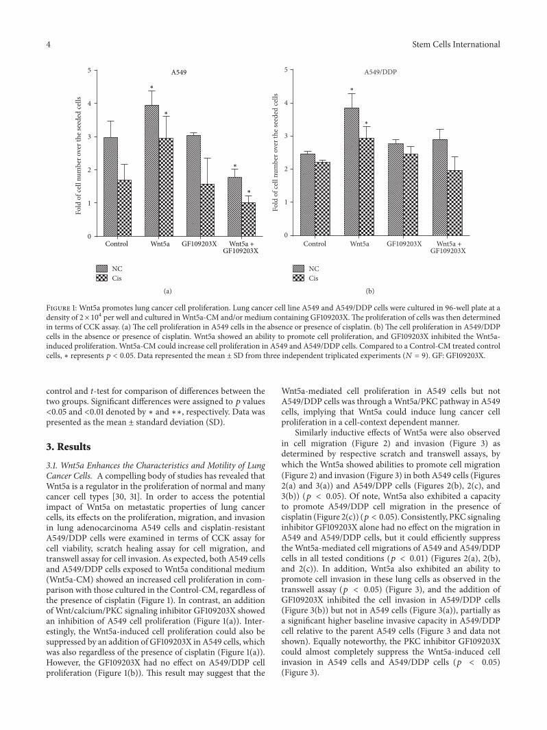
Figure 1: Wnt5a promotes lung cancer cell proliferation. Lung cancer cell line A549 and A549/DDP cells were cultured in 96-well plate at adensity of 2× 10⁴ per well and cultured inWnt5a-CM and/or medium containing GF109203X.The proliferation of cells was then determinedin terms of CCK assay. (a) The cell proliferation in A549 cells in the absence or presence of cisplatin. (b) The cell proliferation in A549/DDPcells in the absence or presence of cisplatin. Wnt5a showed an ability to promote cell proliferation, and GF109203X inhibited the Wnt5a-induced proliferation. Wnt5a-CM could increase cell proliferation in A549 and A549/DDP cells. Compared to a Control-CM treated controlcells, ∗ represents 𝑝 < 0.05. Data represented the mean ± SD from three independent triplicated experiments (𝑁 = 9). GF: GF109203X.
control and t-test for comparison of differences between thetwo groups. Significant differences were assigned to 𝑝 values<0.05 and <0.01 denoted by ∗ and ∗∗, respectively. Data waspresented as the mean ± standard deviation (SD).
3. Results
3.1. Wnt5a Enhances the Characteristics and Motility of LungCancer Cells. A compelling body of studies has revealed thatWnt5a is a regulator in the proliferation of normal and manycancer cell types [30, 31]. In order to access the potentialimpact of Wnt5a on metastatic properties of lung cancercells, its effects on the proliferation, migration, and invasionin lung adenocarcinoma A549 cells and cisplatin-resistantA549/DDP cells were examined in terms of CCK assay forcell viability, scratch healing assay for cell migration, andtranswell assay for cell invasion. As expected, both A549 cellsand A549/DDP cells exposed to Wnt5a conditional medium(Wnt5a-CM) showed an increased cell proliferation in com-parison with those cultured in the Control-CM, regardless ofthe presence of cisplatin (Figure 1). In contrast, an additionof Wnt/calcium/PKC signaling inhibitor GF109203X showedan inhibition of A549 cell proliferation (Figure 1(a)). Inter-estingly, the Wnt5a-induced cell proliferation could also besuppressed by an addition of GF109203X in A549 cells, whichwas also regardless of the presence of cisplatin (Figure 1(a)).However, the GF109203X had no effect on A549/DDP cellproliferation (Figure 1(b)). This result may suggest that the
Wnt5a-mediated cell proliferation in A549 cells but notA549/DDP cells was through aWnt5a/PKC pathway in A549cells, implying that Wnt5a could induce lung cancer cellproliferation in a cell-context dependent manner.
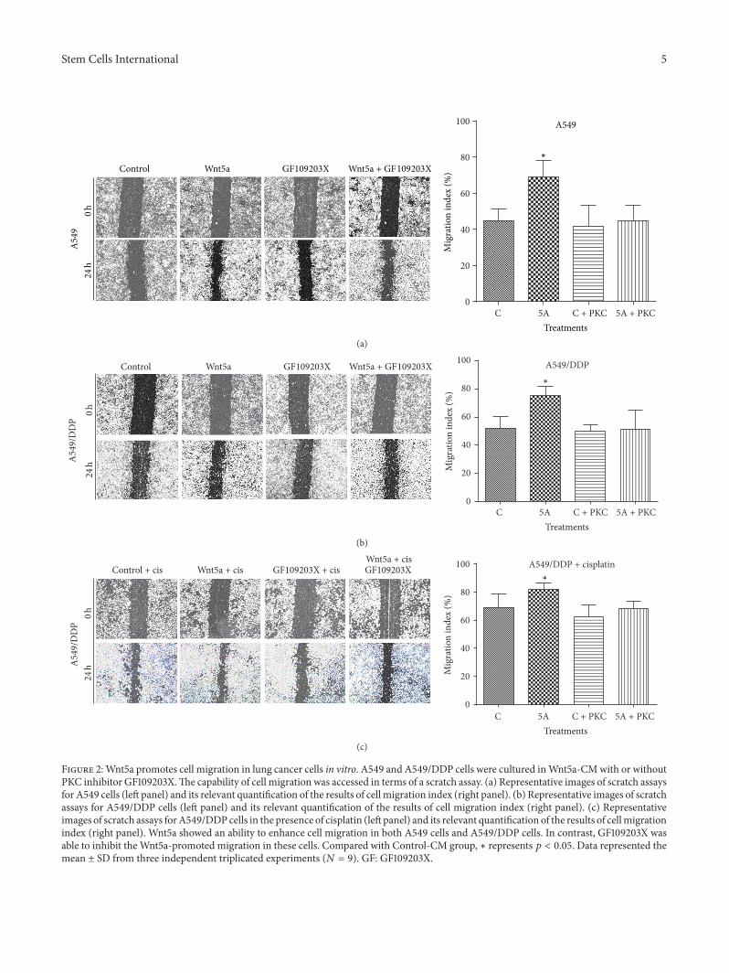
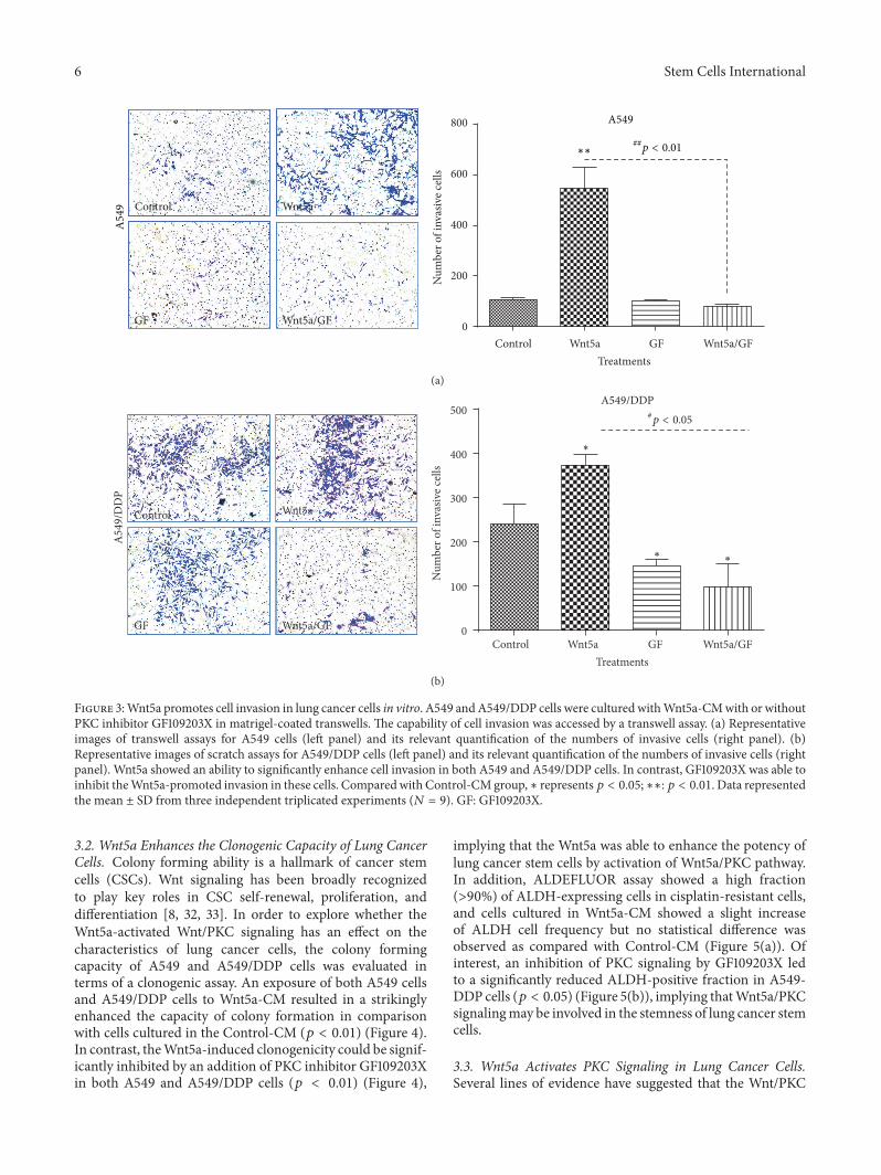
Similarly inductive effects of Wnt5a were also observedin cell migration (Figure 2) and invasion (Figure 3) asdetermined by respective scratch and transwell assays, bywhich the Wnt5a showed abilities to promote cell migration(Figure 2) and invasion (Figure 3) in both A549 cells (Figures2(a) and 3(a)) and A549/DPP cells (Figures 2(b), 2(c), and3(b)) (𝑝 < 0.05). Of note, Wnt5a also exhibited a capacityto promote A549/DDP cell migration in the presence ofcisplatin (Figure 2(c)) (𝑝 < 0.05). Consistently, PKC signalinginhibitor GF109203X alone had no effect on the migration inA549 and A549/DDP cells, but it could efficiently suppressthe Wnt5a-mediated cell migrations of A549 and A549/DDPcells in all tested conditions (𝑝 < 0.01) (Figures 2(a), 2(b),and 2(c)). In addition, Wnt5a also exhibited an ability topromote cell invasion in these lung cells as observed in thetranswell assay (𝑝 < 0.05) (Figure 3), and the addition ofGF109203X inhibited the cell invasion in A549/DDP cells(Figure 3(b)) but not in A549 cells (Figure 3(a)), partially asa significant higher baseline invasive capacity in A549/DDPcell relative to the parent A549 cells (Figure 3 and data notshown). Equally noteworthy, the PKC inhibitor GF109203Xcould almost completely suppress the Wnt5a-induced cellinvasion in A549 cells and A549/DDP cells (𝑝 < 0.05)(Figure 3).
Stem Cells International 5
C 5A C + PKC 5A + PKC0
20
40
60
80
100 A549
∗
Mig
ratio
n in
dex
(%)
Treatments
0h
A549
24h
Control Wnt5a Wnt5a + GF109203XGF109203X
(a)
C 5A C + PKC 5A + PKC0
20
40
60
80
100 A549/DDP∗
Mig
ratio
n in
dex
(%)
Treatments
0h
24hA549
/DD
P
Control Wnt5a Wnt5a + GF109203XGF109203X
(b)
C 5A C + PKC 5A + PKC0
20
40
60
80
100 A549/DDP + cisplatin∗
Mig
ratio
n in
dex
(%)
Treatments
0h
24hA549
/DD
P
Control + cis Wnt5a + cisWnt5a + cisGF109203XGF109203X + cis
(c)
Figure 2:Wnt5a promotes cell migration in lung cancer cells in vitro. A549 and A549/DDP cells were cultured inWnt5a-CMwith or withoutPKC inhibitor GF109203X.The capability of cell migration was accessed in terms of a scratch assay. (a) Representative images of scratch assaysfor A549 cells (left panel) and its relevant quantification of the results of cell migration index (right panel). (b) Representative images of scratchassays for A549/DDP cells (left panel) and its relevant quantification of the results of cell migration index (right panel). (c) Representativeimages of scratch assays forA549/DDPcells in the presence of cisplatin (left panel) and its relevant quantification of the results of cellmigrationindex (right panel). Wnt5a showed an ability to enhance cell migration in both A549 cells and A549/DDP cells. In contrast, GF109203X wasable to inhibit theWnt5a-promoted migration in these cells. Compared with Control-CM group, ∗ represents 𝑝 < 0.05. Data represented themean ± SD from three independent triplicated experiments (𝑁 = 9). GF: GF109203X.
6 Stem Cells International
Control
Control
Wnt5a
Wnt5a
GF
GF
Wnt5a/GF
Wnt5a/GF 0
200
400
600
800
Treatments
Num
ber o
f inv
asiv
e cel
ls
∗∗
A549A549
##p < 0.01
(a)
Control Wnt5a
GF Wnt5a/GFControl Wnt5a GF Wnt5a/GF
Treatments
Num
ber o
f inv
asiv
e cel
ls
0
100
200
300
400
500
∗
∗ ∗
A549/DDP
A549
/DD
P
#p < 0.05
(b)
Figure 3:Wnt5a promotes cell invasion in lung cancer cells in vitro. A549 andA549/DDP cells were cultured withWnt5a-CMwith or withoutPKC inhibitor GF109203X in matrigel-coated transwells. The capability of cell invasion was accessed by a transwell assay. (a) Representativeimages of transwell assays for A549 cells (left panel) and its relevant quantification of the numbers of invasive cells (right panel). (b)Representative images of scratch assays for A549/DDP cells (left panel) and its relevant quantification of the numbers of invasive cells (rightpanel). Wnt5a showed an ability to significantly enhance cell invasion in both A549 and A549/DDP cells. In contrast, GF109203X was able toinhibit theWnt5a-promoted invasion in these cells. Comparedwith Control-CMgroup,∗ represents𝑝 < 0.05;∗∗:𝑝 < 0.01. Data representedthe mean ± SD from three independent triplicated experiments (𝑁 = 9). GF: GF109203X.
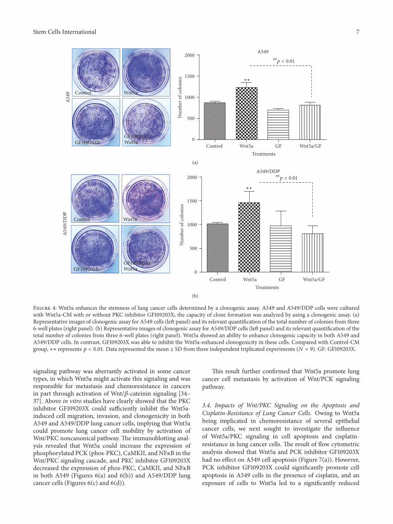
3.2. Wnt5a Enhances the Clonogenic Capacity of Lung CancerCells. Colony forming ability is a hallmark of cancer stemcells (CSCs). Wnt signaling has been broadly recognizedto play key roles in CSC self-renewal, proliferation, anddifferentiation [8, 32, 33]. In order to explore whether theWnt5a-activated Wnt/PKC signaling has an effect on thecharacteristics of lung cancer cells, the colony formingcapacity of A549 and A549/DDP cells was evaluated interms of a clonogenic assay. An exposure of both A549 cellsand A549/DDP cells to Wnt5a-CM resulted in a strikinglyenhanced the capacity of colony formation in comparisonwith cells cultured in the Control-CM (𝑝 < 0.01) (Figure 4).In contrast, theWnt5a-induced clonogenicity could be signif-icantly inhibited by an addition of PKC inhibitor GF109203Xin both A549 and A549/DDP cells (𝑝 < 0.01) (Figure 4),
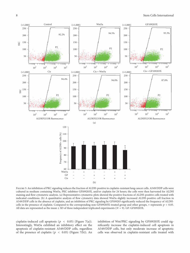
implying that the Wnt5a was able to enhance the potency oflung cancer stem cells by activation of Wnt5a/PKC pathway.In addition, ALDEFLUOR assay showed a high fraction(>90%) of ALDH-expressing cells in cisplatin-resistant cells,and cells cultured in Wnt5a-CM showed a slight increaseof ALDH cell frequency but no statistical difference wasobserved as compared with Control-CM (Figure 5(a)). Ofinterest, an inhibition of PKC signaling by GF109203X ledto a significantly reduced ALDH-positive fraction in A549-DDP cells (𝑝 < 0.05) (Figure 5(b)), implying thatWnt5a/PKCsignalingmay be involved in the stemness of lung cancer stemcells.
3.3. Wnt5a Activates PKC Signaling in Lung Cancer Cells.Several lines of evidence have suggested that the Wnt/PKC
Stem Cells International 7
Control Wnt5a GF Wnt5a/GF0
500
1000
1500
2000
Treatments
Num
ber o
f col
onie
s ∗∗
A549
A54
9 Control Wnt5a
GF109203XGF109203X/Wnt5a
##p < 0.01
(a)
Control Wnt5a GF Wnt5a/GFTreatments
Num
ber o
f col
onie
s
0
500
1000
1500
2000
∗∗
A549/DDP
A549
/DD
P
Control Wnt5a
GF109203XGF109203X/Wnt5a
##p < 0.01
(b)
Figure 4: Wnt5a enhances the stemness of lung cancer cells determined by a clonogenic assay. A549 and A549/DDP cells were culturedwith Wnt5a-CM with or without PKC inhibitor GF109203X; the capacity of clone formation was analyzed by using a clonogenic assay. (a)Representative images of clonogenic assay for A549 cells (left panel) and its relevant quantification of the total number of colonies from three6-well plates (right panel). (b) Representative images of clonogenic assay for A549/DDP cells (left panel) and its relevant quantification of thetotal number of colonies from three 6-well plates (right panel). Wnt5a showed an ability to enhance clonogenic capacity in both A549 andA549/DDP cells. In contrast, GF109203X was able to inhibit the Wnt5a-enhanced clonogenicity in these cells. Compared with Control-CMgroup, ∗∗ represents 𝑝 < 0.01. Data represented the mean ± SD from three independent triplicated experiments (𝑁 = 9). GF: GF109203X.
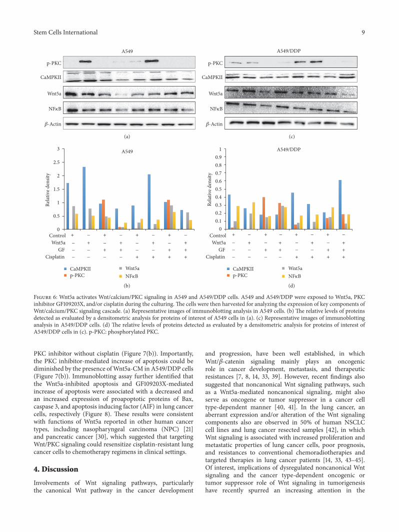
signaling pathway was aberrantly activated in some cancertypes, in which Wnt5a might activate this signaling and wasresponsible for metastasis and chemoresistance in cancersin part through activation of Wnt/𝛽-cateinin signaling [34–37]. Above in vitro studies have clearly showed that the PKCinhibitor GF109203X could sufficiently inhibit the Wnt5a-induced cell migration, invasion, and clonogenicity in bothA549 and A549/DDP lung cancer cells, implying that Wnt5acould promote lung cancer cell mobility by activation ofWnt/PKC noncanonical pathway. The immunoblotting anal-ysis revealed that Wnt5a could increase the expression ofphosphorylated PCK (phos-PKC), CaMKII, and NF𝜅B in theWnt/PKC signaling cascade, and PKC inhibitor GF109203Xdecreased the expression of phos-PKC, CaMKII, and NF𝜅Bin both A549 (Figures 6(a) and 6(b)) and A549/DDP lungcancer cells (Figures 6(c) and 6(d)).
This result further confirmed that Wnt5a promote lungcancer cell metastasis by activation of Wnt/PCK signalingpathway.
3.4. Impacts of Wnt/PKC Signaling on the Apoptosis andCisplatin-Resistance of Lung Cancer Cells. Owing to Wnt5abeing implicated in chemoresistance of several epithelialcancer cells, we next sought to investigate the influenceof Wnt5a/PKC signaling in cell apoptosis and cisplatin-resistance in lung cancer cells. The result of flow cytometricanalysis showed that Wnt5a and PCK inhibitor GF109203Xhad no effect on A549 cell apoptosis (Figure 7(a)). However,PCK inhibitor GF109203X could significantly promote cellapoptosis in A549 cells in the presence of cisplatin, and anexposure of cells to Wnt5a led to a significantly reduced
8 Stem Cells InternationalSS
CSS
C
Cis + Wnt5a
Control
Cis Cis + GF109203X
Wnt5a GF109203X
92.2%
87.0%94.0%94.4%
94.5% 93.3%
P2 P2 P2
P2 P2P2
(×1,000)250
200
150
100
50
(×1,000)250
200
150
100
50
(×1,000)250
200
150
100
50
(×1,000)250
200
150
100
50
(×1,000)250
200
150
100
50
(×1,000)250
200
150
100
50
102 103 104 105 102 103 104 105 102 103 104 105
102 103 104 105102 103 104 105102 103 104 105
ALDEFLUOR fluorescence ALDEFLUOR fluorescenceALDEFLUOR fluorescence(a)
60
70
80
90
100
110
120
∗
Freq
uenc
y of
ALD
H+
cells
(%)
+
+
+
+
+
+
+
−
−
−
−
− −
−
−
−
−
−
Wnt5aGFCis
(b)
Figure 5: An inhibition of PKC signaling reduces the fraction of ALDH-positive in cisplatin-resistant lung cancer cells. A549/DDP cells werecultured in medium containing Wnt5a, PKC inhibitor GF109203X, and/or cisplatin for 24 hours; the cells were then harvested for ALDHstaining and flow cytometric analysis. (a) Representative cytometric plots showed the positive fractions of ALDH-positive cells treated withindicated conditions. (b) A quantitative analysis of flow cytometry data showed Wnt5a slightly increased ALDH-positive cell fraction inA549/DDP cells in the absence of cisplatin, and an inhibition of PKC signaling by GF109203 significantly reduced the frequency of ALDH-cells in the presence of cisplatin. Compared to the corresponding non-GF109203X-treated group and other groups, ∗ represents 𝑝 < 0.05.All data are represented as the mean ± SD of three independent triplicated experiments (𝑁 = 9). GF: GF109203X.
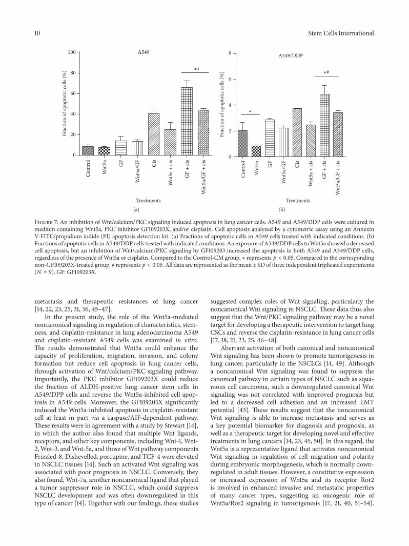
cisplatin-induced cell apoptosis (𝑝 < 0.05) (Figure 7(a)).Interestingly, Wnt5a exhibited an inhibitory effect on theapoptosis of cisplatin-resistant A549/DDP cells, regardlessof the presence of cisplatin (𝑝 < 0.05) (Figure 7(b)). An
inhibition of Wnt/PKC signaling by GF109203X could sig-nificantly increase the cisplatin-induced cell apoptosis inA549/DDP cells, but only moderate increase of apoptoticcells was observed in cisplatin-resistant cells treated with
Stem Cells International 9
ControlWnt5a
GFCisplatin
ControlWnt5a
GFCisplatin
Rela
tive d
ensit
y
Rela
tive d
ensit
y
p-PKC
CaMPKII
Wnt5a
0
0.5
1
1.5
2
2.5
3
CaMPKIIp-PKC
Wnt5a
00.10.20.30.40.50.60.70.80.9
1A549
A549 A549/DDP
A549/DDP
(c)(a)
(d)(b)
+
−
−
−
−
++
+
+
+−
+
−
−
+
+
+
−
−
+
−
++
−
+
+−
−
−
−
+
−
+
−
−
−
−
++
+
+
+−
+
−
−
+
+
+
−
−
+
−
++
−
+
+−
−
−
−
+
−
NF𝜅B
NF𝜅BCaMPKIIp-PKC
Wnt5aNF𝜅B
𝛽-Actin
p-PKC
CaMPKII
Wnt5a
NF𝜅B
𝛽-Actin
Figure 6: Wnt5a activates Wnt/calcium/PKC signaling in A549 and A549/DDP cells. A549 and A549/DDP were exposed to Wnt5a, PKCinhibitor GF109203X, and/or cisplatin during the culturing. The cells were then harvested for analyzing the expression of key components ofWnt/calcium/PKC signaling cascade. (a) Representative images of immunoblotting analysis in A549 cells. (b) The relative levels of proteinsdetected as evaluated by a densitometric analysis for proteins of interest of A549 cells in (a). (c) Representative images of immunoblottinganalysis in A549/DDP cells. (d) The relative levels of proteins detected as evaluated by a densitometric analysis for proteins of interest ofA549/DDP cells in (c). p-PKC: phosphorylated PKC.
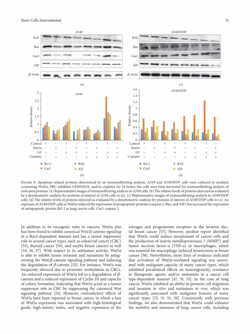
PKC inhibitor without cisplatin (Figure 7(b)). Importantly,the PKC inhibitor-mediated increase of apoptosis could bediminished by the presence ofWnt5a-CM in A549/DDP cells(Figure 7(b)). Immunoblotting assay further identified thatthe Wnt5a-inhibited apoptosis and GF109203X-mediatedincrease of apoptosis were associated with a decreased andan increased expression of proapoptotic proteins of Bax,caspase 3, and apoptosis inducing factor (AIF) in lung cancercells, respectively (Figure 8). These results were consistentwith functions of Wnt5a reported in other human cancertypes, including nasopharyngeal carcinoma (NPC) [21]and pancreatic cancer [30], which suggested that targetingWnt/PKC signaling could resensitize cisplatin-resistant lungcancer cells to chemotherapy regimens in clinical settings.
4. Discussion
Involvements of Wnt signaling pathways, particularlythe canonical Wnt pathway in the cancer development
and progression, have been well established, in whichWnt/𝛽-catenin signaling mainly plays an oncogenicrole in cancer development, metastasis, and therapeuticresistances [7, 8, 14, 33, 39]. However, recent findings alsosuggested that noncanonical Wnt signaling pathways, suchas a Wnt5a-mediated noncanonical signaling, might alsoserve as oncogene or tumor suppressor in a cancer celltype-dependent manner [40, 41]. In the lung cancer, anaberrant expression and/or alteration of the Wnt signalingcomponents also are observed in 50% of human NSCLCcell lines and lung cancer resected samples [42], in whichWnt signaling is associated with increased proliferation andmetastatic properties of lung cancer cells, poor prognosis,and resistances to conventional chemoradiotherapies andtargeted therapies in lung cancer patients [14, 33, 43–45].Of interest, implications of dysregulated noncanonical Wntsignaling and the cancer type-dependent oncogenic ortumor suppressor role of Wnt signaling in tumorigenesishave recently spurred an increasing attention in the
10 Stem Cells International
0
20
40
60
80
100
Treatments
Frac
tion
of ap
opto
tic ce
lls (%
)
A549
Con
trol
Wnt5
a
Wnt5
a/G
F
GF
Cis
Wnt5
a + ci
s
GF
+ ci
s
Wnt5
a/G
F +
cis
∗#
(a)Treatments
0
2
4
6
8
Frac
tion
of ap
opto
tic ce
lls (%
)
A549/DDP
Con
trol
Wnt5
a
GF
Wnt5
a/G
F
Cis
Wnt5
a + ci
s
GF
+ ci
s
Wnt5
a/G
F +
cis
∗
∗#
(b)
Figure 7: An inhibition of Wnt/calcium/PKC signaling induced apoptosis in lung cancer cells. A549 and A549/DDP cells were cultured inmedium containing Wnt5a, PKC inhibitor GF109203X, and/or cisplatin. Cell apoptosis analyzed by a cytometric assay using an AnnexinV-FITC/propidium iodide (PI) apoptosis detection kit. (a) Fractions of apoptotic cells in A549 cells treated with indicated conditions. (b)Fractions of apoptotic cells inA549/DDP cells treatedwith indicated conditions. An exposure ofA549/DDP cells toWnt5a showed a decreasedcell apoptosis, but an inhibition of Wnt/calcium/PKC signaling by GF109203 increased the apoptosis in both A549 and A549/DDP cells,regardless of the presence ofWnt5a or cisplatin. Compared to the Control-CM group, ∗ represents 𝑝 < 0.05. Compared to the correspondingnon-GF109203X-treated group, # represents 𝑝 < 0.05. All data are represented as themean ± SD of three independent triplicated experiments(𝑁 = 9). GF: GF109203X.
metastasis and therapeutic resistances of lung cancer[14, 22, 23, 25, 31, 36, 45–47].
In the present study, the role of the Wnt5a-mediatednoncanonical signaling in regulation of characteristics, stem-ness, and cisplatin-resistance in lung adenocarcinoma A549and cisplatin-resistant A549 cells was examined in vitro.The results demonstrated that Wnt5a could enhance thecapacity of proliferation, migration, invasion, and colonyformation but reduce cell apoptosis in lung cancer cells,through activation of Wnt/calcium/PKC signaling pathway.Importantly, the PKC inhibitor GF109203X could reducethe fraction of ALDH-positive lung cancer stem cells inA549/DPP cells and reverse the Wnt5a-inhibited cell apop-tosis in A549 cells. Moreover, the GF109203X significantlyinduced the Wnt5a-inhibited apoptosis in cisplatin-resistantcell at least in part via a caspase/AIF-dependent pathway.These results were in agreement with a study by Stewart [14],in which the author also found that multiple Wnt ligands,receptors, and other key components, includingWnt-1, Wnt-2,Wnt-3, andWnt-5a, and those ofWnt pathway componentsFrizzled-8, Dishevelled, porcupine, and TCF-4 were elevatedin NSCLC tissues [14]. Such an activated Wnt signaling wasassociated with poor prognosis in NSCLC. Conversely, theyalso found, Wnt-7a, another noncanonical ligand that playeda tumor suppressor role in NSCLC, which could suppressNSCLC development and was often downregulated in thistype of cancer [14]. Together with our findings, these studies
suggested complex roles of Wnt signaling, particularly thenoncanonical Wnt signaling in NSCLC. These data thus alsosuggest that the Wnt/PKC signaling pathway may be a noveltarget for developing a therapeutic intervention to target lungCSCs and reverse the cisplatin-resistance in lung cancer cells[17, 18, 21, 23, 25, 46–48].
Aberrant activation of both canonical and noncanonicalWnt signaling has been shown to promote tumorigenesis inlung cancer, particularly in the NSCLCs [14, 49]. Althougha noncanonical Wnt signaling was found to suppress thecanonical pathway in certain types of NSCLC such as squa-mous cell carcinoma, such a downregulated canonical Wntsignaling was not correlated with improved prognosis butled to a decreased cell adhesion and an increased EMTpotential [43]. These results suggest that the noncanonicalWnt signaling is able to increase metastasis and serves asa key potential biomarker for diagnosis and prognosis, aswell as a therapeutic target for developing novel and effectivetreatments in lung cancers [14, 23, 45, 50]. In this regard, theWnt5a is a representative ligand that activates noncanonicalWnt signaling in regulation of cell migration and polarityduring embryonic morphogenesis, which is normally down-regulated in adult tissues. However, a constitutive expressionor increased expression of Wnt5a and its receptor Ror2is involved in enhanced invasive and metastatic propertiesof many cancer types, suggesting an oncogenic role ofWnt5a/Ror2 signaling in tumorigenesis [17, 21, 40, 51–54].
Stem Cells International 11
Bcl2
Cas3
Bax
AIF
Rela
tive d
ensit
y
Rela
tive d
ensit
y
ControlWnt5a
GFCisplatin
ControlWnt5a
GFCisplatin
0
0.5
1
1.5
2
2.5
3
Bcl-2Cas3
BAXAIF
Bcl-2Cas3
BAXAIF
0
0.2
0.4
0.6
0.8
1
1.2
1.4
1.6
A549
A549
A549/DDP
A549/DDP
+
−
−
−
−
++
+
+
+−
+
−
−
+
+
+
−
−
+
−
++
−
+
+−
−
−
−
+
−
+
−
−
−
−
++
+
+
+−
+
−
−
+
+
+
−
−
+
−
++
−
+
+−
−
−
−
+
−
𝛽-Actin
Bcl2
Cas3
Bax
AIF
𝛽-Actin
(c)(a)
(d)(b)
Figure 8: Apoptosis related proteins determined by an immunoblotting analysis. A549 and A549/DDP cells were cultured in mediumcontaining Wnt5a, PKC inhibitor GF109203X, and/or cisplatin for 24 hours; the cells were then harvested for immunoblotting analysis ofindicated proteins. (a) Representative images of immunoblotting analysis in A549 cells. (b)The relative levels of proteins detected as evaluatedby a densitometric analysis for proteins of interest of A549 cells in (a). (c) Representative images of immunoblotting analysis in A549/DDPcells. (d) The relative levels of proteins detected as evaluated by a densitometric analysis for proteins of interest of A549/DDP cells in (c). Anexposure of A549/DDP cells toWnt5a reduced the expression of proapoptotic proteins (caspase 3, Bax, and AIF) but increased the expressionof antiapoptotic protein Bcl-2 in lung cancer cells. Cas3: caspase 3.
In addition to its oncogenic roles in cancers, Wnt5a alsohas been found to inhibit canonical Wnt/𝛽-catenin signalingin a Ror2-dependent manner and has a tumor suppressorrole in several cancer types, such as colorectal cancer (CRC)[55], thyroid cancer [56], and maybe breast cancers as well[34, 36, 57]. With respect to its antitumor activity, Wnt5ais able to inhibit tumor invasion and metastasis by antag-onizing the Wnt/𝛽-catenin signaling pathway and inducingthe degradation of 𝛽-catenin [32]. For instance, Wnt5a wasfrequently silenced due to promoter methylation in CRCs.An enforced expression of Wnt5a led to a degradation of 𝛽-catenin and a reduced expression of Cyclin D1 and capacityof colony formation, indicating that Wnt5a acted as a tumorsuppressor role in CRC by suppressing the canonical Wntsignaling pathway [55]. However, contradictory effects ofWnt5a have been reported in breast cancer, in which a lossof Wnt5a expression was associated with high-histologicalgrade, high-mitotic index, and negative expression of the
estrogen and progesterone receptors in the invasive duc-tal breast cancer [57]. However, another report identifiedthat Wnt5a could induce invasiveness of cancer cells andthe production of matrix metalloproteinase-7 (MMP7) andtumor necrosis factor-𝛼 (TNF-𝛼) in macrophages, whichwas essential for macrophage-induced invasiveness in breastcancer [58]. Nevertheless, more lines of evidence indicatedthat activation of Wnt5a-mediated signaling was associ-ated with malignant capacity of many cancer types, whichexhibited paradoxical effects on tumorigenicity, resistanceto therapeutic agents, and/or metastasis in a cancer celltype-dependent manner [47, 51, 53]. In the case of lungcancer, Wnt5a exhibited an ability to promote cell migrationand invasion in vitro and metastasis in vivo, which wassignificantly associated with malignant features of manycancer types [23, 51, 53, 59]. Consistently with previousfindings, we also demonstrated that Wnt5a could enhancethe mobility and stemness of lung cancer cells, including
12 Stem Cells International
migration, invasion, colony formation, and chemoresistance,through a Wnt/PKC pathway.
Indeed, an early microarray analysis for gene expressionprofiling demonstrated that Wnt5a could directly promotemelanoma cell invasion and motility via inducing expressionof Slug, which in turn promotes EMT gene expression ina PKC-dependent manner [60, 61]. Such a Wnt5a/PKC-promoted EMT was also found in epithelial ovarian cancer[18] and oral squamous cell carcinoma (OSCC) [20]. In theepithelial ovarian cancer, an increased expression of Wnt5aand PKC𝛼 was significantly correlated with metastasis of thedisease, and the PKC𝛼 inhibitor could reduce the metastaticcapacity [18]; in OSCC, Wnt5a was found to promote themigration and invasion of cancer cells through activation ofnoncanonical Wnt/Calcium/PKC pathway [20]. In addition,a recent high-throughput gene expression profiling furtherrevealed that Wnt5a signaling was upregulated in highlymetastatic nasopharyngeal carcinoma (NPC) cells and tis-sues, in which Wnt5a was able to activate PKC signaling andform a positive Wnt5a and phosphorylated PKC loop to pro-mote the stemness characteristics of NPC cells, leading to anenhanced metastatic features and tumorigenicity in vitro andin vivo [21]. In lung cancer,Whang et al. also found that expo-sure of human bronchial epithelia (HBE) cells to cigarettesmoke condensate showed an activated Wnt5a/PKC/Aktsignaling and increased cell proliferation and clonogenicity.A higher expression of Wnt5a was also found in tumortissues of smokers relative to matched normal tissues. Ablockage of Wnt5a/PKC led to a significantly decreased cellviability and clonogenicity, along with an increased apoptosisvia the downregulation of Bcl2 and induction of cleavedpoly-ADP-ribose polymerase [23]. In addition, Wnt5a wastranscribed based onmultiple mechanisms, including Notch,Hedgehog, TGF𝛽, and NF-𝜅B signaling cascades [62]. Inagreement with these findings, our results also suggested thataWnt5a/PKC-mediated caspase-dependent apoptosis signal-ing pathway and the NF-𝜅B signaling cascade were involvedin the metastatic potentials, stemness, and chemoresistancein A549 and cisplatin-resistant A549/DDP NSCLC cells. Ofimportance, an inhibition of PKC signaling byGF109203X ledto a significantly decreased fraction ofALDH-expressing cellsinA549/DDP cells in the presence of cisplatin.TheALDHhasbeen recognized as a cancer stem cellmarker in several cancertypes, including the lung cancer [63–68].This finding impliesthat targeting of PKC pathway may reverse chemoresistancein part by reducing stemness of CSC in lung cancer cells.
Apart from the oncogenic role of Wnt signaling in cancermetastasis and invasion, dysregulations of Wnt signalingpathways, particularly the canonical Wnt pathway, havebeen demonstrated to contribute to therapeutic resistancesin many cancer types [31, 37, 44, 69–72]. In this regard,Wnt5a was also found to modulate cell cycle progression andcontributes to the chemoresistance in pancreatic cancer cells[70] and regulates ABCB1 expression in multidrug-resistantbreast cancer cells through activation of the noncanonicalPKA/beta-catenin pathway [48]. In the NSCLC cells, acti-vation of Wnt pathway was previously demonstrated to beassociated with resistance to platinum-based chemotherapy[37, 50, 73]. Cisplatin-resistant NSCLC cells had increased
expression of Wnt pathway genes. An exposure of these cellsto a Wnt inhibitor led to an inhibited cell proliferation andsensitized NSCLC cells to chemotherapeutic agent docetaxel[74] and cisplatin [75], along with a downregulated expres-sion of Wnt signaling target genes. In this study, we showedthat a blockage of Wnt/PKC signaling could increase thebaseline apoptosis and cisplatin-induced apoptosis inNSCLCA549 cells and cisplatin-resistant A549/DDP cells, suggestingthat targetingWnt/PKCnoncanonicalWnt signalingmay be apotential therapeutic strategy to circumvent chemoresistancein lung cancer treatment.
5. Conclusion
In conclusion, in the present study, we examined the potentialofWnt5a-mediated noncanonicalWnt signaling in themobil-ity and chemoresistance of NSCLC cells and stemness of lungCSCs. The results demonstrated that Wnt5a could promoteproliferation, migration, invasion, and colony formation, aswell as inhibit cell apoptosis in both A549 and cisplatin-resistant A549/DDP NSCLC cells. Moreover, a blockage ofWnt5a signaling by PKC inhibitor GF109203X could strik-ingly reduce the mobility and increase both the baselineapoptosis and the cisplatin-induced apoptosis in A549 andA549/DDP cells and reverse the inhibitory function ofWnt5a in cell apoptosis. Mechanistically, theWnt5a activatedWnt5a/PKC signaling cascade and led to activation of endo-plasmic reticulum (ER) release of Ca2+, PKC, and CaMKII,which in turn activated NF𝜅B, subsequently promoted stem-ness, and inhibited apoptosis in NSCLC cells (Figure 9).This study thus suggests that Wnt5a/PKC signaling may playan important role in the metastasis, chemoresistance, andstemness of lung CSCs, which may provide a novel clue forunderstanding the crucial role of noncanonicalWnt signalingin lung carcinogenesis and therapeutic resistances.
Abbreviations
ABCB: ATP-binding cassette, subfamily BALDH: Aldehyde dehydrogenaseBAAA: Bodipy-aminoacetaldehydeCaMKII: Calmodulin-dependent protein kinase IICSC: Cancer stem cellDEAB: N,N-DiethylaminobenzaldehydeEMT: Epithelial-mesenchymal transitionNLK: Nemo-like kinaseNFAT: Nuclear factor of activated T-cellsNSCLC: Non-small-cell lung cancerPCP: Planar cell polarityPKC: Protein kinase CTAK1: Transforming growth factor beta-activated
kinase 1.
Disclosure
Theauthors are solely responsible for the contents andwritingof this paper.

Stem Cells International 13
StimulationInhibition
CamKII
Wnt5aWnt5a
PKC
Cytokine
PKC inhibitor
Wnt5a
Wnt5a
Stem cell properties
Apoptosis
W
NF𝜅B
NF𝜅B
Ca2+
Ca2+Ca2+Ca2+
Ca2+Ca2+
Ca2+
Ror2
Target genes:Bcl-2, caspase 3,
and so forth
Ca2+ pathway
Dishevelled
Figure 9: A schematic model to summarize Wnt5/PKC signaling pathway in lung cancer. Wnt5a activates PKC signaling and leads toendoplasmic reticulum (ER) release of Ca2+; PKC and CaMKII are activated, which in turn activate NF𝜅B. Subsequently, the activated NF𝜅Bsignaling promotes stem cell properties and inhibits apoptosis in lung cancer. CaMK: calcium/calmodulin kinase; FZD: Frizzled; PKC: proteinkinase C; Ror: receptor tyrosine kinase-like orphan receptor.
Competing Interests
The authors declare that they have no competing interests.
Authors’ Contributions
JinxiHe andXiaoming Liu conceived and designed the exper-iments; Jiali Yang, Kangjian Zhang, and JunWei analyzed thedata and drafted the manuscript; Jiali Yang, Kangjian Zhang,Jing Xue, Jun Wei, Jing Li, Juan Shi, Yongzhao Zhu, and JuanChen performed experiments and acquired data; Jiali Yang,Jinxi He, and Xiaoming Liu interpreted data and criticallyrevised the manuscript. All authors read and approved thefinal version of themanuscript. Jiali Yang andKangjianZhangcontributed equally.
Acknowledgments
This work was supported by grants from Natural ScienceFoundation of Ningxia NZ15277 to Jinxi He and NZ16155 toJiali Yang, a starting grant (XM2015090) from the NingxiaMedical University to Jiali Yang, and a grant from the
National Natural Science Foundation of China (31472191) toXiaoming Liu.
References
[1] R. L. Siegel, K. D. Miller, and A. Jemal, “Cancer statistics, 2015,”CA: A Cancer Journal for Clinicians, vol. 65, no. 1, pp. 5–29, 2015.
[2] F. Han, J. He, F. Li et al., “Emerging roles of MicroRNAsin EGFR-targeted therapies for lung cancer,” BioMed ResearchInternational, vol. 2015, Article ID 672759, 10 pages, 2015.
[3] A. K. Dubey, U. Gupta, and S. Jain, “Epidemiology of lungcancer and approaches for its prediction: a systematic reviewand analysis,”Chinese Journal of Cancer, vol. 35, no. 1, p. 71, 2016.
[4] J. Milanowski and K. Szmygin-Milanowska, “Treatment ofnon-small cell lung cancer—where we are?” Pneumonologia iAlergologia Polska, vol. 81, no. 1, pp. 55–60, 2013.
[5] L. Yan and L. Xu, “Global efforts in conquering lung cancer inChina,” Chinese Journal of Cancer, vol. 34, no. 7, pp. 320–322,2015.
[6] Z.H. Siddik, “Cisplatin:mode of cytotoxic action andmolecularbasis of resistance,” Oncogene, vol. 22, no. 47, pp. 7265–7279,2003.
14 Stem Cells International
[7] J. N. Anastas and R. T. Moon, “WNT signalling pathways astherapeutic targets in cancer,” Nature Reviews Cancer, vol. 13,no. 1, pp. 11–26, 2013.
[8] N. Takebe, L. Miele, P. J. Harris et al., “Targeting Notch,Hedgehog, and Wnt pathways in cancer stem cells: clinicalupdate,” Nature Reviews Clinical Oncology, vol. 12, no. 8, pp.445–464, 2015.
[9] M. C. Florian, K. J. Nattamai, K. Dorr et al., “A canonical tonon-canonical Wnt signalling switch in haematopoietic stem-cell ageing,” Nature, vol. 503, no. 7476, pp. 392–396, 2013.
[10] X. Lim and R. Nusse, “Wnt signaling in skin development,homeostasis, and disease,” Cold Spring Harbor Perspectives inBiology, vol. 5, no. 2, 2013.
[11] C. E. Ford, S. S. Qian Ma, A. Quadir, and R. L. Ward, “The dualrole of the novelWnt receptor tyrosine kinase, ROR2, in humancarcinogenesis,” International Journal of Cancer, vol. 133, no. 4,pp. 779–787, 2013.
[12] R. A. Winn, M. Van Scoyk, M. Hammond et al., “Antitu-morigenic effect of Wnt 7a and Fzd 9 in non-small cell lungcancer cells is mediated through ERK-5-dependent activationof peroxisome proliferator-activated receptor 𝛾,”The Journal ofBiological Chemistry, vol. 281, no. 37, pp. 26943–26950, 2006.
[13] D. J. Stewart, D. W. Chang, Y. Ye et al., “Wnt signaling pathwaypharmacogenetics in non-small cell lung cancer,” Pharmacoge-nomics Journal, vol. 14, no. 6, pp. 509–522, 2014.
[14] D. J. Stewart, “Wnt signaling pathway in non-small cell lungcancer,” Journal of the National Cancer Institute, vol. 106, no. 1,Article ID djt356, 2014.
[15] C.-L. Huang, D. Liu, J. Nakano et al., “Wnt5a expression isassociatedwith the tumor proliferation and the stromal vascularendothelial growth factor—an expression in non-small-celllung cancer,” Journal of Clinical Oncology, vol. 23, no. 34, pp.8765–8773, 2005.
[16] S. Lejeune, E. L. Huguet, A. Hamby, R. Poulsom, and A.L. Harris, “Wnt5a cloning, expression, and up-regulation inhuman primary breast cancers,” Clinical Cancer Research, vol.1, no. 2, pp. 215–222, 1995.
[17] C. Peng, X. Zhang, H. Yu, D. Wu, and J. Zheng, “Wnt5a as apredictor in poor clinical outcome of patients and a mediatorin chemoresistance of ovarian cancer,” International Journal ofGynecological Cancer, vol. 21, no. 2, pp. 280–288, 2011.
[18] H. Qi, B. Sun, X. Zhao et al., “Wnt5a promotes vasculogenicmimicry and epithelial-mesenchymal transition via proteinkinase C𝛼 in epithelial ovarian cancer,” Oncology Reports, vol.32, no. 2, pp. 771–779, 2014.
[19] T. Saitoh, T. Mine, and M. Katoh, “Frequent up-regulationof WNT5A mRNA in primary gastric cancer,” InternationalJournal of Molecular Medicine, vol. 9, no. 5, pp. 515–519, 2002.
[20] Z. Prgomet, L. Axelsson, P. Lindberg, and T. Andersson,“Migration and invasion of oral squamous carcinoma cellsis promoted by WNT5A, a regulator of cancer progression,”Journal of Oral Pathology and Medicine, vol. 44, no. 10, pp. 776–784, 2015.
[21] L. Qin, Y.-T. Yin, F.-J. Zheng et al., “WNT5A promotes stemnesscharacteristics in nasopharyngeal carcinoma cells leading tometastasis and tumorigenesis,” Oncotarget, vol. 6, no. 12, pp.10239–10252, 2015.
[22] A. Kikuchi, H. Yamamoto, A. Sato, and S. Matsumoto, “Wnt5a:its signalling, functions and implication in diseases,” ActaPhysiologica, vol. 204, no. 1, pp. 17–33, 2012.
[23] Y. M. Whang, U. Jo, J. S. Sung et al., “Wnt5a is associated withcigarette smoke-related lung carcinogenesis via protein kinaseC,” PLoS ONE, vol. 8, no. 1, article e53012, 2013.
[24] M.-I. L. Kang, A. R. Baker, C. R. Dextras, S. M. Cabarcas,M. R. Young, and N. H. Colburn, “Targeting of noncanonicalWnt5a signaling by AP-1 blocker dominant-negative jun whenit inhibits skin carcinogenesis,” Genes and Cancer, vol. 3, no. 1,pp. 37–50, 2012.
[25] Z. Debebe and W. K. Rathmell, “Ror2 as a therapeutic target incancer,” Pharmacology and Therapeutics, vol. 150, pp. 143–148,2015.
[26] X. Wu, G. Deng, M. Li et al., “Wnt/𝛽-Catenin signalingreduces Bacillus Calmette-Guerin-induced macrophage necro-sis through a ROS -mediated PARP/AIF-dependent pathway,”BMC Immunology, vol. 16, no. 1, article 16, 2015.
[27] Q. Fu, Y. Du, C. Yang et al., “An oncogenic role of miR-592 in tumorigenesis of human colorectal cancer by targetingForkhead Box O3A (FoxO3A),” Expert Opinion on TherapeuticTargets, vol. 20, no. 7, pp. 771–782, 2016.
[28] H. Li, C. Li, R. Dai et al., “Expression of acetylated histone 3 inthe spinal cord and the effect ofmorphine on inflammatory painin rats,” Neural Regeneration Research, vol. 7, no. 7, pp. 517–522,2012.
[29] Y. Li, J. Shi, J. Yang et al., “A Wnt/𝛽-catenin negative feedbackloop represses TLR-triggered inflammatory responses in alve-olar epithelial cells,” Molecular Immunology, vol. 59, no. 2, pp.128–135, 2014.
[30] H. Bo, L. Gao, Y. Chen, J. Zhang, and M. Zhu, “Upregulationof the expression of Wnt5a promotes the proliferation ofpancreatic cancer cells in vitro and in a nude mouse model,”Molecular Medicine Reports, vol. 13, no. 2, pp. 1163–1171, 2016.
[31] L. J. Vuga, A. Ben-Yehudah, E. Kovkarova-Naumovski et al.,“WNT5A is a regulator of fibroblast proliferation and resistanceto apoptosis,” American Journal of Respiratory Cell and Molecu-lar Biology, vol. 41, no. 5, pp. 583–589, 2009.
[32] M. Katoh and M. Katoh, “STAT3-induced WNT5A signalingloop in embryonic stem cells, adult normal tissues, chronicpersistent inflammation, rheumatoid arthritis and cancer(Review),” International Journal of Molecular Medicine, vol. 19,no. 2, pp. 273–278, 2007.
[33] X. Zhang, Y. Lou, H. Wang et al., “Wnt signaling regulatesthe stemness of lung cancer stem cells and its inhibitorsexert anticancer effect on lung cancer SPC-A1 cells,” MedicalOncology, vol. 32, no. 4, article 95, 2015.
[34] T. Pukrop, F. Klemm, T. Hagemann et al., “Wnt 5a signaling iscritical for macrophage-induced invasion of breast cancer celllines,” Proceedings of the National Academy of Sciences of theUnited States of America, vol. 103, no. 14, pp. 5454–5459, 2006.
[35] S. K. Dissanayake and A. T. Weeraratna, “Detecting PKC phos-phorylation as part of the Wnt/calcium pathway in cutaneousmelanoma,”Methods inMolecular Biology, vol. 468, pp. 157–172,2008.
[36] C. Henry, A. Quadir, N. J. Hawkins et al., “Expression of thenovel Wnt receptor ROR2 is increased in breast cancer andmay regulate both 𝛽-catenin dependent and independent Wntsignalling,” Journal of Cancer Research and Clinical Oncology,vol. 141, no. 2, pp. 243–254, 2015.
[37] Y. Gao, Z. Liu, X. Zhang et al., “Inhibition of cytoplasmic GSK-3𝛽 increases cisplatin resistance through activation of Wnt/𝛽-catenin signaling in A549/DDP cells,” Cancer Letters, vol. 336,no. 1, pp. 231–239, 2013.
Stem Cells International 15
[38] A. De, “Wnt/Ca2+ signaling pathway: a brief overview,” ActaBiochimica et Biophysica Sinica, vol. 43, no. 10, pp. 745–756, 2011.
[39] J. Espada, M. B. Calvo, S. Dıaz-Prado, and V. Medina, “Wntsignalling and cancer stem cells,” Clinical and TranslationalOncology, vol. 11, no. 7, pp. 411–427, 2009.
[40] M. Endo, M. Nishita, M. Fujii, and Y. Minami, “Insight into therole of Wnt5a-induced signaling in normal and cancer cells,”International Review of Cell and Molecular Biology, vol. 314, pp.117–148, 2015.
[41] Y. Yuan, C. C. Niu, G. Deng et al., “TheWnt5a/Ror2 noncanon-ical signaling pathway inhibits canonicalWnt signaling in K562cells,” International Journal of Molecular Medicine, vol. 27, no. 1,pp. 63–69, 2011.
[42] G. Akiri, M. M. Cherian, S. Vijayakumar, G. Liu, A. Bafico, andS. A. Aaronson, “Wnt pathway aberrations including autocrineWnt activation occur at high frequency in human non-small-cell lung carcinoma,” Oncogene, vol. 28, no. 21, pp. 2163–2172,2009.
[43] D. Bartis, V. Csongei, A. Weich et al., “Down-regulation ofcanonical and up-regulation of non-canonical wnt signallingin the carcinogenic process of squamous cell lung carcinoma,”PLoS ONE, vol. 8, no. 3, Article ID e57393, 2013.
[44] A. Nakata, R. Yoshida, R. Yamaguchi et al., “Elevated 𝛽-cateninpathway as a novel target for patients with resistance to EGFreceptor targeting drugs,” Scientific Reports, vol. 5, article 13076,2015.
[45] C. Li, S. Bellusci, Z. Borok, and P.Minoo, “Non-canonicalWNTsignalling in the lung,” Journal of Biochemistry, vol. 158, no. 5, pp.355–365, 2015.
[46] R. J.MacLeod,M.Hayes, and I. Pacheco, “Wnt5a secretion stim-ulated by the extracellular calcium-sensing receptor inhibitsdefectiveWnt signaling in colon cancer cells,”American Journalof Physiology—Gastrointestinal and Liver Physiology, vol. 293,no. 1, pp. G403–G411, 2007.
[47] N. Zhu, L. Qin, Z. G. Luo, Q. Guo, L. Y. Yang, and D. F. Liao,“Challenging role of Wnt5a and its signaling pathway in cancermetastasis (Review),” Experimental and Therapeutic Medicine,vol. 8, no. 1, pp. 3–8, 2014.
[48] T.-H. Hung, S.-C. Hsu, C.-Y. Cheng et al., “Wnt5A regulatesABCB1 expression in multidrug-resistant cancer cells throughactivation of the non-canonical PKA/𝛽-catenin pathway,”Onco-target, vol. 5, no. 23, pp. 12273–12290, 2014.
[49] T. S. Gujral, M. Chan, L. Peshkin, P. K. Sorger, M.W. Kirschner,and G. Macbeath, “A noncanonical frizzled2 pathway regulatesepithelial-mesenchymal transition and metastasis,” Cell, vol.159, no. 4, pp. 844–856, 2014.
[50] Y. Gao, C. Song, L. Hui et al., “Overexpression of RNF146 innon-small cell lung cancer enhances proliferation and invasionof tumors through the wnt/𝛽-catenin signaling pathway,” PLoSONE, vol. 9, no. 1, Article ID e85377, 2014.
[51] C. Lu, X. Wang, H. Zhu, J. Feng, S. Ni, and J. Huang, “Over-expression of ROR2 and Wnt5a cooperatively correlates withunfavorable prognosis in patients with non-small cell lungcancer,” Oncotarget, vol. 6, no. 28, pp. 24912–24921, 2015.
[52] M. P. O’Connell, J. L. Fiori, M. Xu et al., “The orphan tyrosinekinase receptor, ROR2, mediatesWnt5A signaling in metastaticmelanoma,” Oncogene, vol. 29, no. 1, pp. 34–44, 2010.
[53] L. Yao, B. Sun, X. Zhao et al., “Overexpression of Wnt5a pro-motes angiogenesis in NSCLC,” BioMed Research International,vol. 2014, Article ID 832562, 8 pages, 2014.
[54] S. L. McDonald and A. Silver, “The opposing roles of Wnt-5ain cancer,” British Journal of Cancer, vol. 101, no. 2, pp. 209–214,2009.
[55] J. Ying, H. Li, J. Yu et al., “WNT5A exhibits tumor-suppressiveactivity through antagonizing theWnt/𝛽-catenin signaling, andis frequently methylated in colorectal cancer,” Clinical CancerResearch, vol. 14, no. 1, pp. 55–61, 2008.
[56] N. Kremenevskaja, R. von Wasielewski, A. S. Rao, C. Schofl,T. Andersson, and G. Brabant, “Wnt-5a has tumor suppressoractivity in thyroid carcinoma,” Oncogene, vol. 24, no. 13, pp.2144–2154, 2005.
[57] M. Jonsson, J. Dejmek, P.-O. Bendahl, and T. Andersson, “Lossof Wnt-5a protein is associated with early relapse in invasiveductal breast carcinomas,” Cancer Research, vol. 62, no. 2, pp.409–416, 2002.
[58] A. Kikuchi and H. Yamamoto, “Tumor formation due toabnormalities in the 𝛽-catenin-independent pathway of Wntsignaling,” Cancer Science, vol. 99, no. 2, pp. 202–208, 2008.
[59] Y. Huang, G. Liu, B. Zhang, G. Xu, W. Xiong, and H. Yang,“Wnt-5a regulates proliferation in lung cancer cells,” OncologyReports, vol. 23, no. 1, pp. 177–181, 2010.
[60] M. Bittner, P. Meltzer, Y. Chen et al., “Molecular classification ofcutaneous malignant melanoma by gene expression profiling,”Nature, vol. 406, no. 6795, pp. 536–540, 2000.
[61] S. K. Dissanayake, M. Wade, C. E. Johnson et al., “TheWnt5A/protein kinase C pathway mediates motility inmelanoma cells via the inhibition of metastasis suppressorsand initiation of an epithelial to mesenchymal transition,” TheJournal of Biological Chemistry, vol. 282, no. 23, pp. 17259–17271,2007.
[62] M. Katoh and M. Katoh, “Transcriptional mechanisms ofWNT5A based on NF-𝜅B, Hedgehog, TGF𝛽, and Notch signal-ing cascades,” International Journal of Molecular Medicine, vol.23, no. 6, pp. 763–769, 2009.
[63] L. Cortes-Dericks, L. Froment, R. Boesch, R. A. Schmid,and G. Karoubi, “Cisplatin-resistant cells in malignant pleuralmesothelioma cell lines show ALDHhighCD44+ phenotype andsphere-forming capacity,”BMCCancer, vol. 14, no. 1, article 304,2014.
[64] A. L. de Aberasturi, M. Redrado, M. Villalba et al., “TMPRSS4induces cancer stem cell-like properties in lung cancer cells andcorrelates with ALDH expression in NSCLC patients,” CancerLetters, vol. 370, no. 2, pp. 165–176, 2016.
[65] Z. Li, Y. Xiang, L. Xiang, Y. Xiao, F. Li, and P. Hao, “ALDHmaintains the stemness of lung adenoma stem cells by suppress-ing the notch/CDK2/CCNE pathway,” PLoS ONE, vol. 9, no. 3,Article ID e92669, 2014.
[66] C. Shao, J. P. Sullivan, L. Girard et al., “Essential role of aldehydedehydrogenase 1A3 for the maintenance of non-small cell lungcancer stem cells is associated with the STAT3 pathway,”ClinicalCancer Research, vol. 20, no. 15, pp. 4154–4166, 2014.
[67] R. Suresh, S. Ali, A. Ahmad, P. A. Philip, and F. H. Sarkar, “Therole of cancer stem cells in recurrent and drug-resistant lungcancer,” Advances in Experimental Medicine and Biology, vol.890, pp. 57–74, 2016.
[68] H. Tomita, K. Tanaka, T. Tanaka et al., “Aldehyde dehydrogenase1A1 in stem cells and cancer,”Oncotarget, vol. 7, no. 10, pp. 11018–11032, 2016.
[69] J. N. Anastas, R. M. Kulikauskas, T. Tamir et al., “WNT5Aenhances resistance of melanoma cells to targeted BRAFinhibitors,” Journal of Clinical Investigation, vol. 124, no. 7, pp.2877–2890, 2014.
16 Stem Cells International
[70] W. Wei, H.-H. Sun, N. Li et al., “WNT5A modulates cellcycle progression and contributes to the chemoresistance inpancreatic cancer cells,” Hepatobiliary and Pancreatic DiseasesInternational, vol. 13, no. 5, pp. 529–538, 2014.
[71] A. B. Nagaraj, P. Joseph, O. Kovalenko et al., “Critical role ofWnt/𝛽-catenin signaling in driving epithelial ovarian cancerplatinum resistance,”Oncotarget, vol. 6, no. 27, pp. 23720–23734,2015.
[72] Y. Togashi, H. Hayashi, M. Terashima et al., “Inhibition of 𝛽-Catenin enhances the anticancer effect of irreversible EGFR-TKI in EGFR-mutated non-small-cell lung cancer with aT790M mutation,” Journal of Thoracic Oncology, vol. 10, no. 1,pp. 93–101, 2015.
[73] J. Zhao, M. Z. Ma, H. Ren et al., “Anti-HDGF targets cancer andcancer stromal stem cells resistant to chemotherapy,” ClinicalCancer Research, vol. 19, no. 13, pp. 3567–3576, 2013.
[74] H.-Q. Wang, M.-L. Xu, J. Ma, Y. Zhang, and C.-H. Xie,“Frizzled-8 as a putative therapeutic target in human lungcancer,” Biochemical and Biophysical Research Communications,vol. 417, no. 1, pp. 62–66, 2012.
[75] J. Okamoto, T. Hirata, Z. Chen et al., “EMX2 is epigeneticallysilenced and suppresses growth in human lung cancer,” Onco-gene, vol. 29, no. 44, pp. 5969–5975, 2010.

Submit your manuscripts athttp://www.hindawi.com
Hindawi Publishing Corporationhttp://www.hindawi.com Volume 2014
Anatomy Research International
PeptidesInternational Journal of
Hindawi Publishing Corporationhttp://www.hindawi.com Volume 2014
Hindawi Publishing Corporation http://www.hindawi.com
International Journal of
Volume 2014
Zoology
Hindawi Publishing Corporationhttp://www.hindawi.com Volume 2014
Molecular Biology International
GenomicsInternational Journal of
Hindawi Publishing Corporationhttp://www.hindawi.com Volume 2014
The Scientific World JournalHindawi Publishing Corporation http://www.hindawi.com Volume 2014
Hindawi Publishing Corporationhttp://www.hindawi.com Volume 2014
BioinformaticsAdvances in
Marine BiologyJournal of
Hindawi Publishing Corporationhttp://www.hindawi.com Volume 2014
Hindawi Publishing Corporationhttp://www.hindawi.com Volume 2014
Signal TransductionJournal of
Hindawi Publishing Corporationhttp://www.hindawi.com Volume 2014
BioMed Research International
Evolutionary BiologyInternational Journal of
Hindawi Publishing Corporationhttp://www.hindawi.com Volume 2014
Hindawi Publishing Corporationhttp://www.hindawi.com Volume 2014
Biochemistry Research International
ArchaeaHindawi Publishing Corporationhttp://www.hindawi.com Volume 2014
Hindawi Publishing Corporationhttp://www.hindawi.com Volume 2014
Genetics Research International
Hindawi Publishing Corporationhttp://www.hindawi.com Volume 2014
Advances in
Virolog y
Hindawi Publishing Corporationhttp://www.hindawi.com
Nucleic AcidsJournal of
Volume 2014
Stem CellsInternational
Hindawi Publishing Corporationhttp://www.hindawi.com Volume 2014
Hindawi Publishing Corporationhttp://www.hindawi.com Volume 2014
Enzyme Research
Hindawi Publishing Corporationhttp://www.hindawi.com Volume 2014
International Journal of
Microbiology