Embed Size (px)
Citation preview
Wnt5a cooperates with canonical Wnts to generatemidbrain dopaminergic neurons in vivo and instem cellsEmma R. Anderssona,b,1, Carmen Saltóa,1, J. Carlos Villaescusaa, Lukas Cajaneka, Shanzheng Yanga, Lenka Bryjovaa,c,d,Irina I. Nagye, Seppo J. Vainioe, Carmen Ramireza,f, Vitezslav Bryjaa,c,d, and Ernest Arenasa,2
aLaboratory of Molecular Neurobiology, Department of Medical Biochemistry and Biophysics, Center of Developmental Biology for Regenerative Medicine,and bDepartment of Cell and Molecular Biology, Karolinska Institute, Stockholm, 171 77 Sweden; cDepartment of Cytokinetics, Institute of Biophysics,Academy of Sciences of the Czech Republic, and dInstitute of Experimental Biology, Faculty of Science, Masaryk University, 611 37, Brno, Czech Republic;eBiocenter Oulu, Oulu Center for Cell Matrix Research, Department of Medical Biochemistry and Molecular Biology, University of Oulu, 90014, Oulu, Finland;and fLaboratory of Cellular and Molecular Biology of Stem Cells, Centro Regional de Investigaciones Biomédicas, Universidad de Castilla La Mancha, 02006Albacete, Spain
Edited by Thomas C. Südhof, Stanford University School of Medicine, Stanford, CA, and approved December 12, 2012 (received for review May 20, 2012)
Wnts are a family of secreted proteins that regulate multiple stepsof neural development and stem cell differentiation. Two of them,Wnt1 and Wnt5a, activate distinct branches of Wnt signaling andindividually regulate different aspects of midbrain dopaminergic(DA) neuron development. However, several of their functions andinteractions remain to be elucidated. Here, we report that loss ofWnt1 results in loss of Lmx1a and Ngn2 expression, as well asagenesis of DA neurons in the midbrain floor plate. Remarkably, afew ectopic DA neurons still emerge in the basal plate of Wnt1−/−
mice, where Lmx1a is ectopically expressed. These results indicatethat Wnt1 orchestrates DA specification and neurogenesis in vivo.Analysis of Wnt1−/−;Wnt5a−/− mice revealed a greater loss ofNurr1+ cells and DA neurons than in single mutants, indicating thatWnt1 and Wnt5a interact genetically and cooperate to promotemidbrain DA neuron development in vivo. Our results unravela functional interaction between Wnt1 and Wnt5a resulting inenhanced DA neurogenesis. Taking advantage of these findings,we have developed an application of Wnts to improve the gener-ation of midbrain DA neurons from neural and embryonic stemcells. We thus show that coordinated Wnt actions promote DAneuron development in vivo and in stem cells and suggest thatcoordinated Wnt administration can be used to improve DA dif-ferentiation of stem cells and the development of stem cell-basedtherapies for Parkinson’s disease.
ES cell | Wnt3a | Mash1 | Foxa2 | Pitx3
Wnts are a large family of lipid-modified glycoproteins thatare evolutionarily conserved and serve multiple functions
in development, tissue homeostasis, and disease (1–3). Theseproteins work as ligands that bind to and activate a growingnumber of membrane-bound receptors that in turn activate nu-merous signaling pathways, including Wnt/β-catenin, Wnt/planarcell polarity (PCP)/small GTPase, and Wnt/Ca2+ pathways (4–6).By activating several of these pathways, Wnts control a widevariety of essential functions in diverse tissues, including thenervous system. For example, Wnts are known to control neuralpatterning, morphogenesis, polarity, proliferation, differentia-tion, survival, neuritogenesis, axonogenesis, and synaptogenesis(3, 7–12).Several Wnts and their signaling components are expressed in
the developing ventral midbrain (VM) (13–15). These includeWnt1, Wnt2, and Wnt3a, which activate the Wnt/β-cateninpathway (14, 16), and Wnt5a, which activates the Wnt/Rac1pathway in dopaminergic (DA) cells (17). Wnt1-knockout miceshow a partial segmental deletion of the midbrain and hindbrainregions (18, 19) resulting from multiple sequential defects, in-cluding altered Otx2 and Pitx3 expression, reduced progenitorproliferation, and death of midbrain DA neurons (18–22). This
phenotype is in stark contrast with the phenotype of the Wnt5a−/−
mice, in which progenitor proliferation is enhanced, Nurr1+
precursors are in excess, and a nearly normal number of tyrosinehydroxylase-positive (TH+) cells are mispositioned by a conver-gent extension defect [lateral expansion and anterior–posterior(A–P) shortening of the VM] (17). Similarly, in vitro studies haveshown that Wnt1 activates Wnt/β-catenin signaling and regulatesthe expression of Lmx1a and Otx2 in mouse ES cells (23) andacts on DA progenitors to promote proliferation and (to a lesserextent) DA differentiation (14, 24, 25). In contrast, Wnt5a,a Wnt that activates Wnt/Rac1 signaling in DA cells, promotesVM morphogenesis and DA differentiation (17, 26). We, andothers, have shown that canonical Wnts such as Wnt1 or Wnt3aactivate Wnt/β-catenin signaling and promote midbrain DAneurogenesis both in vitro (24, 27, 28), and in vivo (29, 30), inpart by negatively regulating Sonic hedgehog (Shh) in the mid-brain floor plate (FP) (30–32). However, it also has beenreported that an excess of Wnt/β-catenin signaling leads toa defect in the differentiation of Nurr1+ DA neuroblasts anda decrease in the number of midbrain DA neurons (32). Theseresults indicate that the level of Wnt/β-catenin signaling is criticalin regulating DA neuron development. Surprisingly, the defectgenerated by overactivation of Wnt/β-catenin signaling is notrescued by administration of Shh but instead is rescued by Wnt5a(32). These data led us to hypothesize that Wnt/β-catenin sig-naling may need to be in balance with Wnt5a, at least during DAprecursor differentiation. To test this hypothesis, we examinedwhether Wnt1 and Wnt5a interact genetically and competefunctionally or cooperate to generate midbrain DA neurons invivo. Our analysis of Wnt1−/−;Wnt5a−/− mice revealed, first, thatWnt1 is the Wnt required for midbrain DA specification andneurogenesis and, second, that Wnt1 and Wnt5a interact genet-ically and cooperate to promote midbrain DA neurogenesis invivo. Based on these findings, we developed a Wnt protocol thatimproves the DA differentiation of both neural and ES cells.
Author contributions: E.R.A., C.S., J.C.V., L.C., C.R., V.B., and E.A. designed research; E.R.A.,C.S., J.C.V., L.C., S.Y., L.B., and C.R. performed research; I.I.N. and S.J.V. contributed newreagents/analytic tools; E.R.A., C.S., J.C.V., L.C., S.Y., C.R., V.B., and E.A. analyzed data; andE.R.A. and E.A. wrote the paper.
The authors declare no conflict of interest.
This article is a PNAS Direct Submission.
Freely available online through the PNAS open access option.1E.R.A. and C.S. contributed equally to this work.2To whom correspondence should be addressed. E-mail: [email protected].
See Author Summary on page 2450 (volume 110, number 7).
This article contains supporting information online at www.pnas.org/lookup/suppl/doi:10.1073/pnas.1208524110/-/DCSupplemental.
E602–E610 | PNAS | Published online January 16, 2013 www.pnas.org/cgi/doi/10.1073/pnas.1208524110
Dow
nloa
ded
by g
uest
on
Apr
il 23
, 202
0
We suggest that differentiation protocols incorporating criticalaspects of both Wnt/β-catenin–dependent and –independentpathways can contribute to current efforts to develop stem cell-based therapies for Parkinson’s disease.
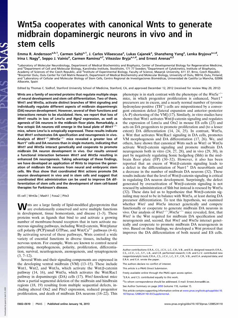
ResultsWnt1 Is Required for DA Neurogenesis and to Specify the Midbrain FPas a Neurogenic Region. Recent reports have indicated that Wnt/β-catenin signaling is required for midbrain DA neurogenesis(30, 31), but it is not known which of the multiple canonical Wntsexpressed in the VM (13–15) is/are required for DA neuro-genesis. In our study we focused on Wnt1 because Wnt1−/− mice,unlike Wnt2−/− mice, for instance (16), show a strong sequentialmidbrain and DA neuron phenotype (18–22). Because DAneurons are born in the midbrain FP, we first examined the ex-pression of the FP and basal plate (BP) markers, Shh and theShh-target gene, Foxa2, both of which are required for DAneuron development (31, 33–35). In situ hybridization (ISH)revealed that the expression patterns of Shh and Foxa2 weredelayed, as previously described in β-catenin−/− mice (31). In-deed, we found a delay in the lateral expansion of the Shh andFoxa2 expression domains (Fig. 1A, asterisks) and in the down-regulation of Shh in the FP (Fig. 1A, arrow), as well as weakerlevels of Foxa2 in Wnt1−/− mice at embryonic day (E) 11.5 (Fig.1A). Surprisingly, no TH+ DA neurons were present in the FP ofWnt1−/− mice at E12, and only a few DA neurons arose in anectopic lateral position in the Foxa2+ BP, which at this stageshowed normal Foxa2 protein levels (Fig. 1B). Moreover,Lmx1a, a Lim homeobox transcription factor required for thespecification of DA neurons (36), was also absent from the FPand was ectopically expressed in the BP ofWnt1−/− mice at E11.5(Fig. 1C). Previous in vitro experiments have indicated thatβ-catenin regulates Lmx1a via an auto-regulatory loop (23) andthat deletion of β-catenin results in ectopic expression of Lmx1ain vivo (30). Because Wnt1 activates Wnt/β-catenin signaling, ourresults indicate that it is Wnt1 that regulates the expression levelsand position of Lmx1a in vivo. Interestingly, examination of thelateral Lmx1a+ BP region revealed that it is the site where thefew midbrain DA neurons are present in Wnt1−/− mice (Fig. 1C).We therefore asked whether the lateral displacement of Lmx1a+
and TH+ cells results from an inversion of the BP and FP domainsand examined the expression and distribution of Nkx6.1, whichnormally is expressed in the BP and alar plate of the midbrain(33, 37). Although a partial reduction in the number of Nkx6.1+
cells was detected at E11.5 and E12.5 (Fig. S1A), Nkx6.1+ cellswere found laterally, in the correct position. Similarly, the ex-pression pattern of Wnt5a in the medial and basal plates of theVM was not disrupted in Wnt1−/− mice at E11.5 (Fig. S1B).These results, together with the medial expression of Shh andFoxa2 (Fig. 1A), indicated that the midbrain FP and BP domainswere not inverted in Wnt1−/− mice but that gene expression inthese compartments is altered.Because the midbrain FP contained no Lmx1a+ or TH+ cells,
we then asked whether overall neurogenesis was impaired andexamined the expression of proneural genes in the VM FP ofWnt1−/− mice at E11.5. We have shown previously that Ngn2 isrequired for DA neuron development and can be partiallyreplaced by Mash1 (38). Interestingly, the expression of bothNgn2 and Mash1 was abolished in the midbrain FP, and Ngn2expression increased in the dorsal midbrain and particularly inthe BP of Wnt1−/− mice (Fig. 1D). These results suggested thatWnt1 controls neurogenesis in the midbrain and led us to ex-amine whether any neurons are found in the FP of Wnt1−/− mice.Staining with Topro3 (a nuclear marker) first revealed that theWT midbrain FP consisted of nearly 20 cell diameters in themidline (in ventricular, intermediate, and marginal zones) atE12.5, whereas only about five cell diameters (in the ventricularzone) were found in Wnt1−/− mice (Fig. 1E). Staining for Tuj1,
a pan-neuronal marker, and Topro3, a nuclear marker, revealedno double Tuj+/Topro+ cell bodies in the FP, indicating that theFP region of Wnt1−/− mice contains no newborn neurons andthat only commissural Tuj1+ fibers are present (Fig. 1E).Moreover, no Nurr1+ DA neuroblasts were detected in the FP.In fact, the only cell bodies found in the FP ofWnt1−/− mice werethose of Sox2+ ventricular zone neuroepithelial cells (Fig. 1F)and Glast+ radial glia (22). Interestingly, a reduced number ofNurr1+ DA neuroblasts also were found in an ectopic lateralposition in the BP (Fig. 1F). Thus, our results indicate that Wnt1is required not only for the expression of Lmx1a and proneuralgenes (Mash1 and Ngn2) in the midbrain FP but also for DAspecification and neurogenesis.
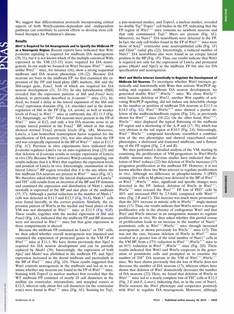
Wnt1 and Wnt5a Interact Genetically to Regulate the Development ofMidbrain DA Neurons. To investigate whether Wnt1 interacts ge-netically and functionally with Wnts that activate Wnt/PCP sig-naling and regulate midbrain DA neuron development, wegenerated double Wnt1−/−;Wnt5a−/− mice. We chose Wnt5a−/−
mice because deletion of Wnt11, another Wnt capable of acti-vating Wnt/PCP signaling, did not induce any detectable changein the number or position of midbrain DA neurons at E11.5 orE14.5 (Fig. S2). Wnt1−/−Wnt5a+/+ mice showed a partial de-letion of the midbrain/hindbrain region (Fig. 2A), as previouslyshown for Wnt1−/− mice (18–22). On the other hand, Wnt1+/+;Wnt5a−/− mice displayed the typical flattening of the midbrainFP region and a shortening of the A–P axis (17, 39), which wasvery obvious in the tail region at E10.5 (Fig. 2A). Interestingly,Wnt1−/−Wnt5a−/− compound knockouts resembled a combina-tion of the two phenotypes and showed exacerbated caudalphenotypes, a shortened and truncated midbrain, and a flatten-ing of the FP region (Fig. 2 A and B).We then performed a detailed analysis of the VM, starting by
examining the proliferation of VM progenitors in Wnt1/Wnt5adouble -mutant mice. Previous studies have indicated that de-letion of Wnt1 reduces (22) but deletion of Wnt5a increases (17)progenitor proliferation in the midbrain FP. However, it is notknown whether these two Wnts interact to regulate proliferationin vivo. Although no difference in phospho-histone 3 (PH3)staining (for cells in M-phase) was detected in the BP of Wnt1−/−
or Wnt1−/−;Wnt5a−/− mice, significant and specific changes weredetected in the FP. Indeed, deletion of Wnt5a in Wnt1−/−;Wnt5a−/− mice rescued the Wnt1−/− FP loss of PH3+ cells by40% and increased PH3 by 2.5-fold, compared with Wnt1−/−
mice (Fig. 2 B and C). This increase was much more pronouncedthan the 20% increase in mitotic cells in Wnt5a−/− single-mutantmice (17). Thus, our results indicate that Wnt5a serves a strongerproliferative role in the absence of endogenous Wnt1 and thatWnt1 and Wnt5a interact in an antagonistic manner to regulateproliferation in vivo. We then asked whether this partial rescueof proliferation leads to an increase in the number of Nurr1+
postmitotic cells in Wnt1−/−;Wnt5a−/− mice through increasedneurogenesis, as shown previously for Wnt5a−/− mice (17). Thiswas not the case, because deletion of Wnt5a in Wnt1−/− miceresulted in a greater loss of the total number of Nurr1+ cells inthe VM BP, from a 57% reduction in Wnt1−/−;Wnt5a+/+ mice toan 81% reduction in Wnt1−/−;Wnt5a−/− mice (Fig. 2D). Theseresults indicated that Wnt1 and Wnt5a cooperate in the gener-ation of postmitotic cells and prompted us to examine thenumber of TH+ DA neurons in the VM of Wnt1−/−;Wnt5a−/−
mice. We have shown previously that the loss of Wnt5a does notdecrease the number of DA neurons (17), whereas others haveshown that deletion of Wnt1 dramatically decreases the numberof DA neurons (22). Here, we found that deletion of Wnt5a inWnt1−/− mice led to a nearly complete loss of TH+ cells at E12.5(Fig. 2 E and F, Lower), indicating that, as in the case of Nurr1,Wnt5a worsens the Wnt1 phenotype and cooperates positivelywith Wnt1 to regulate DA neurogenesis. Moreover, although
Andersson et al. PNAS | Published online January 16, 2013 | E603
NEU
ROSC
IENCE
PNASPL
US
Dow
nloa
ded
by g
uest
on
Apr
il 23
, 202
0
Nurr1+, TH+, or Lmx1a+ cells always were found in the mid-brain FP of WT mice, none of them were present in the FP ofWnt1−/−Wnt5a+/+, or Wnt1−/−Wnt5a−/− mice. Combined, thesedata show that Wnt1 and Wnt5a interact genetically in a complexmanner resulting in both antagonistic and synergistic functions inthe developing VM, as shown by the opposing regulation ofproliferation and the cooperative regulation of DA neurogenesisby Wnt1 and Wnt5a.The phenotype described above showed that deletion of
Wnt5a aggravates the neurogenesis phenotype of Wnt1−/− mice.Therefore we next decided to examine whether the opposite alsois true, i.e., whether deletion of Wnt1 worsens typical Wnt5a−/−
phenotypes such as morphogenesis defects caused by alterationsin convergent extension. We previously have reported that de-letion of Wnt5a leads to a broadening of the ventricular cavity atthe midbrain level that results in a flattening of the ventricularhinge point of the VM (17). Here we found that deletion ofWnt1leads to the opposite phenotype, an elongation of the ventricularcavity and of the VM hinge point. Interestingly, compoundWnt1−/−;Wnt5a−/− mutants showed a Wnt5a−/−-like phenotype,with a severe broadening of the ventricular cavity and flatteningof the VM hinge point (Fig. 2F, arrows). We next examinedwhether other Wnt5a morphogenesis phenotypes, such as the15% reduction in the A–P length of the TH+ domain (17), weremodified in Wnt1−/−;Wnt5a−/− mice. Surprisingly, we found thatthe A–P distribution of the TH+ midbrain DA domain, whichwas shortened by 55% in Wnt1−/− mice (Fig. 2G), was reducedfurther by 85% in Wnt1−/−;Wnt5a−/− mice (Fig. 2G). Moreover,when the mediolateral and dorsoventral axes were examined,TH+ cells occupied more lateral and dorsal positions in theWnt1−/−;Wnt5a−/− mice than in either of the single mutants(arrowheads in Fig. 2F). Thus, our data show that the combineddeletion of Wnt1 and Wnt5a results in a greater alteration ofconvergent extension (shorter A–P domain as well as lateral andventral DA cell distribution), indicating that Wnt1 cooperatespositively with Wnt5a in regulating morphogenetic movements inthe midbrain.In sum, our results show a complex interaction between Wnt5a
and Wnt1 in which the function of Wnt5a in morphogenesis ispotentiated by Wnt1, and the function of Wnt1 in neurogenesis,but not in proliferation, is potentiated by Wnt5a. We thus de-cided to examine whether combined administration of Wntproteins to stem cells, at the right time point of differentiationand in the correct sequence, could be used to improve currentprotocols for the DA differentiation of both neural stem cellsand ES cells.
Wnts Cooperate to Improve DA Neuron Development in Neural StemCells. Wnt1 overexpression or Wnt1-conditioned media havebeen used previously to activate Wnt/β-catenin signaling (40–42)and to promote DA differentiation of midbrain progenitors (14)or mouse ES (mES) cells (23). However, to date, it has not beenpossible to obtain pure Wnt1 protein capable of activating Wnt/β-catenin signaling. Indeed, Wnt1 protein (purified by us orcommercially available) did not activate Wnt/β-catenin signaling,as assessed by diverse assays such as Lrp6 phosphorylation, de-phosphorylation of β-catenin, or activation of the TOPFLASH
Fig. 1. Wnt1 is required for ventral midbrain FP neurogenesis. (A) Shh andFoxa2mRNA expression in the VM ofWnt1−/− mice is delayed compared withWT mice at E11.5; their expression is lost in lateral positions (*), and themedial down-regulation of Shh in WT mice (arrow) is not detected inWnt1−/−
mice. (B) Foxa2 protein and TH are found throughout the VM of WT mice atE12, whereas TH+ DA neurons are severely reduced in number and are foundsolely in the lateral BP in Wnt1−/− mice. The boxed regions in the left panelsdemarcate the regions that are magnified in the panels at the far right.
V = ventricle. (C) TH+ cells are found only in Lmx1a+ domains, which arelaterally displaced from the FP to the BP in Wnt1−/− mice. The boxed regionsin the left panels demarcate the regions that are magnified in the panels atthe far right. (D) The expression of the proneural factors Mash1 and Ngn2 islost from the FP and is displaced laterally in Wnt1−/− mice at E11.5. (E) AtE12.5, the FP ofWnt1−/− mice shows a reduced ventricular zone and containsno neuronal somas (Tuj+Topro3+ cells). (F) Although the WT FP containsSox2+ progenitors and Nurr1+ postmitotic cells, the Wnt1−/− FP shows fewerSox2+ progenitors, and all Nurr1+ cells are found in the BP.
E604 | www.pnas.org/cgi/doi/10.1073/pnas.1208524110 Andersson et al.
Dow
nloa
ded
by g
uest
on
Apr
il 23
, 202
0
reporter in DA cells (Fig. S3). This lack of activation is in con-trast with other purified Wnt proteins such as Wnt3a or Wnt5a,which have been shown to activate Wnt signaling and to exertbiologically relevant activities (26, 43). Given the current un-availability of purified active Wnt1 protein for our experiments,we turned our attention to Wnt3a, a protein capable of activatingWnt/β-catenin signaling in DA cells (Fig. S3) (14). We thus ex-amined whether it was possible to substitute Wnt1 for Wnt3a asa source of Wnt/β-catenin activation in our stem cell-differenti-ation protocols. We started by devising a three-step protocol to
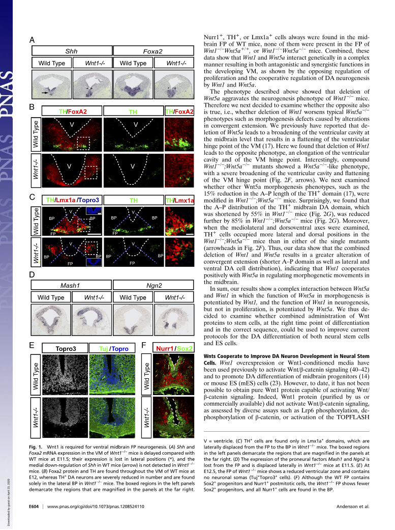
treat midbrain progenitor/neural stem cells, expanded as neu-rospheres (Fig. 3A), in which we first used Wnt3a for 3 d topromote progenitor proliferation and specification, followed by3 d without Wnts to eliminate all remaining Wnt3a, and finally3 d with Wnt5a to activate the Wnt/PCP/Rac1 pathway, promoteprogenitor cell-cycle exit, and direct Nurr1+ precursor differen-tiation into TH+ DA neurons (17). Although treatment withWnt3a alone at the indicated time point (Fig. 3A) increased thenumber of TuJ1+ neurons, Wnt5a alone or Wnt3a and Wnt5adid not affect the number of neurons in the culture (Fig. 3 B,
Fig. 2. Wnt1/Wnt5a double-mutant mice reveal redundant and nonredundant functions during ventral midbrain development. (A) Wnt1+/−;Wnt5a+/− micewere mated to produce WT, Wnt1−/−;Wnt5a+/+ (labeled Wnt1−/−), Wnt1+/+;Wnt5a−/− (labeled Wnt5a−/−), and Wnt1−/−;Wnt5a−/− mice. At E10.5, Wnt1−/− micedisplay the previously described phenotypes of midbrain deletion and a less distinct isthmus (yellow arrowhead), whereas Wnt5a−/− mice display A–Pshortening which is most obvious in the tail region (white arrow). Wnt1−/−;Wnt5a−/− double-mutant mice resemble a combination of the two singleknockouts, with a shortened A–P axis and a significantly shorter midbrain region (dashed lines). (B and C) Proliferation, assessed by staining for the mitoticmarker PH3, is specifically reduced in the FP but not the BP of Wnt1−/− mice. Panels show whole midbrain (Top Row) and magnifications of the BP and FP(Middle Row). The areas demarcated by red dotted boxes are magnified in the bottom row. The reduction in the numbers of PH3+ cells in the FP of Wnt1−/−
mice is partially rescued by loss of Wnt5a. (D) The total number of Nurr1+ cells decreases to a greater extent in double mutants than in single mutants or WTlittermates. (E and F) TH+ cells are dramatically reduced in number and are laterally displaced (white arrowheads in F) from the FP to the BP in Wnt1−/−;Wnt5a+/+ mice. These defects are worsened in Wnt1−/−;Wnt5a−/− mice, in which even fewer cells are found (E), and they are positioned further dorsolaterallyin the BP (white arrowheads in F). The VM hinge point (invagination of the ventricle; green arrow in F) is elongated inWnt1−/− Wnt5a+/+ mice and flattened inWnt1−/−;Wnt5a−/− mice. (G) The decrease of the A–P length of the TH-expressing midbrain domain observed in Wnt1−/− mice is exacerbated by loss of Wnt5a.*P < 0.05, **P < 0.01, ***P < 0.001.
Andersson et al. PNAS | Published online January 16, 2013 | E605
NEU
ROSC
IENCE
PNASPL
US
Dow
nloa
ded
by g
uest
on
Apr
il 23
, 202
0
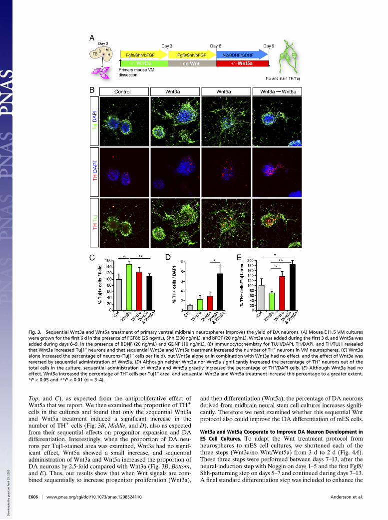
Top, and C), as expected from the antiproliferative effect ofWnt5a that we report. We then examined the proportion of TH+
cells in the cultures and found that only the sequential Wnt3aand Wnt5a treatment induced a significant increase in thenumber of TH+ cells (Fig. 3B, Middle, and D), also as expectedfrom their sequential effects on progenitor expansion and DAdifferentiation. Interestingly, when the proportion of DA neu-rons per Tuj1-stained area was examined, Wnt3a had no signif-icant effect, Wnt5a showed a small increase, and sequentialadministration of Wnt3a and Wnt5a increased the proportion ofDA neurons by 2.5-fold compared with Wnt3a (Fig. 3B, Bottom,and E). Thus, our results show that when Wnt signals are com-bined sequentially to increase progenitor proliferation (Wnt3a),
and then differentiation (Wnt5a), the percentage of DA neuronsderived from midbrain neural stem cell cultures increases signifi-cantly. Therefore we next examined whether this sequential Wntprotocol also could improve the DA differentiation of mES cells.
Wnt3a and Wnt5a Cooperate to Improve DA Neuron Development inES Cell Cultures. To adapt the Wnt treatment protocol fromneurospheres to mES cell cultures, we shortened each of thethree steps (Wnt3a/no Wnt/Wnt5a) from 3 d to 2 d (Fig. 4A).These three steps were performed between days 7–13, after theneural-induction step with Noggin on days 1–5 and the first Fgf8/Shh-patterning step on days 5–7 and continued during days 7–13.A final standard differentiation step was included to enhance the
Fig. 3. Sequential Wnt3a and Wnt5a treatment of primary ventral midbrain neurospheres improves the yield of DA neurons. (A) Mouse E11.5 VM cultureswere grown for the first 6 d in the presence of FGF8b (25 ng/mL), Shh (300 ng/mL), and bFGF (20 ng/mL). Wnt3a was added during the first 3 d, and Wnt5a wasadded during days 6–9, in the presence of BDNF (20 ng/mL) and GDNF (10 ng/mL). (B) Immunocytochemistry for TUJ1/DAPI, TH/DAPI, and TH/TUJ1 revealedthat Wnt3a increased Tuj1+ neurons and that sequential Wnt3a and Wnt5a treatment increased the number of TH+ neurons in VM neurospheres. (C) Wnt3aalone increased the percentage of neurons (Tuj1+ cells per field), but Wnt5a alone or in combination with Wnt3a had no effect, and the effect of Wnt3a wasreversed by sequential administration of Wnt5a. (D) Although neither Wnt3a nor Wnt5a significantly increased the percentage of TH+ neurons out of thetotal cells in the culture, sequential administration of Wnt3a and Wnt5a greatly increased the percentage of TH+/DAPI cells. (E) Although Wnt3a had noeffect, Wnt5a increased the percentage of TH+ cells per Tuj1+ area, and sequential Wnt3a and Wnt5a treatment increase this percentage to a greater extent.*P < 0.05 and **P < 0.01 (n = 3–4).
E606 | www.pnas.org/cgi/doi/10.1073/pnas.1208524110 Andersson et al.
Dow
nloa
ded
by g
uest
on
Apr
il 23
, 202
0
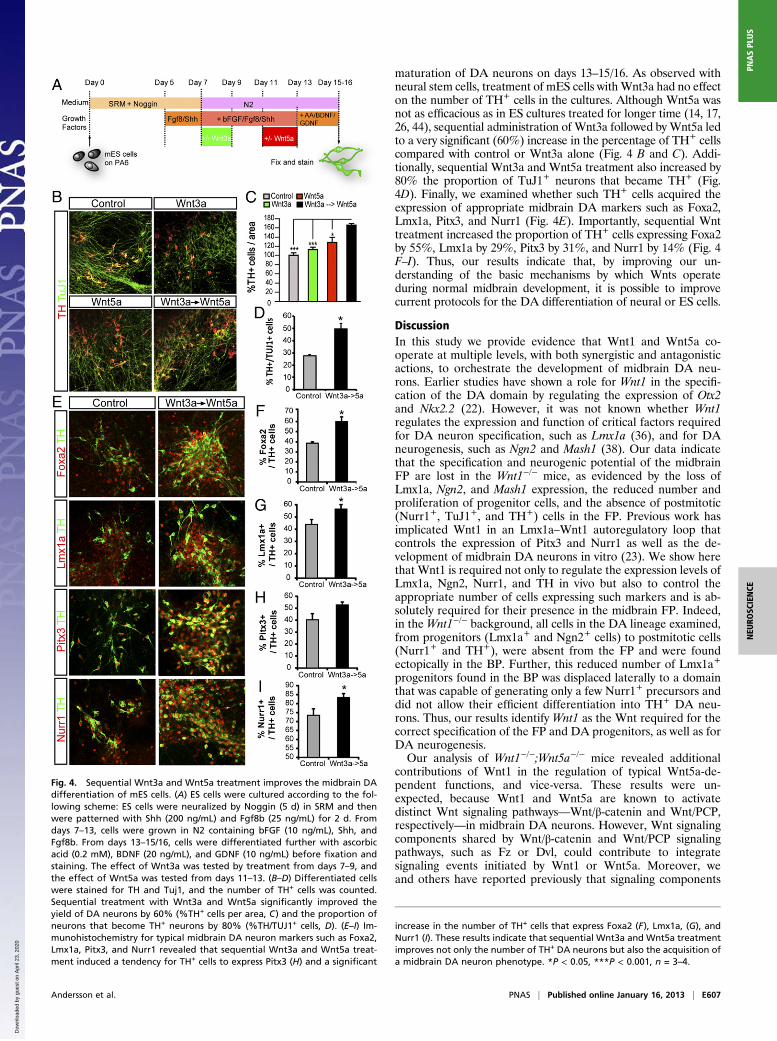
maturation of DA neurons on days 13–15/16. As observed withneural stem cells, treatment of mES cells with Wnt3a had no effecton the number of TH+ cells in the cultures. Although Wnt5a wasnot as efficacious as in ES cultures treated for longer time (14, 17,26, 44), sequential administration of Wnt3a followed by Wnt5a ledto a very significant (60%) increase in the percentage of TH+ cellscompared with control or Wnt3a alone (Fig. 4 B and C). Addi-tionally, sequential Wnt3a and Wnt5a treatment also increased by80% the proportion of TuJ1+ neurons that became TH+ (Fig.4D). Finally, we examined whether such TH+ cells acquired theexpression of appropriate midbrain DA markers such as Foxa2,Lmx1a, Pitx3, and Nurr1 (Fig. 4E). Importantly, sequential Wnttreatment increased the proportion of TH+ cells expressing Foxa2by 55%, Lmx1a by 29%, Pitx3 by 31%, and Nurr1 by 14% (Fig. 4F–I). Thus, our results indicate that, by improving our un-derstanding of the basic mechanisms by which Wnts operateduring normal midbrain development, it is possible to improvecurrent protocols for the DA differentiation of neural or ES cells.
DiscussionIn this study we provide evidence that Wnt1 and Wnt5a co-operate at multiple levels, with both synergistic and antagonisticactions, to orchestrate the development of midbrain DA neu-rons. Earlier studies have shown a role for Wnt1 in the specifi-cation of the DA domain by regulating the expression of Otx2and Nkx2.2 (22). However, it was not known whether Wnt1regulates the expression and function of critical factors requiredfor DA neuron specification, such as Lmx1a (36), and for DAneurogenesis, such as Ngn2 and Mash1 (38). Our data indicatethat the specification and neurogenic potential of the midbrainFP are lost in the Wnt1−/− mice, as evidenced by the loss ofLmx1a, Ngn2, and Mash1 expression, the reduced number andproliferation of progenitor cells, and the absence of postmitotic(Nurr1+, TuJ1+, and TH+) cells in the FP. Previous work hasimplicated Wnt1 in an Lmx1a–Wnt1 autoregulatory loop thatcontrols the expression of Pitx3 and Nurr1 as well as the de-velopment of midbrain DA neurons in vitro (23). We show herethat Wnt1 is required not only to regulate the expression levels ofLmx1a, Ngn2, Nurr1, and TH in vivo but also to control theappropriate number of cells expressing such markers and is ab-solutely required for their presence in the midbrain FP. Indeed,in the Wnt1−/− background, all cells in the DA lineage examined,from progenitors (Lmx1a+ and Ngn2+ cells) to postmitotic cells(Nurr1+ and TH+), were absent from the FP and were foundectopically in the BP. Further, this reduced number of Lmx1a+
progenitors found in the BP was displaced laterally to a domainthat was capable of generating only a few Nurr1+ precursors anddid not allow their efficient differentiation into TH+ DA neu-rons. Thus, our results identify Wnt1 as the Wnt required for thecorrect specification of the FP and DA progenitors, as well as forDA neurogenesis.Our analysis of Wnt1−/−;Wnt5a−/− mice revealed additional
contributions of Wnt1 in the regulation of typical Wnt5a-de-pendent functions, and vice-versa. These results were un-expected, because Wnt1 and Wnt5a are known to activatedistinct Wnt signaling pathways—Wnt/β-catenin and Wnt/PCP,respectively—in midbrain DA neurons. However, Wnt signalingcomponents shared by Wnt/β-catenin and Wnt/PCP signalingpathways, such as Fz or Dvl, could contribute to integratesignaling events initiated by Wnt1 or Wnt5a. Moreover, weand others have reported previously that signaling components
Fig. 4. Sequential Wnt3a and Wnt5a treatment improves the midbrain DAdifferentiation of mES cells. (A) ES cells were cultured according to the fol-lowing scheme: ES cells were neuralized by Noggin (5 d) in SRM and thenwere patterned with Shh (200 ng/mL) and Fgf8b (25 ng/mL) for 2 d. Fromdays 7–13, cells were grown in N2 containing bFGF (10 ng/mL), Shh, andFgf8b. From days 13–15/16, cells were differentiated further with ascorbicacid (0.2 mM), BDNF (20 ng/mL), and GDNF (10 ng/mL) before fixation andstaining. The effect of Wnt3a was tested by treatment from days 7–9, andthe effect of Wnt5a was tested from days 11–13. (B–D) Differentiated cellswere stained for TH and Tuj1, and the number of TH+ cells was counted.Sequential treatment with Wnt3a and Wnt5a significantly improved theyield of DA neurons by 60% (%TH+ cells per area, C) and the proportion ofneurons that become TH+ neurons by 80% (%TH/TUJ1+ cells, D). (E–I) Im-munohistochemistry for typical midbrain DA neuron markers such as Foxa2,Lmx1a, Pitx3, and Nurr1 revealed that sequential Wnt3a and Wnt5a treat-ment induced a tendency for TH+ cells to express Pitx3 (H) and a significant
increase in the number of TH+ cells that express Foxa2 (F), Lmx1a, (G), andNurr1 (I). These results indicate that sequential Wnt3a and Wnt5a treatmentimproves not only the number of TH+ DA neurons but also the acquisition ofa midbrain DA neuron phenotype. *P < 0.05, ***P < 0.001, n = 3–4.
Andersson et al. PNAS | Published online January 16, 2013 | E607
NEU
ROSC
IENCE
PNASPL
US
Dow
nloa
ded
by g
uest
on
Apr
il 23
, 202
0
initially assigned to the Wnt/β-catenin pathway, such as the Lrp6coreceptor, can contribute to convergent extension and Wnt/PCP signaling (45, 46). Thus, multiple mechanisms couldaccount for the interaction between these two pathways. In ourstudy, we found that deletion ofWnt1 worsened all typical Wnt/PCPmorphogenesis phenotypes found in Wnt5a−/− mice: increasedmediolateral and dorsoventral distribution of DA neurons in theVM, and severe A–P shortening of the TH+ domain. These dataindicated thatWnt1 cooperates withWnt5a to regulate Wnt5a/PCP-dependent functions in the developing VM. We also found thatdeletion of Wnt5a worsened Wnt1−/− phenotypes, such as thedecrease in DA neurogenesis and differentiation, as shown bya greater decrease in both Nurr1+ and TH+ cells in vivo. Thisresult also was unexpected, because deletion of Wnt5a partiallyrescues the proliferation defect in Wnt1−/− mice (this study) andincreases the number of Nurr1+ cells (17). Thus, the analysis ofWnt1−/−Wnt5a−/− mice uncovered two cooperative functions ofWnt1 and Wnt5a, whereby Wnt1 contributes to Wnt5a-depen-dent morphogenesis and Wnt5a contributes to Wnt1-dependentneurogenesis.Shh and Fgf8 have been described previously as being
expressed at the cross-section between the FP and isthmus andhave been demonstrated to regulate the development of VM DAneurons in a coordinated manner (47, 48) and to promote thedifferentiation of stem cells into midbrain DA neurons (28, 32,44, 49–51). Similarly, Wnt1 and Wnt5a are coexpressed and in-tersect in defined spatial and temporal patterns in the developingmidbrain: Wnt1 is expressed in the midbrain as early as E8 (52)and is found in two lateral bands flanking the FP from E10.5–12.5 (22), and Wnt5a is expressed in the VM, including the FP,from E9.5–13.5 (17). In this context, our study showing thatWnt1 and Wnt5a cooperate to promote DA neuron de-velopment provides the in vivo functional basis for the molecularintersection between Wnt1/β-catenin signaling and Wnt5a/PCPsignaling. These findings led us to propose that intersectionsbetween Wnts, Shh, and Fgf8, are key elements of the molecularmorphogen logic that controls the development of midbrain DAneurons. This concept was tested further and verified in stem cellcultures, where we found that the combined administration ofthese factors improved the DA differentiation of neural stemand ES cells. In our protocol, in addition to Shh and Fgf8b, weused Wnt3a to activate Wnt/β-catenin signaling, followed byWnt5a to activate Wnt/PCP signaling. Previous data in the lit-erature have shown that Wnt3a promotes proliferation of neuralstem cells and DA progenitors (14, 53) and that activation ofWnt/β-catenin signaling with GSK3β inhibitors enhances DAneurogenesis in DA progenitors (24), mES cells (32), and humanES cells (28). In addition, Wnt5a treatment has been found toinhibit proliferation and to promote DA differentiation of ro-dent neural and ES cells (14, 17, 26, 44). To date, however,a combined treatment providing adequate temporal activation ofboth pathways has not been developed. Our results show thatappropriate temporal and sequential administration of Wntssuccessfully improves the DA differentiation of VM neural stemcells, grown as neurospheres, and of ES cells. Sequential Wnt3aand Wnt5a treatment increased the percentage of TH+ neuronsin midbrain neurosphere cultures. We also found that sequentialWnt treatment increased the number of ES cells that differen-tiate into TH+ cells and acquire expression of transcriptionfactors critical for midbrain DA neuron development, such asFoxa2, Lmx1a, Pitx3, and Nurr1. These results confirmed thatsequential Wnt3a and Wnt5a treatment promoted DA differ-entiation and the acquisition of a true midbrain DA neuronphenotype. Thus, our data suggest that it is possible to replaceWnt1 by Wnt3a, at least partially, to activate β-catenin signalingand promote DA progenitor proliferation. Future availability ofpurified and active Wnt1 protein will allow testing to determine
whether Wnt1 protein may offer additional improvements in DAdifferentiation protocols, as compared with Wnt3a.The positive results obtained with neural and ES cells support
the idea that Wnt proteins could be used for therapeutic pur-poses, as previously suggested for hematopoietic stem cells (43).In the case of DA neurons, the main therapeutic target would beParkinson’s disease, a neurodegenerative disorder resulting inthe demise of adult midbrain DA neurons. The implementationof protocols such as the one described here for the generation ofmidbrain DA neurons from stem cells thus may be useful for cell-replacement therapy (54, 55) and in assays for drug discovery,taking advantage of either human ES cells or patient-derivedinduced pluripotent stem cells (56–58).In sum, our study identifies a genetic interaction betweenWnt1
andWnt5a, that controls different stages of midbrain DA neurondevelopment in an antagonistic or cooperative manner. AlthoughWnt1 and Wnt5a competed to promote or inhibit DA progenitorproliferation, respectively, Wnt1 cooperated with Wnt5a in regu-lating morphogenesis, and Wnt5a collaborated with Wnt1 topromote DA neurogenesis. Importantly, these concepts allowed usto improve current protocols for the DA differentiation of stemcells, opening the door for the development of novel therapies forParkinson’s disease.
Materials and MethodsAnimals, Immunohistochemistry, and ISH. Wnt1+/− (21), Wnt5a+/− (39),Wnt11−/− (59), and CD1 mice (Charles River) were housed, bred, and treatedin accordance with protocols approved by the local ethics committees(Stockholm’s Norra Djurförsöketiska Nämnd N154/06, N135/08, N145/09, andN273/11). All mutant mice were kept on a C57bl/6 background.Wnt1+/− micewere bred with Wnt5a+/− mice to generate Wnt1+/−Wnt5a+/− mice, whichwere obtained at the expected Mendelian proportions (expected = 25%,actual = 24.86%, n = 185). For embryo analyses, heterozygous mice of therelevant genotype were mated overnight, and noon of the day the plug wasconsidered E0.5. Embryos were dissected out of the uterine horns in ice-coldPBS, fixed in 4% (wt/wt) paraformaldehyde (PFA) for 4 h to overnight, cry-oprotected in 20–30% sucrose, and frozen in Tissue-Tek Optimum CuttingTemperature (OCT) compound (Sakura Fine-Tek) on dry ice. Serial coronal14-μm sections of the brain were obtained on a cryostat. Immunohisto-chemistry and ISH were carried out as previously described (14). Probes andantibodies are described below. Immunohistochemistry and ISH were visu-alized with a Zeiss HBO100 microscope or Zeiss LSM 510 Confocal Micro-scope, collected with a C4742-95 Hamamatsu camera, and processed withOpenLab software (PerkinElmer), Photoshop (Adobe), and/or ImageJ (http://imagej.nih.gov/ij/). Figures were assembled in Illustrator (Adobe).
Antibodies and Probes. The RNA probes for Shh (60), Foxa2 (17), and Ngn2and Mash1 (38) have been described previously.
Rabbit anti-Nurr1 (Nr4a2) (1:1,000; Santa Cruz Biotechnology), rabbit anti-TH (1:500–2000; Pel-Freeze), rabbit anti–phospho-histone3 (1:400; Cell Sig-naling), mouse anti-β III tubulin (1:1,000–1:2,000; Promega), anti-Foxa2 [1:20;4C7; Developmental Studies Hybridoma Bank (DSHB)], anti-Nkx6.1 (F55A10;DSHB), anti-Lmx1a (1:1,000; gift from Mike German, University of California,San Francisco, CA), anti-Sox2 (1:500; Millipore), anti-Wnt5a (1:250; R&D Sys-tems), and Cy2-, Cy3-, or Rhodamine-coupled secondary antibodies (1:250;Jackson ImmunoResearch) were used. Some sections or cells were counter-stained with DAPI (500 ng/mL) (Sigma), Topro3 (Invitrogen), or Alexa 488phalloidin (Molecular Probes, Invitrogen).
Quantification of TH+ or Nurr1+ Cells in Knockout Mice. E11.5 and E12.5 brainswere sectioned serially at 14 μm to two slides. Because both anti-TH and anti-Nurr1 are rabbit antibodies, one slide was stained for TH and the other forNurr1. TH+ cells were counted in every second 14-μm section (i.e., every 28μm) through the entire mesodiencephalon from anterior to posterior, andthe total number of TH+ cells was calculated by multiplying the cell counts by2. On average, 18 sections per WT slide contained TH+ cells, whereasWnt1−/−
Wnt5a+/+ and Wnt1−/−Wnt5a+/− slides contained eight sections with TH+
cells, and Wnt1−/−Wnt5a−/− slides contained two or three sections with TH+
cells. Because Nurr1 is expressed further anteriorly and posteriorly than TH,Nurr1+ cells were counted in three sections per brain (28 μm apart) withinthe central segment of the Th+ domain in each genotype. The total numberof Nurr1+ cells then was calculated by correcting for the total number of
E608 | www.pnas.org/cgi/doi/10.1073/pnas.1208524110 Andersson et al.
Dow
nloa
ded
by g
uest
on
Apr
il 23
, 202
0
sections containing TH+ DA neurons. All values represent the mean of threeor four animals per genotype, ± SEM.
Statistical Analyses in Knockout Mice. Statistical analysis was performed withGraphPad Prism. Differences between knockout animals or culture conditionswere analyzed using ANOVA, with Bonferroni’s post hoc test. Significantdifferences were assumed at *P < 0.05, **P < 0.01, and ***P < 0.001.GraphPad Prism (GraphPad Software) was used for statistical analyses.
Neurosphere Cultures. Mouse E11.5 ventral midbrain tissue was dissected outin ice-cold PBS/0.2% glucose. The tissue was dissociated in N2 medium (MEM,F12 medium, Hepes, N2 supplement and Glutamine, all from Life Technol-ogies) by trituration through flame-narrowed Pasteur pipettes. The cellsuspension then was plated at 1 × 10⁵ cells/cm2 (100,000 cells/mL) on un-coated Petri flasks (BD Falcon) in complete medium consisting of 200–300ng/mL Shh (R&D Systems), 25 ng/mL FGF8b (R&D Systems), and 20 ng/mLbasis FGF (bFGF; R&D Systems) in N2. The design of the experiments usingWnt3a and/or Wnt5a in neurospheres was as follows: First, neurosphereswere cultured with or without Wnt3a (100–300 ng/mL) (R&D Systems) in N2with Shh, FGF8, and bFGF. After 3 d, cells were collected, washed with Wntvehicle (CHAPS 0.05%; Tamro), and plated at a density of 100,000 cells/mLwithout adding Wnts. After a second passage, cells were plated and dif-ferentiated with or without Wnt5a (100 ng/mL) (R&D Systems) (see schemein Fig. 3A). At day 9, cells were fixed in 4% (wt/vol) PFA and processed forimmunocytochemistry as described below.
ES Cell Culture. mES R1cell cultures were cultured on gelatinized plates inKO-DMEM (Life Technologies) supplemented with 15% (vol/vol) Knockoutserum replacement (SRM; Life Technologies), 2 mM L-glutamine (Life Tech-nologies), 1% nonessential amino acids (VWR), 0.1 mM β-mercaptoethanol(Sigma), 1,000 U/mL leukemia inhibitory factor (ESGRO; Chemicon/Millipore),and 10,000 U/mL penicillin/streptomycin (Life Technologies). Stromal PA6cells were cultured in α-minimum essential medium (Life Technologies)containing 10% (vol/vol) FBS (Life Technologies), 2 mM L-glutamine (LifeTechnologies), and 10,000 U/mL penicillin/streptomycin (Life Technologies)and were mitotically inactivated before use with 1 μg/mL mitomycin C(Roche) overnight at 37 °C. All cell lines were maintained at 37 °C, 5% CO2
and 95% humidity. mES cells were plated at low density (100 cells/cm2) ona confluent layer of PA6 cells in 24-well plates and were grown in SRM andNoggin (300 ng/mL; R&D Systems). At day 5, 200 ng/mL Shh and 25 ng/mLFGF8b were added to the medium. After 7 d, cultures were switched to N2medium in the presence of Shh, FGF8, and FGF2 (10 ng/mL), with or without100 ng/mL Wnt3a (R&D Systems). At day 9, Wnt3a was withdrawn, cells werewashed with CHAPS for 1 h, and fresh medium was added. Two days later(on day 11) 100 ng/mL Wnt5a (R&D Systems) was added with fresh medium.Four conditions were tested: (a) control with no Wnt addition; (b) Wnt3aalone added at day 7 and then removed at day 9; (c) Wnt5a alone addedfrom days 11–13; and (d) Wnt3a added at day 7, cells washed at day 9, andWnt5a added from days 11–13. Shh, FGF8, bFGF, and Wnt5a were removedat day 13 from the conditions in which they had been added. Cultures thenwere differentiated until day 15 in N2 medium containing 0.2 mM ascorbicacid, 20 ng/mL BDNF (R&D Systems), and 10 ng/mL glial cell-derived neuro-trophic factor (GDNF; R&D Systems). At day 15 cells were fixed in 4% PFAand processed for immunocytochemistry as described below.
Immunocytochemistry. PFA-fixed cells were washed in PBS and blocked in5% (vol/vol) normal goat serum/PBS for 1 h at room temperature. Primary
antibodies were diluted in PBS (pH 7.4), 0.3% Triton X-100, and 1% (wt/vol)BSA, and incubations were carried out overnight at 4 °C. After washes,incubations with the appropriate Alexa-conjugated secondary antibodies(1:500; Life Technologies) were carried out for 2 h at room temperature. Thefollowing antibodies were used: mouse monoclonal anti–β-tubulin III (TuJ1)(1:1,000; Promega), rabbit polyclonal anti-TH (1:500; Pel-Freeze) or mousemonoclonal anti-TH (1:500; Sigma or 1:400; Immunostar), rabbit polyclonalanti-Nurr1 (1:250; Santa Cruz), rabbit polyclonal anti-Pitx3 (1:50; Invitrogen),rabbit polyclonal anti-Lmx1a (1:500; a gift from M. German), and rabbitpolyclonal anti-Foxa2 (1:500; Cell Signaling Technology).
Cell Counts in Vitro. For neurosphere cultures, Hoechst staining (Roche) wasused and TH+, Tuj1+, and Hoechst-positive cells were counted in 10–12 pre-determined fields along the x axis of the well. For each field, up to nine z-planes were analyzed. Each condition was analyzed in duplicate to qua-druplicate determinations for every experiment, and three to four in-dependent experiments were performed for every condition.
For ES cell cultures, TH+ cells per area were counted in 12 colonies per well,in three wells per condition, and in four independent experiments. To correctfor variations in colony size, the number of TH+ cells was referred to the areaoccupied by the colonies. The percentage of TH+/Tuj1+ cells also was countedin 8–10 fields along the x axis of the well in two wells per experiment andthree experiments per condition. Finally, the number of TH+ cells expressingmidbrain DA markers (Foxa2, Lmx1a, Pitx3, and Nurr1) was counted in 8–12fields, in duplicate wells and three independent experiments.
All data are expressed as mean ± SEM. Statistical analysis was performedby t-test or ANOVA with post hoc tests.
Wnt Signaling. SN4741 cells were transfected with 500 ng SuperTOP-FLASH/SuperFOP-FLASH and 50 ng of Renilla-expressing vector pRL-TK Luc (Prom-ega) using Superfect (Qiagen) according to the manufacturer’s instructions.Twelve hours after transfection, cells were stimulated with 50–100 ng/mLrecombinant Wnt3a (R&D Systems) and/or 50–500ng/mL Wnt1 (Preprotech)for 24 h. Reporter activity was measured using the Dual-Luciferase Reporterassay (Promega). Each experiment was repeated three times, and each ex-perimental condition was measured in duplicate. Background luminescence(signal from lysis buffer without any cells) was subtracted from each sam-ple. Luciferase activity of SuperTOP/FOP-FLASH was normalized to Renillaluciferase signal.
Sample preparation, Western blot analysis, and signal detection wasperformed as previously described (61). Primary antibodies used were rabbitpolyclonal anti-Phospho-LRP5/6 (1:1,000; Cell Signaling), mouse monoclonalanti–β-actin (1:5,000; BD Transduction Laboratories), and mouse monoclonalanti–active β-catenin (1:1,000; Millipore). HRP-conjugated anti-mouse andanti-rabbit secondary antibodies, A7282 and A6667 respectively, were pur-chased from Sigma, and were used at 1:5,000.
ACKNOWLEDGMENTS. This work was supported by grants from the SwedishFoundation for Strategic Research (Center of Excellence in DevelopmentalBiology and Successful Research Leaders Program); Swedish Research CouncilGrants VR2008:2811, VR2008:3287, and VR2011:3116; the Center of Excel-lence in Developmental Biology for Regenerative Medicine); the KarolinskaInstitute (Thematic Center in Stem Cells and Regenerative Medicine); theEuropean Commission (Neurostemcell and DDPD-Genes); and Parkinsonfon-den. J.C.V. was supported by a Federation of European Biochemical SocietiesLong-Term Fellowship. V.B. is supported by the Czech Science Foundation(204/09/0498, 301/11/0747).
1. Chien AJ, Conrad WH, Moon RT (2009) A Wnt survival guide: From flies to human
disease. J Invest Dermatol 129(7):1614–1627.2. Sugimura R, Li L (2010) Noncanonical Wnt signaling in vertebrate development, stem
cells, and diseases. Birth Defects Res C Embryo Today 90(4):243–256.3. Inestrosa NC, Arenas E (2010) Emerging roles of Wnts in the adult nervous system. Nat
Rev Neurosci 11(2):77–86.4. Grigoryan T, Wend P, Klaus A, Birchmeier W (2008) Deciphering the function of
canonical Wnt signals in development and disease: Conditional loss- and gain-
of-function mutations of beta-catenin in mice. Genes Dev 22(17):2308–2341.5. Angers S, Moon RT (2009) Proximal events in Wnt signal transduction. Nat Rev Mol
Cell Biol 10(7):468–477.6. Wu J, Mlodzik M (2009) A quest for the mechanism regulating global planar cell
polarity of tissues. Trends Cell Biol 19(7):295–305.7. Hur EM, Zhou FQ (2010) GSK3 signalling in neural development. Nat Rev Neurosci
11(8):539–551.8. Wu H, Xiong WC, Mei L (2010) To build a synapse: Signaling pathways in
neuromuscular junction assembly. Development 137(7):1017–1033.
9. Cerpa W, Toledo EM, Varela-Nallar L, Inestrosa NC (2009) The role of Wnt signaling in
neuroprotection. Drug News Perspect 22(10):579–591.10. Fradkin LG, Dura JM, Noordermeer JN (2010) Ryks: New partners for Wnts in the
developing and regenerating nervous system. Trends Neurosci 33(2):84–92.11. Freese JL, Pino D, Pleasure SJ (2010) Wnt signaling in development and disease.
Neurobiol Dis 38(2):148–153.12. Salinas PC, Zou Y (2008) Wnt signaling in neural circuit assembly. Annu Rev Neurosci
31:339–358.13. Rawal N, et al. (2006) Dynamic temporal and cell type-specific expression of
Wnt signaling components in the developing midbrain. Exp Cell Res 312(9):
1626–1636.14. Castelo-Branco G, et al. (2003) Differential regulation of midbrain dopaminergic
neuron development by Wnt-1, Wnt-3a, and Wnt-5a. Proc Natl Acad Sci USA 100(22):
12747–12752.15. Fischer T, Guimera J, Wurst W, Prakash N (2007) Distinct but redundant expression of
the Frizzled Wnt receptor genes at signaling centers of the developing mouse brain.
Neuroscience 147(3):693–711.
Andersson et al. PNAS | Published online January 16, 2013 | E609
NEU
ROSC
IENCE
PNASPL
US
Dow
nloa
ded
by g
uest
on
Apr
il 23
, 202
0
16. Sousa KM, et al. (2010) Wnt2 regulates progenitor proliferation in the developingventral midbrain. J Biol Chem 285(10):7246–7253.
17. Andersson ER, et al. (2008) Wnt5a regulates ventral midbrain morphogenesis and thedevelopment of A9-A10 dopaminergic cells in vivo. PLoS ONE 3(10):e3517.
18. Thomas KR, Capecchi MR (1990) Targeted disruption of the murine int-1 proto-oncogene resulting in severe abnormalities in midbrain and cerebellar development.Nature 346(6287):847–850.
19. McMahon AP, Bradley A (1990) The Wnt-1 (int-1) proto-oncogene is required fordevelopment of a large region of the mouse brain. Cell 62(6):1073–1085.
20. Danielian PS, McMahon AP (1996) Engrailed-1 as a target of the Wnt-1 signallingpathway in vertebrate midbrain development. Nature 383(6598):332–334.
21. McMahon AP, Joyner AL, Bradley A, McMahon JA (1992) The midbrain-hindbrainphenotype of Wnt-1-/Wnt-1- mice results from stepwise deletion of engrailed-expressing cells by 9.5 days postcoitum. Cell 69(4):581–595.
22. Prakash N, et al. (2006) A Wnt1-regulated genetic network controls the identity andfate of midbrain-dopaminergic progenitors in vivo. Development 133(1):89–98.
23. Chung S, et al. (2009) Wnt1-lmx1a forms a novel autoregulatory loop and controlsmidbrain dopaminergic differentiation synergistically with the SHH-FoxA2 pathway.Cell Stem Cell 5(6):646–658.
24. Castelo-Branco G, Rawal N, Arenas E (2004) GSK-3beta inhibition/beta-cateninstabilization in ventral midbrain precursors increases differentiation into dopamineneurons. J Cell Sci 117(Pt 24):5731–5737.
25. Rawal N, et al. (2009) Parkin protects dopaminergic neurons from excessive Wnt/beta-catenin signaling. Biochem Biophys Res Commun 388(3):473–478.
26. Schulte G, et al. (2005) Purified Wnt-5a increases differentiation of midbraindopaminergic cells and dishevelled phosphorylation. J Neurochem 92(6):1550–1553.
27. Cajánek L, et al. (2009) Wnt/beta-catenin signaling blockade promotes neuronalinduction and dopaminergic differentiation in embryonic stem cells. Stem Cells27(12):2917–2927.
28. Kriks S, et al. (2011) Dopamine neurons derived from human ES cells efficientlyengraft in animal models of Parkinson’s disease. Nature 480(7378):547–551.
29. Chilov D, Sinjushina N, Saarimäki-Vire J, Taketo MM, Partanen J (2010) beta-Cateninregulates intercellular signalling networks and cell-type specific transcription in thedeveloping mouse midbrain-rhombomere 1 region. PLoS ONE 5(6):e10881.
30. Tang M, Miyamoto Y, Huang EJ (2009) Multiple roles of beta-catenin in controllingthe neurogenic niche for midbrain dopamine neurons. Development 136(12):2027–2038.
31. Joksimovic M, et al. (2009) Wnt antagonism of Shh facilitates midbrain floor plateneurogenesis. Nat Neurosci 12(2):125–131.
32. Tang M, et al. (2010) Interactions of Wnt/beta-catenin signaling and sonic hedgehogregulate the neurogenesis of ventral midbrain dopamine neurons. J Neurosci 30(27):9280–9291.
33. Hynes M, Poulsen K, Tessier-Lavigne M, Rosenthal A (1995) Control of neuronaldiversity by the floor plate: Contact-mediated induction of midbrain dopaminergicneurons. Cell 80(1):95–101.
34. Kittappa R, Chang WW, Awatramani RB, McKay RD (2007) The foxa2 gene controlsthe birth and spontaneous degeneration of dopamine neurons in old age. PLoS Biol5(12):e325.
35. Ferri AL, et al. (2007) Foxa1 and Foxa2 regulate multiple phases of midbraindopaminergic neuron development in a dosage-dependent manner. Development134(15):2761–2769.
36. Andersson E, et al. (2006) Identification of intrinsic determinants of midbraindopamine neurons. Cell 124(2):393–405.
37. Hynes M, Rosenthal A (1999) Specification of dopaminergic and serotonergic neuronsin the vertebrate CNS. Curr Opin Neurobiol 9(1):26–36.
38. Kele J, et al. (2006) Neurogenin 2 is required for the development of ventral midbraindopaminergic neurons. Development 133(3):495–505.
39. Yamaguchi TP, Bradley A, McMahon AP, Jones S (1999) A Wnt5a pathway underliesoutgrowth of multiple structures in the vertebrate embryo. Development 126(6):1211–1223.
40. Young CS, Kitamura M, Hardy S, Kitajewski J (1998) Wnt-1 induces growth, cytosolicbeta-catenin, and Tcf/Lef transcriptional activation in Rat-1 fibroblasts. Mol Cell Biol18(5):2474–2485.
41. Hinck L, Nelson WJ, Papkoff J (1994) Wnt-1 modulates cell-cell adhesion inmammalian cells by stabilizing beta-catenin binding to the cell adhesion proteincadherin. J Cell Biol 124(5):729–741.
42. Peifer M, Sweeton D, Casey M, Wieschaus E (1994) wingless signal and Zeste-white 3kinase trigger opposing changes in the intracellular distribution of Armadillo.Development 120(2):369–380.
43. Willert K, et al. (2003) Wnt proteins are lipid-modified and can act as stem cell growthfactors. Nature 423(6938):448–452.
44. Parish CL, et al. (2008) Wnt5a-treated midbrain neural stem cells improve dopaminecell replacement therapy in parkinsonian mice. J Clin Invest 118(1):149–160.
45. Bryja V, et al. (2009) The extracellular domain of Lrp5/6 inhibits noncanonical Wntsignaling in vivo. Mol Biol Cell 20(3):924–936.
46. Tahinci E, et al. (2007) Lrp6 is required for convergent extension during Xenopusgastrulation. Development 134(22):4095–4106.
47. Hynes M, et al. (1995) Induction of midbrain dopaminergic neurons by Sonichedgehog. Neuron 15(1):35–44.
48. Ye W, Shimamura K, Rubenstein JL, Hynes MA, Rosenthal A (1998) FGF and Shhsignals control dopaminergic and serotonergic cell fate in the anterior neural plate.Cell 93(5):755–766.
49. Barberi T, et al. (2003) Neural subtype specification of fertilization and nucleartransfer embryonic stem cells and application in parkinsonian mice. Nat Biotechnol21(10):1200–1207.
50. Lonardo E, et al. (2010) A small synthetic cripto blocking Peptide improves neuralinduction, dopaminergic differentiation, and functional integration of mouseembryonic stem cells in a rat model of Parkinson’s disease. Stem Cells 28(8):1326–1337.
51. Sanchez-Pernaute R, et al. (2008) Parthenogenetic dopamine neurons from primateembryonic stem cells restore function in experimental Parkinson’s disease. Brain 131(Pt 8):2127–2139.
52. Davis CA, Joyner AL (1988) Expression patterns of the homeo box-containing genesEn-1 and En-2 and the proto-oncogene int-1 diverge during mouse development.Genes Dev 2(12B):1736–1744.
53. Kalani MY, et al. (2008) Wnt-mediated self-renewal of neural stem/progenitor cells.Proc Natl Acad Sci USA 105(44):16970–16975.
54. Arenas E (2010) Towards stem cell replacement therapies for Parkinson’s disease.Biochem Biophys Res Commun 396(1):152–156.
55. Lindvall O, Kokaia Z (2009) Prospects of stem cell therapy for replacing dopamineneurons in Parkinson’s disease. Trends Pharmacol Sci 30(5):260–267.
56. Kiskinis E, Eggan K (2010) Progress toward the clinical application of patient-specificpluripotent stem cells. J Clin Invest 120(1):51–59.
57. Nguyen HN, et al. (2011) LRRK2 mutant iPSC-derived DA neurons demonstrateincreased susceptibility to oxidative stress. Cell Stem Cell 8(3):267–280.
58. Soldner F, et al. (2011) Generation of isogenic pluripotent stem cells differingexclusively at two early onset Parkinson point mutations. Cell 146(2):318–331.
59. Majumdar A, Vainio S, Kispert A, McMahon J, McMahon AP (2003) Wnt11 and Ret/Gdnf pathways cooperate in regulating ureteric branching during metanephrickidney development. Development 130(14):3175–3185.
60. Puelles E, et al. (2004) Otx2 regulates the extent, identity and fate of neuronalprogenitor domains in the ventral midbrain. Development 131(9):2037–2048.
61. Bryja V, et al. (2005) Abnormal development of mouse embryoid bodies lackingp27Kip1 cell cycle regulator. Stem Cells 23(7):965–974.
E610 | www.pnas.org/cgi/doi/10.1073/pnas.1208524110 Andersson et al.
Dow
nloa
ded
by g
uest
on
Apr
il 23
, 202
0