Embed Size (px)
Citation preview
Hindawi Publishing CorporationThe Scientific World JournalVolume 2013, Article ID 417413, 8 pageshttp://dx.doi.org/10.1155/2013/417413
Research ArticleVEGF Promotes Proliferation of Human GlioblastomaMultiforme Stem-Like Cells through VEGF Receptor 2
Chengshi Xu,1 Xing Wu,1 and Jianhong Zhu1,2
1 Department of Neurosurgery, Huashan Hospital, Fudan University, No. 12 Middle Wulumuqi Road, Shanghai 200040, China2National Key Laboratory for Medical Neurobiology, Institutes of Brain Science, Shanghai Medical College, Fudan University,No. 130 Dong’an Road, Shanghai 200032, China
Correspondence should be addressed to Jianhong Zhu; [email protected]
Received 4 January 2013; Accepted 3 February 2013
Academic Editors: L. Dirix and R. Hamamoto
Copyright © 2013 Chengshi Xu et al. This is an open access article distributed under the Creative Commons Attribution License,which permits unrestricted use, distribution, and reproduction in any medium, provided the original work is properly cited.
Cancer stem-like cells, which have been described as tumor-initiating cells or tumor-propagating cells, play a crucial role inour fundamental understanding of glioblastoma multiforme (GBM) and its recurrence. GBM is a lethal cancer, characterizedby florid vascularization and aberrantly elevated vascular endothelial growth factor (VEGF). VEGF promotes tumorigenesis andangiogenesis of human GBM stem-like cells (GBSCs). However, whether and how VEGF contributes to GBSCs proliferationremain largely uncertain. In this study, human GBSCs were isolated from surgical specimens of glioblastoma and cultured inmedium favored for stem cell growth. Neural Colony-Forming Cell Assay and ATP assay were performed to measure GBSCproliferation under normoxia (20% O
2) and hypoxia (1% O
2). Our observations demonstrate that exogenous VEGF stimulates
GBSC proliferation in a dose-dependentmanner via VEGFReceptor 2 (VEGFR2); while VEGFReceptor 1 (VEGFR1) has a negativefeedback effect on VEGFR2 when cells were exposed to higher concentration of VEGF. These results suggest that suppressingVEGFR2-dependent GBSC proliferation is a potentially therapeutic strategy in GBM.
1. Introduction
Angiogenesis and tumorigenesis are prominent features ofglioblastoma multiforme (GBM). One common thread thatconnects angiogenesis and tumorigenesis may be vascularendothelial growth factor (VEGF or VEGF-A), which wasidentified on the basis of its vascular effects [1]. In addition,it has been considered as an important signaling molecule inthe nervous system [2, 3].
GBM, the most common primary malignant brain tumoramong adults, is characterized by widely spread invasive-ness, tumour necrosis, and angiogenesis. Surgical resection,while being effective in removing the primary lesion, cannotremove all of the micrometastases seeded by the migratingglioblastoma cells which, in turn, have been proposed to beglioblastoma stem-like cells (GBSCs) [4, 5]. Thus, genetic,mutational, and proteomic profiling of GBSCs might providecritical indication on the therapeutic targets that may beunique for this small, yet lethal subpopulation of tumor cells.
On the one hand, cancer stem cells are maintained withina specialmicroenvironment, known as niche, which regulatesstem cell proliferation and cell-fate decision. GBSCs areindeed maintained within vascular niches that mimic theneural stem cell niche [6]. Endothelial cells may impact thebiology of cancer stem cells in the tumor microenvironmentby directly interacting with tumor cells [7]. In addition,endothelial cells produce various cytokines, including HGF,VEGF, PDGF, and PIGF. These cytokines stimulate the self-renewal and survival of adjacent cancer stem cells [8, 9].On the other hand, GBM grows faster than the vasculature,thus leading to an avascular environment deficient of oxygen,leading to hypoxic conditions. Hypoxia can stimulate VEGFsecretion through activation of hypoxia-inducible transcrip-tion factors (HIFs) [10–12]. VEGF upregulation is associatedwith a poor response to treatment and poor prognosis. Asdescribed previously, VEGF is secreted by endothelial cells,and hypoxia can promote the secretion of VEGF through theHIF pathway. It has been confirmed that the level of VEGF is
2 The Scientific World Journal
elevated in GBM, which promotes tumorigenesis and angio-genesis of human GBSCs [13, 14]. Moreover, a number of pre-vious studies have linked VEGF to the proliferation of neuralstem cell (NSC) and have shown thatGBSCs share some com-mon features with NSC [15, 16]. Hence, VEGF may also playan important role in the survival and proliferation of GBSCs.
VEGF Receptor 1 (VEGFR1, Flt1) and VEGFR2 (KDR/Flk1) are expressed on the cell surface of the human GBSCs.VEGFR2 appears to mediate almost all of the known cellularresponses to VEGF. The function of VEGFR1 is to modulateVEGFR2 signaling; also VEGFR1may act as a decoy receptor,sequestering VEGF from VEGFR2 binding [17, 18].
Collectively, we hypothesize that VEGF can promotethe proliferation of GBSCs through VEGFR2 under bothnormoxic and hypoxic conditions. In order to address thishypothesis, we used a population of stem-like cells derivedfrom patients diagnosed with GBM to study the effect ofVEGF on GBSCs proliferation and its related molecularmechanisms.
2. Results and Discussion
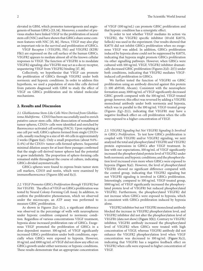
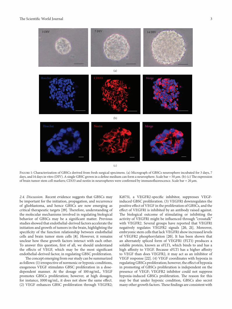
2.1. Glioblastoma Stem-Like Cells Were Derived from Glioblas-tomaMultiforme. CD133 has been successfully used to enrichputative cancer stem cells. After dissociation of nonadherenttumor spheres, CD133+ cells were identified and enriched byfluorescence-activated cell sorting (FACS). Upon replating atone cell per well, GBSCs spheres formed from single CD133+cells, usually reaching to a size of 40–60 cells in approximately2 weeks (Figure 1(a)). Only a small proportion (about 9.6%–11.4%) of the CD133+ tumor cells formed spheres. Sequentialminimal dilution assays for at least three passages confirmedthat the single-cell-derived tumor spheres had the potentialto grow indefinitely. The proportion of sphere-forming cellsremained stable throughout the course of culture, indicatingGBSCs divided asymmetrically.
GBSCs spheres were found to express brain tumor stemcell markers, CD133 and nestin, which were examined byimmunofluorescence (Figures 1(b) and 1(c)).
2.2. VEGFPromotes GBSCs Proliferation throughVEGFR2 butNotVEGFR1. Theeffect ofVEGFonGBSCs proliferationwastested by Neural Colony-Forming Cell Assay. In addition, toconfirm the proliferative phenomenon, which we observedunder the microscope, an ATP assay was performed tomeasure GBSC proliferation.
As shown in Figures 2(a)–2(c), a significant differencewas observed in the percentage of wells with neurospheresunder hypoxic condition compared to normoxic condi-tion. Regardless of various concentrations VEGF treatment,hypoxia alone increased proliferation rate of GBSCs. Exoge-nous VEGF promoted the proliferation of GBSCs in adose-dependent manner. 100 ng/mL of VEGF significantlyincreased GBSCs proliferation under both conditions, espe-cially when GBSCs were exposed to hypoxia. However,10 ng/mL and 1000 ng/mLofVEGFdid not show any effect onGBSCs growth under either normoxic or hypoxic conditions.These results demonstrate that an appropriate concentration
of VEGF (100 ng/mL) can promote GBSC proliferation andthat hypoxic condition has a synergistic effect.
In order to test whether VEGF mediates its action viaVEGFR2, the VEGFR2 specific inhibitor (10 nM Ki8751,Selleck) was used in the experiment. Our results showed thatKi8751 did not inhibit GBSCs proliferation when no exoge-nous VEGF was added. In addition, GBSCs proliferationinduced by hypoxia alone could not be suppressed by Ki8751,indicating that hypoxia might promote GBSCs proliferationvia other signalling pathways. However, when GBSCs werecultured with 100 ng/mL VEGF, VEGFR2 inhibitor dramati-cally decreased GBSC proliferation (Figures 2(d)–2(f)) underboth conditions, indicating that VEGFR2 mediates VEGF-induced cell proliferation in GBSCs.
We further tested the function of VEGFR1 on GBSCproliferation using an antibody directed against this protein(1 : 100 ab9540, Abcam). Consistent with the neurosphereformation assay, 1000 ng/mL of VEGF significantly decreasedGBSC growth compared with the 100 ng/mL VEGF-treatedgroup; however, this effect was reversed by adding a VEGFR1monoclonal antibody under both normoxia and hypoxia,which was in parallel to the 100 ng/mL VEGF-treated group(Figures 2(g)–2(i)), indicating that VEGFR1 may have anegative feedback effect on cell proliferation when the cellswere exposed to a higher concentration of VEGF.
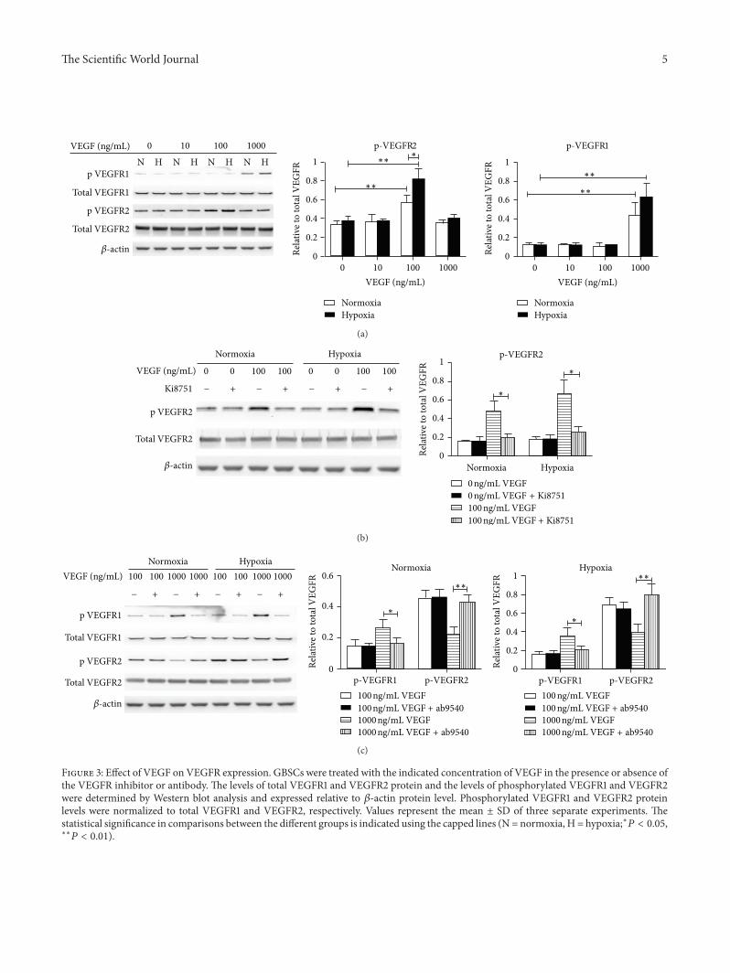
2.3. VEGFR2 Signaling but Not VEGFR1 Signaling Is Involvedin GBSCs Proliferation. To test how GBSCs proliferation iscoupled with VEGFR1 and/or VEGFR2 signaling, we nextanalyzed the total and phosphorylatedVEGFR1 andVEGFR2protein expressions in GBSCs after VEGF treatment. Inline with our expectations, 100 ng/mL of VEGF significantlyincreased the phosphorylated protein level of VEGFR2 underboth normoxic andhypoxic conditions; and the phosphoryla-tion level increased even more when GBSCs were exposed tohypoxia (Figure 3(a)). However, the level of phosphorylatedVEGFR1 showed no significant difference compared withthe control group, indicating that VEGFR2 signaling butnot VEGFR1 signaling is involved in GBSCs proliferation.Interestingly, compared to 100 ng/mL VEGF-treated group,1000 ng/mL of VEGF significantly increased the phosphory-lated protein level of VEGFR1 but reduced phosphorylatedVEGFR2. Furthermore, the phosphorylated VEGFR2 didnot increase when GBSCs were exposed to hypoxia, whichis consistent with GBSCs proliferation induced by hypoxiaalone.
VEGFR2 inhibitor but notVEGFR1monoclonal antibodyblocked the increase in VEGFR2 phosphorylation level, andVEGFR2 inhibitor did not alter the phosphorylation level ofVEGFR1 (data not show) (Figure 3(b)). Contrary to VEGFR2inhibitor, VEGFR1 antibody increased the phosphorylatedlevel of VEGFR2 when GBSCs were treated with highconcentration of VEGF, whereas VEGFR1 antibody did notenhance the VEGFR2 phosphorylation level when VEGFconcentration was decreased to 100 ng/mL (Figure 3(c)),indicating that VEGFR1 has a negative feedback effect onVEGFR2 when cells were exposed to higher concentration ofVEGF.
The Scientific World Journal 3
7 DIV3 DIV 14 DIV
(a)
Hoechst CD133 Merge
(b)
Hoechst MergeNestin
(c)
Figure 1: Characterization of GBSCs derived from fresh surgical specimens. (a) Micrograph of GBSCs neurosphere incubated for 3 days, 7days, and 14 days in vitro (DIV). A single GBSC grown in a define medium can form a neurosphere. Scale bar = 50 𝜇m. (b)-(c)The expressionof brain tumor stem cell markers; CD133 and nestin in neurospheres were confirmed by immunofluorescence. Scale bar = 20𝜇m.
2.4. Discussion. Recent evidence suggests that GBSCs maybe important for the initiation, propagation, and recurrenceof glioblastoma, and hence GBSCs are now emerging ascritical therapeutic targets [19]. Therefore, understanding ofthe molecular mechanisms involved in regulating biologicalbehavior of GBSCs may be a significant matter. Previousstudies showed that endothelial-derived factors accelerate theinitiation and growth of tumors in the brain, highlighting thespecificity of the function relationship between endothelialcells and brain tumor stem cells [8]. However, it remainsunclear how these growth factors interact with each other.To answer this question, first of all, we should understandthe effects of VEGF, which may be the most significantendothelial-derived factor, in regulating GBSC proliferation.
The concept emerging fromour study can be summarizedas follows: (1) irrespective of normoxic or hypoxic conditions,exogenous VEGF stimulates GBSC proliferation in a dose-dependent manner. At the dosage of 100 ng/mL, VEGFpromotes GBSCs proliferation; however, at high dosages,for instance, 1000 ng/mL, it does not show the same effect.(2) VEGF enhances GBSC proliferation through VEGFR2;
Ki8751, a VEGFR2-specific inhibitor, suppresses VEGF-induced GBSC proliferation. (3) VEGFR1 downregulates thepositive effect of VEGF in the proliferation of GBSCs, and theeffect of VEGFR1 is inhibited by an antibody raised against.The biological outcome of stimulating or inhibiting theactivity of VEGFR1 might be influenced through “crosstalk”with VEGFR2. Several groups have reported that VEGFR1negatively regulates VEGFR2 signals [20, 21]. Moreover,embryonic stem cells that lack VEGFR1 show increased levelsof VEGFR2 phosphorylation [20]. It has been shown thatan alternately spliced form of VEGFR1 (FLT1) produces asoluble protein, known as sFLT1, which binds to and has ahigh affinity to VEGF. Because sFLT1 has a higher affinityto VEGF than does VEGFR2, it may act as an inhibitor ofVEGF response [22]. (4) VEGF coordinates with hypoxia inregulatingGBSCs proliferation; however, the effect of hypoxiain promoting of GBSCs proliferation is independent on thepresence of VEGF; VEGFR2 inhibitor could not suppresshypoxia-induced GBSCs proliferation. The reason for thismay be that under hypoxic condition, GBSCs also secretmany other growth factors.These findings are consistent with

4 The Scientific World Journal
∗∗
∗∗
∗
∗
HypoxiaNormoxia
250
200
150
100
50
0
∗
∗
HypoxiaNormoxia
Well
s with
neu
rosp
here
s (%
)
25
20
15
10
5
0
∗∗
∗
∗∗
HypoxiaNormoxia
250
200
150
100
50
0
∗∗
∗∗
∗
HypoxiaNormoxia
250
200
150
100
50
00 10 100 1000
∗∗∗∗
∗∗
∗
∗
∗
∗
∗25
20
15
10
5
0
25
20
15
10
5
00 10 100 1000
NormoxiaHypoxia
Normoxia Hypoxia Normoxia Hypoxia
Normoxia Hypoxia
Normoxia Hypoxia
Well
s with
neu
rosp
here
s (%
)
Well
s with
neu
rosp
here
s (%
)
(a) (d) (g)
(b) (e)
(c) (f)
(h)
(i)
VEGF (ng/mL)
VEGF (ng/mL)
Prol
ifera
tion
(% n
orm
oxia
cont
rol)
Prol
ifera
tion
(% n
orm
oxia
cont
rol)
Prol
ifera
tion
(% n
orm
oxia
cont
rol)
0 ng/mLVEGF
0 ng/mLVEGF
10 ng/mLVEGF
100 ng/mLVEGF
100 ng/mLVEGF
1000 ng/mLVEGF
1000 ng/mLVEGF
100 ng/mL VEGF+ ab9540
0ng/mL VEGF+ Ki8751
100ng/mL VEGF+ Ki8751
100ng/mL VEGF
1000ng/mL VEGF+ ab9540
0 ng/mL VEGF + Ki8751
100 ng/mL VEGF + Ki8751
0 ng/mL VEGF
100 ng/mL VEGF
0 ng/mL VEGF + Ki8751
100 ng/mL VEGF + Ki8751
0 ng/mL VEGF
100 ng/mL VEGF
100 ng/mL VEGF100 ng/mL VEGF + ab95401000 ng/mL VEGF1000 ng/mL VEGF + ab9540
100 ng/mL VEGF100 ng/mL VEGF + ab95401000 ng/mL VEGF1000 ng/mL VEGF + ab9540
0 VEGF
Figure 2: Effect of VEGF on GBSCs proliferation. GBSCs were treated with indicated concentration of VEGF in the presence or absenceof VEGFR inhibitors, followed by Neural Colony-Forming Cell Assay and cell viability analysis using ATP assay. (a)–(c) dose effect ofVEGF on GBSCs proliferation. (d)–(f) effect of VEGFR2 inhibition on GBSCs proliferation. (g)–(i) effect of VEGFR1 inhibition on GBSCsproliferation. Panels (a), (d), and (g) representative images of neurospheres formed in neurosphere formation assays. Panels (b), (e), and (h)GBSC proliferation was tested using a Neural Colony-Forming Cell Assay; all data are represented as the percent of wells with neurospheres.Panels (c), (f), and (i) GBSCs proliferation was tested using ATP assay; all data are represented as a percentage of the normoxia control. Thestatistical significance in comparisons between the different groups is indicated using the capped lines (∗𝑃 < 0.05, ∗∗𝑃 < 0.01).
The Scientific World Journal 5
VEGF (ng/mL) 0 10 100 1000
N H N H N H N H
𝛽-actin
1
0.8
0.6
0.4
0.2
0
∗∗
∗∗
∗∗∗∗
∗
VEGF (ng/mL)0 10 100 1000
VEGF (ng/mL)0 10 100 1000
p-VEGFR2 p-VEGFR1
HypoxiaNormoxia
HypoxiaNormoxia
Relat
ive t
o to
tal V
EGFR
1
0.8
0.6
0.4
0.2
0Relat
ive t
o to
tal V
EGFRp VEGFR1
Total VEGFR1
p VEGFR2
Total VEGFR2
(a)
1
0.8
0.6
0.4
0.2
0Relat
ive t
o to
tal V
EGFR
p-VEGFR2
∗
∗
HypoxiaNormoxia
0 0 0 0100 100 100 100
− + − + − + − +
Normoxia Hypoxia
Ki8751
𝛽-actin
VEGF (ng/mL)
0 ng/mL VEGF + Ki8751
100 ng/mL VEGF + Ki8751
0 ng/mL VEGF
100 ng/mL VEGF
p VEGFR2
Total VEGFR2
(b)
100 100 1000 1000 100 100 1000 1000
− + − + − + − +
Normoxia Hypoxia Normoxia HypoxiaVEGF (ng/mL)
p-VEGFR1 p-VEGFR1p-VEGFR2 p-VEGFR2
𝛽-actin
1
0.8
0.6
0.4
0.2
0
∗∗∗∗
∗∗
Relat
ive t
o to
tal V
EGFR
0.6
0.4
0.2
0Relat
ive t
o to
tal V
EGFR
100 ng/mL VEGF100 ng/mL VEGF + ab95401000 ng/mL VEGF1000 ng/mL VEGF + ab9540
100 ng/mL VEGF100 ng/mL VEGF + ab95401000 ng/mL VEGF1000 ng/mL VEGF + ab9540
p VEGFR1
Total VEGFR1
p VEGFR2
Total VEGFR2
(c)
Figure 3: Effect of VEGF on VEGFR expression. GBSCs were treated with the indicated concentration of VEGF in the presence or absence ofthe VEGFR inhibitor or antibody. The levels of total VEGFR1 and VEGFR2 protein and the levels of phosphorylated VEGFR1 and VEGFR2were determined by Western blot analysis and expressed relative to 𝛽-actin protein level. Phosphorylated VEGFR1 and VEGFR2 proteinlevels were normalized to total VEGFR1 and VEGFR2, respectively. Values represent the mean ± SD of three separate experiments. Thestatistical significance in comparisons between the different groups is indicated using the capped lines (N = normoxia, H = hypoxia;∗𝑃 < 0.05,∗∗𝑃 < 0.01).
6 The Scientific World Journal
Bao et al, who showed that high level of VEGF, produced byCD133+ human glioblastoma cells, might contribute to theirtumour-initiating capacity [23]. This novel finding enhancesour understanding on the mechanism by which VEGF inregulating GBSCs proliferation and may provide insight intothe regulation of GBSC by VEGF signaling.
The prognosis for GBM is very poor, and novel treatmentstrategies are urgently needed. GBM is a highly vasculartumor; a result of its increased expression of VEGF duringprogression is compared with other brain tumors [24, 25].Increased levels of VEGF in GBM accelerate vascular prolif-eration and exacerbate the disease [26]. Based on our presentdata, we propose that VEGF may be the dual targets of notonly the tumor vessels but also the tumor stem cells. Multipletreatment modalities have targeted VEGF and VEGFRs dueto their significant roles in regulating angiogenic processesand GBSCs proliferation. In glioma patients, anti-VEGFand VEGFR2 inhibitors are commonly used to target theVEGF-VEGFR2 signaling cascade. However, despite sometransient positive therapeutic effects, the efficacy of these twostrategies has been disappointing [27–31]. Antiangiogenictherapy leads to devascularization that limits tumor growth,but the benefits of angiogenesis inhibitors are typically tran-sient, and resistance often develops [27]. In addition, someantiangiogenic agents have been shown to promote tumorgrowth and metastasis [29]. Multiple mechanisms may beinvolved, and induction of hypoxia which promotes GBSCproliferation might be one of the these mechanisms [29,32]. Collectively, our results demonstrate that hypoxia canstimulate GBSC proliferation, which cannot be suppressedby VEGFR inhibitors. We believe that not only VEGF butalso some other growth factor signaling contributes to theenhanced GBSC proliferation under a hypoxic condition,but VEGF is not essential. With prolonged antiangiogenictreatment, tumors develop progressive hypoxia, which maybe a central factor in promoting tumor resistance to therapyand ultimately tumor progression. The strong correlationbetween GBSC proliferation and the level of hypoxia furthersupports the idea that hypoxia is an important initiator oftumor growth and metastasis.
In summary, our results have many implications forclinical practice. VEGF plays a positive role in regulatingangiogenesis and tumorigenesis; as a result, VEGF inhibitionis a potential effective treatment strategy in GBM. Our find-ings emphasize the requirement to target pathways involvedin the development of resistance to antiangiogenic treatment,such as hypoxia.
3. Experimental Section
3.1. GBM Tissues. Surgical specimens of GBMwere obtainedafter patients’ written consent under a protocol approved bythe institution’s Institutional Review Board.The neuropatho-logical review of gliomas was completed by a neuropathologyspecialist.
3.2. Isolation of Glioma Stem-Like Cells. GBSCs were isolatedfrom primary human brain tumor patient specimens. Fresh
surgical specimens were obtained from 12 patients diagnosedwith glioblastoma multiforme (GBM). Briefly, tumor tissueswere washed and minced with fine scissors into smallfragments, which were disaggregated by Papain DissociationSystem and filtered by 70 𝜇m cell strainer according to themanufacturer’s instructions. Cells were then cultured in stemcell culture medium for at least 4 hours to recover surfaceantigens. Cells were then labeledwithAPC- or PE-conjugatedCD133 antibody and sorted by fluorescence-activated cellsorting (FACS) as described previously [33]. CD133-positivecells were designated as GBSCs, which were resuspendedin serum-free DMEM/F-12 containing human recombinantN2(20 ng/mL; Invitrogen), EGF (20 ng/mL; Invitrogen), and
bFGF (20 ng/mL; Gibco) and then seeded in a nonadherentcell culture flask (5mL per flask) at a density of 5 × 10⁶ livecells per flask. Culture media was changed twice a week, andneurospheres are passaged every 1 or 2 weeks, depending onthe growth rate of each sample. Immunofluorescence stainingto detect the expression of brain tumor stem cell markers wasperformed as described previously [33].
3.3. Neural Colony-Forming Cell Assay. Neurosphere for-mation assays were performed with CD133-positive cellssorted by FACS to single cells per well of 96 well plates ina collagen semisolid matrix (Stem Cell Technologies). Thesemisolid matrix contained serum-free media supplementedwith human recombinant N
2(20 ng/mL; Invitrogen), EGF
(20 ng/mL; Invitrogen), and bFGF (20 ng/mL; Gibco). Vari-ous concentrations of VEGF (0 ng/mL, 10 ng/mL, 100 ng/mL,and 1000 ng/mL; Sigma) in the presence or absence ofVEGFRinhibitors were added as previously described, and cells wereincubated for 14 days in vitro (DIV) under normoxic (20%O2) or hypoxic (1%O
2) condition. In order to induce hypoxia,
cells were cultured in hypoxia chambers (Sanyo). Completereplenishment medium was added into the center of eachNCFC dish once every 3 days during the entire NCFC cultureincubation (14 days). Neurosphere formation was measuredas the percent of wells with neurospheres after 14 days.
3.4. ATP Assay. Primary neurospheres were dispersed with0.025% trypsin andmechanical trituration after trypsin inac-tivation. 10,000 single GBSCs diluted in 100 𝜇L of mediumwere seeded in a 96-well plate and cultured for 24 hrs.The cells were then treated with indicated concentrationsof VEGF in the presence or absence of VEGFR inhibitorsunder normoxic (20% O
2) and hypoxic (1% O
2) conditions.
After 24 hrs of incubation, cells were equilibrated at roomtemperature for 30min, and then 100𝜇L of ATP reagent(CellTiter-Glo luminescent cell viability assay, Promega) wasadded to eachwell. Cell lysis was induced on an orbital shakerfor 2min, then equilibrated at room temperature for 10min,and the luminescent signal was recorded in a PolarStar platereader.
3.5. Western Blotting. For Western blot analysis, cells grownin 6-well plate to confluence were treated as described in thesection on the ATP assay. Cells floating in mediumwere thenharvested, washed twice with ice-cold PBS, and lysed in RIPA
The Scientific World Journal 7
buffer (Sigma) with freshly added protease and phosphataseinhibitor cocktail (Thermo Scientific). Cells were allowedto lyse for 10min on ice and centrifuged at 12,000 g for15min at 4∘C. The supernatant was removed, and proteinconcentrations in the supernatant were determined by BCAprotein assay. A total of 40𝜇g of proteins were loaded on 4–15% SDS polyacrylamide gradient gels and transferred ontoa NC membrane. The membrane was sequentially incubatedwith a 5% fat-free milk for 1 hour at room temperature,a specific primary antibody for total and phosphorylatedVEGFR1 and VEGFR2 (cell signaling) overnight at 4∘C andappropriated HRP-conjugated second antibody for 1 hourat room temperature. Immunoreactive bands were detectedusing the ECL chemiluminescence reagent (GE Healthcare-Amersham Biosciences). The membrane was stripped usingstripping buffer (Amersham Biosciences) and subsequentlylabeled with 𝛽-actin following the standard Western blotprocedures. Densitometry was analyzed using Quantity Onesoftware (Bio-Rad). Densitometry results were either nor-malized to total protein or 𝛽-actin.
3.6. Statistical Analysis. All experiments were repeated atleast three times. The statistical significance of differenceswas evaluated by a one-way analysis of variance (ANOVA)or Student’s t-test (Prism 5; GraphPad Software, San Diego,http://www.graphpad.com/). Descriptive statistics were gen-erated for all quantitative data with presentation of means ±standard error. The level of significance for all comparisonswas 𝑃 < 0.05.
4. Conclusions
VEGF stimulates GBSC proliferation in a dose-dependentmanner via VEGFR2 signaling, and VEGFR1 has a negativefeedback effect on VEGFR2.This novel finding enhances ourunderstanding of the mechanism by which VEGF regulatesGBSC proliferation. In addition, Our results support thehypothesis that the devascularization caused by anti-VEGFtherapy increases tumor hypoxia, and this hypoxia mediatesresistance to antiangiogenic therapy.
Conflict of Interests
The authors declare no conflict of interests.
Acknowledgments
This study was supported by Grants (2010CB945500,2012CB966300, 2009CB941100, 81271003, 30870805,90919002, and 08dj1400503) from the National NatureScience Foundation, Ministry of Science and Technology ofChina, and Shanghai City Science Foundation.
References
[1] D. A.Greenberg andK. Jin, “From angiogenesis to neuropathol-ogy,” Nature, vol. 438, no. 7070, pp. 954–959, 2005.
[2] F. Mackenzie and C. Ruhrberg, “Diverse roles for VEGF-A inthe nervous system,” Development, vol. 139, pp. 1371–1380, 2012.
[3] J. M. Rosenstein and J. M. Krum, “New roles for VEGF in ner-vous tissue—beyond blood vessels,” Experimental Neurology,vol. 187, no. 2, pp. 246–253, 2004.
[4] S. H. Cheshier,M. Y. S. Kalani,M. Lim, L. Ailles, S. L. Huhn, andI. L. Weissman, “A neurosurgeon’s guide to stem cells, cancerstem cells, and brain tumor stem cells,” Neurosurgery, vol. 65,no. 2, pp. 237–249, 2009.
[5] S. G. M. Piccirillo, E. Binda, R. Fiocco, A. L. Vescovi, and K.Shah, “Brain cancer stem cells,” Journal of Molecular Medicine,vol. 87, no. 11, pp. 1087–1095, 2009.
[6] N. Takakura, “Formation and regulation of the cancer stem cellniche,” Cancer Science, vol. 103, pp. 1177–1181, 2012.
[7] T. S. Zhu, M. A. Costello, C. E. Talsma et al., “Endothelial cellscreate a stem cell niche in glioblastoma by providing notchligands that nurture self-renewal of cancer stem-like cells,”Cancer Research, vol. 71, pp. 6061–6072, 2011.
[8] P. Hamerlik, J. D. Lathia, R. Rasmussen et al., “Autocrine VEGF-VEGFR2-neuropilin-1 signaling promotes glioma stem-like cellviability and tumor growth,” The Journal of ExperimentalMedicine, vol. 209, pp. 507–520, 2012.
[9] M. Jinushi, M. Baghdadi, S. Chiba, and H. Yoshiyama, “Reg-ulation of cancer stem cell activities by tumor-associatedmacrophages,” American Journal of Cancer Research, vol. 2, pp.529–539, 2012.
[10] J. M. Heddleston, Z. Li, J. D. Lathia, S. Bao, A. B. Hjelmeland,and J. N. Rich, “Hypoxia inducible factors in cancer stem cells,”British Journal of Cancer, vol. 102, no. 5, pp. 789–795, 2010.
[11] B. Keith and M. C. Simon, “Hypoxia-inducible factors, stemcells, and cancer,” Cell, vol. 129, no. 3, pp. 465–472, 2007.
[12] Z. Li, S. Bao, Q. Wu et al., “Hypoxia-inducible factors regulatetumorigenic capacity of glioma stem cells,” Cancer Cell, vol. 15,no. 6, pp. 501–513, 2009.
[13] C. Folkins, Y. Shaked, S. Man et al., “Glioma tumor stem-like cells promote tumor angiogenesis and vasculogenesis viavascular endothelial growth factor and stromal-derived factor1,” Cancer Research, vol. 69, no. 18, pp. 7243–7251, 2009.
[14] R. Mentlein, F. Forstreuter, H. M. Mehdorn, and J. Held-Feindt,“Functional significance of vascular endothelial growth factorreceptor expression on human glioma cells,” Journal of Neuro-Oncology, vol. 67, no. 1-2, pp. 9–18, 2004.
[15] Z. Xiao, Y. Kong, S. Yang,M. Li, J.Wen, and L. Li, “Upregulationof Flk-1 by bFGF via the ERK pathway is essential for VEGF-mediated promotion of neural stem cell proliferation,” CellResearch, vol. 17, no. 1, pp. 73–79, 2007.
[16] D. Zhao, J. Najbauer, E. Garcia et al., “Neural stem cell tropismto glioma: critical role of tumor hypoxia,” Molecular CancerResearch, vol. 6, no. 12, pp. 1819–1829, 2008.
[17] A. F. Bruns, L. Bao, J. H. Walker, and S. Ponnambalam, “VEGF-A-stimulated signalling in endothelial cells via a dual receptortyrosine kinase system is dependent on co-ordinated traffickingand proteolysis,” Biochemical Society Transactions, vol. 37, no. 6,pp. 1193–1197, 2009.
[18] A. K. Olsson, A. Dimberg, J. Kreuger, and L. Claesson-Welsh,“VEGF receptor signalling—in control of vascular function,”Nature Reviews Molecular Cell Biology, vol. 7, no. 5, pp. 359–371,2006.
[19] C. G. Hadjipanayis and E. G. Van Meir, “Brain cancer propa-gating cells: biology, genetics and targeted therapies,” Trends inMolecular Medicine, vol. 15, no. 11, pp. 519–530, 2009.

8 The Scientific World Journal
[20] D. M. Roberts, J. B. Kearney, J. H. Johnson, M. P. Rosenberg, R.Kumar, and V. L. Bautch, “The vascular endothelial growth fac-tor (VEGF) receptor flt-1 (VEGFR-1) modulates flk-1 (VEGFR-2) signaling during blood vessel formation,” American Journalof Pathology, vol. 164, no. 5, pp. 1531–1535, 2004.
[21] N. C. Kappas, G. Zeng, J. C. Chappell et al., “The VEGFreceptor Flt-1 spatially modulates Flk-1 signaling and bloodvessel branching,” Journal of Cell Biology, vol. 181, no. 5, pp. 847–858, 2008.
[22] S. Sela, S. Natanson-Yaron, E. Zcharia, I. Vlodavsky, S. Yagel,and E. Keshet, “Local retention versus systemic release ofsoluble VEGF receptor-1 are mediated by heparin-binding andregulated by heparanase,” Circulation Research, vol. 108, no. 9,pp. 1063–1070, 2011.
[23] S. Bao, Q. Wu, S. Sathornsumetee et al., “Stem cell-like gliomacells promote tumor angiogenesis through vascular endothelialgrowth factor,” Cancer Research, vol. 66, no. 16, pp. 7843–7848,2006.
[24] A. G. Linkous and E. M. Yazlovitskaya, “Angiogenesis inglioblastoma multiforme: navigating the maze,” Anti-CancerAgents in Medicinal Chemistry, vol. 11, pp. 712–718, 2011.
[25] L. Robles Irizarry, D. Hambardzumyan, I. Nakano, C. L. Glad-son, and M. S. Ahluwalia, “Therapeutic targeting of VEGF inthe treatment of glioblastoma,” Expert Opinion on TherapeuticTargets, vol. 16, pp. 973–984, 2012.
[26] A. S. Chi, A. G. Sorensen, R. K. Jain, and T. T. Batchelor,“Angiogenesis as a therapeutic target in malignant gliomas,”Oncologist, vol. 14, no. 6, pp. 621–636, 2009.
[27] Y. L. Hu, M. DeLay, A. Jahangiri et al., “Hypoxia-inducedautophagy promotes tumor cell survival and adaptation toantiangiogenic treatment in glioblastoma,”Cancer Research, vol.72, pp. 1773–1783, 2012.
[28] A. Bikfalvi,M.Moenner, S. Javerzat, S.North, andM.Hagedorn,“Inhibition of angiogenesis and the angiogenesis/invasion shift,”Biochemical Society Transactions, vol. 39, pp. 1560–1564, 2011.
[29] O. Keunen, M. Johansson, A. Oudin et al., “Anti-VEGF treat-ment reduces blood supply and increases tumor cell invasion inglioblastoma,” Proceedings of the National Academy of Sciencesof the United States of America, vol. 108, pp. 3749–3754, 2011.
[30] A. D. Norden, J. Drappatz, and P. Y. Wen, “Antiangiogenictherapies for high-grade glioma,” Nature Reviews Neurology,vol. 5, no. 11, pp. 610–620, 2009.
[31] M. C. Chamberlain, “Antiangiogenic blockage: a new treatmentfor glioblastoma,” Expert Opinion on Biological Therapy, vol. 8,no. 10, pp. 1449–1453, 2008.
[32] J. M. Heddleston, Q. Wu, M. Rivera et al., “Hypoxia-inducedmixed-lineage leukemia 1 regulates glioma stem cell tumori-genic potential,” Cell Death & Differentiation, vol. 19, pp. 428–439, 2012.
[33] H. Tang, Y. Gong, Y. Mao et al., “Cd133-positive cells mightbe responsible for efficient proliferation of human meningiomacells,” International Journal of Molecular Sciences, vol. 13, pp.6424–6439, 2012.

Submit your manuscripts athttp://www.hindawi.com
Stem CellsInternational
Hindawi Publishing Corporationhttp://www.hindawi.com Volume 2014
Hindawi Publishing Corporationhttp://www.hindawi.com Volume 2014
MEDIATORSINFLAMMATION
of
Hindawi Publishing Corporationhttp://www.hindawi.com Volume 2014
Behavioural Neurology
EndocrinologyInternational Journal of
Hindawi Publishing Corporationhttp://www.hindawi.com Volume 2014
Hindawi Publishing Corporationhttp://www.hindawi.com Volume 2014
Disease Markers
Hindawi Publishing Corporationhttp://www.hindawi.com Volume 2014
BioMed Research International
OncologyJournal of
Hindawi Publishing Corporationhttp://www.hindawi.com Volume 2014
Hindawi Publishing Corporationhttp://www.hindawi.com Volume 2014
Oxidative Medicine and Cellular Longevity
Hindawi Publishing Corporationhttp://www.hindawi.com Volume 2014
PPAR Research
The Scientific World JournalHindawi Publishing Corporation http://www.hindawi.com Volume 2014
Immunology ResearchHindawi Publishing Corporationhttp://www.hindawi.com Volume 2014
Journal of
ObesityJournal of
Hindawi Publishing Corporationhttp://www.hindawi.com Volume 2014
Hindawi Publishing Corporationhttp://www.hindawi.com Volume 2014
Computational and Mathematical Methods in Medicine
OphthalmologyJournal of
Hindawi Publishing Corporationhttp://www.hindawi.com Volume 2014
Diabetes ResearchJournal of
Hindawi Publishing Corporationhttp://www.hindawi.com Volume 2014
Hindawi Publishing Corporationhttp://www.hindawi.com Volume 2014
Research and TreatmentAIDS
Hindawi Publishing Corporationhttp://www.hindawi.com Volume 2014
Gastroenterology Research and Practice
Hindawi Publishing Corporationhttp://www.hindawi.com Volume 2014
Parkinson’s Disease
Evidence-Based Complementary and Alternative Medicine
Volume 2014Hindawi Publishing Corporationhttp://www.hindawi.com