Embed Size (px)
Citation preview
HAL Id: hal-02366087https://hal.archives-ouvertes.fr/hal-02366087
Submitted on 15 Nov 2019
HAL is a multi-disciplinary open accessarchive for the deposit and dissemination of sci-entific research documents, whether they are pub-lished or not. The documents may come fromteaching and research institutions in France orabroad, or from public or private research centers.
L’archive ouverte pluridisciplinaire HAL, estdestinée au dépôt et à la diffusion de documentsscientifiques de niveau recherche, publiés ou non,émanant des établissements d’enseignement et derecherche français ou étrangers, des laboratoirespublics ou privés.
GFAT1 phosphorylation by AMPK promotesVEGF-induced angiogenesis
Darya Zibrova, Franck Vandermoere, Olga Göransson, Mark Peggie, KarinaMariño, Anne Knierim, Katrin Spengler, Cora Weigert, Benoît Viollet,
Nicholas Morrice, et al.
To cite this version:Darya Zibrova, Franck Vandermoere, Olga Göransson, Mark Peggie, Karina Mariño, et al.. GFAT1phosphorylation by AMPK promotes VEGF-induced angiogenesis. Biochemical Journal, PortlandPress, 2017, 474 (6), pp.983-1001. �10.1042/BCJ20160980�. �hal-02366087�
ACCEPTED MANUSCRIPT
GFAT1 phosphorylation by AMPK promotes VEGF-induced angiogenesis
Darya Zibrova, Franck Vandermoere, Olga Göransson, Mark Peggie, Karina Mariño, Anne Knierim, Katrin Spengler, Cora Weigert, Benoit Viollet, Nicholas A. Morrice, Kei
Sakamoto, Regine Heller
Copyright 2016 The Author(s). Use of open access articles is permitted based on the terms of the specific Creative Commons Licence under which the article is published. Archiving of non-open access articles is permitted in accordance with the Archiving Policy of Portland Press (http://www.portlandpresspublishing.com/content/open-access-policy#Archiving).
Cite as Biochemical Journal (2016) DOI: 10.1042/BCJ20160980
BIOCHEMICAL JOURNAL
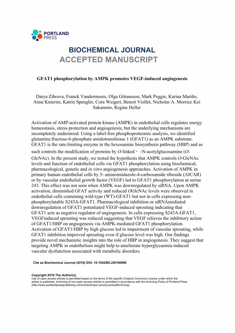
Activation of AMP-activated protein kinase (AMPK) in endothelial cells regulates energy homeostasis, stress protection and angiogenesis, but the underlying mechanisms are incompletely understood. Using a label-free phosphoproteomic analysis, we identified glutamine:fructose-6-phosphate amidotransferase 1 (GFAT1) as an AMPK substrate. GFAT1 is the rate-limiting enzyme in the hexosamine biosynthesis pathway (HBP) and as such controls the modification of proteins by O-linked ዘ-N-acetylglucosamine (O-GlcNAc). In the present study, we tested the hypothesis that AMPK controls O-GlcNAc levels and function of endothelial cells via GFAT1 phosphorylation using biochemical, pharmacological, genetic and in vitro angiogenesis approaches. Activation of AMPK in primary human endothelial cells by 5- aminoimidazole-4-carboxamide riboside (AICAR) or by vascular endothelial growth factor (VEGF) led to GFAT1 phosphorylation at serine 243. This effect was not seen when AMPK was downregulated by siRNA. Upon AMPK activation, diminished GFAT activity and reduced OGlcNAc levels were observed in endothelial cells containing wild-type (WT)-GFAT1 but not in cells expressing non-phosphorylatable S243A-GFAT1. Pharmacological inhibition or siRNAmediated downregulation of GFAT1 potentiated VEGF-induced sprouting indicating that GFAT1 acts as negative regulator of angiogenesis. In cells expressing S243A-GFAT1, VEGFinduced sprouting was reduced suggesting that VEGF relieves the inhibitory action of GFAT1/HBP on angiogenesis via AMPK-mediated GFAT1 phosphorylation. Activation of GFAT1/HBP by high glucose led to impairment of vascular sprouting, while GFAT1 inhibition improved sprouting even if glucose level was high. Our findings provide novel mechanistic insights into the role of HBP in angiogenesis. They suggest that targeting AMPK in endothelium might help to ameliorate hyperglycaemia-induced vascular dysfunction associated with metabolic disorders.
1
GFAT1 phosphorylation by AMPK promotes VEGF-induced angiogenesis
Darya Zibrova1,§, Franck Vandermoere2,§, Olga Göransson3, Mark Peggie4, Karina Mariño5,
Anne Knierim1, Katrin Spengler1, Cora Weigert6,7,8, Benoit Viollet9,10,11, Nicholas A. Morrice12,
Kei Sakamoto13,#,¶, Regine Heller1,¶,*
1Institute of Molecular Cell Biology, Center for Molecular Biomedicine, Jena University
Hospital, 07745 Jena, Germany
2Institut de Génomique Fonctionnelle, CNRS UMR5203, INSERM U1191, Université de
Montpellier, France
3Department of Experimental Medical Sciences, Lund University, 221 84 Lund, Sweden
4Division of Signal Transduction Therapy, College of Life Sciences, University of Dundee,
Dundee DD1 5EH, Scotland, UK
5Laboratorio de Glicómica Funcional y Molecular, Instituto de Biología y Medicina
Experimental, Consejo Nacional de Investigaciones Científicas y Técnicas (IBYME-CONICET),
C1428 Buenos Aires, Argentina
6Division of Pathobiochemistry and Clinical Chemistry, University of Tübingen, 72076
Tübingen, Germany
7Institute for Diabetes Research and Metabolic Diseases of the Helmholtz Zentrum München at
the University of Tübingen, 72076 Tübingen, Germany
8German Center for Diabetes Research (DZD), 85764 Neuherberg, Germany
9INSERM U1016, Institut Cochin, Paris, France
10CNRS UMR 8104, Paris, France
11Université Paris Descartes, Sorbonne Paris Cité, Paris, France
12AB-Sciex, Phoenix House, Centre Park, Warrington WA1 1RX, UK
2
13MRC Protein Phosphorylation and Ubiquitylation Unit, College of Life Sciences, University of
Dundee, Dundee DD1 5EH, Scotland, UK
#Current address: Nestlé Institute of Health Sciences SA, EPFL Innovation Park, bâtiment H,
1015 Lausanne, Switzerland
§Co-first author
¶co-senior author
*Corresponding author: Dr. Regine Heller
Institute for Molecular Cell Biology, Center for Molecular
Biomedicine, Jena University Hospital
Hans-Knöll-Straße 2
07745 Jena
Germany
Telefon +49 (0)3641-9395633
Telefax +49 (0)3641-9395602
E-mail: [email protected]
3
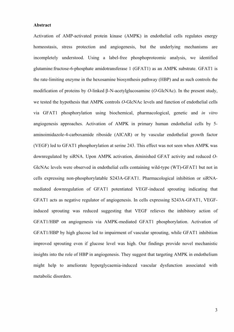
Abstract
Activation of AMP-activated protein kinase (AMPK) in endothelial cells regulates energy
homeostasis, stress protection and angiogenesis, but the underlying mechanisms are
incompletely understood. Using a label-free phosphoproteomic analysis, we identified
glutamine:fructose-6-phosphate amidotransferase 1 (GFAT1) as an AMPK substrate. GFAT1 is
the rate-limiting enzyme in the hexosamine biosynthesis pathway (HBP) and as such controls the
modification of proteins by O-linked -N-acetylglucosamine (O-GlcNAc). In the present study,
we tested the hypothesis that AMPK controls O-GlcNAc levels and function of endothelial cells
via GFAT1 phosphorylation using biochemical, pharmacological, genetic and in vitro
angiogenesis approaches. Activation of AMPK in primary human endothelial cells by 5-
aminoimidazole-4-carboxamide riboside (AICAR) or by vascular endothelial growth factor
(VEGF) led to GFAT1 phosphorylation at serine 243. This effect was not seen when AMPK was
downregulated by siRNA. Upon AMPK activation, diminished GFAT activity and reduced O-
GlcNAc levels were observed in endothelial cells containing wild-type (WT)-GFAT1 but not in
cells expressing non-phosphorylatable S243A-GFAT1. Pharmacological inhibition or siRNA-
mediated downregulation of GFAT1 potentiated VEGF-induced sprouting indicating that
GFAT1 acts as negative regulator of angiogenesis. In cells expressing S243A-GFAT1, VEGF-
induced sprouting was reduced suggesting that VEGF relieves the inhibitory action of
GFAT1/HBP on angiogenesis via AMPK-mediated GFAT1 phosphorylation. Activation of
GFAT1/HBP by high glucose led to impairment of vascular sprouting, while GFAT1 inhibition
improved sprouting even if glucose level was high. Our findings provide novel mechanistic
insights into the role of HBP in angiogenesis. They suggest that targeting AMPK in endothelium
might help to ameliorate hyperglycaemia-induced vascular dysfunction associated with
metabolic disorders.
4
Summary statement
AMPK is an important regulator of endothelial function; however, the underlying mechanisms
are poorly understood. In the present study, we demonstrate that AMPK decreases O-GlcNAc
levels in human endothelial cells via GFAT1 phosphorylation thereby promoting VEGF-induced
angiogenesis.
Short title: GFAT1 mediates pro-angiogenic AMPK effects
Key words: AMPK; angiogenesis; GFAT1; O-GlcNAcylation; phosphoproteomics; VEGF
Abbreviations: ACC, acetyl-CoA carboxylase; AMPK, AMP-activated protein kinase; DON, 6-
diazo-5-oxo-L-norleucine; GFAT, glutamine:fructose-6-phosphate amidotransferase; HBP,
hexosamine biosynthesis pathway; HAS, human serum albumin; HUVEC, human umbilical cord
vein endothelial cells; IMAC, immobilised metal affinity chromatography; MEF, mouse
embryonic fibroblasts; O-GlcNAc, O-linked -N-acetylglucosamine; TiO2, titanium dioxide;
TSC, tuberous sclerosis complex; UDP-GlcNAc, UDP-N-acetylglucosamine; VEGF, vascular
endothelial growth factor; WT, wild-type.
5
Introduction
AMPK is an important component of signalling mechanisms regulating energy and nutrient
metabolism. It is a heterotrimeric serine/threonine protein kinase consisting of catalytic and
regulatory and subunits, each existing as several isoforms [1]. Energy-depriving stresses or
pharmacological agents, which increase AMP/ATP and ADP/ATP ratios, trigger binding of
AMP to the subunit and activate AMPK via a triple mechanism: (i) allosteric activation, (ii)
phosphorylation of threonine 172 on the activation loop of AMPK and (iii) inhibition of
dephosphorylation of threonine 172 [2-4]. The upstream kinases of AMPK include LKB1 in
response to metabolic stress and Ca2+/calmodulin-dependent protein kinase kinase 2 in response
to Ca2+-elevating agonists (reviewed in [1]). AMPK maintains cellular ATP homeostasis by
activating ATP-producing pathways and by inhibiting ATP-consuming pathways via
phosphorylation of target proteins such as acetyl-CoA carboxylase isoforms 1 and 2, which
regulate fatty acid synthesis and oxidation, respectively [5, 6]. Furthermore, it serves as a
metabolic checkpoint for cell growth by inhibiting mammalian target-of-rapamycin complex-1
[7, 8].
AMPK plays an important role in endothelial cells. It is activated by a variety of stimuli
including shear stress, oxidants, hormones, vascular mediators and VEGF (reviewed in [9]).
Activation of AMPK in endothelial cells seems to be associated with regulation of energy
supply, stress protection, maintenance of anti-inflammatory and anti-atherogenic phenotypes and
regulation of angiogenesis [10] but the underlying mechanisms are not fully elucidated. To
further understand cellular functions of AMPK, the identification and characterisation of novel
AMPK substrates is therefore of high importance.
Recent studies suggested GFAT1 as a new AMPK target while GFAT2, predominantly
expressed in the central nervous system [11], was not found in screens for novel AMPK
substrates [12-14]. GFAT1 is ubiquitously expressed and catalyses the formation of
glucosamine-6-phosphate, the first product of the HBP, using fructose-6-phosphate and L-
6
glutamine as substrates. This step is rate-limiting for the synthesis of UDP-N-acetylglucosamine
(UDP-GlcNAc), the end product of the HBP. UDP-GlcNAc is used as donor for adding N-
acetylglucosamine to serine/threonine residues of proteins. This post-translational modification
known as O-GlcNAcylation modulates key biological processes such as transcription, signal
transduction and cytoskeletal reorganisation [15]. GFAT is activated by high extracellular
glucose, and studies using genetically modified mice demonstrated a causative link between
GFAT1 activity and diabetic state [16-18]. Moreover, GFAT1 activity was shown to be elevated
in diabetic patients [19]. The increase in O-GlcNAcylation of proteins as a consequence of
GFAT activation is thought to contribute to the development of insulin resistance [20] and
endothelial dysfunction [21], which is an initial event in atherogenesis and a hallmark of vascular
complications of type 2 diabetes.
Despite the importance of the HBP in providing the substrate for O-GlcNAcylation, the
posttranslational regulation of the key enzyme GFAT is incompletely understood. GFAT1 has
been reported to be an AMPK substrate with serine 243 as putative phosphorylation site, which
was shown to be activating in vitro [13], but inhibitory in certain cellular context [12]. However,
detailed biochemical characterisation of AMPK-dependent GFAT1 phosphorylation and the
functional role of this pathway in physiologically relevant systems have not been established.
Therefore, in the present study we address the role of GFAT1 regulation by AMPK in primary
human endothelial cells. We demonstrate that GFAT1 is a component of the VEGF-AMPK
pathway in endothelial cells and, as such, a mediator of pro-angiogenic effects of AMPK. Via
GFAT1 phosphorylation, AMPK can decrease O-GlcNAcylation of proteins, which may also
contribute to beneficial metabolic outcomes of AMPK activation, thus further underlining the
importance of AMPK as a drug target.
7
Experimental procedures
Materials
Cell culture media and sera were from Lonza. Endothelial mitogen was from Hycultec GmbH.
Human plasma fibrinogen (341576) was purchased from Calbiochem. Proteinase inhibitor
mixture complete, EDTA-free, was acquired from Roche Diagnostics. Mass spectrometry grade
trypsin (Trypsin Gold, V5280) was from Promega. KOD Hot Start Polymerase and pSC-b vector
were purchased from Novagen and Stratagene, respectively. Colloidal Blue and LDS sample
loading buffer were from Life Technologies. DCTM Protein Assay kit was from Bio-Rad
Laboratories. Recombinant human VEGF-165 was purchased from R&D Systems GmbH. The
AMPK activator A-769662 was synthesized either as previously described [22] or obtained from
Abcam Biochemicals (ab120335). 5-aminoimidazole-4-carboxamide riboside (AICAR,
ab120358) was from Abcam Biochemicals. Protein G Sepharose (P3296) and PHOS-Select�™
Iron Affinity Gel were from Sigma-Aldrich. Glutathione sepharose resin and ECLTM Western
Blotting Detection kit were from GE Healthcare. Titansphere 5 m loose beads were from
Hichrom Limited. SAINT-RED was from Synvolux Therapeutics B.V. Unless otherwise
indicated all other reagents were from Sigma-Aldrich.
Antibodies
Total AMPK (#2532), phospho-AMPK (#2531), total ACC (#3676), phospho-ACC (#3661),
and -actin (#4970) antibodies were from Cell Signalling Technology. O-GlcNAc antibody
(CTD110.6 clone, #MMS-248R) was from Biolegend Covance. GFAT1 rabbit antiserum was
generated as described previously [23]. Polyclonal sheep antibody against AMPK 1 and
glutathione S-transferase (GST) antibody were kindly provided by Dr. D. Grahame Hardie
(University of Dundee). Polyclonal sheep antibody against GFAT1 (S702C, 3rd bleed) and site-
specific sheep polyclonal antibody against phospho-(Ser243)-GFAT1 (S343C, 3rd bleed) were
generated in the Division of Signal Transduction Therapy (University of Dundee, UK) by
8
immunization with a full-length human GST-GFAT1 or phosphorylated peptide of the human
sequence (residues 240-251 [RVDS*TTCLFPVE, * indicates the phospho-serine]), respectively.
Horseradish peroxidase (HRP)-conjugated secondary antibody to rabbit and sheep IgG were
from Kirkegaard & Perry Laboratories, Inc. (KPL) and Santa Cruz Biotechnology, Inc.,
respectively. HRP-conjugated secondary antibody to mouse IgM chain (#074-1803) was from
KPL.
Oligos
The primers used for site-directed mutagenesis of GFAT1 were:
MP4146 CTCTCTCGTGTGGACGCCACAACCTGCCTTTTC (forward)
MP4147 GAAAAGGCAGGTTGTGGCGTCCACACGAGAGAG (reverse).
The siRNA duplex oligonucleotides used in this study were based on the human cDNAs
encoding AMPK 1, AMPK 2, and GFAT1. AMPK 1- and AMPK 2-specific SMARTpool
siRNA reagents (M-005027-02-0020 and M-005361-02-0020, respectively) were purchased
from Dharmacon (GE Healthcare). For GFAT1, 5 -GGAGGAUACUGAGACCAUU-3 (sense)
and 3 -CCUCCUAUGACUCUGGUAA-5 (anti-sense) siRNA duplex oligonucleotides, reported
by Jokela et al. [24], were obtained from Sigma. A nonspecific control SMARTpool siRNA (D-
001810-10-20) was from Dharmacon.
Mouse embryonic fibroblasts
WT (AMPK 1+/+ 2+/+) and AMPK-deficient (AMPK 1-/- 2-/-) mouse embryonic fibroblasts
(MEFs) were generated as previously described [25]. The animal study was approved by the
Paris Descartes University ethics committee (no. CEEA34.BV.157.12) and performed under
French authorization to experiment on vertebrates (no. 75�–886) in accordance with the European
guidelines. MEFs were maintained in DMEM containing 4.5 g/l glucose, 10% foetal calf serum
9
(FCS), 2 mmol/l glutamine, 50 U/ml penicillin and 50 µg/ml streptomycin (DMEM culture
medium).
Phosphoproteomic screen for AMPK substrates in cells
After treatment of AMPK 1+/+ 2+/+ and AMPK 1-/- 2-/- MEFs with A-769662 (100 µmol/l, 1
h), protein lysates were obtained and trypsin digested. Phosphopeptides were enriched and
analysed by LC-MS/MS. Subsequently, phosphopeptides (1697 within 742 proteins) identified in
a Mascot search were checked for AMPK consensus sequence. The latter is based on a
compromise between the sequence derived from established AMPK cellular substrates [26] and
an in vitro peptide screen published by Gwinn et al: [M/V/L/F/I]-X(0,1)-[R/H/K]-X(2,3)-[S/T]-
X(3)-[M/V/L/I/F], where X is any amino acid [7]. Matched peptides were relatively quantified
using the height of the extracted ion chromatogram, which was compared between A-769662-
treated AMPK 1+/+/ 2+/+ and AMPK 1-/-/ 2-/- MEFs. The detailed screening is reported in
Supplementary Material, Methods.
Cloning, site directed-mutagenesis
Human GFAT1 (NCBI BC045641) was amplified from IMAGE EST 5298728 using KOD Hot
Start Polymerase, cloned into pSC-b and sequenced to completion. The resulting plasmid was
digested with BamH1 and Not1 and cloned between the same sites into vectors for bacterial
(pGEX6P-1) and mammalian (pEBG6P) expression, encoding proteins with an N-terminal
glutathione S-transferase (GST) tag. Site-directed mutagenesis was performed by the Stratagene
Quickchange method but using KOD Hot Start DNA Polymerase.
For generation of lentiviral particles, cDNA sequences encoding WT- and S243A-GFAT1 were
cut off respective pEBG6P vectors using BamH1 and Not1 and inserted between the same sites
into pCDH-CMV-MCS-EF1-Puro lentivectors (CD510B-1, System Biosciences (SBI)) to
10
generate pCDH-CMV-MCS-EF1-Puro-GFAT1 and pCDH-CMV-MCS-EF1-Puro-S243A-
GFAT1 plasmids for expression of untagged versions of either protein.
In vitro phosphorylation of recombinant GFAT1
WT or S243A mutant of human GST-tagged-GFAT1 were expressed in E. coli and purified
using glutathione sepharose resin. Each recombinant protein (1 µg) was treated with 5 U/ml of
activated recombinant AMPK trimeric complex ( 2 2 1) (kindly provided by Dr. D. Grahame
Hardie, University of Dundee, UK), in 50 mmol/l Tris-HCl, pH 7.5, containing 10 mmol/l MgCl2,
0.1 mmol/l EGTA, 0.1% -mercaptoethanol, and 0.1 mmol/l ATP for 5 min at 30°C. When
radioactive labelling was required, ATP was provided as [ -32P]ATP (0.1 mmol/l, GE
Healthcare; 1,000�–2,000 cpm/pmol in analytical kinase assays, approx. 10,000 cpm/pmol for
phosphorylation site analysis). Reaction was stopped by boiling samples for 5 min in LDS
sample loading buffer. The proteins were subjected to electrophoresis on polyacrylamide gels
followed by Colloidal Blue staining. GFAT1, which was evident as a 110 kDa band after
staining and autoradiography, was excised, and the amount of 32P incorporation was determined
by Cerenkov counting. The stoichiometry of GFAT1 phosphorylation at serine 243 site was
estimated based on the following calculations: [ -32P]ATP had a specific activity of 10,000
cpm/pmol. Since fusion GST-GFAT1 protein weights 104 kDa, 1 µg of this protein used for the
kinase assay, corresponded to 9.6 pmol (1x10⁻⁶/104,000=9.6x10-12 mol). After a kinase assay and
SDS-PAGE, the GFAT band excised from the gel showed a radioactivity count of 32,000 cpm,
meaning that 3.2 pmol (32,000/10,000) of phosphate was transferred to the 9.6 pmol of GFAT
protein, i. e. 33% of GFAT protein copies were phosphorylated after 5 min of kinase assay.
For phosphosite mapping, the protein was reduced with 10 mmol/l DTT, alkylated with
iodoacetamide (50 mmol/l in 0.1 mol/l ammonium bicarbonate) and digested with trypsin (5
g/ml protease in 25 mmol/l triethylammonium bicarbonate). The resulting peptides were
applied to a Vydac 218TP215 C18 column equilibrated with 0.1% trifluoroacetic acid (TFA) and
11
the column was developed with a linear gradient of acetonitrile/0.1% TFA at a flow rate of 0.2
ml/min with 0.1 ml fractions collected. 32P radioactivity was recorded with an on-line monitor.
Identification of phosphorylated peptides present in the radioactive fraction was performed by
mass spectrometry.
Primary endothelial cell culture and treatment conditions
Human umbilical vein endothelial cells (HUVEC) were isolated from anonymously acquired
umbilical cords according to the Declaration of Helsinki �“Ethical principles for Medical
Research Involving Human Subjects�” (1964). The study was approved by the Jena University
Hospital ethics committee (no. 3950-12/13). The donors were informed and gave written consent.
The study comprises data obtained in approximately 50 different HUVEC batches (3-5
individual batches per experimental setting). In general, cells of the first or second passage were
used, for experiments with genetic modification first to third passage cells were included.
HUVEC were prepared and cultured in M199 containing 17.5 % fetal calf serum (FCS), 2.5%
human serum, and 7.5 µg/ml endothelial mitogen as described previously [27]. Prior to
stimulation, HUVEC were serum-starved in M199 containing 0.25% human serum albumin
(HSA) for 4 h. Subsequently, 2 mmol/l AICAR or vehicle was added for 1 h if not specified.
Alternatively, VEGF stimulation (50 ng/ml, 5 min if not specified) was performed in Hepes
buffer (10 mmol/l Hepes, pH 7.4, 145 mmol/l NaCl, 5 mmol/l KCl, 1 mmol/l MgSO4, 10 mmol/l
glucose) supplemented with 1.5 mmol/l CaCl2 and 0.25% HSA. The VEGF concentrations used
in the present study (10 and 50 ng/ml for spheroid and biochemical assays, respectively) were
previously shown to be plateau concentrations for AMPK activation in endothelial cells [10, 28]
and angiogenesis [10]. To investigate glucose-dependency, M199 growth medium containing
either normal (5.5 mmol/l) or high (25 mmol/l) glucose was used. Treatment of HUVEC with
GFAT1 antagonist 6-diazo-5-oxo-L-norleucine (DON) was performed in M199 growth medium
for 24-72 h. The applied DON concentration of 100 µmol/l is in the upper range of what is
12
frequently used in the literature [29, 30] but did not induce adverse effects on HUVEC viability
and proliferation.
Cell lysis and immunoprecipitation
Following the respective treatments, cells were washed with PBS and lysed on ice with buffer
containing 50 mmol/l Tris (pH 7.5), 1 mmol/l EDTA, 1 mmol/l EGTA, 1% (v/v) Triton X-100, 1
mmol/l Na3VO4, 50 mmol/l NaF, 5 mmol/l Na4P2O7, 0.27 mol/l sucrose, 0.1% (v/v) -
mercaptoethanol, 0.2 mmol/l PMSF, 1% complete protease inhibitor cocktail (Roche) and 40
µmol/l PUGNAc (for O-GlcNAc blots). After centrifugation (13,000 x g, 10 min, 4°C) protein
concentrations were determined using Lowry reagents (DCTM Protein Assay kit) and bovine
serum albumin (BSA) as standard.
For immunoprecipitation of recombinant GST-GFAT1, lysates (1 mg protein) were incubated
with 2 µg of anti-GST antibody. Endogenous GFAT was immunoprecipitated from cell lysates
(0.2-1 mg) using 1 µl of GFAT1 rabbit antiserum in the presence of 10 µl of 50% protein G
sepharose for 2 h at 4°C on a rotating wheel. Protein G sepharose beads with bound immune
complexes were recovered at 6,000 g for 1 min. Then, beads were washed sequentially on ice
with lysis buffer plus 0.5 mol/l NaCl, salt-free lysis buffer, and 50 mmol/l Tris-HCl (pH 8.0)
plus 0.1 mmol/l EGTA, twice with each washing medium. The proteins were boiled in 25
l/pellet of 2x Laemmli buffer at 95 C for 5 min and recovered at 16,000 x g for 5 min at room
temperature. The supernatants were analysed by immunoblotting.
GFAT enzymatic activity assay
Endothelial cells were lysed as described in the previous chapter except that 2 mM DTT and 1
mM fructose-6-phosphate were included into the lysis buffer to stabilise the enzyme [31]. To
separate GFAT from other glutaminases present in the cell lysate, GFAT was immunopurified as
detailed above and the activity of GFAT was determined employing a spectrophotometric assay,
13
in which glutaminase activity of GFAT is coupled to the glutamate dehydrogenase reaction [32].
Briefly, GFAT immunoprecipitates were resuspended in 100 l assay mixture consisting of 6
mmol/l fructose-6-phosphate, 10 mmol/l glutamine (saturating concentrations for both
substrates), 0.3 mmol/l 3-acetylpyridine adenine dinucleotide (APAD), 50 mmol/l KCl, 100
mmol/l KH2PO4, (pH 7.5), 1 mmol/l EDTA, and 6 U of glutamate dehydrogenase. The assay was
incubated at 37°C for 2 h with agitation and stopped by centrifugation at 13,000 x g for 1 min to
separate the protein G sepharose-bound enzyme from the substrates. Afterwards, the absorbance
of supernatants due to reduction of APAD to APADH was monitored spectrophotometrically at
365 nm. A standard curve was prepared using 0 �– 25 nmol glutamate [33]. A unit of activity was
defined as 1 nmol of glutamate formed per min. The remaining beads were used to control for
GFAT equality and phosphorylation level in every sample.
Immunoblotting
Cell lysates (30-50 g/lane), immunoprecipitates or samples of kinase reaction (10 ng) were
electrophoretically separated by SDS-PAGE and transferred onto PVDF membranes. The
membranes were blocked for 1 h in TBST buffer (20 mmol/l Tris (pH 7.6), 137 mmol/l NaCl,
0.1% (v/v) Tween-20) containing 5% non-fat dried skimmed milk or 4% BSA (for O-GlcNAc
blots). Membranes were incubated overnight at 4°C with primary antibodies (plus non-
phosphopeptide [RVDSTTCLFPVE] in case of phospho-GFAT1 antibody, 10:1 by mass).
Antibody dilutions were prepared in TBST containing 5% BSA, 5% milk for total and phospho-
GFAT1 blots or 4% BSA for O-GlcNAc blots. Following incubation with respective horseradish
peroxidase-conjugated secondary antibodies for 1 h, signal detection was performed using
enhanced chemiluminescence reagent (ECLTM). Protein bands were quantified by densitometry
using ImageJ software and ratios between phosphoprotein and total protein were calculated if
applicable. For quantification of relative O-GlcNAc levels, every O-GlcNAcylated protein
contributing to a signal of a whole lane was quantified densitomentrically in each condition. The
14
sum of all values within a condition representing relative O-GlcNAcylation level was compared
between distinct conditions.
Genetic manipulations of HUVEC
The RNA interference (RNAi) duplex oligos against AMPK 1, AMPK 2, GFAT1 and non-
targeting control-siRNA were transfected into HUVEC for 72-120 h using the amphiphilic
delivery system SAINT-RED as described previously [33].
For expression of WT-GFAT1 and S243A-GFAT1, HUVEC were transduced using freshly
prepared lentiviral particles and stable transductants were puromycin-selected. For more detailed
description of the procedure, see Supplementary Material, Methods.
Spheroid assay
Spheroids were generated by mixing cells suspended in M199 growth medium (untreated
HUVEC) or in M199 containing 2% FCS (transduced HUVEC) with methyl cellulose (stock
solution 12 mg/ml) at a 4:1 ratio and by incubating 3,000 cells/well overnight in 96-well round-
bottom plates. After washing with Hepes buffer including 0.75 mmol/l CaCl2 (Hepes-Ca2+
buffer), spheroids were seeded onto 24-well plates containing 1.8 mg/ml fibrinogen in 300
µl/well Hepes-Ca2+ buffer. Subsequently, thrombin (0.66 unit/well) was added to induce the
formation of a fibrin gel. After washing out thrombin, spheroids were cultured in M199
containing 2% FCS and 10 or 50 ng/ml VEGF for 24 h (transduced HUVEC) or 48 h. Finally,
spheroids were fixed with 4% paraformaldehyde and viewed by light microscopy. Images were
captured and analysed using cellSensTM image analysis software (Olympus). Analysis of
sprouting was performed with 5-10 spheroids per condition in duplicates. Absolute values of
sprout number and lengths as well as differences of stimulated minus control values were
compared.

15
Statistics
Experimental values were expressed as percentage of control values, set as 100%. Data are
presented as means SEM of 3-5 independent experiments. Single variables were compared
between two groups using unpaired or paired two-tailed Student�’s t-test; p<0.05 was considered
statistically significant. Statistical tests were performed and graphs were plotted using GraphPad
Prism 4 software.
16
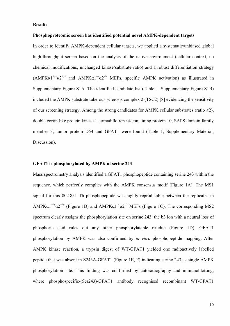
Results
Phosphoproteomic screen has identified potential novel AMPK-dependent targets
In order to identify AMPK-dependent cellular targets, we applied a systematic/unbiased global
high-throughput screen based on the analysis of the native environment (cellular context, no
chemical modifications, unchanged kinase/substrate ratio) and a robust differentiation strategy
(AMPK 1+/+ 2+/+ and AMPK 1-/- 2-/- MEFs, specific AMPK activation) as illustrated in
Supplementary Figure S1A. The identified candidate list (Table 1, Supplementary Figure S1B)
included the AMPK substrate tuberous sclerosis complex 2 (TSC2) [8] evidencing the sensitivity
of our screening strategy. Among the strong candidates for AMPK cellular substrates (ratio 2),
double cortin like protein kinase 1, armadillo repeat-containing protein 10, SAPS domain family
member 3, tumor protein D54 and GFAT1 were found (Table 1, Supplementary Material,
Discussion).
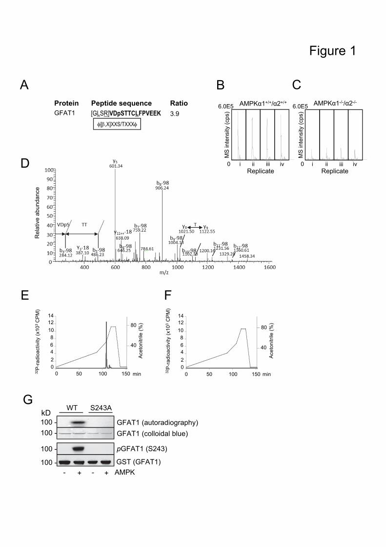
GFAT1 is phosphorylated by AMPK at serine 243
Mass spectrometry analysis identified a GFAT1 phosphopeptide containing serine 243 within the
sequence, which perfectly complies with the AMPK consensus motif (Figure 1A). The MS1
signal for this 802.851 Th phosphopeptide was highly reproducible between the replicates in
AMPK 1+/+ 2+/+ (Figure 1B) and AMPK 1-/- 2-/- MEFs (Figure 1C). The corresponding MS2
spectrum clearly assigns the phosphorylation site on serine 243: the b3 ion with a neutral loss of
phosphoric acid rules out any other phosphorylatable residue (Figure 1D). GFAT1
phosphorylation by AMPK was also confirmed by in vitro phosphopeptide mapping. After
AMPK kinase reaction, a trypsin digest of WT-GFAT1 yielded one radioactively labelled
peptide that was absent in S243A-GFAT1 (Figure 1E, F) indicating serine 243 as single AMPK
phosphorylation site. This finding was confirmed by autoradiography and immunoblotting,
where phosphospecific-(Ser243)-GFAT1 antibody recognised recombinant WT-GFAT1
17
phosphorylated with AMPK but not S243A-GFAT1 (Figure 1G). The stoichiometry of GFAT1
phosphorylation at serine 243 was estimated to be 33%.
GFAT1 was also proven to be a cellular AMPK target since increased GFAT1 phosphorylation
in response to AMPK agonists was seen only in AMPK 1+/+/ 2+/+ but not in AMPK 1-/-/ 2-/-
MEFs, which are completely lacking AMPK activity (Supplementary Figure S2A, B). Of note, a
residual phospho-GFAT1 signal was still observed in AMPK-deficient MEFs, indicating that an
alternative kinase may exist. AMPK-dependent GFAT1 phosphorylation was also observed in
HEK293 cells (Supplementary Figure S2C, D).
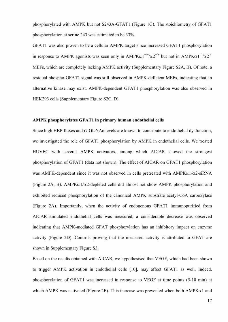
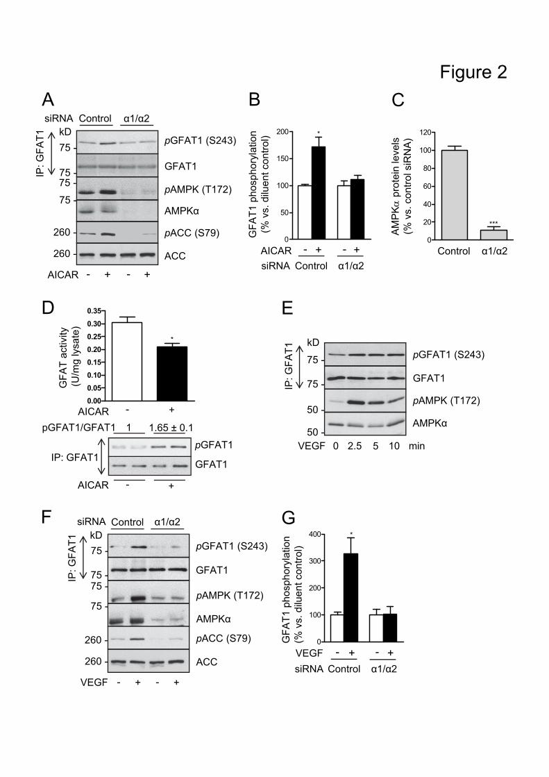
AMPK phosphorylates GFAT1 in primary human endothelial cells
Since high HBP fluxes and O-GlcNAc levels are known to contribute to endothelial dysfunction,
we investigated the role of GFAT1 phosphorylation by AMPK in endothelial cells. We treated
HUVEC with several AMPK activators, among which AICAR showed the strongest
phosphorylation of GFAT1 (data not shown). The effect of AICAR on GFAT1 phosphorylation
was AMPK-dependent since it was not observed in cells pretreated with AMPK 1/ 2-siRNA
(Figure 2A, B). AMPK 1/ 2-depleted cells did almost not show AMPK phosphorylation and
exhibited reduced phosphorylation of the canonical AMPK substrate acetyl-CoA carboxylase
(Figure 2A). Importantly, when the activity of endogenous GFAT1 immunopurified from
AICAR-stimulated endothelial cells was measured, a considerable decrease was observed
indicating that AMPK-mediated GFAT phosphorylation has an inhibitory impact on enzyme
activity (Figure 2D). Controls proving that the measured activity is attributed to GFAT are
shown in Supplementary Figure S3.
Based on the results obtained with AICAR, we hypothesised that VEGF, which had been shown
to trigger AMPK activation in endothelial cells [10], may affect GFAT1 as well. Indeed,
phosphorylation of GFAT1 was increased in response to VEGF at time points (5-10 min) at
which AMPK was activated (Figure 2E). This increase was prevented when both AMPK 1 and
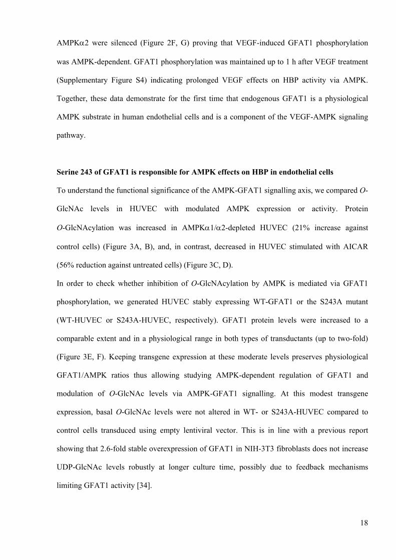
18
AMPK 2 were silenced (Figure 2F, G) proving that VEGF-induced GFAT1 phosphorylation
was AMPK-dependent. GFAT1 phosphorylation was maintained up to 1 h after VEGF treatment
(Supplementary Figure S4) indicating prolonged VEGF effects on HBP activity via AMPK.
Together, these data demonstrate for the first time that endogenous GFAT1 is a physiological
AMPK substrate in human endothelial cells and is a component of the VEGF-AMPK signaling
pathway.
Serine 243 of GFAT1 is responsible for AMPK effects on HBP in endothelial cells
To understand the functional significance of the AMPK-GFAT1 signalling axis, we compared O-
GlcNAc levels in HUVEC with modulated AMPK expression or activity. Protein
O-GlcNAcylation was increased in AMPK 1/ 2-depleted HUVEC (21% increase against
control cells) (Figure 3A, B), and, in contrast, decreased in HUVEC stimulated with AICAR
(56% reduction against untreated cells) (Figure 3C, D).
In order to check whether inhibition of O-GlcNAcylation by AMPK is mediated via GFAT1
phosphorylation, we generated HUVEC stably expressing WT-GFAT1 or the S243A mutant
(WT-HUVEC or S243A-HUVEC, respectively). GFAT1 protein levels were increased to a
comparable extent and in a physiological range in both types of transductants (up to two-fold)
(Figure 3E, F). Keeping transgene expression at these moderate levels preserves physiological
GFAT1/AMPK ratios thus allowing studying AMPK-dependent regulation of GFAT1 and
modulation of O-GlcNAc levels via AMPK-GFAT1 signalling. At this modest transgene
expression, basal O-GlcNAc levels were not altered in WT- or S243A-HUVEC compared to
control cells transduced using empty lentiviral vector. This is in line with a previous report
showing that 2.6-fold stable overexpression of GFAT1 in NIH-3T3 fibroblasts does not increase
UDP-GlcNAc levels robustly at longer culture time, possibly due to feedback mechanisms
limiting GFAT1 activity [34].
19
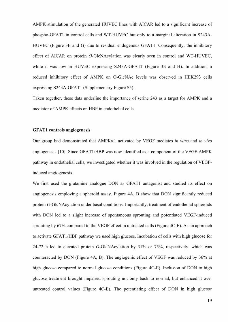
AMPK stimulation of the generated HUVEC lines with AICAR led to a significant increase of
phospho-GFAT1 in control cells and WT-HUVEC but only to a marginal alteration in S243A-
HUVEC (Figure 3E and G) due to residual endogenous GFAT1. Consequently, the inhibitory
effect of AICAR on protein O-GlcNAcylation was clearly seen in control and WT-HUVEC,
while it was low in HUVEC expressing S243A-GFAT1 (Figure 3E and H). In addition, a
reduced inhibitory effect of AMPK on O-GlcNAc levels was observed in HEK293 cells
expressing S243A-GFAT1 (Supplementary Figure S5).
Taken together, these data underline the importance of serine 243 as a target for AMPK and a
mediator of AMPK effects on HBP in endothelial cells.
GFAT1 controls angiogenesis
Our group had demonstrated that AMPK 1 activated by VEGF mediates in vitro and in vivo
angiogenesis [10]. Since GFAT1/HBP was now identified as a component of the VEGF-AMPK
pathway in endothelial cells, we investigated whether it was involved in the regulation of VEGF-
induced angiogenesis.
We first used the glutamine analogue DON as GFAT1 antagonist and studied its effect on
angiogenesis employing a spheroid assay. Figure 4A, B show that DON significantly reduced
protein O-GlcNAcylation under basal conditions. Importantly, treatment of endothelial spheroids
with DON led to a slight increase of spontaneous sprouting and potentiated VEGF-induced
sprouting by 67% compared to the VEGF effect in untreated cells (Figure 4C-E). As an approach
to activate GFAT1/HBP pathway we used high glucose. Incubation of cells with high glucose for
24-72 h led to elevated protein O-GlcNAcylation by 31% or 75%, respectively, which was
counteracted by DON (Figure 4A, B). The angiogenic effect of VEGF was reduced by 36% at
high glucose compared to normal glucose conditions (Figure 4C-E). Inclusion of DON to high
glucose treatment brought impaired sprouting not only back to normal, but enhanced it over
untreated control values (Figure 4C-E). The potentiating effect of DON in high glucose
20
condition was lower than at normal glucose, possibly due to involvement of pathways apart from
HBP into antiangiogenic effects of high glucose (Figure 4C-E).
Since DON also inhibits other glutamine-utilizing enzymes, we secondly applied a genetic
approach to modulate GFAT1. We treated HUVEC with GFAT1-specific siRNA, which led to a
significant downregulation of GFAT1 expression (Figure 5A, B). As a consequence, O-
GlcNAcylation of proteins decreased (32% and 48% decrease compared to controls at 72 h and
120 h post transfection, respectively) (Figure 5A and C). When GFAT1-depleted cells were
utilised in spheroid assays, a trend towards spontaneous sprouting of the capillary-like structures
was observed (Figure 5D-F) similarly to what had been seen with DON. Furthermore, GFAT1
downregulation significantly increased VEGF-induced sprouting by 72% compared to the VEGF
effect in cells treated with control siRNA (Figure 5D-F).
Together, these data demonstrate that VEGF-induced angiogenesis is inhibited by GFAT1/HBP.
GFAT1 phosphorylation at serine 243 mediates VEGF-induced pro-angiogenic effects of
AMPK
The above described data indicate that VEGF via activation of AMPK and subsequent
phosphorylation of GFAT may impair the HBP and thus relieve its inhibitory action on
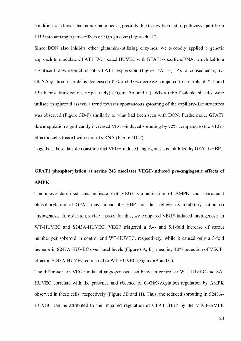
angiogenesis. In order to provide a proof for this, we compared VEGF-induced angiogenesis in
WT-HUVEC and S243A-HUVEC. VEGF triggered a 5.4- and 5.1-fold increase of sprout
number per spheroid in control and WT-HUVEC, respectively, while it caused only a 3-fold
increase in S243A-HUVEC over basal levels (Figure 6A, B), meaning 40% reduction of VEGF-
effect in S243A-HUVEC compared to WT-HUVEC (Figure 6A and C).
The differences in VEGF-induced angiogenesis seen between control or WT-HUVEC and SA-
HUVEC correlate with the presence and absence of O-GlcNAcylation regulation by AMPK
observed in these cells, respectively (Figure 3E and H). Thus, the reduced sprouting in S243A-
HUVEC can be attributed to the impaired regulation of GFAT1/HBP by the VEGF-AMPK

21
pathway. These data provide unequivocal evidence that GFAT1 phosphorylation at serine 243
represents one of the mechanisms underlying pro-angiogenic effects of AMPK in response to
VEGF.
22
Discussion
An increase of the glucose flux through HBP and chronically elevated O-GlcNAcylation of
target proteins is increasingly recognised as an important contributor to the pathogenesis of type
2 diabetes and its cardiovascular complications [35]. However, the regulation of the HBP and the
mechanisms and functions of protein O-GlcNAcylation are poorly characterised. The present
study reveals that AMPK, a key regulator of cellular metabolism and homeostasis, controls HBP
and the abundance of O-GlcNAcylation in endothelial cells via targeting GFAT1, the rate-
limiting enzyme of the HBP, and that this process is a part of the proangiogenic VEGF-AMPK
axis.
GFAT1 was found as an AMPK target in our systematic phosphoproteomic approach aimed at
identifying novel AMPK substrates. In line with this finding two previous studies have already
suggested GFAT1 as an AMPK substrate employing either purified recombinant GFAT1 in vitro
[13] or recombinant GFAT1 expressed in CHO cells [12]. However, the role of AMPK-mediated
GFAT1 phosphorylation in regulating GFAT activity was not clear and the biological
significance of this process in a physiologically relevant system had not been investigated. Our
study confirms serine 243 of GFAT1 as phosphorylation site by tandem MS and as AMPK site
in a range of in vitro experiments using a recombinant GFAT1 preparation. As a novel outcome
of these experiments, we show that serine 243 is a single AMPK site, which is phosphorylated to
a stoichiometry of 0.33 mol/mol. In addition, we validated endogenous GFAT1 as a cellular
AMPK target in WT and AMPK-null MEFs as an unequivocal model for testing AMPK-
dependency, thus establishing GFAT1 as a direct physiological AMPK substrate. Since we
detected basal serine 243 phosphorylation of GFAT1 in AMPK-null MEFs, a second kinase such
as Ca2+/calmodulin-dependent kinase II (CaMKII), which has recently been shown to
phosphorylate GFAT at serine 243 in vitro [13], may share this phosphorylation site with AMPK.
However, phosphorylation signals in AMPK-null MEFS were lower compared to WT MEFs
suggesting that AMPK plays a major role.
23
Enhanced glucose fluxes through the HBP as well as increased protein O-GlcNAcylation are
known to contribute to endothelial dysfunction underlying the development of diabetic
vasculopathies. GFAT activity has been described in primary endothelial cells of different origin
and has been shown to be upregulated by hyperglycaemia [36]. In addition, while expression of
GFAT was barely detected in endothelial cells of healthy human tissues, it was increased in
activated cells suggesting that it may be modulated under pathophysiological conditions [37].
Given these indications, we addressed the role of the AMPK-GFAT1 axis in endothelial cells.
Our study demonstrates for the first time that GFAT1 is a physiological AMPK substrate in
primary human endothelial cells, as VEGF, a major physiological AMPK agonist in endothelial
cells, was able to increase AMPK-dependent GFAT1 phosphorylation. Using AMPK activators
and AMPK-specific siRNA, we revealed an inhibitory effect of AMPK on O-GlcNAc levels,
which is most likely due to inhibition of GFAT1 activity by AMPK-mediated phosphorylation,
since activity of GFAT was decreased in cells treated with the AMPK activator. In line with this,
a recent study showed that metformin and AICAR cause an AMPK-dependent reduction of
UDP-GlcNAc in NIH3T3 cells [38].
To further verify the functional importance of AMPK-dependent GFAT1 phosphorylation, we
performed experiments with cells expressing S243A-GFAT1. In these cells, AMPK activation
led to a lower reduction of cellular O-GlcNAc levels as compared to control cells, thus
confirming the inhibitory role of serine 243 phosphorylation for GFAT1 activity. The fact that
the inhibitory effects of AMPK were only partially prevented by the S243A mutant could be due
to residual endogenous GFAT1 and/or GFAT-independent effects of AMPK on metabolic
branches which supply O-GlcNAc production, e.g. glycolysis or fatty acid oxidation. Our data
are in line with the study of Eguchi et al., who showed that GFAT1 activity was decreased after
activating cellular AMPK by treatment with 2-deoxyglucose [12] indicating a possible inhibitory
role of serine 243 phosphorylation for GFAT1 activity. In contrast, Li et al. observed an
activation of recombinant GFAT1 after serine 243 phosphorylation by AMPK in vitro [13]. This
24
discrepancy may be due to the lack of endogenous regulatory factors, e.g. allosteric regulators of
GFAT1 or different posttranslational modifications, when recombinant proteins are employed.
Importantly, our study extends the study by Eguchi et al. [12] by providing cellular O-
GlcNAcylation data and showing that the AMPK-GFAT1 regulatory axis is coupled to O-
GlcNAc signalling. The observed degree of reduction in protein O-GlcNAcylation after AMPK
activation seems to be moderate, which is in line with 33% stoichiometry of GFAT1
phosphorylation. However, even a modest alteration of O-GlcNAcylation can have functional
consequences as shown for the microtubule-associated protein tau. Changes in tau O-
GlcNAcylation in the range of 20-30% led to significant alteration of its phosphorylation state
and may be involved in tau pathology in the context of Alzheimer disease [39-41]. Given that the
O-GlcNAc machinery is tightly controlled by negative regulatory feedback loops at the level of
GFAT1 [31, 42] and O-GlcNAc transferase (OGT) [43], our data support the view that AMPK
has an important function in controlling O-GlcNAc levels. In line with this, AMPK depletion led
also to de novo O-GlcNAcylation of proteins. Interestingly, AMPK has also been shown to
phosphorylate OGT, the enzyme responsible for O-GlcNAcylation, thereby determining its
substrate selectivity [44]. Thus, AMPK is regulating the O-GlcNAcylation machinery at different
levels.
The major question of the present study was if GFAT1 regulation by AMPK plays a biological
role in endothelial cells. Previous data obtained in our group revealed that AMPK 1 is required
for VEGF-induced in vitro and in vivo angiogenesis [10], but the underlying mechanisms were
completely unknown. The present data suggest that GFAT1 phosphorylation by AMPK
represents a previously unknown pro-angiogenic pathway. Pharmacological inhibition or siRNA-
mediated downregulation of GFAT1 led to increased VEGF-induced sprouting of endothelial
spheroids indicating that inhibition of GFAT1 by AMPK-mediated phosphorylation promotes
angiogenesis. Indeed, when this phosphorylation was prevented by introducing S243A-GFAT1
into endothelial cells, VEGF-induced angiogenesis was decreased. Our data indicate that O-
25
GlcNAcylation patterns essentially modulate the angiogenic response of endothelial cells to
VEGF with high levels of O-GlcNAcylated proteins leading to inhibition of angiogenesis. In line
with this, several studies have correlated O-GlcNAcylation with possible anti-angiogenic effects.
For example, O-GlcNAcylation of the proangiogenic enzyme eNOS induced decreased enzyme
activity [45, 46] and O-GlcNAcylation of Akt was suggested to negatively affect migration and
tube formation of endothelial cells [47]. Furthermore, O-GlcNAcylation of Sp1 leads to elevated
expression of TGF 1 (an inducer of extracellular matrix protein synthesis) and PAI-1 (an
inhibitor of extracellular matrix degradation) [48], while O-GlcNAcylation of Sp3 promotes
angiopoietin-2 expression, which in turn triggers increased expression of ICAM-1 and VCAM-1
[49].
Our data demonstrate that high glucose induces impairment of VEGF-stimulated in vitro
angiogenesis and that this effect was counteracted by pharmacological inhibition of GFAT1 with
DON. These data are in line with a study by Luo et al. who showed that high fat diet or
streptozotocin injections in vivo or glucosamine treatment in vitro reduced sprouting from aortic
rings, which was associated with increased O-GlcNAc tissue levels [47]. In this study, O-
GlcNAcase overexpression prevented the adverse effects of hyperglycaemia on angiogenesis.
Together, our data and the data of Luo et al. demonstrate that stimulation of the HBP and
elevated O-GlcNAcylation of proteins are implicated in high glucose-induced inhibition of
angiogenesis. This in turn may contribute to cardiovascular complications in diabetes such as
impaired wound healing, reduced myocardial perfusion or even organ dysfunction as observed in
the islets of Langerhans (reviewed in [50, 51]).
The interpretation of the current study is limited since the results were obtained in vitro using
HUVEC as a model. Although HUVEC have been widely used to characterise endothelial
functions they may differ from adult cells of different vascular beds and may be influenced by
maternal and foetal factors. However, the HUVEC spheroid model has recently been
characterised as a sensitive tool to study angiogenesis and provides reliable results if it is
26
performed under standardised conditions [52]. In addition to HUVEC we have shown AMPK-
mediated GFAT1 phosphorylation in other cell lines (HEK293, MEFs) suggesting that this
pathway is of general importance. Future experiments need to involve animal models of diabetes
as well as ex vivo methodologies for evaluating endothelial cells from patients [53]. Moreover, to
reveal the causal involvement of reduced O-GlcNAcylation, the respective protein targets need
to be identified and the effect of mutating the sites of modification on protein function needs to
be investigated.
In summary, we conclude that modulation of angiogenesis via interference with the HBP may
help to prevent or ameliorate the clinical sequelae of hyperglycaemia. In this context, targeting
AMPK, which was shown to control HBP via GFAT in our study, may represent a promising
vasculoprotective strategy.
27
Author Contribution
D.Z., F.V., K.S., R.H. conceived the study. F.V. setup, performed and analysed the
phosphoproteomic screen (supervised by N.A.M, K.S.) and carried out in vitro validation
(supervised by K.S.). D.Z. performed and analysed cellular validation (supervised by K.S.) and
experiments using endothelial cells (supervised by R.H). O.G. performed initial cellular
validation of recombinant GFAT1. M.P. performed cloning and generated GFAT1 expression
constructs. K.M. provided valuable advices regarding GFAT1 and sugar nucleotide biology. A.K.
and K.Sp. contributed to generation/characterisation of stable HUVEC lines. C.W. provided
GFAT1 anti-serum and helpful advices. B.V. provided AMPK WT and AMPK-null MEF. D.Z.,
F.V., K.S., R.H. wrote the manuscript. All authors discussed the results and commented on the
manuscript. R.H. is the guarantor of this work and, as such, had full access to all the data in the
study and takes responsibility for the integrity of the data and the accuracy of the data analysis.
Funding
This work was supported by Medical Research Council and the pharmaceutical companies
supporting the Division of Signal Transduction Therapy Unit (AstraZeneca, Boehringer-
Ingelheim, GlaxoSmithKline, Merck KGaA, Janssen Pharmaceutica and Pfizer). F.V. was
funded by grant from RASOR (Radical Solutions for Researching the proteome, Scotland), an
Interdisciplinary Research Collaboration Initiative between the Biotechnology and Biological
Sciences Research Council, the Engineering and Physical Sciences Research Council and the
Scottish Funding Council (UK). R.H. was funded by the DFG (Deutsche
Forschungsgemeinschaft), RTG 1715, subproject 2, and RTG 2155, subproject 13.
Competing Interests
The Authors declare that there are no competing interests associated with the manuscript.

28
Acknowledgements
We thank Elke Teuscher (Institute for Molecular Cell Biology, Center for Molecular
Biomedicine, University Hospital Jena) for excellent assistance with the isolation and culture of
HUVEC. We are grateful to Dr. D. Grahame Hardie (University of Dundee, UK) for providing
recombinant AMPK trimeric complex and AMPK 1 antibody, and to Dr. Jörg Müller (Jena
University Hospital, Germany) for supplying packaging plasmids and lentivectors.
29
References
1 Steinberg, G. R. and Kemp, B. E. (2009) AMPK in Health and Disease. Physiol Rev. 89, 1025-1078
2 Scott, J. W., Ling, N., Issa, S. M., Dite, T. A., O'Brien, M. T., Chen, Z. P., Galic, S., Langendorf, C. G., Steinberg, G. R., Kemp, B. E. and Oakhill, J. S. (2014) Small molecule drug A-769662 and AMP synergistically activate naive AMPK independent of upstream kinase signaling. Chem Biol. 21, 619-627
3 Xiao, B., Sanders, M. J., Underwood, E., Heath, R., Mayer, F. V., Carmena, D., Jing, C., Walker, P. A., Eccleston, J. F., Haire, L. F., Saiu, P., Howell, S. A., Aasland, R., Martin, S. R., Carling, D. and Gamblin, S. J. (2011) Structure of mammalian AMPK and its regulation by ADP. Nature. 472, 230-233
4 Gowans, G. J., Hawley, S. A., Ross, F. A. and Hardie, D. G. (2013) AMP is a true physiological regulator of AMP-activated protein kinase by both allosteric activation and enhancing net phosphorylation. Cell Metab. 18, 556-566
5 Merrill, G. F., Kurth, E. J., Hardie, D. G. and Winder, W. W. (1997) AICA riboside increases AMP-activated protein kinase, fatty acid oxidation, and glucose uptake in rat muscle. Am J Physiol. 273, E1107-1112
6 Munday, M. R., Campbell, D. G., Carling, D. and Hardie, D. G. (1988) Identification by amino acid sequencing of three major regulatory phosphorylation sites on rat acetyl-CoA carboxylase. Eur J Biochem. 175, 331-338
7 Gwinn, D. M., Shackelford, D. B., Egan, D. F., Mihaylova, M. M., Mery, A., Vasquez, D. S., Turk, B. E. and Shaw, R. J. (2008) AMPK phosphorylation of raptor mediates a metabolic checkpoint. Mol Cell. 30, 214-226
8 Inoki, K., Zhu, T. and Guan, K. L. (2003) TSC2 mediates cellular energy response to control cell growth and survival. Cell. 115, 577-590
9 Fisslthaler, B. and Fleming, I. (2009) Activation and signaling by the AMP-activated protein kinase in endothelial cells. Circ Res. 105, 114-127
10 Stahmann, N., Woods, A., Spengler, K., Heslegrave, A., Bauer, R., Krause, S., Viollet, B., Carling, D. and Heller, R. (2010) Activation of AMP-activated protein kinase by vascular endothelial growth factor mediates endothelial angiogenesis independently of nitric-oxide synthase. J Biol Chem. 285, 10638-10652
11 Oki, T., Yamazaki, K., Kuromitsu, J., Okada, M. and Tanaka, I. (1999) cDNA cloning and mapping of a novel subtype of glutamine:fructose-6-phosphate amidotransferase (GFAT2) in human and mouse. Genomics. 57, 227-234
12 Eguchi, S., Oshiro, N., Miyamoto, T., Yoshino, K., Okamoto, S., Ono, T., Kikkawa, U. and Yonezawa, K. (2009) AMP-activated protein kinase phosphorylates glutamine : fructose-6-phosphate amidotransferase 1 at Ser243 to modulate its enzymatic activity. Genes Cells. 14, 179-189
30
13 Li, Y., Roux, C., Lazereg, S., LeCaer, J. P., Laprevote, O., Badet, B. and Badet-Denisot, M. A. (2007) Identification of a novel serine phosphorylation site in human glutamine:fructose-6-phosphate amidotransferase isoform 1. Biochemistry. 46, 13163-13169
14 Banko, M. R., Allen, J. J., Schaffer, B. E., Wilker, E. W., Tsou, P., White, J. L., Villen, J., Wang, B., Kim, S. R., Sakamoto, K., Gygi, S. P., Cantley, L. C., Yaffe, M. B., Shokat, K. M. and Brunet, A. (2011) Chemical genetic screen for AMPKalpha2 substrates uncovers a network of proteins involved in mitosis. Mol Cell. 44, 878-892
15 Hardiville, S. and Hart, G. W. (2014) Nutrient regulation of signaling, transcription, and cell physiology by O-GlcNAcylation. Cell Metab. 20, 208-213
16 Cooksey, R. C., Hebert, L. F., Jr., Zhu, J. H., Wofford, P., Garvey, W. T. and McClain, D. A. (1999) Mechanism of hexosamine-induced insulin resistance in transgenic mice overexpressing glutamine:fructose-6-phosphate amidotransferase: decreased glucose transporter GLUT4 translocation and reversal by treatment with thiazolidinedione. Endocrinology. 140, 1151-1157
17 Hazel, M., Cooksey, R. C., Jones, D., Parker, G., Neidigh, J. L., Witherbee, B., Gulve, E. A. and McClain, D. A. (2004) Activation of the hexosamine signaling pathway in adipose tissue results in decreased serum adiponectin and skeletal muscle insulin resistance. Endocrinology. 145, 2118-2128
18 Hebert, L. F., Jr., Daniels, M. C., Zhou, J., Crook, E. D., Turner, R. L., Simmons, S. T., Neidigh, J. L., Zhu, J. S., Baron, A. D. and McClain, D. A. (1996) Overexpression of glutamine:fructose-6-phosphate amidotransferase in transgenic mice leads to insulin resistance. J Clin Invest. 98, 930-936
19 Srinivasan, V., Sandhya, N., Sampathkumar, R., Farooq, S., Mohan, V. and Balasubramanyam, M. (2007) Glutamine fructose-6-phosphate amidotransferase (GFAT) gene expression and activity in patients with type 2 diabetes: inter-relationships with hyperglycaemia and oxidative stress. Clin Biochem. 40, 952-957
20 Ma, J. and Hart, G. W. (2013) Protein O-GlcNAcylation in diabetes and diabetic complications. Expert Rev Proteomics. 10, 365-380
21 Laczy, B., Hill, B. G., Wang, K., Paterson, A. J., White, C. R., Xing, D., Chen, Y. F., Darley-Usmar, V., Oparil, S. and Chatham, J. C. (2009) Protein O-GlcNAcylation: a new signaling paradigm for the cardiovascular system. Am J Physiol Heart Circ Physiol. 296, H13-28
22 Goransson, O., McBride, A., Hawley, S. A., Ross, F. A., Shpiro, N., Foretz, M., Viollet, B., Hardie, D. G. and Sakamoto, K. (2007) Mechanism of action of A-769662, a valuable tool for activation of AMP-activated protein kinase. J Biol Chem. 282, 32549-32560
23 Weigert, C., Klopfer, K., Kausch, C., Brodbeck, K., Stumvoll, M., Haring, H. U. and Schleicher, E. D. (2003) Palmitate-induced activation of the hexosamine pathway in human myotubes: increased expression of glutamine:fructose-6-phosphate aminotransferase. Diabetes. 52, 650-656
24 Jokela, T. A., Makkonen, K. M., Oikari, S., Karna, R., Koli, E., Hart, G. W., Tammi, R. H., Carlberg, C. and Tammi, M. I. (2011) Cellular content of UDP-N-acetylhexosamines controls hyaluronan synthase 2 expression and correlates with O-linked N-acetylglucosamine modification of transcription factors YY1 and SP1. J Biol Chem. 286, 33632-33640
31
25 Laderoute, K. R., Amin, K., Calaoagan, J. M., Knapp, M., Le, T., Orduna, J., Foretz, M. and Viollet, B. (2006) 5'-AMP-activated protein kinase (AMPK) is induced by low-oxygen and glucose deprivation conditions found in solid-tumor microenvironments. Mol Cell Biol. 26, 5336-5347
26 Towler, M. C. and Hardie, D. G. (2007) AMP-activated protein kinase in metabolic control and insulin signaling. Circ Res. 100, 328-341
27 Heller, R., Unbehaun, A., Schellenberg, B., Mayer, B., Werner-Felmayer, G. and Werner, E. R. (2001) L-ascorbic acid potentiates endothelial nitric oxide synthesis via a chemical stabilization of tetrahydrobiopterin. J Biol Chem. 276, 40-47
28 Reihill, J. A., Ewart, M. A., Hardie, D. G. and Salt, I. P. (2007) AMP-activated protein kinase mediates VEGF-stimulated endothelial NO production. Biochem Biophys Res Commun. 354, 1084-1088
29 Kommaddi, R. P., Dickson, K. M. and Barker, P. A. (2011) Stress-induced expression of the p75 neurotrophin receptor is regulated by O-GlcNAcylation of the Sp1 transcription factor. J Neurochem. 116, 396-405
30 Ostrowski, A. and van Aalten, D. M. (2013) Chemical tools to probe cellular O-GlcNAc signalling. Biochem J. 456, 1-12
31 Broschat, K. O., Gorka, C., Page, J. D., Martin-Berger, C. L., Davies, M. S., Huang Hc, H. C., Gulve, E. A., Salsgiver, W. J. and Kasten, T. P. (2002) Kinetic characterization of human glutamine-fructose-6-phosphate amidotransferase I: potent feedback inhibition by glucosamine 6-phosphate. J Biol Chem. 277, 14764-14770
32 Badet, B., Vermoote, P., Haumont, P. Y., Lederer, F. and LeGoffic, F. (1987) Glucosamine synthetase from Escherichia coli: purification, properties, and glutamine-utilizing site location. Biochemistry. 26, 1940-1948
33 Stahmann, N., Woods, A., Carling, D. and Heller, R. (2006) Thrombin activates AMP-activated protein kinase in endothelial cells via a pathway involving Ca2+/calmodulin-dependent protein kinase kinase beta. Mol Cell Biol. 26, 5933-5945
34 Weigert, C., Brodbeck, K., Lehmann, R., Haring, H. U. and Schleicher, E. D. (2001) Overexpression of glutamine:fructose-6-phosphate-amidotransferase induces transforming growth factor-beta1 synthesis in NIH-3T3 fibroblasts. FEBS Lett. 488, 95-99
35 Buse, M. G. (2006) Hexosamines, insulin resistance, and the complications of diabetes: current status. Am J Physiol Endocrinol Metab. 290, E1-E8
36 Wu, G., Haynes, T. E., Yan, W. and Meininger, C. J. (2001) Presence of glutamine:fructose-6-phosphate amidotransferase for glucosamine-6-phosphate synthesis in endothelial cells: effects of hyperglycaemia and glutamine. Diabetologia. 44, 196-202
37 Nerlich, A. G., Sauer, U., Kolm-Litty, V., Wagner, E., Koch, M. and Schleicher, E. D. (1998) Expression of glutamine:fructose-6-phosphate amidotransferase in human tissues: evidence for high variability and distinct regulation in diabetes. Diabetes. 47, 170-178
38 Ishibashi, Y. and Hirabayashi, Y. (2015) AMP-activated Protein Kinase Suppresses Biosynthesis of Glucosylceramide by Reducing Intracellular Sugar Nucleotides. J Biol Chem. 290, 18245-18260
32
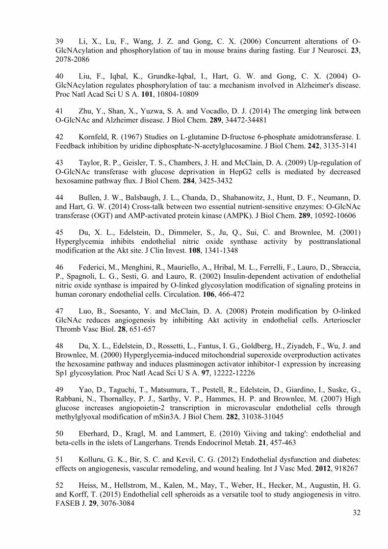
39 Li, X., Lu, F., Wang, J. Z. and Gong, C. X. (2006) Concurrent alterations of O-GlcNAcylation and phosphorylation of tau in mouse brains during fasting. Eur J Neurosci. 23, 2078-2086
40 Liu, F., Iqbal, K., Grundke-Iqbal, I., Hart, G. W. and Gong, C. X. (2004) O-GlcNAcylation regulates phosphorylation of tau: a mechanism involved in Alzheimer's disease. Proc Natl Acad Sci U S A. 101, 10804-10809
41 Zhu, Y., Shan, X., Yuzwa, S. A. and Vocadlo, D. J. (2014) The emerging link between O-GlcNAc and Alzheimer disease. J Biol Chem. 289, 34472-34481
42 Kornfeld, R. (1967) Studies on L-glutamine D-fructose 6-phosphate amidotransferase. I. Feedback inhibition by uridine diphosphate-N-acetylglucosamine. J Biol Chem. 242, 3135-3141
43 Taylor, R. P., Geisler, T. S., Chambers, J. H. and McClain, D. A. (2009) Up-regulation of O-GlcNAc transferase with glucose deprivation in HepG2 cells is mediated by decreased hexosamine pathway flux. J Biol Chem. 284, 3425-3432
44 Bullen, J. W., Balsbaugh, J. L., Chanda, D., Shabanowitz, J., Hunt, D. F., Neumann, D. and Hart, G. W. (2014) Cross-talk between two essential nutrient-sensitive enzymes: O-GlcNAc transferase (OGT) and AMP-activated protein kinase (AMPK). J Biol Chem. 289, 10592-10606
45 Du, X. L., Edelstein, D., Dimmeler, S., Ju, Q., Sui, C. and Brownlee, M. (2001) Hyperglycemia inhibits endothelial nitric oxide synthase activity by posttranslational modification at the Akt site. J Clin Invest. 108, 1341-1348
46 Federici, M., Menghini, R., Mauriello, A., Hribal, M. L., Ferrelli, F., Lauro, D., Sbraccia, P., Spagnoli, L. G., Sesti, G. and Lauro, R. (2002) Insulin-dependent activation of endothelial nitric oxide synthase is impaired by O-linked glycosylation modification of signaling proteins in human coronary endothelial cells. Circulation. 106, 466-472
47 Luo, B., Soesanto, Y. and McClain, D. A. (2008) Protein modification by O-linked GlcNAc reduces angiogenesis by inhibiting Akt activity in endothelial cells. Arterioscler Thromb Vasc Biol. 28, 651-657
48 Du, X. L., Edelstein, D., Rossetti, L., Fantus, I. G., Goldberg, H., Ziyadeh, F., Wu, J. and Brownlee, M. (2000) Hyperglycemia-induced mitochondrial superoxide overproduction activates the hexosamine pathway and induces plasminogen activator inhibitor-1 expression by increasing Sp1 glycosylation. Proc Natl Acad Sci U S A. 97, 12222-12226
49 Yao, D., Taguchi, T., Matsumura, T., Pestell, R., Edelstein, D., Giardino, I., Suske, G., Rabbani, N., Thornalley, P. J., Sarthy, V. P., Hammes, H. P. and Brownlee, M. (2007) High glucose increases angiopoietin-2 transcription in microvascular endothelial cells through methylglyoxal modification of mSin3A. J Biol Chem. 282, 31038-31045
50 Eberhard, D., Kragl, M. and Lammert, E. (2010) 'Giving and taking': endothelial and beta-cells in the islets of Langerhans. Trends Endocrinol Metab. 21, 457-463
51 Kolluru, G. K., Bir, S. C. and Kevil, C. G. (2012) Endothelial dysfunction and diabetes: effects on angiogenesis, vascular remodeling, and wound healing. Int J Vasc Med. 2012, 918267
52 Heiss, M., Hellstrom, M., Kalen, M., May, T., Weber, H., Hecker, M., Augustin, H. G. and Korff, T. (2015) Endothelial cell spheroids as a versatile tool to study angiogenesis in vitro. FASEB J. 29, 3076-3084

33
53 Onat, D., Brillon, D., Colombo, P. C. and Schmidt, A. M. (2011) Human vascular endothelial cells: a model system for studying vascular inflammation in diabetes and atherosclerosis. Curr Diab Rep. 11, 193-202
34
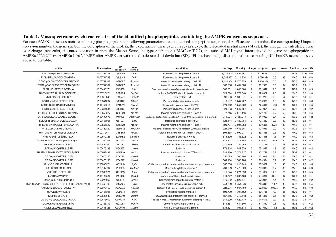
Table 1. Mass spectrometry characteristics of the identified phosphopeptides containing the AMPK consensus sequence. For each AMPK consensus motif-containing phosphopeptide, the following parameters are summarised: the peptide sequence, the IPI accession number, the corresponding Uniprot accession number, the gene symbol, the description of the protein, the experimental mass over charge (m/z exp), the calculated neutral mass (M calc), the charge, the calculated mass over charge (m/z calc), the mass deviation in ppm, the Mascot Score, the type of fraction (IMAC or TiO2), the ratio of MS1 signal intensities of the same phosphopeptide in AMPK 1+/+/ 2+/+ vs. AMPK 1-/-/ 2-/- MEF after AMPK activation and ratio standard deviation (SD). IPI database being discontinued, corresponding UniProtKB accession were added to the table.
peptide IPI accession SP accession
gene symbol description m/z (exp) M (calc) charge m/z (calc) ppm score fraction ratio SD
R.DLYRPLpSSDDLDSVGDSV IPI00761729 Q9JLM8 Dclk1 Double cortin like protein kinase 1 1,016.945 2,031.867 2 1,016.941 3.5 70 TiO2 12.6 0.8 R.DLYRPLpSpSDDLDSVGDSV IPI00761729 Q9JLM8 Dclk1 Double cortin like protein kinase 1 1,056.927 2,111.834 2 1,056.924 2.5 63 IMAC 4.5 0.8
LRPSR.pSAEDLTDGSYDDILNAEQLK IPI00757909 Q9D0L7 Armc10 Armadillo repeat-containing protein 10 1,139.000 2,275.973 2 1,138.994 5.5 119 TiO2 4.0 0.3 LRPSR.pSAEDLTDGSYDDILNAEQLKK IPI00757909 Q9D0L7 Armc10 Armadillo repeat-containing protein 10 802.366 2,404.068 3 802.363 3.1 64 TiO2 3.9 2.1
GLSR.VDpSTTCLFPVEEK.A IPI00406371 P47856 Gfpt1 Glucosamine-fructose-6-phosphate aminotransferase 1 802.851 1,603.684 2 802.849 2.3 67 TiO2 3.9 0.3 R.NTVDLVTTcHIHSpSpSDDEIDFK IPI00719971 G5E8R4 Ppp6r3 Isoform 3 of SAPS domain family member 3 925.024 2,772.043 3 925.022 2.3 37 IMAC 3.2 0.4
VMR.NSApTFKSFEDR IPI00319046 Q9CYZ2 Tpd52l2 Tumor protein D54 691.293 1,380.571 2 691.293 0.9 54 TiO2 3.0 0.1 IRSTR.pSVENLPECGITHEQR IPI00341034 Q8BKC8 Pik4cb Phosphatidylinositol 4-kinase beta 616.937 1,847.787 3 616.936 0.3 37 TiO2 2.6 0.6 MRRR.RpSSDPLGDTASNLGS IPI00453615 Q7TMY8 Huwe1 E3 ubiquitin-protein ligase HUWE1 778.832 1,555.652 2 778.833 -2.0 26 TiO2 2.4 0.5 IRSTR.pSVENLPECGITHEQR IPI00341034 Q8BKC8 Pik4cb Phosphatidylinositol 4-kinase beta 924.903 1,847.787 2 924.901 2.3 63 TiO2 2.4 0.2
FR.IEDpSEPHIPLIDDTDAEDDAPTK IPI00556827 G5E829 Atp2b1 Plasma membrane calcium ATPase 1 872.715 2,615.116 3 872.713 3.2 53 TIO2 2.3 0.4 LVR.KDpSEEEVSLLGNQDIEEGNSR IPI00135975 P15920 Atp6v0a2 Vacuolar proton translocating ATPase 116 kDa subunit a isoform 2 810.024 2,427.044 3 810.022 2.4 58 TiO2 2.3 0.2 LSK.SSpSSPELQTLQDILGDLGDK IPI00468568 Q61037 Tsc2 Tuberous sclerosis 2 isoform 2 728.344 2,182.004 3 728.342 2.7 33 TiO2 2.2 0.1
FR.IEDpSEPHIPLIDDpTDAEDDAPTK IPI00556827 G5E829 Atp2b1 Plasma membrane calcium ATPase 1 899.704 2,695.083 3 899.368 373.5 39 IMAC 2.1 0.1 VR.EEApSDDDMEGDEAVVR IPI00420329 Q6P4T2 Snrnp200 U5 small nuclear ribonucleoprotein 200 kDa helicase 923.840 1,845.661 2 923.838 2.2 70 TiO2 2.1 0.1
R.NTVDLVTTcHIHSpSpSDDEIDFK IPI00719971 G5E8R4 Ppp6r3 Isoform 3 of SAPS domain family member 3 898.368 2,692.077 3 898.366 2.5 59 IMAC 2.0 0.3 RPR.FpSHSYLpSDSDTEAK IPI00649326 B2RRE2 Myo18a Isoform 3 of Myosin-XVIIIa 873.820 1,745.622 2 873.818 1.5 34 IMAC 2.0 0.3
K.LNFAVASRKTFSHELpSDFGLEpST IPI00230108 P27773 Pdia3 Protein disulfide-isomerase A3 679.797 2,715.198 4 679.807 -14.5 32 TiO2 1.8 0.2 GPRGDN.ASpSLEDLVLK IPI00454140 Q6NZR5 Skiv2l superkiller viralicidic activity 2-like 577.789 1,153.563 2 577.789 0.0 29 TiO2 1.8 0.1
LEKR.ApSGQAFELILpSPR IPI00475138 P54227 Stmn1 Stathmin 1 774.848 1,547.679 2 774.847 1.8 45 IMAC 1.8 0.2 FR.IEDpSEPHIPLIDDTDAEDDAPpTKR IPI00556827 G5E829 Atp2b1 Plasma membrane calcium ATPase 1 924.750 2,771.217 3 924.746 3.3 47 TIO2 1.7 0.1
LEK.RApSGQAFELILpSPR IPI00475138 P54227 Stmn1 Stathmin 1 852.899 1,703.780 2 852.897 2.0 56 IMAC 1.7 0.1 LEK.RApSGQAFELILpSPR IPI00475138 P54227 Stmn1 Stathmin 1 568.934 1,703.780 3 568.934 0.2 30 IMAC 1.7 0.2 K.LVpSFHDDpSDEDLLHI IPI00308971 Q07113 Igf2r Cation-independent mannose-6-phosphate receptor precursor 957.883 1,913.749 2 957.882 1.8 43 IMAC 1.6 0.3 LEK.LKpSKEpSLQEAGK IPI00137194 P53986 Slc16a1 Monocarboxylate transporter 1 739.339 1,476.663 2 739.339 0.2 35 IMAC 1.4 0.3 LV.SFHDDpSDEDLLHI IPI00308971 Q07113 Igf2r Cation-independent mannose-6-phosphate receptor precursor 811.824 1,621.630 2 811.822 2.8 25 TiO2 1.3 0.5
LLR.SPpSWEPFR IPI00128522 P14602 Hspb1 Isoform A of Heat-shock protein beta-1 543.727 1,084.438 2 543.226 924.4 47 TiO2 1.2 0.1 R.MScFpSRPSMpSPTPLDR IPI00225062 Q8BTI8 Srrm2 Serine/arginine repetitive matrix protein 2 676.932 2,027.771 3 676.931 1.4 29 IMAC 1.2 0.2
TQVSITAAIPHLKpTpSpTLPPLPLPPLLPGDDDmDpSPKETL IPI00460706 Q14AX6 Crkrs Cdc2-related kinase, arginine/serine-rich 743.359 4,454.046 6 743.348 13.7 33 TiO2 1.0 0.4 VGK.ISVpSSDSVSTLNSEDFV IPI00378156 A2AWA9 Rabgap1 Isoform 1 of Rab GTPase-activating protein 1 623.271 1,864.798 3 622.607 1068.7 31 IMAC 1.0 0.0
IR.HGEpSAWNLENR IPI00457898 Q9DBJ1 Pgam1 Phosphoglycerate mutase 1 696.790 1,391.562 2 696.788 1.9 56 TiO2 1.0 0.1 K.SIFREEpSPLR.I IPI00620639 Q8K019 Bclaf1 BCL2-associated transcription factor 1 isoform 3 657.316 1,312.618 2 657.316 0.5 26 TiO2 0.8 0.2
LGR.EPpSEDSLSGQKGDSVSK IPI00675666 Q9WVR4 Fxr2 Fragile X mental retardation syndrome-related protein 2 610.599 1,828.773 3 610.598 0.7 27 TiO2 0.8 0.1 GMAK.NGpSEADIDESLYSR IPI00123313 Q02053 Ube1x Ubiquitin-activating enzyme E1 X 818.331 1,634.646 2 818.330 0.8 49 TiO2 0.7 0.2 R.IQpSLELDKLGpTpSELL IPI00468726 Q9QWY8 Asap1 ARF1 GTPase-activating protein 633.621 1,897.813 3 633.612 14.3 27 TiO2 0.4 0.2
35
Figure legends
Figure 1. Identification and validation of GFAT1 as direct AMPK target.
(A) GFAT1 phosphopeptide (in bold letters) complies with AMPK consensus sequence [26]
(box; , , X are hydrophobic, basic, and any amino acid, respectively; the residues in brackets
are in any order). (B, C) Extracted ion chromatograms in quadruplicates showing the intensities
of the MS1 signal for the GFAT1 phosphopeptide in AMPK 1+/+ 2+/+ (B) and AMPK 1-/- 2-/-
(C) MEFs. (D) Fragmentation spectrum of the GFAT1 phosphopeptide. (E, F) Recombinant
WT-GFAT1 (E) and S243A-GFAT1 (F) were treated with AMPK for phospho-site mapping. (F)
Autoradiography/colloidal blue staining and immunoblot analysis after kinase reaction.
Figure 2. AMPK activation phosphorylates and inhibits GFAT1 in HUVEC.
(A and F) Immunoblot analysis of HUVEC transfected with non-targeting (control) or
AMPK 1/ 2-specific ( 1/ 2) siRNA and treated with AICAR (A, 2 mmol/l, 1h), VEGF (F, 50
ng/ml, 5 min) or vehicle (-) as control. (B and G) Densitometric analysis of phospho-GFAT1
levels in A and F, respectively. (C) Evaluation of siRNA-mediated AMPK knockdown in A and
F. (D) Upper panel �– determination of glutaminase activity of GFAT immunoprecipitated from
cells treated with AICAR (2 mM) or vehicle (-) for 6 h. Lower panel �– phosphorylation state of
immunoprecipitated GFAT1 analysed after the enzymatic reaction. (E) Immunoblot analysis of
HUVEC treated with 50 ng/ml VEGF. Representative blots are shown; densitometry data are
presented as means ± SEM, n=3-6. GFAT activity was measured in duplicated/condition using
cells from three independent donors. Statistical analysis was performed using unpaired Student�’s
t-test. ***p<0.001, *p<0.05 vs. respective controls.
36
Figure 3. Serine 243 of GFAT1 mediates AMPK effects on O-GlcNAc levels in HUVEC.
(A and C) Immunoblot analysis of HUVEC transfected with non-targeting (control) or
AMPK 1/ 2 ( 1/ 2) siRNA (A) or treated with 2 mmol/l AICAR or vehicle (-) for 6 h (C). (B
and D) Densitometric analysis of O-GlcNAcylation in A and C, respectively. (E) Immunoblot
analysis of control, WT- and S243A-HUVEC treated with 2 mmol/l AICAR or vehicle (-) for 6
h. (F-H) Densitometric analysis of immunoblots shown in (E), total GFAT (F), phospho-GFAT1
(G) and O-GlcNAc levels (H). Representative blots are shown; densitometry data are presented
as means ± SEM, n=3. Statistical analysis was performed using unpaired Student�’s t-test.
*p<0.05, **p<0.01, ***p<0.001 vs. respective vehicle controls; �†p 0.05, §p 0.05 GFAT1 levels
in WT- and SA-HUVEC, respectively, vs. control cells.
Figure 4. Pharmacological inhibition of GFAT1 improves VEGF-induced in vitro
angiogenesis.
(A) Immunoblot analysis of protein O-GlcNAcylation in HUVEC cultured at normal or high
glucose (Glc) in the presence or absence of DON. In parallel, the same samples were run on
another gel and blotted with -actin antibody as internal control. (B) Densitometric analysis of
protein O-GlcNAcylation shown in (A). (C-E) HUVEC spheroids stimulated with VEGF (10
ng/ml, 48 h) under normal or high glucose in the presence or absence of DON. Representative
images of spheroids (C) and analysis of the number of sprouts per spheroid shown as absolute
(D) or as VEGF minus basal values (E). Densitometry data are means ± SEM, n=3. Spheroid
data are means SEM, n=5. Statistical analysis was performed using unpaired Student�’s t-test.
�†p 0.05, �†�†p<0.01 5.5 mM vs. 25 mM Glc, *p<0.05, **p<0.01, ***p<0.001 vs. respective vehicle
control.
37
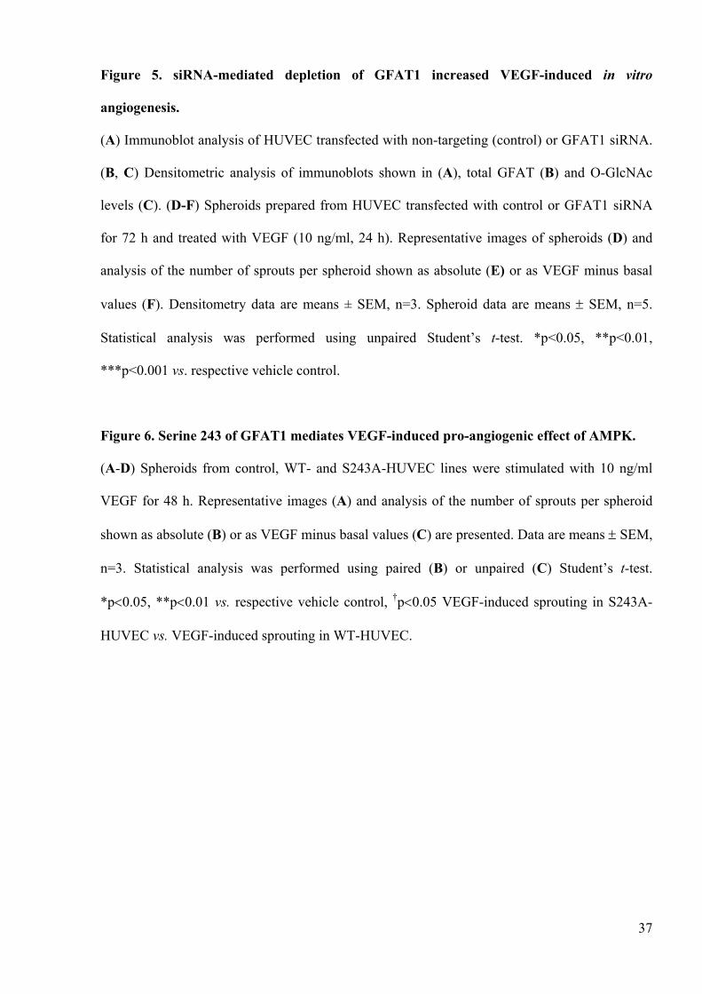
Figure 5. siRNA-mediated depletion of GFAT1 increased VEGF-induced in vitro
angiogenesis.
(A) Immunoblot analysis of HUVEC transfected with non-targeting (control) or GFAT1 siRNA.
(B, C) Densitometric analysis of immunoblots shown in (A), total GFAT (B) and O-GlcNAc
levels (C). (D-F) Spheroids prepared from HUVEC transfected with control or GFAT1 siRNA
for 72 h and treated with VEGF (10 ng/ml, 24 h). Representative images of spheroids (D) and
analysis of the number of sprouts per spheroid shown as absolute (E) or as VEGF minus basal
values (F). Densitometry data are means ± SEM, n=3. Spheroid data are means SEM, n=5.
Statistical analysis was performed using unpaired Student�’s t-test. *p<0.05, **p<0.01,
***p<0.001 vs. respective vehicle control.
Figure 6. Serine 243 of GFAT1 mediates VEGF-induced pro-angiogenic effect of AMPK.
(A-D) Spheroids from control, WT- and S243A-HUVEC lines were stimulated with 10 ng/ml
VEGF for 48 h. Representative images (A) and analysis of the number of sprouts per spheroid
shown as absolute (B) or as VEGF minus basal values (C) are presented. Data are means SEM,
n=3. Statistical analysis was performed using paired (B) or unpaired (C) Student�’s t-test.
*p 0.05, **p 0.01 vs. respective vehicle control, �†p 0.05 VEGF-induced sprouting in S243A-
HUVEC vs. VEGF-induced sprouting in WT-HUVEC.
D
BRatioProtein
GFAT1 3.9Peptide sequence[GLSR]VDpSTTCLFPVEEK
[ ,X]XXS/TXXX
A
Replicate
MS
inte
nsity
(cps
)
0
6.0E5
i ii iii iv
6.0E5
MS
inte
nsity
(cps
)
0 i ii iii ivReplicate
AMPK 1-/-/ 2-/-AMPK 1+/+/ 2+/+
Rel
ativ
e ab
unda
nce
GWT S243A
GFAT1 (colloidal blue)GFAT1 (autoradiography)
pGFAT1 (S243)
AMPK- + - +GST (GFAT1)
E
40
80
Ace
toni
trile
(%)
32P
-rad
ioac
tivity
(x10
3 C
PM
)
min0 50 100 1500
1412108642
min
40
80
Ace
toni
trile
(%)
32P
-rad
ioac
tivity
(x10
3C
PM
)
0 50 100 150
14121086420
F
Figure 1
kD100 -100 -
100 -
100 -
C
A
pGFAT1 (S243)
Figure 2
Control 1/ 2siRNA
*
GFA
T1 p
hosp
hory
latio
n(%
vs.
dilu
ent c
ontro
l)
0
50
100
150
200
AICAR - + - +
AM
PK
prot
ein
leve
ls(%
vs.
con
trol s
iRN
A)
***
Control 1/ 20
20
40
60
80
100
120
GFA
T1 p
hosp
hory
latio
n(%
vs.
dilu
ent c
ontro
l)
*
Control 1/ 2
0
100
200
300
400
VEGF - + - +siRNA
GFAT1
pGFAT1 (S243)
VEGF
IP: G
FAT1
pAMPK (T172)
AMPK
min0 2.5 5 10
75 -
kD
75 -
50 -
50 -
E
B C
FpGFAT1 (S243)
Control 1/ 2
GFAT1
pAMPK (T172)
AMPK
IP: G
FAT1
VEGF - + - +
siRNA
pACC (S79)
ACC
75 -
75 -75 -
75 -
260 -
260 -
kDG
GFAT1
pAMPK (T172)
AMPK
IP: G
FAT1
siRNA Control 1/ 2
pACC (S79)
ACC
75 -
75 -
75 -
75 -
260 -
260 -
kD
AICAR - + - +
D
1.65 ± 0.11
pGFAT1
GFAT1
pGFAT1/GFAT1
*
AICAR - +
GFA
T ac
tivity
(U/m
g ly
sate
)
0.00
0.05
0.10
0.15
0.20
0.25
0.30
0.35
AICAR - +
IP: GFAT1
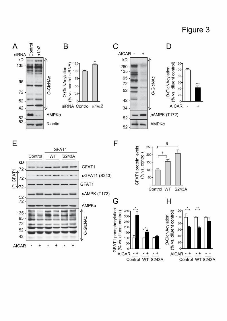
kD135 -
95 -
72 -
52 -
42 -
O-G
lcN
Ac
A
AMPK
-actin
siRNA AICAR
O-G
lcN
Ac
kD
pAMPK (T172)
AMPK
260 -135 -95 -72 -52 -
42 -
34 -
C- +C
ontro
l
1/ 20
25
50
75
100
125 **O
-Glc
NA
cyla
tion
(% v
s. c
ontro
l siR
NA
)
ControlsiRNA
52 -52 -
52 -
52 -
B D1/
2
***
O-G
lcN
Acy
latio
n(%
vs.
dilu
ent c
ontro
l)
AICAR0
20
40
60
80
100
120
- +
S243A0
50
100
150
200
250
GFA
T1 p
rote
in le
vels
(% v
s. c
ontro
l)§
Control WT
�†
F
GFAT1
pGFAT1 (S243)
pAMPK (T172)
AMPK
GFAT1
WT S243AControl
IP: G
FAT1
O-G
lcN
Ac
AICAR - + - + - +
135 -95 -72 -52 -42 -
EGFAT1
72 -
kD
72 -
72 -
52 -72 -
0
50
100
150
200
250
300
350
GFA
T1 p
hosp
hory
latio
n(%
vs.
dilu
ent c
ontro
l)
Control WT S243A
*
*
G
0
20
40
60
80
100
120
O-G
lcN
Acy
latio
n(%
vs.
dilu
ent c
ontro
l)
Control WT S243A
* **
H
AICAR - + - + - + AICAR - + - + - +
Figure 3
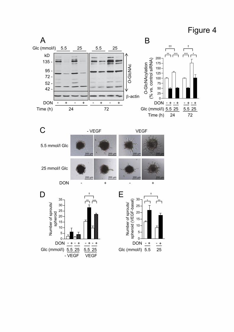
***
AGlc (mmol/l)
DON
5.5 25
kD135 -
95 -72 -52 -42 -
O-G
lcN
Ac
- + - +Time (h) 24 72
- + - +
5.5 25B
E
0
5
10
15
20
25
30 *
Num
ber o
f spr
outs
/sp
hero
id (V
EGF-
basa
l) **
�†
5.5 25+-- +DON
Glc (mmol/l)
D
Num
ber o
f spr
outs
/sp
hero
id
0
5
10
15
20
25
30
35
VEGF- VEGF5.5 25 5.5 25Glc (mmol/l)
DON - + - + - + - +
**
�†
***
Figure 4
- VEGF VEGF
5.5 mmol/l Glc
DON - + - +
C
200 µm 200 µm 200 µm
200 µm 200 µm
200 µm
200 µm 200 µm
25 mmol/l Glc
O-G
lcN
Acy
latio
n(%
vs.
con
trol s
iRN
A)
0255075
100125150175200
DON - + - + - + - +-actin
Time (h) 24 72
�†�† �†
*** ***
5.5 25Glc (mmol/l) 5.5 25
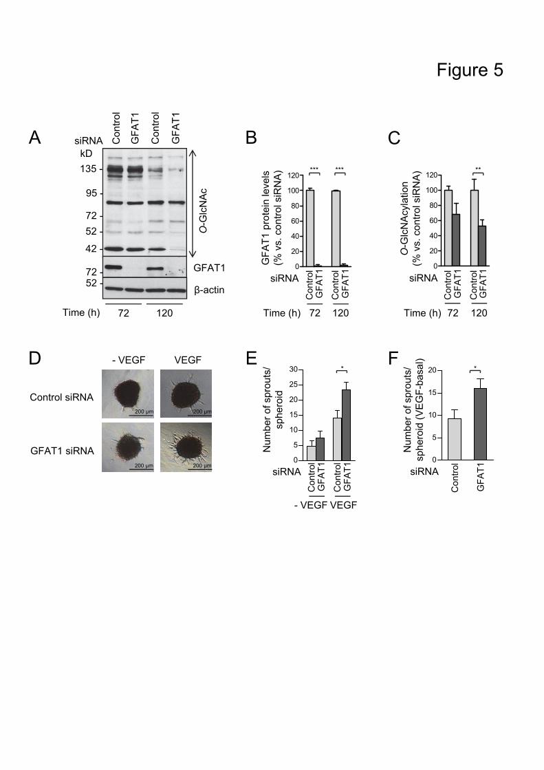
Figure 5
Con
trol
Con
trol
GFA
T1
GFA
T1
GFA
T1
0
5
10
15
20 *
0
5
10
15
20
25
30
Num
ber o
f spr
outs
/sp
hero
id (V
EG
F-ba
sal)
Num
ber o
f spr
outs
/sp
hero
id
VEGF
Control siRNA
GFAT1 siRNA
D - VEGF
- VEGF VEGF
E F*
siRNA
O-G
lcN
Ac
135 -
95 -
72 -52 -
42 -
kD
-actin
Time (h) 72 120
GFAT1
Time (h) 12072
A
52 -72 -
O-G
lcN
Acy
latio
n(%
vs.
con
trol s
iRN
A)
0
20
40
60
80
100
120
siRNA
**
Con
trol
GFA
T1
Con
trol
GFA
T1
Con
trol
GFA
T1
Con
trol
GFA
T1
0
20
40
60
80
100
120
GFA
T1 p
rote
in le
vels
(% v
s. c
ontro
l siR
NA
) *** ***
siRNA
Time (h) 12072
B CGFA
T1
Con
trol
GFA
T1siRNA
Con
trol
Con
trol
siRNA
200 µm 200 µm
200 µm 200 µm
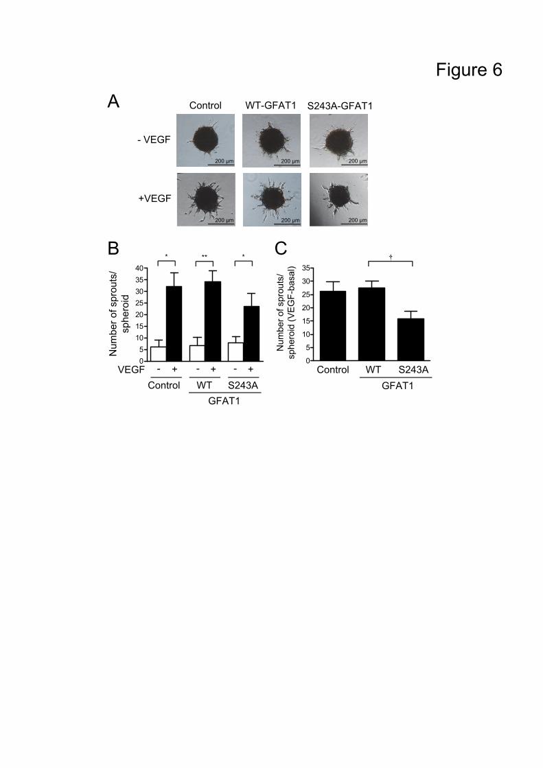
Control
- VEGF
WT-GFAT1 S243A-GFAT1A
+VEGF
05
10152025303540
Figure 6
B
Control WT S243A
Num
ber o
f spr
outs
/sp
hero
id
Num
ber o
f spr
outs
/sp
hero
id (V
EGF-
basa
l)
0
5
10
15
20
25
30
35
Control WT S243A
�†C
* ** *
GFAT1GFAT1
VEGF - + - + - +
200 µm 200 µm 200 µm
200 µm 200 µm 200 µm