Embed Size (px)
Citation preview
Research ArticleThe Relationship of the Facial Nerve to the CondylarProcess: A Cadaveric Study with Implications for OpenReduction Internal Fixation
H. P. Barham,1 P. Collister,2 V. D. Eusterman,2 and A. M. Terella2
1Department of Otolaryngology-Head and Neck Surgery, Louisiana State University Health Sciences Center, 533 Bolivar Street,Suite 566, New Orleans, LA 70112, USA2Department of Otolaryngology-Head and Neck Surgery, University of Colorado, Aurora, CO, USA
Correspondence should be addressed to H. P. Barham; [email protected]
Received 7 June 2015; Revised 6 August 2015; Accepted 19 August 2015
Academic Editor: Jeffrey P. Pearson
Copyright © 2015 H. P. Barham et al. This is an open access article distributed under the Creative Commons Attribution License,which permits unrestricted use, distribution, and reproduction in any medium, provided the original work is properly cited.
Introduction. The mandibular condyle is the most common site of mandibular fracture. Surgical treatment of condylar fracturesby open reduction and internal fixation (ORIF) demands direct visualization of the fracture. This project aimed to investigate theanatomic relationship of the tragus to the facial nerve and condylar process. Materials and Methods. Twelve fresh hemicadaversheads were used. An extended retromandibular/preauricular approach was utilized, with the incision being based parallel to theposterior edge of the ramus. Measurements were obtained from the tragus to the facial nerve and condylar process. Results. Thetemporozygomatic division of the facial nerve was encountered during each approach, crossing the mandible at the condylar neck.The mean tissue depth separating the facial nerve from the condylar neck was 5.5mm (range: 3.5mm–7mm, SD 1.2mm). Theupper division of the facial nerve crossed the posterior border of the condylar process on average 2.31 cm (SD 0.10 cm) anteriorto the tragus. Conclusions. This study suggests that the temporozygomatic division of the facial nerve will be encountered in mostapproaches to the condylar process. As visualization of the relationship of the facial nerve to condyle is often limited, recognitionthat, on average, 5.5mm of tissue separates condylar process from nerve should help reduce the incidence of facial nerve injuryduring this procedure.
1. Introduction
The condylar process has been reported as most common siteof mandibular fractures, accounting for 29% of all mandibu-lar fractures [1]. Surgical treatment of condylar fracturesby open reduction and internal fixation (ORIF) demandsthat internal fixation and anatomic reduction be completedunder direct visualization of the fracture. One challenge ofopen surgery for condylar process fractures is navigatingthe anatomic complexity of the adjacent vital structures,specifically the facial nerve.
Several authors have described the location of the facialnerve in the preauricular area. Despite this, one of themost common complications of open reduction and internal
fixation of subcondylar fractures remains facial nerve paresisand paralysis [2–4].
It is our feeling that the novice surgeon can benefitfrom a system of reference to enable the prediction ofcritical anatomic structures. This system must be based onanatomical landmarks that are (1) easily identifiable, (2) fixedin position during the procedure, and (3) independent ofpatient position [2].
This project aimed to describe pertinent anatomic rela-tionships of the facial nerve in the preauricular region andrelate these findings to ORIF procedures of the subcondylarregion. Specifically, we describe the anatomic relationship ofthe nerve to subcondylar mandible and to easily palpabletopographic landmarks, such as the tragus. We feel that these
Hindawi Publishing CorporationInternational Journal of OtolaryngologyVolume 2015, Article ID 715126, 3 pageshttp://dx.doi.org/10.1155/2015/715126
2 International Journal of Otolaryngology
T-C
T-FN
T-P
(a)
2.20 cm
2.31 cm2.25 cm
(b)
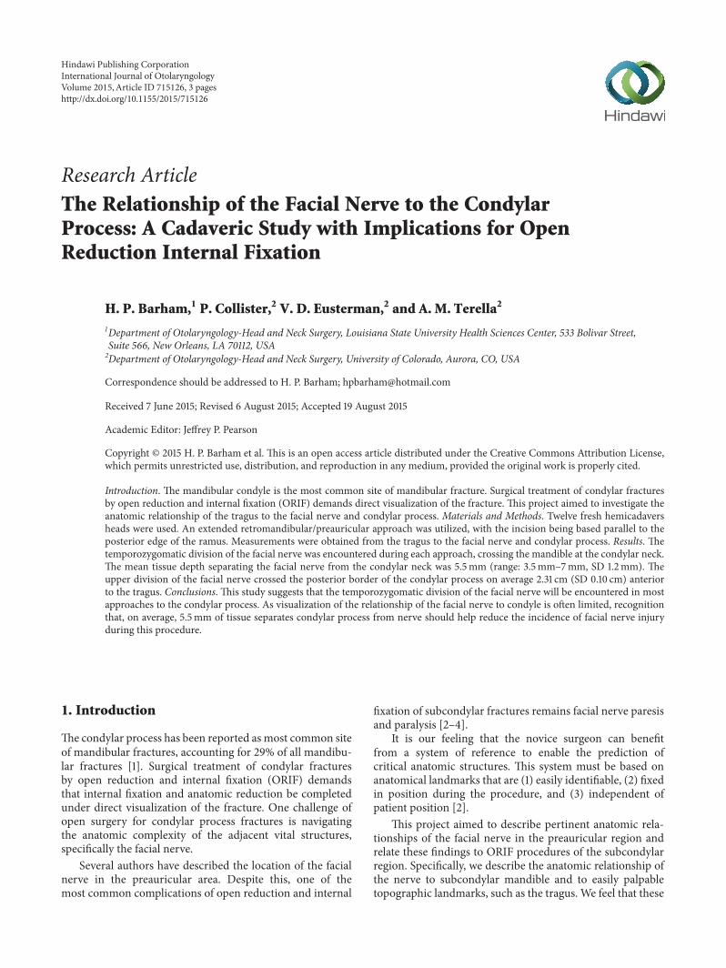
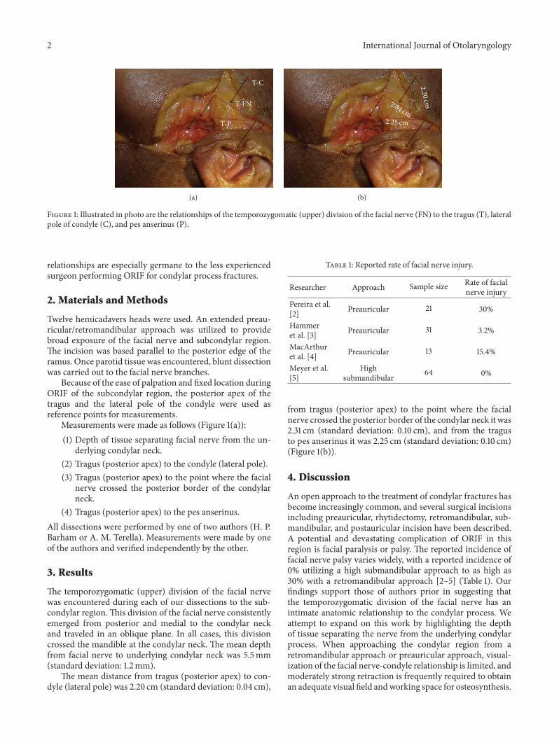
Figure 1: Illustrated in photo are the relationships of the temporozygomatic (upper) division of the facial nerve (FN) to the tragus (T), lateralpole of condyle (C), and pes anserinus (P).
relationships are especially germane to the less experiencedsurgeon performing ORIF for condylar process fractures.
2. Materials and Methods
Twelve hemicadavers heads were used. An extended preau-ricular/retromandibular approach was utilized to providebroad exposure of the facial nerve and subcondylar region.The incision was based parallel to the posterior edge of theramus. Once parotid tissue was encountered, blunt dissectionwas carried out to the facial nerve branches.
Because of the ease of palpation and fixed location duringORIF of the subcondylar region, the posterior apex of thetragus and the lateral pole of the condyle were used asreference points for measurements.
Measurements were made as follows (Figure 1(a)):(1) Depth of tissue separating facial nerve from the un-
derlying condylar neck.(2) Tragus (posterior apex) to the condyle (lateral pole).(3) Tragus (posterior apex) to the point where the facial
nerve crossed the posterior border of the condylarneck.
(4) Tragus (posterior apex) to the pes anserinus.All dissections were performed by one of two authors (H. P.Barham or A. M. Terella). Measurements were made by oneof the authors and verified independently by the other.
3. Results
The temporozygomatic (upper) division of the facial nervewas encountered during each of our dissections to the sub-condylar region. This division of the facial nerve consistentlyemerged from posterior and medial to the condylar neckand traveled in an oblique plane. In all cases, this divisioncrossed the mandible at the condylar neck. The mean depthfrom facial nerve to underlying condylar neck was 5.5mm(standard deviation: 1.2mm).
The mean distance from tragus (posterior apex) to con-dyle (lateral pole) was 2.20 cm (standard deviation: 0.04 cm),
Table 1: Reported rate of facial nerve injury.
Researcher Approach Sample size Rate of facialnerve injury
Pereira et al.[2] Preauricular 21 30%
Hammeret al. [3] Preauricular 31 3.2%
MacArthuret al. [4] Preauricular 13 15.4%
Meyer et al.[5]
Highsubmandibular 64 0%
from tragus (posterior apex) to the point where the facialnerve crossed the posterior border of the condylar neck it was2.31 cm (standard deviation: 0.10 cm), and from the tragusto pes anserinus it was 2.25 cm (standard deviation: 0.10 cm)(Figure 1(b)).
4. Discussion
An open approach to the treatment of condylar fractures hasbecome increasingly common, and several surgical incisionsincluding preauricular, rhytidectomy, retromandibular, sub-mandibular, and postauricular incision have been described.A potential and devastating complication of ORIF in thisregion is facial paralysis or palsy. The reported incidence offacial nerve palsy varies widely, with a reported incidence of0% utilizing a high submandibular approach to as high as30% with a retromandibular approach [2–5] (Table 1). Ourfindings support those of authors prior in suggesting thatthe temporozygomatic division of the facial nerve has anintimate anatomic relationship to the condylar process. Weattempt to expand on this work by highlighting the depthof tissue separating the nerve from the underlying condylarprocess. When approaching the condylar region from aretromandibular approach or preauricular approach, visual-ization of the facial nerve-condyle relationship is limited, andmoderately strong retraction is frequently required to obtainan adequate visual field andworking space for osteosynthesis.
International Journal of Otolaryngology 3
Although the temporozygomatic (upper) division of the facialnerve should not be encountered during the submandibularor high submandibular approaches, the nerve is retractedlaterally and easily stretched when attempting to achievean ample working space and optical field. On average, only5.5mm of tissue separates the condylar process from thenerve. The surgeon must appreciate that blind and aggressivelateral or superior retraction of overlying soft tissue in thisregion can easily result in stretch injury and neuropraxis.Understanding this close relationship should help reduce theincidence of facial nerve injury during ORIF of the condylarregion.
Additionally, on average, the pes anserinus of the facialnerve was located approximately 2.25 cm anterior-inferiorto the tragus, while the facial nerve crossed the posteriorborder of the mandible on average 2.31 cm anterior-inferiorto the tragus.Themeasurements and relationship of the facialnerve from this study should allow for nerve position to beestimated using the tragus and palpated posterior border ofthe mandible.
It is our opinion that the use of a palpable landmark isof greatest utility to the novice surgeon, less experienced inthis region. Techniques and measurements to predict nervelocation are only estimates and cannot replace the need forprecise anatomic understanding and cautious dissection inthe condylar region. Further, theymust be interpreted under-standing the inherent, well documented anatomic variationof the facial nerve.
Several studies have demonstrated efficacy of techniquesfor locating the facial nerve, with the work of de Ruet al. being the most complete and showing the single bestanatomic landmark for locating the facial nerve trunk to bethe tympanomastoid fissure (TMF), usually within 3mm ofthis landmark [6]. These findings were confirmed by Patherand Osman. However, Pather and Osman noted that theTMF was not an ideal landmark because it often lay behindthe sturdy tendon of the sternocleidomastoid muscle, thusrequiring a complex dissection [7]. These techniques areexcellent in localizing the nerve during dissection but do nothelp provide a preoperative estimate of nerve location in thecondylar region.
Limitations to this study include those that are commonto any cadaveric anatomic study. Tissues operated uponfollowing a traumatic insult may undergo distortion due toedema or disruption of soft tissues. Presumably, a swellingprocess would increase distances between structures if uni-formly distributed, so it may not significantly change thesurgeon’s operative strategy. Further, it is acknowledged thateven careful anatomic dissection could lead to distortionof tissue in our specimens, thus affecting measurements.Lastly, our limited sample size enabled calculation of standarddeviations, but not an evaluation of anatomic variation.
5. Conclusions
The temporozygomatic (upper) division of the facial nervehas an intimate relationship to the condylar process. It iscritical to understanding both the course of the nerve andthe depth of tissue separating it from the condylar neck. Soft
tissue retraction to optimize the optical field can easily stretchthe nerve resulting in neuropraxis. Understanding this closerelationship should help reduce the incidence of facial nerveinjury during ORIF of the condylar region.
Further the novice surgeon, less experienced in thesubcondylar region, can benefit from estimates of facial nervelocation using easily palpable topographic landmarks. Wesuggest that the tragus and lateral pole of the condyle canserve this function.
Ethical Approval
This study is IRB exempt.
Conflict of Interests
The authors declare that there is no conflict of interestsregarding the publication of this paper.
References
[1] R. A. Olson, R. J. Fonseca, D. L. Zeitler, and D. B. Osbon,“Fractures of the mandible: a review of 580 cases,” Journal ofOral and Maxillofacial Surgery, vol. 40, no. 1, pp. 23–28, 1982.
[2] J. A. Pereira, A. Meri, J. M. Potau, A. Prats-Galino, J. J. Sancho,and A. Sitges-Serra, “A simple method for safe identification ofthe facial nerve using palpable landmarks,” Archives of Surgery,vol. 139, no. 7, pp. 745–748, 2004.
[3] B. Hammer, P. Schier, and J. Prein, “Osteosynthesis of condylarneck fractures: a review of 30 patients,” British Journal of Oraland Maxillofacial Surgery, vol. 35, no. 4, pp. 288–291, 1997.
[4] C. J. MacArthur, P. J. Donald, J. Knowles, and H. C. Moore,“Open reduction-fixation of mandibular subcondylar fractures.A review,” Archives of Otolaryngology—Head and Neck Surgery,vol. 119, no. 4, pp. 403–406, 1993.
[5] C. Meyer, S. Zink, B. Chatelain, and A. Wilk, “Clinical expe-rience with osteosynthesis of subcondylar fractures of themandible using TCP plates,” Journal of Cranio-MaxillofacialSurgery, vol. 36, no. 5, pp. 260–268, 2008.
[6] J. A. de Ru, P. P. G. van Benthem, R. L. A. W. Bleys, H. Lubsen,and G.-J. Hordijk, “Landmarks for parotid gland surgery,” TheJournal of Laryngology and Otology, vol. 115, no. 2, pp. 122–125,2001.
[7] N. Pather andM.Osman, “Landmarks of the facial nerve: impli-cations for parotidectomy,” Surgical and Radiologic Anatomy,vol. 28, no. 2, pp. 170–175, 2006.

Submit your manuscripts athttp://www.hindawi.com
Stem CellsInternational
Hindawi Publishing Corporationhttp://www.hindawi.com Volume 2014
Hindawi Publishing Corporationhttp://www.hindawi.com Volume 2014
MEDIATORSINFLAMMATION
of
Hindawi Publishing Corporationhttp://www.hindawi.com Volume 2014
Behavioural Neurology
EndocrinologyInternational Journal of
Hindawi Publishing Corporationhttp://www.hindawi.com Volume 2014
Hindawi Publishing Corporationhttp://www.hindawi.com Volume 2014
Disease Markers
Hindawi Publishing Corporationhttp://www.hindawi.com Volume 2014
BioMed Research International
OncologyJournal of
Hindawi Publishing Corporationhttp://www.hindawi.com Volume 2014
Hindawi Publishing Corporationhttp://www.hindawi.com Volume 2014
Oxidative Medicine and Cellular Longevity
Hindawi Publishing Corporationhttp://www.hindawi.com Volume 2014
PPAR Research
The Scientific World JournalHindawi Publishing Corporation http://www.hindawi.com Volume 2014
Immunology ResearchHindawi Publishing Corporationhttp://www.hindawi.com Volume 2014
Journal of
ObesityJournal of
Hindawi Publishing Corporationhttp://www.hindawi.com Volume 2014
Hindawi Publishing Corporationhttp://www.hindawi.com Volume 2014
Computational and Mathematical Methods in Medicine
OphthalmologyJournal of
Hindawi Publishing Corporationhttp://www.hindawi.com Volume 2014
Diabetes ResearchJournal of
Hindawi Publishing Corporationhttp://www.hindawi.com Volume 2014
Hindawi Publishing Corporationhttp://www.hindawi.com Volume 2014
Research and TreatmentAIDS
Hindawi Publishing Corporationhttp://www.hindawi.com Volume 2014
Gastroenterology Research and Practice
Hindawi Publishing Corporationhttp://www.hindawi.com Volume 2014
Parkinson’s Disease
Evidence-Based Complementary and Alternative Medicine
Volume 2014Hindawi Publishing Corporationhttp://www.hindawi.com