Embed Size (px)
Citation preview
Research ArticleThe Influence of DNA Configuration onthe Direct Strand Break Yield
M. A. Bernal,1 C. E. deAlmeida,2 S. Incerti,3 C. Champion,3 V. Ivanchenko,4,5 and Z. Francis6
1Gleb Wataghin Institute of Physics, State University of Campinas, 13083-859 Campinas, SP, Brazil2Radiological Sciences Laboratory, State University of Rio de Janeiro, 20550-900 Rio de Janeiro, RJ, Brazil3Centre d’Etudes Nucleaires de Bordeaux-Gradignan, University of Bordeaux, 33175 Gradignan, France4Ecoanalytica, Moscow 119899, Russia5Geant4 Associates International Ltd., West Yorkshire HX7 7BT, UK6Department of Physics, Faculty of Sciences, Saint Joseph University, Beirut 1107 2050, Lebanon
Correspondence should be addressed to M. A. Bernal; [email protected]
Received 16 October 2014; Revised 11 January 2015; Accepted 28 January 2015
Academic Editor: Chen Yanover
Copyright © 2015 M. A. Bernal et al. This is an open access article distributed under the Creative Commons Attribution License,which permits unrestricted use, distribution, and reproduction in any medium, provided the original work is properly cited.
Purpose. To study the influence ofDNAconfiguration on the direct damage yield.No indirect effect has been accounted for.Methods.The GEANT4-DNA code was used to simulate the interactions of protons and alpha particles with geometrical models of the A-,B-, and Z-DNA configurations. The direct total, single, and double strand break yields and site-hit probabilities were determined.Certain features of the energy deposition process were also studied. Results. A slight increase of the site-hit probability as a functionof the incident particle linear energy transfer was found for each DNA configuration. Each DNA form presents a well-defined site-hit probability, independently of the particle linear energy transfer. Approximately 70% of the inelastic collisions and ∼60% of theabsorbed dose are due to secondary electrons. These fractions are slightly higher for protons than for alpha particles at the sameincident energy. Conclusions. The total direct strand break yield for a given DNA form depends weakly on DNA conformationtopology. This yield is practically determined by the target volume of the DNA configuration. However, the double strand breakyield increases with the packing ratio of the DNA double helix; thus, it depends on the DNA conformation.
1. Introduction
Themechanisms bywhich ionizing radiation induces damagein DNA are very complex and multifaceted, going throughphysical, physicochemical, and biological stages, from thechronological point of view.This damage may lead to variousbiological effects, from the cellular to the organic level.Thanks mainly to the vertiginous growth in computationpower during the past two decades, simulation approacheshave become a powerful tool to study very complex phenom-ena.Theyhave been applied to the radiation-DNA interactionprocess, which has been studied by combining aMonte Carlocode for the radiation transport simulation, a geneticmaterialgeometrical model, and a biophysical model to mimic DNAdamage induction after a particle interaction (see [1, 2] and
references therein). In atomistic DNA geometrical models[3], the volume of the target to be impacted by the radiation toinduce a strand break is defined by the union of all the atomsmaking up the sugar-phosphate groups.This target definitionwill be used throughout this document. In this case, the atomsize can be estimated by the corresponding van der Waalsradius.
Several geometrical models used to simulate the inter-action of ionizing particles with DNA have been published.A short review of these models can be found elsewhere [4].Friedland et al. [5] have developed the most sophisticatedgeometrical model presented thus far. However, some modelparameters are adjusted to reproduce observed damage yieldsfor 60Co radiation (see [6]). The model is then applied toother radiation qualities. The effective target volume in that
Hindawi Publishing CorporationComputational and Mathematical Methods in MedicineVolume 2015, Article ID 417501, 8 pageshttp://dx.doi.org/10.1155/2015/417501
2 Computational and Mathematical Methods in Medicine
work is the union of all the atoms that form the sugar-phosphate group. This volume could be slightly different foreach DNA form, according to the preliminary results wehave obtained using the corresponding DNA structures withatomic resolution.
There are three main DNA configurations: A-, B-, andZ-DNA [7]. B-DNA is the canonical and predominant pre-sentation of this nucleic acid, and its morphology was firstdescribed by Watson and Crick [8]. The A-DNA confor-mation is thought to be related to DNA-drug and DNA-protein interactions andmay share with the B-DNA form theresponsibility for genome structure and function (see [9] andreferences therein). Very recently,Whelan et al. have reportedan important amount of reversible A- to B-DNA transitionsin live bacterial cells by the use of Fourier transform infraredspectroscopy [10]. These authors also mention that A-DNAmay be involved in the resistance of some bacteria to thedamage induced by UV radiation. In contrast to A- and B-DNA, Z-DNA is a left-handed double helix macromolecule.It appears during certain physiological cellular processesand decays in B-DNA [11]. The human genetic material hasapproximately 100 000 copies of potential Z-DNA sequences[12]. It has also been observed that these Z-DNA sequencesinduce genome instability that produces DSB in certainhuman tumors and that theymight be related to transcriptionactivation, which is related to gene expression (see [13] andreferences therein). In this sense, a series of works [14–16]has noted that the structure of DNA and its binding to othermacromolecules play an important role in the radiosensitivityof DNA due to the attack of OH∙ radicals. However, theseworks only addressed the indirect DNA damage caused bythis chemical species and did not study the relation of thedirect damage and the DNA structure. Although the indirecteffects play amajor role in theDNAdamage, their importancedecreases as the LET of the incident particles increases,decreasing from approximately 65% of the damage for 60Coradiation (LET∼0.4 keV/𝜇m) to approximately 50% for a LETof 70 keV/𝜇m(see Figure 2 of [6]).We did not account for thiskind of damage in the current work as we preferred to leavethis issue until our atomistic DNA models are ready for use.Atomistic models would allow a more rigorous treatment ofthe indirect effects [5].
Semsarha et al. [17] have recently published a studyon the influence of DNA conformation on strand breakyields after 60Co irradiation, including direct and indirecteffects, although the latter has been roughly accounted for.They included the A-, B-, and Z-DNA forms and alsodetermined microdosimetric quantities such as the meanspecific imparted energy. In fact, they used a previouslycalculated 60Co electron spectrum to irradiate the region ofinterest uniformly instead of using the corresponding pri-mary photon beam. Many experimental works have reporteddamage yields after the impact of ionizing radiation, but thedifficulties associated with the determination of the preciseDNA conformation during such experiments do not allowthese yields to be obtained as a function of this conformation.This issue is one of the reasonswe do not have any experimen-tal reference for comparison to the results of this work.
In a previous work [19], the physical causes of the totaldirect strand break yield invariance with respect to theincident particle type and energy were investigated. In thatwork, a B-DNA representationwasmodeled, and it was foundthat this behavior results from the combination of quasicon-stant number of inelastic events per unit absorbed dose andsite-hit probability. This probability accounts for the chancethat a sugar-phosphate group has to be hit by an energydeposition event. Furthermore, the site-hit probability seemsto be determined by geometrical factors associated with theDNAmodel. However, only oneDNA configurationwas usedin that study, and thus the influence of DNA conformationon the site-hit probability could not be investigated. Most ofthe DNA geometrical models developed to determine directeffects on DNA by Monte Carlo simulations are based on theB-DNA form (see, e.g., [3, 20]).
This work is intended to study the influence of the DNAconfiguration on the site-hit probability when protons andalpha particles impact the DNA. In addition, the direct total,single, and double strand break yields are also determinedand analyzed. Some aspects involved in the process ofenergy deposition by charged particles are also studied.The differences between the volumes of the sugar-phosphategroups for the A-, B-, and Z-DNA forms have been enhancedsomewhat arbitrarily to study how these volumes influencethe damage yields. These differences should be large enoughwhen relatively compared to the uncertainties associatedwith the determined quantities. Thus, the damage yieldsreported in this work should not be treated as absolute valuesnor be compared to experimental or previous simulatedanalog results. We simply want to understand how DNAconformation influences these yields.
As far as we know, this study is the first investigation ofthe influence of DNA configuration on the direct damageprobability due to ion impact.
All the uncertainties reported in this work represent onestandard deviation of the mean.
2. Methods and Materials
2.1. The GEANT4-DNA Package. The GEANT4-DNA pack-age (v.9.4) [21] has been used to simulate the chargedparticle transport in liquid water. Details on the latestdevelopments of this package can be found elsewhere [22].Ionization, excitation, and charge transfer processes havebeen accounted for during the interaction of the nonnegativecharge states of hydrogen and helium projectiles with liquidwater. Ionizations, excitations, and elastic scattering weretaken into account for electrons. Heavy charged particleswere transported down to 1 keV and electrons down to ∼9 eV.
2.2. DNA Geometrical Model. The three main DNA geo-metrical configurations (A, B, and Z) were studied in thiswork.TheB-DNAgeometricalmodel developed in a previouswork [18] was adapted to the other two variants. It is worthnoting that this model accounts for five organization levelsof the human genetic material: nucleotide pairs, the doublehelix, nucleosomes, and the 10 nm and 30 nm chromatin
Computational and Mathematical Methods in Medicine 3
A-DNA
0.23
2.46
0.76
1.02.55
B-DNA
0.33
1.2
3.2
1.02.37
Z-DNA
0.773.0
0.25
0.6
1.84
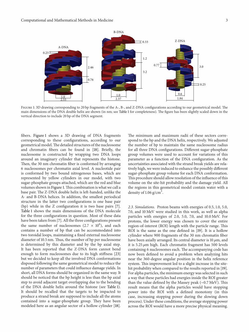
Figure 1: 3D drawing corresponding to 20 bp fragments of the A-, B-, and Z-DNA configurations according to our geometrical model. Themain dimensions of the DNA double helix are shown (in nm; see Table 1 for completeness). The figure has been slightly scaled down in thevertical direction to include 20 bp of the DNA segment.
fibers. Figure 1 shows a 3D drawing of DNA fragmentscorresponding to these configurations, according to ourgeometricalmodel.Thedetailed structures of the nucleosomeand chromatin fibers can be found in [18]. Briefly, thenucleosome is constructed by wrapping two DNA loopsaround an imaginary cylinder that represents the histone.Then, the 30 nm chromatin fiber is conformed by arranging6 nucleosomes per chromatin axial level. A nucleotide pairis conformed by two bound nitrogenous bases, which arerepresented by yellow cylinders in our model, with twosugar-phosphate groups attached, which are the red and bluevolumes shown in Figure 1.This combination is what we call abase pair. The Z-DNA double helix is left-handed, unlike theA- and B-DNA helices. In addition, the smallest periodicalstructure in the latter two configurations is one base pair(bp) while in the Z configuration it is two base pairs [7].Table 1 shows the main dimensions of the DNA moleculefor the three configurations in question. Most of these datahave been taken from [7]. All the three configurations presentthe same number of nucleosomes (2.7 × 10⁶), and eachcontains a number of bp that can be accommodated intotwo toroidal loops, maintaining a fixed external nucleosomediameter of 10.5 nm.Thus, the number of bp per nucleosomeis determined by this diameter and by the bp axial step.It has been reported that the Z-DNA form cannot bendenough to form nucleosomes due to its high stiffness [23]but we decided to keep all the involved DNA conformationsdisposed following the same geometrical model to reduce thenumber of parameters that could influence damage yields. Inshort, all DNA forms should be organized in the same way. Itshould be noticed that the bp height is less than the bp axialstep to avoid adjacent target overlapping due to the bendingof the DNA double helix around the histone (see Table 1).It should be recalled that the targets to be impacted toproduce a strand break are supposed to include all the atomscontained into a sugar-phosphate group. They have beenmodeled here as an angular sector of a hollow cylinder [18].
The minimum and maximum radii of these sectors corre-spond to the bp and the DNA helix, respectively. We adjustedthe number of bp to maintain the same nucleosome radiusfor all three DNA configurations. Different sugar-phosphategroup volumes were used to account for variations of thisparameter as a function of the DNA configuration. As theuncertainties associated with the strand break yields are rela-tively high, we were induced to enhance the possibly differentsugar-phosphate group volume for each DNA conformation.This procedure should allow resolution of the influence of thisvolume on the site-hit probability and the damage yield. Allthe regions in this geometrical model contain water with adensity of 1.06 g/cm3.
2.3. Simulations. Proton beams with energies of 0.5, 1.0, 5.0,7.0, and 10MeV were studied in this work, as well as alphaparticles with energies of 2.0, 5.0, 7.0, and 10.0MeV. Forprotons, the lower energy was chosen to cover the entireregion-of-interest (ROI) length with the particle range. ThisROI is the same as the one defined in [19]. It is a hollowcylinder where 900 fragments of the 30 nm chromatin fiberhave been axially arranged. Its central diameter is 10 𝜇m, andit is 5.25 𝜇m high. Each chromatin fragment has 500 levelscontaining 6 nucleosomes each. The accompanying code hasnow been defined to avoid a problem when analyzing hitsnear the 360-degree angular position in the helix referencesystem. This improvement led to a slight increase in the site-hit probability when compared to the results reported in [19].For alpha particles, theminimum energy was selected in sucha way that these particles had energies inside the ROI greaterthan the value defined by the Massey peak (∼0.7MeV). Thisresult means that the alpha particles would have stoppingpower into the ROI with a defined monotony (in thiscase, increasing stopping power during the slowing downprocess). Under these conditions, the average stopping poweracross the ROI would have a more precise physical meaning.
4 Computational and Mathematical Methods in Medicine
Table 1: Dimensions of the main DNA structures for the threeconfigurations studied in this work. Most of these data wereextracted from [7]. The target volumes were determined accordingto these dimensions and the geometrical model used (see [18] fordetails). All dimensions are shown in nm, unless otherwise stated.
Features DNA configurationsA B Z
Helix orientation Right-handed Right-handed Left-handedDNA diameter 2.55 2.37 1.84bp diameter 1.0 1.0 0.6bp axial step 0.23 0.33 0.38Helix pitch 2.46 3.2 3.0bp/helix turn 10.7 10 12bp/nucleosome 286 198 172Helix axial shift 0.76 1.2 0.77Target angular aperture 87∘ 73∘ 60∘
Target height 0.119 0.183 0.249Total number of bp 7.72 × 10
85.35 × 10
84.64 × 10
8
Target volume (nm3) 0.12 0.13 0.10
With these energies, the LET in themiddle of the ROI for pro-tons and alpha particles ranges within 4.8–66.9 keV/𝜇m and58.0–235.0 keV/𝜇m, respectively.With this energy choice, theproton and alpha particle LET ranges overlap so that thestrand break yield for two different particles at the same LETcan be investigated.
As in our previous work [19], ion beams impact thesurface of a semi-infinite water phantom in which the ROI isplaced at 2.6𝜇m depth, with its axis normal to the phantomsurface. This practice is a common one when irradiating cellcultures with heavy charged particles [6]. Primary particlesimpinge the phantomnormally and are uniformly distributedwithin the annulus defined by the projection of the ROI onthe phantom surface. A single strand break (SSB) is recordedif an event occurs within a target, defined previously, withan energy transfer of ≥8.23 eV. As the minimum possibleinelasticity during inelastic events (ionization, excitation, andcharge transfer) is 9 eV in this GEANT4-DNA version, anyevent can potentially cause a SSB because energy transfers arealways greater than or equal to the inelasticity of the reaction.
Thus, the strand break yields determined in this workmust be analyzed from a relative point of view. That is, theintent is not to calculate absolute yields but to study thesequantities for a few DNA configurations as a function ofthe beam LET. As before, a double strand break (DSB) isaccounted for if two SSBs are produced on opposite helicesand separated from each other by no more than 10 bp.Complex DSBs have not been accounted for in this work,and this kind of damage could be counted as two adjacentDSBs. It should be noted that the aim of this work is notto determine absolute damage yields but to study how theseyields are influenced by the DNA configuration. If one targetis impacted 𝑛 times, then 𝑛 SSBs are counted because we wantto study the target-hit probability as a function of the incidentand the secondary particle type and energy. The total strand
break (TSB) yield is determined by using the total numberof single strand breaks induced, including those leading toDSBs. The number of inelastic events due to primary andsecondary particleswas calculated, fromwhich the fraction ofthe number of events and the absorbed dose due to electronswere found. The site-hit probability was determined as theratio between the total number of strand breaks and the totalnumber of the inelastic events. This probability was studiedfor the three DNA configurations shown above as a functionof the particle type and LET.
The number of histories simulated for each particle-energy combination was chosen in such a way that theabsorbed dose within the ROI was close to 100Gy. Inour previous work [19], strand break yields and site-hitprobabilities were determined for a fixed dose value, thatis, 100Gy. Following this procedure, the history of the lastprimary particle, which would reach this dose value insidethe ROI, was cut so that a portion of the primary and/orsecondary particle events were not accounted for duringthese calculations. This cut increased the uncertainties of thequantities in question. However, these quantities are nowdetermined by completely following, including all secondaryelectrons, a fixed number of histories that would amount toan absorbed dose close to 100Gy. The number of simulatedhistories ranges from 4 to 200, corresponding to the 2MeValpha particle and 10MeV proton cases, respectively.
3. Results and Discussion
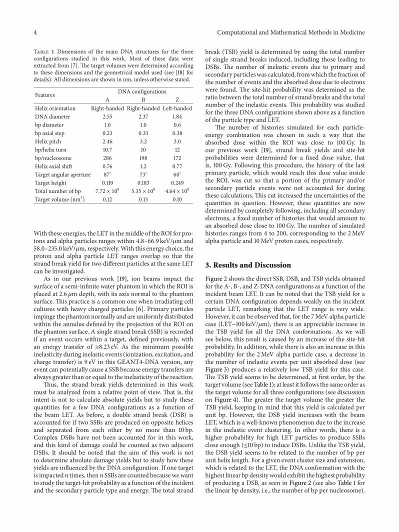
Figure 2 shows the direct SSB, DSB, and TSB yields obtainedfor the A-, B-, and Z-DNA configurations as a function of theincident beam LET. It can be noted that the TSB yield for acertain DNA configuration depends weakly on the incidentparticle LET, remarking that the LET range is very wide.However, it can be observed that, for the 7MeV alpha particlecase (LET∼100 keV/𝜇m), there is an appreciable increase inthe TSB yield for all the DNA conformations. As we willsee below, this result is caused by an increase of the site-hitprobability. In addition, while there is also an increase in thisprobability for the 2MeV alpha particle case, a decrease inthe number of inelastic events per unit absorbed dose (seeFigure 3) produces a relatively low TSB yield for this case.The TSB yield seems to be determined, at first order, by thetarget volume (see Table 1); at least it follows the same order asthe target volume for all three configurations (see discussionon Figure 4). The greater the target volume the greater theTSB yield, keeping in mind that this yield is calculated perunit bp. However, the DSB yield increases with the beamLET, which is a well-known phenomenon due to the increasein the inelastic event clustering. In other words, there is ahigher probability for high LET particles to produce SSBsclose enough (≤10 bp) to induce DSBs. Unlike the TSB yield,the DSB yield seems to be related to the number of bp perunit helix length. For a given event cluster size and extension,which is related to the LET, the DNA conformation with thehighest linear bp densitywould exhibit the highest probabilityof producing a DSB, as seen in Figure 2 (see also Table 1 forthe linear bp density, i.e., the number of bp per nucleosome).
Computational and Mathematical Methods in Medicine 5D
SB y
ield
(GyG
bp)−
1TS
B yi
eld
(GyG
bp)−
1SS
B yi
eld
(GyG
bp)−
1
LET (keV/𝜇m)10 100 1000
LET (keV/𝜇m)10 100 1000
LET (keV/𝜇m)10 100 1000
10
15
20
25
80
100
120
140
160
60
80
100
120
140
160
A-DNA, protonsB-DNA, protonsZ-DNA, protons
A-DNA, alpha part.B-DNA, alpha part.Z-DNA, alpha part.
Figure 2: Direct total, single, and double strand break yields as afunction of LET for protons and alpha particles impacting on A-,B-, and Z-DNA configurations.
It is expected that the SSB yield decreases with the beamLET for increasing DSB and quasiconstant TSB yields, butfluctuations are observed for very high LET values due to theinterplay of the site-hit probability and the number of energydeposition events per unit absorbed dose. There is a kindof saturation in the DSB yield for the 2MeV alpha particlecase but this saturation is due to a relatively low numberof inelastic events per unit absorbed dose for this radiationquality, which will be discussed below (see Figure 3). It isnoteworthy that Friedland et al. [6] also reported a saturationin the total DSB yield, including indirect damage, for heavyions with LET above 200 keV/𝜇m.
It should also be noted in Figure 2 (bottom graph) thatthe DSB yields for the two lowest alpha particle LET valuesare statistically similar, which occurs for all three DNAconfigurations. In addition, the directDSB yield for protons is
0 2 4 6 8 10Projectile energy (MeV)
500
1000
1500
2000
2500
3000
3500
Num
ber o
f ine
lasti
c eve
nts/
Gy
Alpha particles, totalProtons, total
Alpha particles, due to eProtons, due to e
−
−
Figure 3: Number of inelastic events per unit absorbed doseproduced by protons and alpha particles and their correspondingsecondary electrons.
10 100 10000.01
0.02
0.03
0.04
0.05
Site
-hit
prob
abili
ty
LET (keV/𝜇m)
A-DNA, protonsB-DNA, protonsZ-DNA, protons
A-DNA, alpha part.B-DNA, alpha part.Z-DNA, alpha part.
Figure 4: Site-hit probabilities for the three DNA configurationsstudied in this work, as a function of the incident beam LET.
higher than the yield for alpha particles at similar LET values.As clustered ionizations are more efficient in causing DSBthan sparsely distributed ionizations, it could be deducedfrom these results that light ions would be more efficient forproducing ionization clusters than heavier particles at thesame LET.This result was also found in a previous work [24].
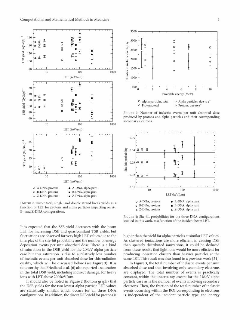
In Figure 3, the total number of inelastic events per unitabsorbed dose and that involving only secondary electronsare displayed. The total number of events is practicallyconstant, within the uncertainty, except for the 2MeV alphaparticle case as is the number of events involving secondaryelectrons. Then, the fraction of the total number of inelasticevents occurring within the ROI corresponding to electronsis independent of the incident particle type and energy

6 Computational and Mathematical Methods in Medicine
(or LET). This result could be explained by the weak depen-dency of the mean energy of the electrons produced by lightions on the ion LET (see [19, 25]). In other words, secondary(first generation) electron spectra for different primary par-ticle LET values are similar, at least for projectile energiesabove a few hundreds of keV/u. The differences are mainlydue to the maximum electron energy, for which the spectralfrequency is generally a few orders of magnitude lower thanthe value corresponding to low electron energies (∼10 eV).The absorbed dose is calculated by adding those energydepositsmade by particles within the volume in question, andthus, it could be expected that, for a quasiconstant number ofevents per unit absorbed dose, the mean energy deposit perinelastic event (𝜀) would depend little on the particle type andenergy (or LET). In a previous work [26], it was shown that𝜀 is quasiconstant for protons and alpha particles impactingon liquid water, increasing by approximately 20% when theprojectile energy goes from 10 keV to 10MeV, that is, increasesby three orders of magnitude. For electrons this quantitydepends somewhat more strongly on the particle energythan for ions. However, the mean energy of the secondaryelectrons directly produced by ion impact is approximately50 eV and 𝜀 shows a little change when electrons slow downfrom this energy to approximately 9 eV. These points are thephysical reasons for the quasiconstant primary and secondarynumber of events per unit absorbed dose.
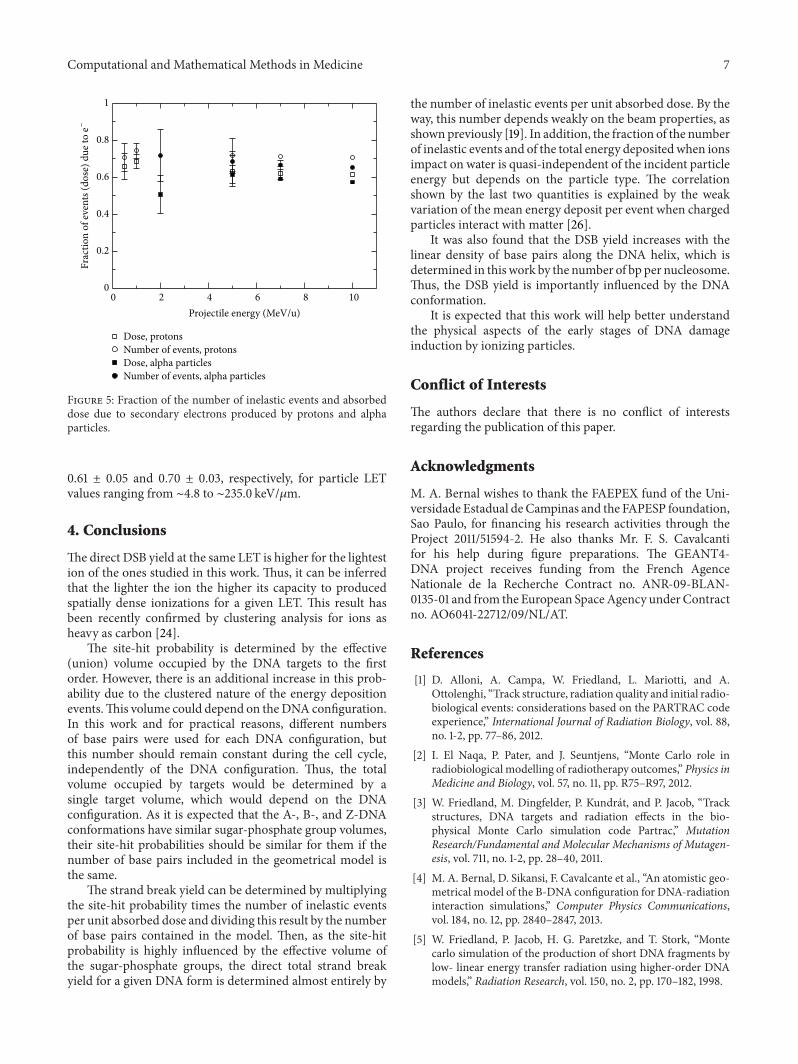
The site-hit probability for all the DNA configurationsand for each particle energy and type was determined bydividing the total number of strand breaks by the numberof targets. The results of these calculations are shown inFigure 4, in which it is observed that the site-hit probabilityfluctuates around a well-defined value for each DNA config-uration. However, there is an increment in this probability asthe particle LET increases. To study this behavior, the site-hit probability was also determined by distributing eventsuniformly through the ROI. That is, the positions of theenergy deposition events were randomly distributed insidethe ROI. In this situation, the site-hit probabilities for the A-,B-, and Z-DNA conformations were 0.038 ± 0.001, 0.0285 ±0.0009, and 0.0184 ± 0.0006, respectively. These probabilitiesshould be equivalent to the ratio of the volume occupied byall the targets and the volume defined by the ROI. Theseratios are 0.038, 0.029, and 0.018 for the A-, B-, and Z-DNAmodels, respectively. These results represent a solid consis-tency check on our geometrical models and the associatedcomputational codes. Lines have been drawn in Figure 4 torepresent these theoretically predicted site-hit probabilities.These ratios and the average site-hit probabilities determinedfor each DNA configuration and primary particle are shownin Table 2. According to these results, the site-hit probabilityis determined by the volume ratio defined just above, atfirst order. The total volume occupied by all the targets issimply the target volume times the number of targets in theROI. Thus, the important feature for the site-hit probabilityis the target volume, which is the sugar-phosphate groupin this case. As shown in Figure 4, practically all the site-hit probabilities are above the predicted theoretical value foreach DNA conformation. This fact can be attributed to theclustering of the energy deposition events. It should be noted
Table 2: Average site-hit probability for each DNA configuration-particle combination. Corresponding values predicted from thevolume ratio defined in the text are also shown.
DNAconfiguration Protons Alpha part. Pred. value
A 0.040 ± 0.002 0.040 ± 0.004 0.038B 0.030 ± 0.001 0.030 ± 0.003 0.029Z 0.0188 ± 0.0008 0.0194 ± 0.002 0.018
that this overestimation tends to increase as the LET of theincident particles increases, where clustering is enhanced.
It seems that the conformation of theDNAhas little influ-ence on the total strand break yield; instead, the target volumeis the key factor. However, this conformation is importantfor the DSB yield, where the distance between adjacent bpplays the main role. In conjunction with the quasiconstantnumber of events per unit absorbed dose, this point leadsto the quasiconstant TSB yield shown in Figure 2. In a realDNA structure, the volume of the target would be determinedby the radius of the corresponding atoms, which can betaken as their van der Waals radii, to a first approximation.In this case, a homogeneous medium has been consideredfor simulations so that the target/ROI volume ratio is agood estimate of the site-hit probability. However, the relativemacroscopic cross sections (or interaction probability perunit path length) should be accounted for in a model withregions of different chemical composition. That is, a regionwith a relatively high cross section would appear larger andwould receive a relatively higher number of events. Ourresults seem to conflict with the ones reported by Semsarhaet al. [17], who found higher site-hit probabilities for smallertarget sizes. However, they defined the hit probability as theratio of the number of hits within the strands to the onesoccurring within the whole DNA volume. According to ourunderstanding, this probability should be equal to the ratio ofthe volume occupied by the sugar-phosphate groups plus thehydration shell to the volume of the whole DNA cylinder. Wehave calculated this ratio for the dimensions shown in Figure2 of [17] and found values of 0.85, 0.81, and 0.71 for the A-, B-,and Z-DNA conformations. These results are similar to thevalues shown in Table 5 of [17], except for the B-DNA case.These discrepancies should be further investigated.
Finally, Figure 5 shows the fraction of the number ofinelastic events and absorbed dose involving the secondaryelectrons produced by protons and alpha particles. Approx-imately 70% of all the inelastic collisions and ∼60% of theabsorbed dose are due to secondary electrons.The fraction ofevents and the absorbed dose due to secondary electrons forprotons are slightly higher than for alpha particles at the sameincident energy.This result could be explained by the fact thatprotons produce electrons with higher average energy thanalpha particles at the same ion energy. Protons are lighter thanalpha particles and thus can transfermore energy, on average,to bound electrons than alpha particles with the same energy.The values shown in this figure depend weakly on the particletype and energy (or LET). The fraction of dose and numberof inelastic events due to secondary electrons are within
Computational and Mathematical Methods in Medicine 7
0 2 4 6 8 100
0.2
0.4
0.6
0.8
1
Dose, protonsNumber of events, protonsDose, alpha particlesNumber of events, alpha particles
Projectile energy (MeV/u)
Frac
tion
of ev
ents
(dos
e) d
ue to
e−
Figure 5: Fraction of the number of inelastic events and absorbeddose due to secondary electrons produced by protons and alphaparticles.
0.61 ± 0.05 and 0.70 ± 0.03, respectively, for particle LETvalues ranging from ∼4.8 to ∼235.0 keV/𝜇m.
4. Conclusions
Thedirect DSB yield at the same LET is higher for the lightestion of the ones studied in this work. Thus, it can be inferredthat the lighter the ion the higher its capacity to producedspatially dense ionizations for a given LET. This result hasbeen recently confirmed by clustering analysis for ions asheavy as carbon [24].
The site-hit probability is determined by the effective(union) volume occupied by the DNA targets to the firstorder. However, there is an additional increase in this prob-ability due to the clustered nature of the energy depositionevents.This volume could depend on theDNA configuration.In this work and for practical reasons, different numbersof base pairs were used for each DNA configuration, butthis number should remain constant during the cell cycle,independently of the DNA configuration. Thus, the totalvolume occupied by targets would be determined by asingle target volume, which would depend on the DNAconfiguration. As it is expected that the A-, B-, and Z-DNAconformations have similar sugar-phosphate group volumes,their site-hit probabilities should be similar for them if thenumber of base pairs included in the geometrical model isthe same.
The strand break yield can be determined by multiplyingthe site-hit probability times the number of inelastic eventsper unit absorbed dose and dividing this result by the numberof base pairs contained in the model. Then, as the site-hitprobability is highly influenced by the effective volume ofthe sugar-phosphate groups, the direct total strand breakyield for a given DNA form is determined almost entirely by
the number of inelastic events per unit absorbed dose. By theway, this number depends weakly on the beam properties, asshownpreviously [19]. In addition, the fraction of the numberof inelastic events and of the total energy depositedwhen ionsimpact on water is quasi-independent of the incident particleenergy but depends on the particle type. The correlationshown by the last two quantities is explained by the weakvariation of the mean energy deposit per event when chargedparticles interact with matter [26].
It was also found that the DSB yield increases with thelinear density of base pairs along the DNA helix, which isdetermined in this work by the number of bp per nucleosome.Thus, the DSB yield is importantly influenced by the DNAconformation.
It is expected that this work will help better understandthe physical aspects of the early stages of DNA damageinduction by ionizing particles.
Conflict of Interests
The authors declare that there is no conflict of interestsregarding the publication of this paper.
Acknowledgments
M. A. Bernal wishes to thank the FAEPEX fund of the Uni-versidade Estadual de Campinas and the FAPESP foundation,Sao Paulo, for financing his research activities through theProject 2011/51594-2. He also thanks Mr. F. S. Cavalcantifor his help during figure preparations. The GEANT4-DNA project receives funding from the French AgenceNationale de la Recherche Contract no. ANR-09-BLAN-0135-01 and from the European SpaceAgency under Contractno. AO6041-22712/09/NL/AT.
References
[1] D. Alloni, A. Campa, W. Friedland, L. Mariotti, and A.Ottolenghi, “Track structure, radiation quality and initial radio-biological events: considerations based on the PARTRAC codeexperience,” International Journal of Radiation Biology, vol. 88,no. 1-2, pp. 77–86, 2012.
[2] I. El Naqa, P. Pater, and J. Seuntjens, “Monte Carlo role inradiobiological modelling of radiotherapy outcomes,” Physics inMedicine and Biology, vol. 57, no. 11, pp. R75–R97, 2012.
[3] W. Friedland, M. Dingfelder, P. Kundrat, and P. Jacob, “Trackstructures, DNA targets and radiation effects in the bio-physical Monte Carlo simulation code Partrac,” MutationResearch/Fundamental and Molecular Mechanisms of Mutagen-esis, vol. 711, no. 1-2, pp. 28–40, 2011.
[4] M. A. Bernal, D. Sikansi, F. Cavalcante et al., “An atomistic geo-metrical model of the B-DNA configuration for DNA-radiationinteraction simulations,” Computer Physics Communications,vol. 184, no. 12, pp. 2840–2847, 2013.
[5] W. Friedland, P. Jacob, H. G. Paretzke, and T. Stork, “Montecarlo simulation of the production of short DNA fragments bylow- linear energy transfer radiation using higher-order DNAmodels,” Radiation Research, vol. 150, no. 2, pp. 170–182, 1998.
8 Computational and Mathematical Methods in Medicine
[6] W. Friedland, P. Bernhardt, P. Jacob et al., “Simulation ofDNA damage after proton and low LET irradiation,” RadiationProtection Dosimetry, vol. 99, no. 1–4, pp. 99–102, 2002.
[7] R. E. Dickerson, H. R. Drew, B. N. Conner, R. M. Wing, A. V.Fratini, andM. L. Kopka, “The anatomy of A-, B-, and Z-DNA,”Science, vol. 216, no. 4545, pp. 475–485, 1982.
[8] J. D. Watson and F. H. C. Crick, “Molecular structure of nucleicacids: a structure for deoxyribose nucleic acid,” Nature, vol. 171,no. 4356, pp. 737–738, 1953.
[9] K. M. Knee, S. B. Dixit, C. E. Aitken, S. Ponomarev, D.L. Beveridge, and I. Mukerji, “Spectroscopic and moleculardynamics evidence for a sequential mechanism for the A-to-Btransition in DNA,” Biophysical Journal, vol. 95, no. 1, pp. 257–272, 2008.
[10] D. R. Whelan, T. J. Hiscox, J. I. Rood, K. R. Bambery, D.McNaughton, and B. R. Wood, “Detection of an en masseand reversible B- to A-DNA conformational transition inprokaryotes in response to desiccation,” Journal of the RoyalSociety Interface, vol. 11, no. 97, Article ID 20140454, 2014.
[11] S. C. Ha, K. Lowenhaupt, A. Rich, Y.-G. Kim, and K. K. Kyeong,“Crystal structure of a junction between B-DNA and Z-DNAreveals two extruded bases,”Nature, vol. 437, no. 7062, pp. 1183–1186, 2005.
[12] T. Ohyama, “The role of unusual DNA structures in chromatinorganization for transcription,” in DNA Conformation andTranscription, Molecular Biology Intelligence Unit, pp. 177–188,Springer, New York, NY, USA, 2005.
[13] J. Zhao, A. Bacolla, G. Wang, and K. M. Vasquez, “Non-B DNAstructure-induced genetic instability and evolution,” Cellularand Molecular Life Sciences, vol. 67, no. 1, pp. 43–62, 2010.
[14] L. Tartier, V.Michalik,M. Spotheim-Maurizot, A. R. Rahmouni,R. Sabattier, and M. Charlier, “Radiolytic signature of Z-DNA,”Nucleic Acids Research, vol. 22, no. 25, pp. 5565–5570, 1994.
[15] V. Michalik, M. Spotheim-Maurizot, and M. Charlier, “Calcu-lated radiosensitivities of different forms of DNA in solution,”Nuclear Instruments and Methods in Physics Research Section B:Beam Interactions with Materials and Atoms, vol. 105, no. 1–4,pp. 328–331, 1995.
[16] M. Spotheim-Maurizot and M. Davıdkova, “Radiation damageto DNA in DNA-protein complexes,” Mutation Research—Fundamental and Molecular Mechanisms of Mutagenesis, vol.711, no. 1-2, pp. 41–48, 2011.
[17] F. Semsarha, B. Goliaei, G. Raisali, H. Khalafi, and L. Mirza-khanian, “An investigation on the radiation sensitivity of DNAconformations to 60Co gamma rays by using Geant4 toolkit,”Nuclear Instruments and Methods in Physics Research Section B:Beam Interactions withMaterials and Atoms, vol. 323, pp. 75–81,2014.
[18] M. A. Bernal and J. A. Liendo, “An investigation on thecapabilities of the PENELOPE MC code in nanodosimetry,”Medical Physics, vol. 36, no. 2, pp. 620–625, 2009.
[19] M. A. Bernal, C. E. Dealmeida, C. Sampaio, S. Incerti, C.Champion, and P. Nieminen, “The invariance of the total directDNAstrand break yield,”Medical Physics, vol. 38, no. 7, pp. 4147–4153, 2011.
[20] E. Pomplun, “A new DNA target model for track structurecalculations and its first application of I-125 Auger electrons,”International Journal of Radiation Biology, vol. 59, no. 3, pp. 625–642, 1991.
[21] S. Incerti, A. Ivanchenko, M. Karamitros et al., “Comparisonof GEANT4 very low energy cross section models with experi-mental data in water,” Medical Physics, vol. 37, no. 9, pp. 4692–4708, 2010.
[22] S. Incerti, “The GEANT4-DNA project,” http://geant4-dna.org/.
[23] M. M. Garner and G. Felsenfeld, “Effect of Z-DNA on nucleo-some placement,” Journal of Molecular Biology, vol. 196, no. 3,pp. 581–590, 1987.
[24] Z. Francis, S. Incerti, V. Ivanchenko et al., “Monte Carlosimulation of energy-deposit clustering for ions of the sameLETin liquidwater,”Physics inMedicine andBiology, vol. 57, no. 1, pp.209–224, 2012.
[25] L. H. Toburen, W. E. Wilson, and R. J. Popowich, “Secondaryelectron emission from ionization of water vapor by 0.3 to 2.0-MeV He+ and He2+ ions,” Radiation Research, vol. 82, no. 1, pp.27–44, 1980.
[26] M.A. Bernal, “Evaluation of themean energy deposit during theimpact of charged particles on liquid water,” Physics inMedicineand Biology, vol. 57, no. 7, pp. 1745–1757, 2012.

Submit your manuscripts athttp://www.hindawi.com
Stem CellsInternational
Hindawi Publishing Corporationhttp://www.hindawi.com Volume 2014
Hindawi Publishing Corporationhttp://www.hindawi.com Volume 2014
MEDIATORSINFLAMMATION
of
Hindawi Publishing Corporationhttp://www.hindawi.com Volume 2014
Behavioural Neurology
EndocrinologyInternational Journal of
Hindawi Publishing Corporationhttp://www.hindawi.com Volume 2014
Hindawi Publishing Corporationhttp://www.hindawi.com Volume 2014
Disease Markers
Hindawi Publishing Corporationhttp://www.hindawi.com Volume 2014
BioMed Research International
OncologyJournal of
Hindawi Publishing Corporationhttp://www.hindawi.com Volume 2014
Hindawi Publishing Corporationhttp://www.hindawi.com Volume 2014
Oxidative Medicine and Cellular Longevity
Hindawi Publishing Corporationhttp://www.hindawi.com Volume 2014
PPAR Research
The Scientific World JournalHindawi Publishing Corporation http://www.hindawi.com Volume 2014
Immunology ResearchHindawi Publishing Corporationhttp://www.hindawi.com Volume 2014
Journal of
ObesityJournal of
Hindawi Publishing Corporationhttp://www.hindawi.com Volume 2014
Hindawi Publishing Corporationhttp://www.hindawi.com Volume 2014
Computational and Mathematical Methods in Medicine
OphthalmologyJournal of
Hindawi Publishing Corporationhttp://www.hindawi.com Volume 2014
Diabetes ResearchJournal of
Hindawi Publishing Corporationhttp://www.hindawi.com Volume 2014
Hindawi Publishing Corporationhttp://www.hindawi.com Volume 2014
Research and TreatmentAIDS
Hindawi Publishing Corporationhttp://www.hindawi.com Volume 2014
Gastroenterology Research and Practice
Hindawi Publishing Corporationhttp://www.hindawi.com Volume 2014
Parkinson’s Disease
Evidence-Based Complementary and Alternative Medicine
Volume 2014Hindawi Publishing Corporationhttp://www.hindawi.com