Embed Size (px)
Citation preview
Research ArticleSenior Dance Experience, Cognitive Performance,and Brain Volume in Older Women
Claudia Niemann,1,2 Ben Godde,1,3,4 and Claudia Voelcker-Rehage1,2,4
1 Jacobs Center on Lifelong Learning and Institutional Development, Jacobs University, 28759 Bremen, Germany2Institute of Human Movement Science and Health, Technische Universitat Chemnitz, 09126 Chemnitz, Germany3Department of Psychology & Methods, Jacobs University, 28759 Bremen, Germany4Center for Cognitive Science, Bremen University, 28359 Bremen, Germany
Correspondence should be addressed to Claudia Voelcker-Rehage; [email protected]
Received 29 January 2016; Accepted 20 July 2016
Academic Editor: Stuart C. Mangel
Copyright © 2016 Claudia Niemann et al. This is an open access article distributed under the Creative Commons AttributionLicense, which permits unrestricted use, distribution, and reproduction in any medium, provided the original work is properlycited.
Physical activity is positively related to cognitive functioning and brain volume in older adults. Interestingly, different types ofphysical activity vary in their effects on cognition and on the brain. For example, dancing has become an interesting topic in agingresearch, as it is a popular leisure activity among older adults, involving cardiovascular and motor fitness dimensions that can bepositively related to cognition. However, studies on brain structure are missing. In this study, we tested the association of long-term senior dance experience with cognitive performance and gray matter brain volume in older women aged 65 to 82 years. Wecompared nonprofessional senior dancers (𝑛 = 28) with nonsedentary control group participants without any dancing experience(𝑛 = 29), who were similar in age, education, IQ score, lifestyle and health factors, and fitness level. Differences neither in thefour tested cognitive domains (executive control, perceptual speed, episodic memory, and long-termmemory) nor in brain volume(VBMwhole-brain analysis, region-of-interest analysis of the hippocampus) were observed. Results indicate thatmoderate dancingactivity (1-2 times per week, on average) has no additional effects on gray matter volume and cognitive functioning when a certainlifestyle or physical activity and fitness level are reached.
1. Introduction
Over the past few decades, research has shown that physicalactivity benefits cognitive performance in older adults (e.g.,review article by Bherer et al. [1]), affirming that tasks mea-suring executive functions and controlled processes profitthe most (see meta-analysis by Colcombe and Kramer [2]).Furthermore, it has been demonstrated that physical activityalso diminishes the risk of suffering from dementia [3] andbenefits hippocampal volume and memory performance [4–6].
The majority of these studies focus on cardiovascularactivities, such as walking or bicycling. However, motor fit-ness and coordination training have also been shown tobenefit cognitive function [7, 8] and brain volume [9] inolder adults (see review articles by Erickson et al. [10]and Voelcker-Rehage and Niemann [11]). Motor fitnessrepresents the skill-related components of physical fitness,
which are based on information processing [11]. It refersto the use of the senses (e.g., sight and hearing) for coor-dinated abilities, together with body parts for performingmotor tasks smoothly and accurately [12]. Examples ofthis include reaction speed, hand-eye coordination, balance,and agility. Training that aims to improve motor fitnessis called coordination training. Using dancing as a multi-modal type of physical activity that addresses cardiovascu-lar, coordinative, and cognitive demands while providingan attractive leisure activity for older adults may be aneffective means to improve cognitive and brain functionin this population. We hypothesized that a combinationof cardiovascular and coordinative demands would add tothe common effects of cardiovascular activity on cognitiveperformance and brain volume and maximize its impact onneuroplasticity and cognition (for a study on additive effectsof cardiovascular activity and environmental enrichment in
Hindawi Publishing CorporationNeural PlasticityVolume 2016, Article ID 9837321, 10 pageshttp://dx.doi.org/10.1155/2016/9837321
2 Neural Plasticity
animals, see Fabel et al. [13]). However, to date, only asmall number of studies have investigated the effects of reg-ular dancing activity on cognitive performance with mixedresults. None of these studies included neurophysiologicalmeasures.
A first prospective study by Verghese and colleagues[14] showed leisure dancing to be associated with a reducedrisk of developing dementia. A similar cross-sectional studyconducted a few years later, however, did not confirm thisresult. Adults aged 80 years (standard deviation [SD] = 6.5years) who engaged in multiple years of nonprofessionalsocial dancing activity did not reveal better cognitive perfor-mance in the domains of memory (Free and Cued SelectiveReminding Test), verbal fluency, and executive control (DigitSpan Task, Digit Symbol Substitution Test, BlockDesign Test,and Trail Making Test), in comparison to nondancers [15].Similarly, a pilot study of 13 healthy older women (mean(M) age = 68 years; SD = 8.6 years) did not demonstrateimprovements in cognitive performance measured usingthe Mini-Mental State Examination (MMSE) after a 12-week jazz dance intervention [16]. Recent studies showmore positive results. Older adults (M age = 71.69; SD = 1.15years) with long-term dancing experience exhibited bettercognitive performance in fluid intelligence (Raven StandardProgressiveMatrices) and attention (Geriatric ConcentrationTest), in comparison to age-matched inactive controls [17].Furthermore, the same research group observed an increasein performance in an overall index of cognition (comprisedof concentration, attention, and nonverbal learning) in olderadults participating in a six-month dancing intervention[18].
To our knowledge, no past research has investigated therelationship between dancing and brain volume in olderadults. Additionally, previous studies in this field often com-pared samples that were dissimilar in measures of lifestyle(e.g., social participation), physical activity behaviour, and fit-ness, which may have influenced results. Comparing the cog-nitive effects of senior dancers against a nonsedentary controlgroup allows for the assessment of the impact of dancingexperience against a “normative” (physically) active lifestyle.As cognitive decline during late adulthood occurs in parallelwith brain volume shrinkage [19, 20], interventions thatcould diminish age-related brain volume loss are of specificinterest.Thus, we were keen to explore whether older womenwith long-term dancing experience in a senior dancingclass showed better cognitive performance and gray matter(GM) volume, in comparison to nonsedentary age-matchedcontrols. Based on previous research performed separately oncardiovascular and coordinated activity and in animals, weexpected better performance in the dancing group as com-pared to the control group, in cognitive aspects that are par-ticularly affected by increasing age, including episodic mem-ory, long-term memory, executive control, and perceptualspeed as basic components of fluid intelligence [21]. At thebrain level, we assumed a positive effect of long-term dancingexperience on GM volume, especially in the hippocampus[6].
2. Materials and Methods
2.1. Participants. We recruited 57 healthy older women (Mage = 72.92 years; SD = 4.50; range = 65–81 years) inBremen, Germany, through the member registry of theGerman Federal Association of Senior Dance (Bundesver-band Seniorentanz e.V.) and newspaper advertisements. Werestricted data collection to women only, due to the failure torecruit enough age-matched male senior dancers in Bremenand to homogenize the sample. Participants were screened forany history of cardiovascular disease, neurological disorders(e.g., self-report of neurological diseases, such as brain tumor,Parkinson’s disease, or stroke), other motor or cognitiverestriction (a score of less than 27 on the MMSE [22]), ormetal implants. All subjects participated voluntarily in thestudy and provided written, informed consent for the proce-dures.The studywas approved by the Ethics Committee of theGerman Psychological Society and conformed to the ethicalprinciples for medical research involving human subjectsoutlined by the World Medical Association Declaration ofHelsinki.
To avoid or control for the confounders present in manyprevious studies, participants were given questionnaires todetermine their demographics, handedness, health, habit-ual physical activity level, and social participation. Generalintelligence (IQ score) was measured with a test batteryfrom the Berlin Aging Study [21], which included five testsrepresenting five primary intellectual abilities: (a) perceptualspeed (Identical Pictures Test), (b) reasoning (test of FiguralAnalogies), (c)memory (Paired-Associate Learning Test), (d)verbal fluency (Naming Animals), and (e) verbal knowledge(Vocabulary Test). Performance scores were transformed intoT scores (M = 50, SD = 10) and a mean intelligence index(IQ score) was calculated to correct for differences in generalintelligence during the analyses (cf. Colcombe et al. [23]).Habitual physical activity was measured using the Germanversion of the Baecke et al. [24] questionnaire [25] andwas expressed in kilocalories/kilo body weight expended perweek (kcal/kg∗wk) by leisure time and physical activities,according to the recommendations from Ainsworth et al.([26, 27] range between 10.14 and 30.76 kcal). Energy expen-diture in relation to dancing activity was excluded (seebelow). Social participation was measured according to thequestionnaire of participation in 17 everyday activities byAartsen and colleagues [28] and was expressed as the sum ofself-reported social activities over the previousmonths (rangebetween 44 and 76 activities).
Half of the sample (𝑛 = 28) included current members ofa senior dancing class of the German Federal Association ofSeniorDance that were active for at least five years (minimumdancing activity of one time per week). Mean senior dancingexperience was 13.38 years (range = 5–34 years; SD = 7.73years). Senior female dancers participated in dancing classesa mean of 1.59 times per week (range = 1–4 times/week;SD = 0.89) for at least 60 minutes, resulting in approximately6.70 kcal/kg∗wk spent on dancing activity. The other half ofthe sample (𝑛 = 29) did not have any dancing experience andserved as a nonsedentary control group. Both groups werestatistically similar to one another in terms of measures of
Neural Plasticity 3
Table 1: Demographic and health information (M and SD) forsenior dancers (𝑛 = 28) and control group (𝑛 = 29) participants.
Characteristic Senior dancers Control group𝑝
M SD M SDAge 73.10 4.32 72.73 4.13 .74Education 12.70 2.36 13.09 2.96 .59IQ score 50.54 4.72 51.61 4.90 .40BMI 26.29 2.76 25.01 3.54 .13Hypertension 0.46 0.51 0.38 0.49 .53ERT 0.29 0.46 0.41 0.50 .32Subjective health 3.72 0.64 3.79 0.73 .71Social participation 60.82 6.95 59.02 7.64 .36Living condition 0.36 0.49 0.59 0.50 .09Physical activity 18.93 7.34 17.89 7.85 .61Cardiovascular fitness 19.28 3.74 20.18 4.48 .41Motor fitness 0.08 0.67 −0.08 0.61 .44Note: age (average age in years), education (years of formal education), IQscore, BMI (bodymass index), hypertension (proportion of participants whowere diagnosed with hypertensive disorders), ERT (estrogen replacementtherapy), proportion of participantswho recieved estrogen replacement ther-apy, social participation, living condition (proportion of participants whoreported living with others), physical activity (other than dancing activity;in kcal/kg∗wk), cardiovascular fitness (VO
2peak mL/kg), and motor fitness
(overall index of z-scores).
age, years of formal education, IQ score, BMI, hypertension,estrogen replacement therapy, subjective health, living condi-tion, social participation, physical activity (dancing activitynot included), and cardiovascular and motor fitness levels(see Table 1 for M and SD, all variables 𝑝 > .09). Bothgroups were engaged in similar physical activities, asidefrom dancing (cf. above). Main activities across the wholesample were bicycling, walking, doing gymnastics or watergymnastics, and swimming.
2.2. Fitness Assessments. Cardiovascular fitness was assessedby spiroergometry (ZAN 600 from nSpire Health based inOberthulba, Germany), which is a measurement system ofoxygen consumption and indirect calorimetric assessment.Participants were required to obtain consent from theirfamily physician before commencing cardiovascular fitnesstesting. During spiroergometry, participants completed asubmaximal graded exercise test (three minutes of warmingup at 10 watts, followed by an increase of 10 watts/min duringthe test, and a five-minute cooldown) on a bicycle ergometer(Ergoline Ergoselect 100P rpm-independent cycle ergometermanufactured in Bitz, Germany). A medical doctor, in coop-eration with a certified sports scientist, secured a ten-leadelectrocardiography (ECG) fully digital stress system (Kiss,GE Healthcare, Munich, Germany) to continuously monitorrespiration, heart rate, blood pressure, and electrocardiogra-phy during the testing protocol. The mean value of oxygenconsumption (VO
2) at the highest complete performance
level achieved by the participants, expressed as VO2peak
mL/kg, was used for data analysis. The test was stopped aftereither participants maintained a respiratory exchange ratio of1 for at least 30 s, due to volitional exhaustion or risk factorsoccurred. Protocols that were terminated because of risk
factors (e.g., blood pressure above 230/115mmHg, cardiacarrhythmia) or due to volitional fatigue that occurred beforethe respiratory quotient reached 1 were not used for analysis.
Motor fitness was assessed according to the study ofVoelcker-Rehage et al. [7], by using a heterogeneous batteryof eight motor tests representing three different domains ofmotor fitness: movement and reaction speed, balance, andfinemotor coordination.Movement speedwasmeasured usingthree tests: Foot Tapping Test, Agility Test, and 30 s ChairStand Test. During the Foot Tapping Test [29], participantssat on a chair and tapped both feet simultaneously across amarker on the floor in front of them (the number of tapswithin 20 s was counted, with the best of the two trialsselected for analysis). The Foot-Up-and-Go Test by Adrian[30] was used to assess agility. Participants sat on a chair,stood up, walked around a cone eight feet in front of them,and then sat back down on the chair (the best out of threetrials was selected for analysis). During the 30 s ChairStand Test [31], participants began by sitting on a chair andcontinuously got up and sat down (the number of get-upswithin 30 s was counted and used for analysis).Reaction speedwas assessed by the Stick-Fall Test [32], in which participantsgripped a falling stick as soon as possible with their dominantand nondominant hand (three trials with right and left hand;falling distance in cmwasmeasured, and themean of the besttrial of the right and left hand was used for data analysis).Balance was measured by three tests: the Backwards BeamWalk Test and the One-Leg Stand with eyes open and closed.In the Backwards Beam Walk Test [33], participants walkedbackwards on three balance beams of differing widths: 6 cm,4.5 cm, and 3 cm (the number of steps on each beam wascounted with a maximum of eight steps per beam). Threetrials per beam were performed and the steps of all ninetrials were added together for a total score). In the One-LegStand [34], participants looked straight ahead while standingon one leg with the other slightly flexed. Compensatorymovements of the arms and the lifted leg, but not of thestanding leg, were accepted. Participants performed threetrials standing on the right and left leg each at a time (durationof standing in seconds with a maximum of 20 s was noted foreach trial, and themean of the best trial with the right and leftlegs was used for data analysis).TheOne-Leg Stand with eyesclosed was performed accordingly. Fine motor coordinationwas measured by the Purdue Pegboard Test [35]. During thistest, participants placed as many pegs as possible into theholes of a pegboard in 30 s. Participants performed this taskthree times with each hand separately and then with bothhands simultaneously (the mean value from the best of thethree trials per condition was used for data analysis).
An overall index of motor fitness (mean of the z-trans-formed individual performances within the three domains)was calculated from the individual performances in the threefitness dimensions: speed (mean of the four tests), balance(mean of the three tests), and fine motor coordination.
2.3. Cognitive Assessments. Memory was measured by theGerman version of the Auditory Verbal Learning and Mem-ory Test (AVLMT, [36]). For assessing episodic memory per-formance, participants were asked to remember and recall
4 Neural Plasticity
as many words as possible after hearing a list of 15 words.The same list was presented in five consecutive trials forapproximately 10 to 15 minutes; the total number of wordsrecalled over the five trials was used for data analysis with amaximum value of 75. Long-term memory performance wasassessed using a delayed recall trial after approximately 20 to30 minutes (participants underwent further cognitive testingduring this time) without additional presentation of the list(the difference between the number of recalled words for thefifth trial and the number of recalled words for the delayedtrial was used for data analysis; the lower the difference, thebetter the performance; see also [36]).
Executive control was measured by a modified version ofthe Flanker Task with three response conditions, as reportedelsewhere [37]. Participants were asked to identify a coloredtarget in the center (red or green) surrounded by fourdistractors (congruent condition: same color; incongruentcondition: competing color; neutral condition: different color(blue)) by pressing a button with either the left index finger(digit N on a German keyboard for red) or right indexfinger (digit X on a German keyboard for green). Participantsperformed three blocks of 50 Flanker items, each presented inrandom order. Further task parameters were a fixation cross-exposure time of 300ms, an intermediate blank period of200ms, a stimulus duration of 500ms, a reaction period untila response was made or 1000ms has passed, a mean randomtrial variance of 150ms, and an interblock break duration of10 s. Participants performed 20 practice trials prior to testingto ensure the task was understood.
Performance of the Flanker Task was measured by speed(i.e., reaction time of correct incongruent trials) and accuracy(i.e., percentage of correct incongruent trials). To offsetspeed against accuracy, a standardized performance index (q-score) was calculated based on reaction time and responseaccuracymeasures in the incongruent condition [38]: q-scoreis equal to IQ standardized mean reaction time (RT correctresponses/IQ standardized percentage of correct responses)with IQ standardized scores having a mean of 100 and astandard deviation of 15. A lower q-score represents faster,more accurate performance on the test.
Perceptual speed was measured by a Visual Search Taskthat used filled and unfilled squares and circles as stimuli[39].We used the conjunction search condition, where a filledcircle (target) had to be found among 2, 8, or 14 unfilled circlesand filled squares (both distractors) appearing on the blackscreen of a computer monitor. Each participant performedfour blocks of 50 trials each in a randomized order, withpossible combinations of three display sizes (2, 8, or 14 items),target present or absent, and eight target locations (four innerand four outer quadrants, which mattered in target-presentconditions only). Further task parameters were a fixationcross-exposure time of 300ms, an intermediate blank periodof 200ms, a stimulus duration until a response was madeor 5000ms had passed, a mean random trial variance of150ms, and an interblock break duration of 30 s. Presenceof the target was indicated when participants pressed thedigit N on a German keyboard with the right index fingeras quickly and as accurately as possible; absence of the targetwas indicated when participants pressed the digit X on the
keyboard with the left index finger. Participants performed20 practice trials (14 items present) prior to testing to ensurethat they understood the task.
Performance of a Visual Search Task was measured byspeed (i.e., the reaction time of correct 14-item trials) andaccuracy (i.e., the percentage of correct answers for 14-itemtrials). As above, standardized performance indices (q-score)for the 14-item condition were calculated from reaction time(only correct responses) and response accuracy [38].
These cognitive domains were chosen for the currentanalyses for two reasons: the meta-analysis by Colcombe andKramer [2] showed that executive control and controlledprocesses (measured using a Visual Search Task) benefit themost from physical activity and hippocampal volume andmemory performance are of particular interest with regardto effects of physical activity in older adults (e.g., [4, 40–42]).
2.4. Study Design. Each participant completed themotor andcognitive tests within two laboratory sessions of approxi-mately 1.5 h each. All tasks were administered in a fixed order.On a separate day, structural T1-weighted anatomical brainscans were collected using a 3-Tesla MRI scanner (MPRAGEsequence, TR of 1900ms, 192 slices with 1 × 1 × 1mm3isotropic resolution). This was part of a larger MRI protocol,which lasted a total of about 45 minutes.
2.5. MRI Data Processing and Analysis
2.5.1. Whole-Brain Voxel-Based Morphometry. We usedSPM8 (Statistical Parametric Mapping Version 8; WellcomeTrust Centre for Neuroimaging, University College London,London, UK) running under MATLAB version 7.12(MathWorks, Sherborn,MA,USA)with help from theVBM8toolbox (Structural BrainMappingGroup, University of Jena,Jena, Germany; http://dbm.neuro.uni-jena.de/vbm/down-load/) for preprocessing of the T1-weighted images anddetermination of voxel-based volume differences betweensenior dancers and control group participants across thewhole brain. We applied standard VBM8 routines anddefault parameters. The “new segmentation” preprocessingprocedure implemented in VBM8 consists of an iterativecombination of corrections for bias-field inhomogeneity,high dimensional spatial DARTEL (Diffeomorphic Anatomi-cal Registration Through Exponentiated Lie Algebra)normalization into MNI (Montreal Neurological Institute)space, tissue classification into GM, white matter (WM), andcerebrospinal fluid (CSF). In addition, a modulation stepmultiplies GM images by the local value derived fromthe deformation field, thereby preserving within-voxelvolumes that may have been altered during the nor-malization step (VBM8 toolbox manual: http://dbm.neuro.uni-jena.de/vbm/download/). The modulated GM volumeswere then smoothed with an isotropic Gaussian kernel of8mm full width at half maximum (FWHM) to accommodateinexact spatial normalization. The normalized, modulated,and smoothed GM images were used in the statisticalanalysis.
Previous research has shown that region-of-interest-(ROI-) based analysis is more sensitive in finding volume
Neural Plasticity 5
differences in the medial-temporal lobe region betweenstudy groups [10, 43]. Thus, hippocampal volume determi-nation was separately performed with the help of Neu-roQuant� software (CorTechs Labs Inc., San Diego, CA;http://www.cortechslabs.com/). The T1-weighted image ofeach participant was uploaded to the NeuroQuant server,where the brain imaging data were processed and analyzed.NeuroQuant uses a computer-automated analysis involvingseveral preprocessing steps (e.g., skull stripping; inflating thebrain to a spherical shape; mapping the spherical brain toa common spherical space shared with the Talairach Atlasbrain; identification of brain regions; and deflation of thepatient’s brain back to its original shape while retaining theidentifying information for brain segments). The advantageof this analysis is that it measures volumes of brain structuresfor several (mainly subcortical) regions. Mapping outputswere visually inspected for segmentation irregularities; noscans had to be excluded from analysis. We used the absolutehippocampal volumes determined by NeuroQuant (left andright hemispheres, separately) and adjusted the raw data forintracranial volume (ICV) to account for head size differencesas recommended by Raz and colleagues [44]. We usedmanual morphometry volume determination for ICV, thesame adjustment procedure as within our previous studies[6].
2.6. Statistical Analyses. Although both groups were sta-tistically similar in demographic and health variables, weincluded age (due to the wide age range in this sample) andIQ score (as general intelligence has been proven to impactbrain volume, cf. Ritchie et al. [45] and Basten et al. [46])as covariates in all subsequent analyses to correct for brainvolume differences relevant to age and general intelligence.
2.6.1. Cognitive Tasks and Hippocampal Volume. Multivariateanalyses of covariance (MANCOVA) were used to test fordifferences between senior dancers and control group par-ticipants in regard to either cognitive performance (Analysis1: group as an independent variable; the four cognitive tasksof episodic memory, long-term memory, executive control,and perceptual speed as dependent variables) or hippocampalvolume (Analysis 2: group as an independent variable; twohippocampal volumes of right and left hemispheres as depen-dent variables). Effect sizes were calculated by partial eta-square (p𝜂2), expressing the amount of variance explainedin the dependent variables by the respective effect. For allanalyses, the significance level was set to 𝛼 = .05.
2.6.2. Voxel-Based Morphometry Data. A group comparisonof GM volumes was performed using both voxel- and cluster-level inference within the framework of the general linearmodel (GLM), with an absolute threshold of 0.2 to avoidpossible partial volume effects near the border between GMand WM. As outlined above, age and general intelligence(IQ score) were included in the model as covariates. One-sided two-sample t-tests were performed voxelwise to test forlocal volume differences between senior dancers and controlgroup participants across the whole brain separately for both
directions (senior dancers > control groups; senior dancers <control group). We reported effects for clusters of voxelsexceeding a statistical threshold at a voxel level of 𝑝 < .001(uncorrected) and additionally reported the familywise error(FWE) correction value of the cluster level (correction formultiple comparisons).
2.6.3. Association between Brain Volume and Cognitive Per-formance. To examine whether brain volume was associatedwith cognitive performance, we performed a 2-tailed Pearsonproduct-moment correlation analysis to test whether theICV-adjusted left and right hippocampal volumes correlatedwith individual performances in the cognitive tasks. Thecorrelation analysis was, again, controlled for age and IQscore. For significant correlations, followup linear regressionanalyses were used to determine the percentage of explainedvariance on cognitive performance within the respectivebrain region.
3. Results
3.1. Cognitive Tasks. The MANCOVA did not reveal anysignificant benefit for female senior dancers in the fourcognitive tasks covering perceptual speed, executive control,episodic memory, and long-term memory in comparison tononsedentary age-matched control participants (see Table 2for M, SD, F-, and p values of the respective MANCOVAs).
3.2. Hippocampal Volume. The MANCOVA with the ICV-adjusted hippocampal volume of the left and right hemi-spheres revealed no differences between senior dancers andnonsedentary control group participants (see Table 3 for M,SD, F-, and p values of the respective MANCOVAs).
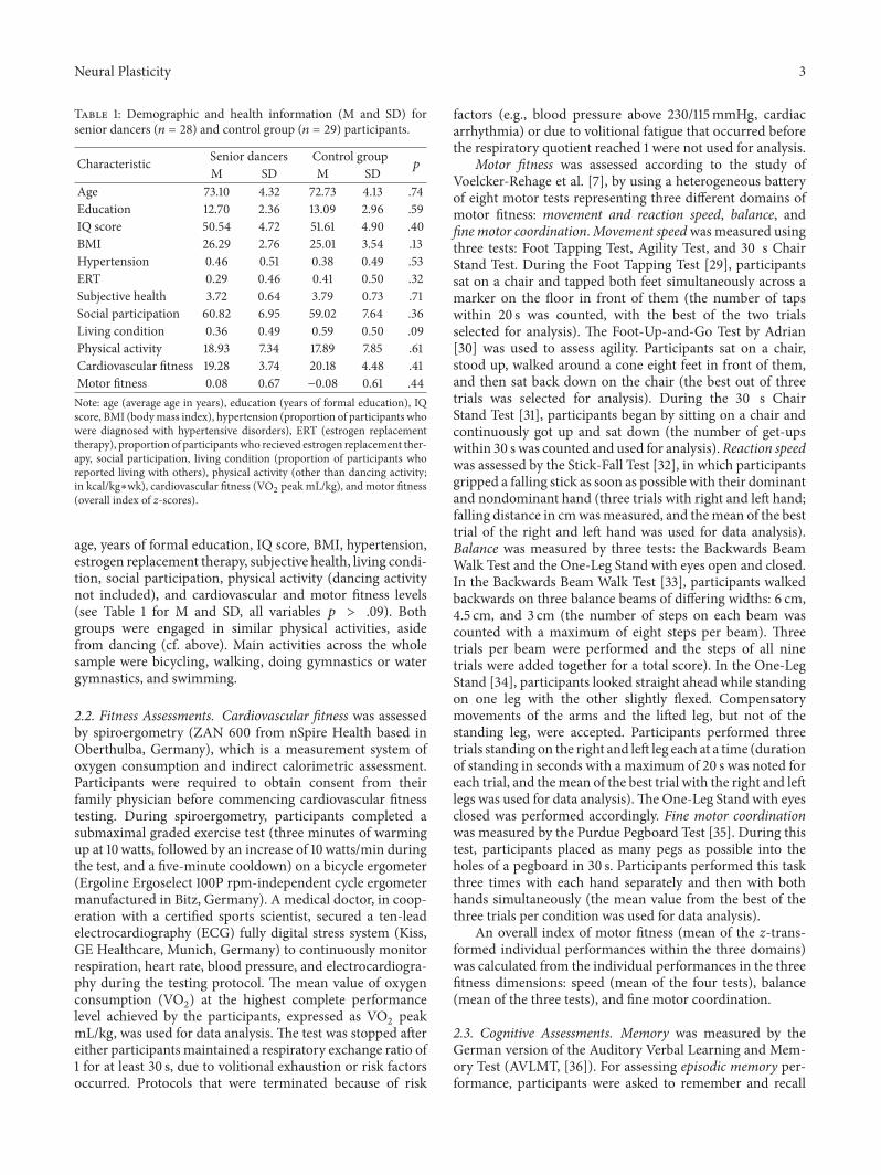
3.3. Voxel-BasedMorphometry Data. T-contrasts revealed noclusters in which senior dancers showed larger GM volumethan controls when results were controlled for familywiseerrors. However, the uncorrected analysis clusters in frontalbrain regions tended to be larger in senior dancers than incontrols (see Figure 1, Table 4). When we tested the oppositecontrast—whether brain volume was larger in control groupparticipants than senior dancers—no cluster was found (datanot shown).
3.4. Additional Correlation Analysis. Across both groups,Pearson product-moment correlation analysis revealed thatICV-adjusted volumes of the left and right hippocampi werenegatively correlated with long-term memory performance,indicating a positive relationship between brain volumeand cognitive performance (see Table 5). Followup linearregression analyses revealed that individual volume of thehippocampus (both hemispheres together) explained 15.1% ofvariance in long-term memory performance (left hippocam-pus: B = −0.07; SE B = 0.90; 𝛽 = −.12; 𝑝 = .45; righthippocampus: B = −1.49; SE B = 0.75; 𝛽 = −.31; 𝑝 = .05).
4. Discussion
In this study, we investigated whether long-term senior danc-ing experience in older women aged 65 to 82 years was
6 Neural Plasticity
Table 2: M and SD for performance in the four cognitive tasks separated for senior dancers (𝑛 = 28) and controls (𝑛 = 29), as well as F- andp values and effect sizes of the respective MANCOVA with age and IQ score as covariates.
Cognitive task Group M SD F(1,55) 𝑝 p𝜂2
Flanker Task (q-score) Senior dancers 1.01 0.26 0.62 .44 .01Control group 1.05 0.28
Visual Search Task (q-score) Senior dancers 1.06 0.39 0.64 .43 .01Control group 1.00 0.27
Memory encoding (sum of 5 trials) Senior dancers 53.00 8.03 0.32 .58 .01Control group 54.59 6.21
Long-term memory (trial 5 – trialdelayed)Senior dancers 2.43 2.17 3.01 .09 .05Control group 1.45 1.57
0
1
2
3
4z = 30
x = 0 y = 50
Figure 1: Regions where gray matter volume was larger (non-significant on cluster level) in senior dancers than control groupparticipants (see also Table 4).
associated with better cognitive performance in the domainsof episodic memory, long-term memory, executive control,and perceptual speed and whether larger GM volume mightexplain these expected cognitive benefits. In contrast toour expectations, we did not find female senior dancersto show better cognitive performances than nonsedentarycontrols. Similarly, in the VBM analysis, no brain regionsrevealed larger volume in senior dancers in comparison tothe control group of similar age, education, IQ score, lifestyleand health factors, and fitness levels. Again, ROI-basedanalysis of hippocampal volume did not show any differencesbetween both groups. Across the whole sample, hippocampalvolume explained 15.1% of variance in long-term memoryperformance.
As outlined in the introduction, findings on dancingactivity and cognitive functioning in older adults are mixed.Our results align with a cross-sectional study by Verghese[15], who did not find better performance in memory (FreeandCued Selective RemindingTest) and several testsmeasur-ing executive control for experienced dancers in comparison
Table 3: M and SD for ICV-adjusted hippocampal volume of theleft and right hemispheres separated for senior dancers (𝑛 = 28) andcontrols (𝑛 = 29), as well as F- and p values and effect sizes of therespective MANCOVA with age and IQ score as covariates.
Brain volume(in cm3) Group M SD F(1,55) 𝑝 p𝜂2
Lefthippocampus
Senior dancers 3.53 0.34 1.50 .23 .03Control group 3.64 0.33
Righthippocampus
Senior dancers 3.81 0.38 0.54 .47 .01Control group 3.91 0.42
to nondancers.Wewere also not able to demonstrate any cog-nitive benefits of multiple years of nonprofessional dancingactivity in these domains, most notably for episodic memoryand perceptual speed (Table 2). This result is in contrastto Kattenstroth and colleagues [17], who revealed betterattentional performance (Nonverbal Geriatric ConcentrationTest [AKT]), as well as higher scores in a taskmeasuring fluidintelligence (Raven Standard Progressive Matrices) in olderadults with long-term dancing experience. As we (as well as[15]) did not measure attention and used other tasks to assessdomains of fluid intelligence, one possible explanation mightbe that cognitive benefits from long-term dancing activity inlate adulthood are highly task-specific.
A further explanation for the dissimilar findings inrelation to past studies might be based on the activity levelof the control group. For example, in a study by Kattenstrothet al. [17], the control group was an inactive sample. Theyreported no regular sporting activities and revealed lowerscores in everyday competence, as well as significantly lowerperformance in the domains of posture, balance, and motorperformance than the older dancers group. In contrast, ourcontrol group participants were similarly active to dancerswith respect to engaging in physical activities like walking,bicycling, or doing gymnastics and also revealed comparablescores in social participation (Table 1). Likelymore importantis the observation that, despite the senior dancers grouphaving a higher weekly energy expenditure based on physicalactivity (regular physical activity plus dancing activity), bothgroups did not differ in cardiovascular and motor fitness(Table 1). This indicates that the additional dancing activitywas not directly associated with improved cardiovascular
Neural Plasticity 7
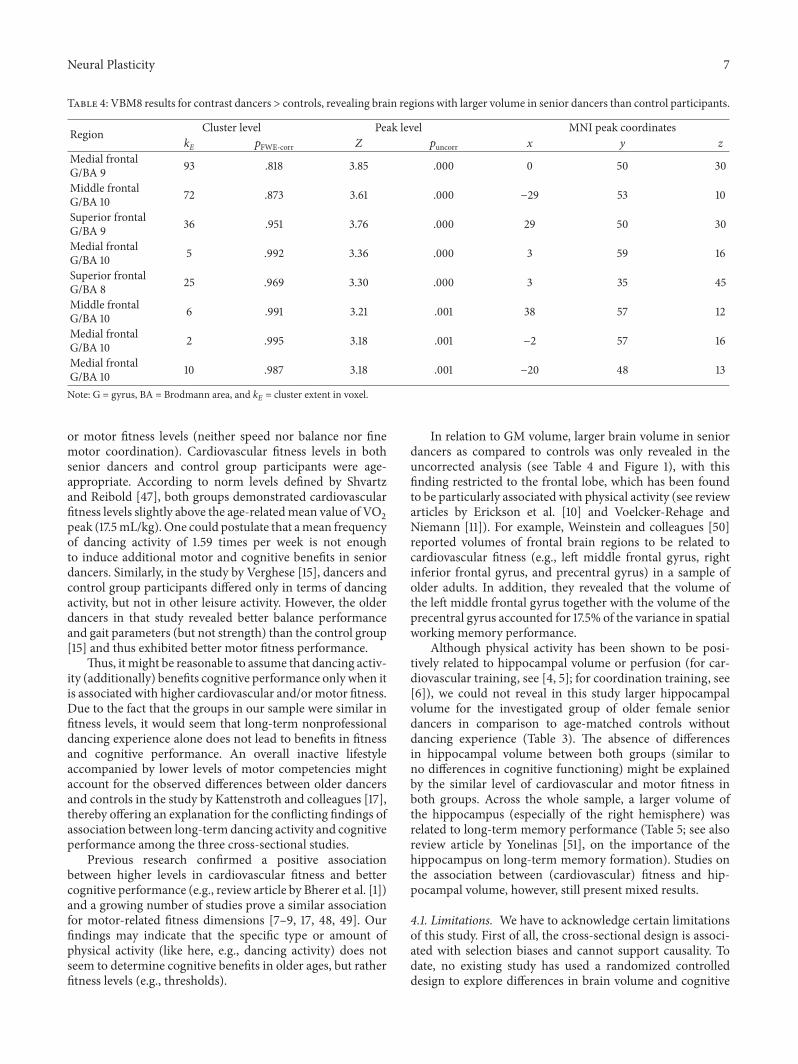
Table 4: VBM8 results for contrast dancers > controls, revealing brain regions with larger volume in senior dancers than control participants.
Region Cluster level Peak level MNI peak coordinateskE 𝑝FWE-corr Z 𝑝uncorr x y z
Medial frontalG/BA 9 93 .818 3.85 .000 0 50 30
Middle frontalG/BA 10 72 .873 3.61 .000 −29 53 10
Superior frontalG/BA 9 36 .951 3.76 .000 29 50 30
Medial frontalG/BA 10 5 .992 3.36 .000 3 59 16
Superior frontalG/BA 8 25 .969 3.30 .000 3 35 45
Middle frontalG/BA 10 6 .991 3.21 .001 38 57 12
Medial frontalG/BA 10 2 .995 3.18 .001 −2 57 16
Medial frontalG/BA 10 10 .987 3.18 .001 −20 48 13
Note: G = gyrus, BA = Brodmann area, and kE = cluster extent in voxel.
or motor fitness levels (neither speed nor balance nor finemotor coordination). Cardiovascular fitness levels in bothsenior dancers and control group participants were age-appropriate. According to norm levels defined by Shvartzand Reibold [47], both groups demonstrated cardiovascularfitness levels slightly above the age-relatedmean value of VO
2
peak (17.5mL/kg). One could postulate that amean frequencyof dancing activity of 1.59 times per week is not enoughto induce additional motor and cognitive benefits in seniordancers. Similarly, in the study by Verghese [15], dancers andcontrol group participants differed only in terms of dancingactivity, but not in other leisure activity. However, the olderdancers in that study revealed better balance performanceand gait parameters (but not strength) than the control group[15] and thus exhibited better motor fitness performance.
Thus, itmight be reasonable to assume that dancing activ-ity (additionally) benefits cognitive performance only when itis associated with higher cardiovascular and/ormotor fitness.Due to the fact that the groups in our sample were similar infitness levels, it would seem that long-term nonprofessionaldancing experience alone does not lead to benefits in fitnessand cognitive performance. An overall inactive lifestyleaccompanied by lower levels of motor competencies mightaccount for the observed differences between older dancersand controls in the study by Kattenstroth and colleagues [17],thereby offering an explanation for the conflicting findings ofassociation between long-termdancing activity and cognitiveperformance among the three cross-sectional studies.
Previous research confirmed a positive associationbetween higher levels in cardiovascular fitness and bettercognitive performance (e.g., review article by Bherer et al. [1])and a growing number of studies prove a similar associationfor motor-related fitness dimensions [7–9, 17, 48, 49]. Ourfindings may indicate that the specific type or amount ofphysical activity (like here, e.g., dancing activity) does notseem to determine cognitive benefits in older ages, but ratherfitness levels (e.g., thresholds).
In relation to GM volume, larger brain volume in seniordancers as compared to controls was only revealed in theuncorrected analysis (see Table 4 and Figure 1), with thisfinding restricted to the frontal lobe, which has been foundto be particularly associated with physical activity (see reviewarticles by Erickson et al. [10] and Voelcker-Rehage andNiemann [11]). For example, Weinstein and colleagues [50]reported volumes of frontal brain regions to be related tocardiovascular fitness (e.g., left middle frontal gyrus, rightinferior frontal gyrus, and precentral gyrus) in a sample ofolder adults. In addition, they revealed that the volume ofthe left middle frontal gyrus together with the volume of theprecentral gyrus accounted for 17.5% of the variance in spatialworking memory performance.
Although physical activity has been shown to be posi-tively related to hippocampal volume or perfusion (for car-diovascular training, see [4, 5]; for coordination training, see[6]), we could not reveal in this study larger hippocampalvolume for the investigated group of older female seniordancers in comparison to age-matched controls withoutdancing experience (Table 3). The absence of differencesin hippocampal volume between both groups (similar tono differences in cognitive functioning) might be explainedby the similar level of cardiovascular and motor fitness inboth groups. Across the whole sample, a larger volume ofthe hippocampus (especially of the right hemisphere) wasrelated to long-term memory performance (Table 5; see alsoreview article by Yonelinas [51], on the importance of thehippocampus on long-term memory formation). Studies onthe association between (cardiovascular) fitness and hip-pocampal volume, however, still present mixed results.
4.1. Limitations. We have to acknowledge certain limitationsof this study. First of all, the cross-sectional design is associ-ated with selection biases and cannot support causality. Todate, no existing study has used a randomized controlleddesign to explore differences in brain volume and cognitive
8 Neural Plasticity
Table 5: Pearson product-moment correlation coefficient (r) andsignificance level (p) between individual cognitive performances inthe four cognitive tasks and individual brain volumes of the left andright hippocampi.
Cognitivetask
Left hippocampus Right hippocampusr p r p
Flanker Task(𝑞-score) .01 .46 −.04 .38
Visual SearchTask(𝑞-score)
.07 .31 −.02 .46
Memoryencoding <−.01 .49 .04 .39
Long-termmemory −.30 .01 −.38 <.01
performance based on senior dance activity in older adults.Therefore, additional research using randomized controlledtrials to test the efficacy of dancing on functional andstructural MRI measures in activity-matched older adultsis warranted. Secondly, sample size in the current study ismoderate (𝑁 = 57), but comparable to other investigationsstudying the impact of physical fitness on MRI parametersin older adults (cf. Bugg and Head [52], Colcombe et al. [2];Colcombe et al. [23]). Thus, we assume that the reported nullresults are not due to a lack of statistical power. Moreover,cardiovascular and motor fitness parameters did not differbetween long-term senior dancers and control group partici-pants. The physical activity level of both groups differed onlyin that senior dancers participated in a senior dancing classan additional 1.59 times per week, on average. Knowing thatfitness level has been shown to positively impact functionaland structural MRI parameters might be a precondition fordemonstrating differences in physical activity effects in anolder adult’s brain.
5. Conclusions
In this study, we investigated the association of long-termsenior dance experience in older women on cognitive func-tioning and brain volume, in comparison to control groupparticipants who lack dancing experience but are similarlyactive in other leisure activities and reveal comparable fitnesslevels. Findings showed that senior dancers did not benefit inthe cognitive domains of executive control, perceptual speed,episodic memory, and long-term memory performance.Moreover, dancing experience showed no effects on GMvolume. These results might be explained by the comparablefitness levels (cardiovascular fitness and motor fitness) ofboth study groups.We assume that differences in fitness levelsor minimum (threshold) fitness levels have more impact oncognitive functioning and brain volume than the type ofperformed physical activity.
Competing Interests
The authors declare that there are no competing interestsregarding the publication of this paper.
Authors’ Contributions
Claudia Niemann conceived, designed, and performed theexperiments, analyzed the data, and wrote the paper. ClaudiaVoelcker-Rehage conceived and designed the experiments,supported data analysis, and wrote the paper. Ben Goddesupported data analysis and proofread the paper.
Acknowledgments
This project was supported by a grant from the Robert BoschFoundation (32.5.3310.0041.0) to Claudia Niemann and fromthe German Association of Senior Dance e.V. to ClaudiaVoelcker-Rehage and Claudia Niemann. The authors wouldlike to thank Lisa Breidenbach, Gabriele Hellenthal, JoonNikolasHille,MalinaHobbie, and LenaHubner for their helpwith data collection; Peter Erhard and JochenHirsch for theirhelp with planning and conducting the MRI scans; and alltest subjects for participating in the study. The publicationcosts of this article were funded by the German ResearchFoundation/DFG and the Technische Universitat Chemnitzin the funding programme Open Access Publishing.
References
[1] L. Bherer, K. I. Erickson, and T. Liu-Ambrose, “Physical exerciseand brain functions in older adults,” Journal of Aging Research,vol. 2013, Article ID 197326, 2 pages, 2013.
[2] S. Colcombe and A. F. Kramer, “Fitness effects on the cognitivefunction of older adults: a meta-analytic study,” PsychologicalScience, vol. 14, no. 2, pp. 125–130, 2003.
[3] M. Hamer and Y. Chida, “Physical activity and risk of neurode-generative disease: a systematic review of prospective evidence,”Psychological Medicine, vol. 39, no. 1, pp. 3–11, 2009.
[4] K. I. Erickson,M.W.Voss, R. S. Prakash et al., “Exercise trainingincreases size of hippocampus and improves memory,” Proceed-ings of the National Academy of Sciences of the United States ofAmerica, vol. 108, no. 7, pp. 3017–3022, 2011.
[5] A. Maass, S. Duzel, M. Goerke et al., “Vascular hippocampalplasticity after aerobic exercise in older adults,” MolecularPsychiatry, vol. 20, no. 5, pp. 585–593, 2015.
[6] C. Niemann, B. Godde, and C. Voelcker-Rehage, “Not only car-diovascular, but also coordinative exercise increases hippocam-pal volume in older adults,” Frontiers in Aging Neuroscience, vol.6, article 170, 2014.
[7] C. Voelcker-Rehage, B. Godde, and U. M. Staudinger, “Physicaland motor fitness are both related to cognition in old age,”TheEuropean Journal ofNeuroscience, vol. 31, no. 1, pp. 167–176, 2010.
[8] C. Voelcker-Rehage, B. Godde, and U. M. Staudinger, “Car-diovascular and coordination training differentially improvecognitive performance and neural processing in older adults,”Frontiers in Human Neuroscience, vol. 5, article 26, 2011.
[9] C. Niemann, B. Godde, U. M. Staudinger, and C. Voelcker-Rehage, “Exercise-induced changes in basal ganglia volume andcognition in older adults,” Neuroscience, vol. 281, pp. 147–163,2014.
[10] K. I. Erickson, R. L. Leckie, and A. M. Weinstein, “Physicalactivity, fitness, and graymatter volume,”Neurobiology of Aging,vol. 35, supplement 2, pp. S20–S28, 2014.
[11] C. Voelcker-Rehage and C. Niemann, “Structural and func-tional brain changes related to different types of physical activity
Neural Plasticity 9
across the life span,” Neuroscience and Biobehavioral Reviews,vol. 37, no. 9, pp. 2268–2295, 2013.
[12] US Department of Health and Human Services, Physical Activ-ity and Health: A Report of the Surgeon General, Edited by USDepartment ofHealth andHuman Services, Centers forDiseaseControl and Prevention, National Center for Chronic DiseasePrevention and Health Promotion, US Department of Healthand Human Services, Atlanta, Ga, USA, 1996.
[13] K. Fabel, S. A. Wolf, D. Ehninger, H. Babu, P. Leal-Galicia,and G. Kempermann, “Additive effects of physical exercise andenvironmental enrichment on adult hippocampal neurogenesisin mice,” Frontiers in Neuroscience, vol. 3, article 50, 2009.
[14] J. Verghese, R. B. Lipton, M. J. Katz et al., “Leisure activities andthe risk of dementia in the elderly,”The New England Journal ofMedicine, vol. 348, no. 25, pp. 2508–2516, 2003.
[15] J. Verghese, “Cognitive and mobility profile of older socialdancers,” Journal of the American Geriatrics Society, vol. 54, no.8, pp. 1241–1244, 2006.
[16] P. T. Alpert, S. K. Miller, H. Wallmann et al., “The effect ofmodified jazz dance on balance, cognition, and mood in olderadults,” Journal of the American Academy of Nurse Practitioners,vol. 21, no. 2, pp. 108–115, 2009.
[17] J.-C. Kattenstroth, I. Kolankowska, T. Kalisch, and H. R. Dinse,“Superior sensory, motor, and cognitive performance in elderlyindividuals with multi-year dancing activities,” Frontiers inAging Neuroscience, vol. 2, article 31, 2010.
[18] J.-C. Kattenstroth, T. Kalisch, S. Holt, M. Tegenthoff, and H. R.Dinse, “Six months of dance intervention enhances postural,sensorimotor, and cognitive performance in elderly withoutaffecting cardio-respiratory functions,” Frontiers in Aging Neu-roscience, vol. 5, article 5, 2013.
[19] A. M. Fjell and K. B. Walhovd, “Structural brain changes inaging: courses, causes and cognitive consequences,” Reviews inthe Neurosciences, vol. 21, no. 3, pp. 187–221, 2010.
[20] N. Raz, U. Lindenberger, K. M. Rodrigue et al., “Regionalbrain changes in aging healthy adults: general trends, individualdifferences and modifiers,” Cerebral Cortex, vol. 15, no. 11, pp.1676–1689, 2005.
[21] S.-C. Li, U. Lindenberger, B. Hommel, G. Aschersleben, W.Prinz, and P. B. Baltes, “Transformations in the couplingsamong intellectual abilities and constituent cognitive processesacross the life span,” Psychological Science, vol. 15, no. 3, pp. 155–163, 2004.
[22] M. F. Folstein, S. E. Folstein, and P. R. McHugh, “‘Mini-mentalstate’. A practical method for grading the cognitive state ofpatients for the clinician,” Journal of Psychiatric Research, vol.12, no. 3, pp. 189–198, 1975.
[23] S. J. Colcombe, A. F. Kramer, K. I. Erickson et al., “Cardiovas-cular fitness, cortical plasticity, and aging,” Proceedings of theNational Academy of Sciences of the United States of America,vol. 101, no. 9, pp. 3316–3321, 2004.
[24] J. A. H. Baecke, J. Burema, and J. E. R. Frijters, “A short ques-tionnaire for the measurement of habitual physical activity inepidemiological studies,”American Journal of Clinical Nutrition,vol. 36, no. 5, pp. 936–942, 1982.
[25] P. Wagner and R. Singer, “Ein fragebogen zur erfassungder habituellen korperlichen aktivitat verschiedener bevolker-ungsgruppen,” Sportwissenschaft, vol. 33, no. 4, pp. 383–397,2003.
[26] B. E.Ainsworth,W. L.Haskell,M.C.Whitt et al., “Compendiumof physical activities: an update of activity codes and MET
intensities,”Medicine and Science in Sports and Exercise, vol. 32,no. 9, supplement, pp. S498–S504, 2000.
[27] B. E. Ainsworth, W. L. Haskell, A. S. Leon et al., “Compendiumof physical activities: classification of energy costs of humanphysical activities,”Medicine and Science in Sports and Exercise,vol. 25, no. 1, pp. 71–74, 1993.
[28] M. J. Aartsen, C. H. M. Smits, T. van Tilburg, K. C. P. M.Knipscheer, and D. J. H. Deeg, “Activity in older adults: cause orconsequence of cognitive functioning? A longitudinal study oneveryday activities and cognitive performance in older adults,”The Journals of Gerontology, Series B: Psychological Sciences andSocial Sciences, vol. 57, no. 2, pp. P153–P162, 2002.
[29] C. Voelcker-Rehage and O. Wiertz, Die Lernfahigkeit sportmo-torischer Fertigkeiten im Lichte der Entwicklungspsychologie derLebensspanne, Universitat Bielefeld, Bielefeld, Germany, 2003.
[30] M. J. Adrian, “Flexibility in the aging adult,” in Exercises andAging: The Scientific Basic, E. L. Smith and R. C. Serfass, Eds.,pp. 45–58, Enslow Publishers, Hillside, NJ, USA, 1981.
[31] R. E. Rikli and C. J. Jones, Senior Fitness Test Manual. Develop-ment and Validation of a Functional Fitness Test for Community-Residing Older Adults, Human Kinetics, Champaign, Ill, USA,2001.
[32] W. Ehrler and M. Huth, “Korperliche leistungsfahigkeit 43- bis45-jahriger und 62- bis 64-jahriger imVergleich,” inAspekte derEntwicklung imMittleren undHoheren Lebensalter, P.Martin, K.U. Ettrich,U. Lehr,D. Roether, andA. Fischer-Cyrulies, Eds., pp.220–334, Steinkopf, Darmstadt, Germany, 2000.
[33] E. J. Kiphard and F. Schilling, Korperkoordinationstest furKinder, Beltz, Weinheim, Germany, 1974.
[34] C. Ekdahl, G. B. Jarnlo, and S. I. Andersson, “Standing balancein healthy subjects. Evaluation of a quantitative test batteryon a force platform,” Scandinavian Journal of RehabilitationMedicine, vol. 21, no. 4, pp. 187–195, 1989.
[35] J. Tiffin and E. J. Asher, “The Purdue Pegboard: norms andstudies of reliability and validity,” Journal of Applied Psychology,vol. 32, no. 3, pp. 234–247, 1948.
[36] C. Helmstaedter, M. Lendt, and S. Lux, Verbaler Lern- undMerkfahigkeitstest, Hogrefe, Gottingen, Germany, 2001.
[37] A. H. Winneke, B. Godde, E.-M. Reuter, S. Vieluf, and C.Voelcker-Rehage, “The association between physical activityand attentional control in younger and older middle-agedadults: an ERP study,” The Journal of Gerontopsychology andGeriatric Psychiatry, vol. 25, no. 4, pp. 207–221, 2012.
[38] C. T. Gualtieri and L. G. Johnson, “A computerized test batterysensitive to mild and severe brain injury,” Medscape GeneralMedicine, vol. 10, no. 4, article 90, 2008.
[39] B. Hommel, K. Z. H. Li, and S.-C. Li, “Visual search across thelife span,”Developmental Psychology, vol. 40, no. 4, pp. 545–558,2004.
[40] K. I. Erickson, R. S. Prakash, M. W. Voss et al., “Aerobic fitnessis associated with hippocampal volume in elderly humans,”Hippocampus, vol. 19, no. 10, pp. 1030–1039, 2009.
[41] A. Floel, R. Ruscheweyh, K. Kruger et al., “Physical activity andmemory functions: are neurotrophins and cerebral gray mattervolume the missing link?” NeuroImage, vol. 49, no. 3, pp. 2756–2763, 2010.
[42] R. Ruscheweyh, C. Willemer, K. Kruger et al., “Physical activityand memory functions: an interventional study,” Neurobiologyof Aging, vol. 32, no. 7, pp. 1304–1319, 2011.
[43] S.M.Hayes, J. P. Hayes,M. Cadden, andM.Verfaellie, “A reviewof cardiorespiratory fitness-related neuroplasticity in the agingbrain,” Frontiers in Aging Neuroscience, vol. 5, article 31, 2013.
10 Neural Plasticity
[44] N. Raz, K. M. Rodrigue, K. M. Kennedy, D. Head, F. Gunning-Dixon, and J. D. Acker, “Differential aging of the humanstriatum: longitudinal evidence,” American Journal of Neurora-diology, vol. 24, no. 9, pp. 1849–1856, 2003.
[45] S. J. Ritchie, T. Booth,M. d. C. Valdes Hernandez et al., “Beyonda bigger brain: multivariable structural brain imaging andintelligence,” Intelligence, vol. 51, pp. 47–56, 2015.
[46] U. Basten, K. Hilger, and C. J. Fiebach, “Where smart brains aredifferent: a quantitative meta-analysis of functional and struc-tural brain imaging studies on intelligence,” Intelligence, vol. 51,pp. 10–27, 2015.
[47] E. Shvartz and R. C. Reibold, “Aerobic fitness norms for malesand females aged 6 to 75 years: a review,” Aviation Space andEnvironmental Medicine, vol. 61, no. 1, pp. 3–11, 1990.
[48] N. Berryman, L. Bherer, S. Nadeau et al., “Executive functions,physical fitness and mobility in well-functioning older adults,”Experimental Gerontology, vol. 48, no. 12, pp. 1402–1409, 2013.
[49] N. Berryman, L. Bherer, S. Nadeau et al., “Multiple roads lead toRome: combined high-intensity aerobic and strength trainingvs. gross motor activities leads to equivalent improvement inexecutive functions in a cohort of healthy older adults,”Age, vol.36, no. 5, article 9710, 2014.
[50] A. M. Weinstein, M. W. Voss, R. S. Prakash et al., “The associa-tion between aerobic fitness and executive function is mediatedby prefrontal cortex volume,” Brain, Behavior, and Immunity,vol. 26, no. 5, pp. 811–819, 2012.
[51] A. P. Yonelinas, “The hippocampus supports high-resolutionbinding in the service of perception, working memory andlong-term memory,” Behavioural Brain Research, vol. 254, pp.34–44, 2013.
[52] J. M. Bugg and D. Head, “Exercise moderates age-related atro-phy of the medial temporal lobe,”Neurobiology of Aging, vol. 32,no. 3, pp. 506–514, 2011.

Submit your manuscripts athttp://www.hindawi.com
Neurology Research International
Hindawi Publishing Corporationhttp://www.hindawi.com Volume 2014
Alzheimer’s DiseaseHindawi Publishing Corporationhttp://www.hindawi.com Volume 2014
International Journal of
ScientificaHindawi Publishing Corporationhttp://www.hindawi.com Volume 2014
Hindawi Publishing Corporationhttp://www.hindawi.com Volume 2014
BioMed Research International
Hindawi Publishing Corporationhttp://www.hindawi.com Volume 2014
Research and TreatmentSchizophrenia
The Scientific World JournalHindawi Publishing Corporation http://www.hindawi.com Volume 2014
Hindawi Publishing Corporationhttp://www.hindawi.com Volume 2014
Neural Plasticity
Hindawi Publishing Corporationhttp://www.hindawi.com Volume 2014
Parkinson’s Disease
Hindawi Publishing Corporationhttp://www.hindawi.com Volume 2014
Research and TreatmentAutism
Sleep DisordersHindawi Publishing Corporationhttp://www.hindawi.com Volume 2014
Hindawi Publishing Corporationhttp://www.hindawi.com Volume 2014
Neuroscience Journal
Epilepsy Research and TreatmentHindawi Publishing Corporationhttp://www.hindawi.com Volume 2014
Hindawi Publishing Corporationhttp://www.hindawi.com Volume 2014
Psychiatry Journal
Hindawi Publishing Corporationhttp://www.hindawi.com Volume 2014
Computational and Mathematical Methods in Medicine
Depression Research and TreatmentHindawi Publishing Corporationhttp://www.hindawi.com Volume 2014
Hindawi Publishing Corporationhttp://www.hindawi.com Volume 2014
Brain ScienceInternational Journal of
StrokeResearch and TreatmentHindawi Publishing Corporationhttp://www.hindawi.com Volume 2014
Neurodegenerative Diseases
Hindawi Publishing Corporationhttp://www.hindawi.com Volume 2014
Journal of
Cardiovascular Psychiatry and NeurologyHindawi Publishing Corporationhttp://www.hindawi.com Volume 2014