Embed Size (px)
Citation preview
Nannini et al. BMC Cancer 2014, 14:685http://www.biomedcentral.com/1471-2407/14/685
RESEARCH ARTICLE Open Access
Integrated genomic study of quadruple-WTGIST (KIT/PDGFRA/SDH/RAS pathway wild-typeGIST)Margherita Nannini1, Annalisa Astolfi2, Milena Urbini2, Valentina Indio2, Donatella Santini3, Michael C Heinrich4,Christopher L Corless5, Claudio Ceccarelli3, Maristella Saponara2, Anna Mandrioli1, Cristian Lolli1, Giorgio Ercolani6,Giovanni Brandi1, Guido Biasco1,2 and Maria A Pantaleo1,2*
Abstract
Background: About 10-15% of adult gastrointestinal stromal tumors (GIST) and the vast majority of pediatric GISTdo not harbour KIT or platelet-derived growth factor receptor alpha (PDGFRA) mutations (J Clin Oncol 22:3813–3825,2004; Hematol Oncol Clin North Am 23:15–34, 2009). The molecular biology of these GIST, originally defined as KIT/PDGFRA wild-type (WT), is complex due to the existence of different subgroups with distinct molecular hallmarks,including defects in the succinate dehydrogenase (SDH) complex and mutations of neurofibromatosis type 1 (NF1), BRAF,or KRAS genes (RAS-pathway or RAS-P).In this extremely heterogeneous landscape, the clinical profile and molecular abnormalities of the small subgroupof WT GIST suitably referred to as quadruple wild-type GIST (quadrupleWT or KITWT/PDGFRAWT/SDHWT/RAS-PWT)remains undefined. The aim of this study is to investigate the genomic profile of KITWT/PDGFRAWT/SDHWT/RAS-PWT
GIST, by using a massively parallel sequencing and microarray approach, and compare it with the genomic profileof other GIST subtypes.
Methods: We performed a whole genome analysis using a massively parallel sequencing approach on a total of16 GIST cases (2 KITWT/PDGFRAWT/SDHWT and SDHBIHC+/SDHAIHC+, 2 KITWT/PDGFRAWT/SDHAmut and SDHBIHC-/SDHAIHC- and 12 cases of KITmut or PDGFRAmut GIST). To confirm and extend the results, whole-genome geneexpression analysis by microarray was performed on 9 out 16 patients analyzed by RNAseq and an additional 20GIST patients (1 KITWT/PDGFRAWT SDHAmut GIST and 19 KITmut or PDGFRAmut GIST). The most impressive data werevalidated by quantitave PCR and Western Blot analysis.
Results: We found that both cases of quadrupleWT GIST had a genomic profile profoundly different from botheither KIT/PDGFRA mutated or SDHA-mutated GIST. In particular, the quadrupleWT GIST tumors are characterized bythe overexpression of molecular markers (CALCRL and COL22A1) and of specific oncogenes including tyrosine andcyclin- dependent kinases (NTRK2 and CDK6) and one member of the ETS-transcription factor family (ERG).(Continued on next page)
* Correspondence: [email protected] of Specialized, Experimental and Diagnostic Medicine,Sant’Orsola-Malpighi Hospital, University of Bologna, Via Massarenti 9, 40138Bologna, Italy2“Giorgio Prodi” Cancer Research Center, University of Bologna, Bologna, ItalyFull list of author information is available at the end of the article
© 2014 Nannini et al.; licensee BioMed Central Ltd. This is an Open Access article distributed under the terms of the CreativeCommons Attribution License (http://creativecommons.org/licenses/by/2.0), which permits unrestricted use, distribution, andreproduction in any medium, provided the original work is properly credited. The Creative Commons Public DomainDedication waiver (http://creativecommons.org/publicdomain/zero/1.0/) applies to the data made available in this article,unless otherwise stated.
Nannini et al. BMC Cancer 2014, 14:685 Page 2 of 12http://www.biomedcentral.com/1471-2407/14/685
(Continued from previous page)
Conclusion: We report for the first time an integrated genomic picture of KITWT/PDGFRAWT/SDHWT/RAS-PWT GIST,using massively parallel sequencing and gene expression analyses, and found that quadrupleWT GIST have anexpression signature that is distinct from SDH-mutant GIST as well as GIST harbouring mutations in KIT or PDGFRA.Our findings suggest that quadrupleWT GIST represent another unique group within the family of gastrointestintalstromal tumors.
Keywords: Gastrointestinal stromal tumors (GIST), Wild-type, KIT, PDGFRA, Succinate dehydrogenase, SDHA, RAS,QuadrupleWT
BackgroundAbout 10-15% of adult gastrointestinal stromal tumors (GIST)and the vast majority of pediatric GIST do not harbourKIT or platelet-derived growth factor receptor alpha(PDGFRA) mutations [1,2]. These GIST were originallydefined as KIT/PDGFRA wild-type (KITWT/PDGFRAWT)and generally are less sensitive to tyrosine-kinase inhibi-tors [3-5]. Their molecular biology is heterogeneous asevidence by the existence of different subgroups withdistinct molecular abnormalities (Figure 1). KITWT/PDGFRAWT GIST can be divided into two main groupsaccording to the succinate dehydrogenase subunit B(SDHB) immunohistochemical status (IHC): SDHB posi-tive (SDHBIHC+), or type 1 GIST which, includes neuro-fibromatosis type 1 (NF1)-mutated GIST and somesporadic KITWT/PDGFRAWT GIST. The second group ofKITWT/PDGFRAWT, called as type 2 GIST, is character-ized by a lack of SDHB protein expression (SDHBIHC-). Insome cases SDHBIHC- is due to germline and/or de novomutations of any of the four SDH subunits (SDHAmut)[6-8]. The SDHBIHC- includes additional subgroups thatcan be distinguished on the basis of the SDHA IHC status,which strictly correlates with the presence of SDHA-inactivating mutations (SDHAmut) [9-16]. In particular,SDHBIHC-/SDHAIHC- GIST include a subgroup of youngadult women patients with a well defined clinical and bio-logical profile, generally characterized by the gastric primarytumour localization, a predominantly mixed epithelioid andspindle cell morphology, diffuse IHC positivity for KIT anddiscovered on gastrointestinal stromal tumours 1 (DOG1),frequent lymph node metastases, and an indolent course ofdisease even if metastasis is present [17]. Moreover, theyare characterized by overexpression of the insulin growthfactor 1 receptor (IGF1R) [18-21]. On the contrary,SDHBIHC-, but SDHAIHC+ subgroup include 1) cases of syn-dromic GIST arising from the Carney-Stratakis Syndrome(CSS), that are characterized by SDHB, SDHC or SDHD in-activating mutations (SDHBmut, SDHCmut, or SDHDmut);and 2) cases of Carney Triad (CT), that lack SDHx-muta-tions [6,22-24]. More rarely, SDHBIHC-/SDHAIHC+ sub-group may include sporadic KITWT/PDGFRAWT GISTcharacterized by SDHB, −C or D mutations (most of themgermline, and in few cases by SDHA mutations), arising
mainly from the stomach, with a lesser female prevalence,but histologically similar to SDHAIHC- GIST [15].The SDHBIHC+ subgroup includes cases of NF1-mu-
tated GIST, that are commonly intestinal, multifocal andhave an IGF1R negative staining, and also sporadicKITWT/PDGFRAWT GIST, arising in the adult from anypart of gastrointestinal tract [15,21,25]. In about 15% ofcases of sporadic KITWT/PDGFRAWT GIST there maybe an activating mutation in BRAF or, more rarely, RAS[26-28]. Taken together, cases of BRAF, RAS, or NF1mutant GIST can be referred to as RAS-pathway (RAS-P)mutant GIST (RAS-Pmut).In this extremely heterogeneous landscape, the clinical
profile and molecular abnormalities of the small sub-group of WT GIST suitably referred to as quadruplewild-type GIST (quadrupleWT or KITWT/PDGFRAWT/SDHWT/RAS-PWT) remains undefined [29]. The aim ofthis study is to investigate the genomic profile ofKITWT/PDGFRAWT/SDHWT/RAS-PWT GIST, by using amassively parallel sequencing and microarray approach,and compare it with the genomic profile of other GISTsubtypes.
Results and discussionWhole-Transcriptome Paired-End RNA Sequencing andcopy number analysisWhole-Transcriptome Paired-End RNA Sequencing wasperformed on a total of 16 GIST samples, of which 2were KITWT/PDGFRAWT without SDH-inactivating mu-tations and SDHBIHC+/SDHAIHC+ (GIST_133 andGIST_127), 2 were KITWT/PDGFRAWT/SDHAmut andSDHBIHC-/SDHAIHC- (GIST_7 and GIST_10), and 12were KITmut or PDGFRAmut. The principal componentanalysis showed that both GIST_133 and GIST_127(KITWT/PDGFRAWT/SDHWT and SDHBIHC+/SDHAIHC+)are characterized by a gene expression profile profoundlydifferent from both GIST_7 and GIST_10 (KITWT/PDGFRAWT/SDHAmut and SDHBIHC/SDHAIHC), whileclustering in proximity of a subset of KITmut or PDGFRAmut
GIST (Figure 2A).To investigate the presence of novel mutations or
small ins/del in the whole coding regions of KIT andPDGFRA we analyzed whole transcriptome sequencing

Figure 1 The complexity of KITWT/PDGFRAWT GIST molecular biology. KITWT/PDGFRAWT GIST could be firstly divided two main groupaccording to the SDHB immunohistochemical status: SDHBIHC+ (including NF1-mutated GIST and sporadic KITWT/PDGFRAWT GIST with or withoutKRAS/BRAF mutations) and SDHBIHC- or SDH-deficient GIST. The latter could be further divided according to the SDHA immunohistochemical status:SDHBIHC-/SDHAIHC- GIST (pediatric type or young adult GIST characterized by germline or somatic inactivating SDHA mutations) and SDHBIHC/SDHAIHC+ GIST (including Carney-Stratakis Syndrome-related GIST, characterized by germline or somatic inactivating SDHB, −C, −D mutations,Carney Triad-related GIST that lack SDHx mutations, and sporadic KITWT/PDGFRAWT GIST, characterized by germline or somatic inactivating SDHB, −C, −Dmutations and SDHA mutations, reported in only three cases [15]. In red the subset of KITWT/PDGFRAWT GIST referred to as quadrupleWT GIST (KITWT/PDGFRAWT/SDHWT/RAS-PWT), that represent the subject of this study.
Figure 2 Principal Component Analysis (PCA) performed on samples analyzed with RNA-seq (Figure 2A) and microarray (Figure 2B).In both cases the patients with SDHA mutations are arranged in a strongly separated cluster (yellow points), as were the KITWT/PDGFRAWT/SDHWT/RAS-PWT samples (red point) although closer to KIT or PDGFRA mutated (respectively blue and green point).
Nannini et al. BMC Cancer 2014, 14:685 Page 3 of 12http://www.biomedcentral.com/1471-2407/14/685
Nannini et al. BMC Cancer 2014, 14:685 Page 4 of 12http://www.biomedcentral.com/1471-2407/14/685
data for single nucleotide variant (SNV) and found noprivate or cryptic mutations. Moreover, no NF-1, BRAF,RAS mutations were found by whole transcriptome se-quencing. Therefore, the GIST from these two patientswere KITWT/PDGFRAWT/SDHWT/RAS-PWT, or quadru-pleWT GIST. Analysis of deleterious mutations fromwhole transcriptome sequencing did not identify anyknown oncogenic event or shared alteration in the twopatients (Additional file 1: Table S1). Copy number ana-lysis was performed on the two KITWT/PDGFRAWT/SDHWT/RAS-PWT GIST: GIST_133 showed no genomicimbalances, while GIST_127 harbors several macroscopiccytogenetic alterations, including loss of chromosomearms 14q and 22q frequently observed in KIT/PDGFRAmutated GIST.
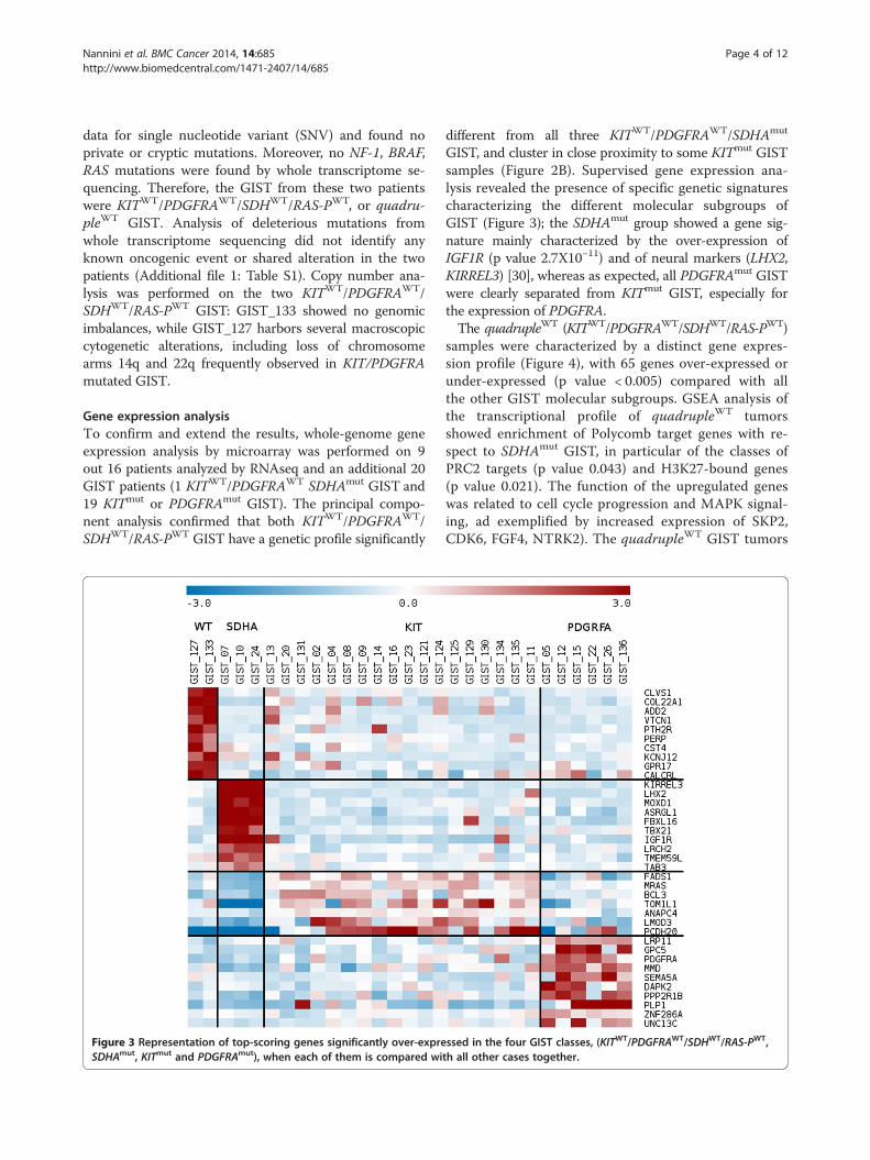
Gene expression analysisTo confirm and extend the results, whole-genome geneexpression analysis by microarray was performed on 9out 16 patients analyzed by RNAseq and an additional 20GIST patients (1 KITWT/PDGFRAWT SDHAmut GIST and19 KITmut or PDGFRAmut GIST). The principal compo-nent analysis confirmed that both KITWT/PDGFRAWT/SDHWT/RAS-PWT GIST have a genetic profile significantly
Figure 3 Representation of top-scoring genes significantly over-expreSDHAmut, KITmut and PDGFRAmut), when each of them is compared wi
different from all three KITWT/PDGFRAWT/SDHAmut
GIST, and cluster in close proximity to some KITmut GISTsamples (Figure 2B). Supervised gene expression ana-lysis revealed the presence of specific genetic signaturescharacterizing the different molecular subgroups ofGIST (Figure 3); the SDHAmut group showed a gene sig-nature mainly characterized by the over-expression ofIGF1R (p value 2.7X10⁻¹¹) and of neural markers (LHX2,KIRREL3) [30], whereas as expected, all PDGFRAmut GISTwere clearly separated from KITmut GIST, especially forthe expression of PDGFRA.The quadrupleWT (KITWT/PDGFRAWT/SDHWT/RAS-PWT)
samples were characterized by a distinct gene expres-sion profile (Figure 4), with 65 genes over-expressed orunder-expressed (p value < 0.005) compared with allthe other GIST molecular subgroups. GSEA analysis ofthe transcriptional profile of quadrupleWT tumorsshowed enrichment of Polycomb target genes with re-spect to SDHAmut GIST, in particular of the classes ofPRC2 targets (p value 0.043) and H3K27-bound genes(p value 0.021). The function of the upregulated geneswas related to cell cycle progression and MAPK signal-ing, ad exemplified by increased expression of SKP2,CDK6, FGF4, NTRK2). The quadrupleWT GIST tumors
ssed in the four GIST classes, (KITWT/PDGFRAWT/SDHWT/RAS-PWT,th all other cases together.

Figure 4 Unsupervised hierarchical clustering representation of differential expressed genes (P-value < 0.005) in KITWT/PDGFRAWT/SDHWT/RAS-PWT GIST with respect to the other GIST classes (SDHxmut, KITmut and PDGFRAmut).
Nannini et al. BMC Cancer 2014, 14:685 Page 5 of 12http://www.biomedcentral.com/1471-2407/14/685
are characterized by the overexpression of molecularmarkers (CALCRL and COL22A1) and of specific oncogenesincluding tyrosine and cyclin- dependent kinases (NTRK2and CDK6) and one member of the ETS-transcriptionfactor family (ERG). Overexpression of CALCRL, COL22A1,NTRK2 (TrkB) and of the ETS-transcription factor ERGwas confirmed by quantitative PCR, showing that only theKITWT/PDGFRAWT/SDHWT/RAS-PWT GIST subgroupexpressed these molecular markers and possible therapeutictargets (Figure 5). NTRK2 protein expression level was alsoevaluated by Western Blot analysis and its overexpressionin quadrupleWT GIST was confirmed (Additional file 2:Figure S1). No mutations, gene fusions or amplificationswere identified in NTRK2 and ERG.
DiscussionThe pathogenesis and underlying biology of KITWT/PDGFRAWT with intact SDH complex (SDHxWT) and
non-mutated RAS-pathway members (RAS-PWT) suit-ably referred to as quadrupleWT GIST remains un-defined. In the present study, we performed a wholegenome analysis using a massively parallel sequencingapproach on a total of 16 GIST cases that included 2KITWT/PDGFRAWT/SDHWT and SDHBIHC+/SDHAIHC+,, 2KITWT/PDGFRAWT/SDHAmut and SDHBIHC-/SDHAIHC-
and 12 cases of KITmut or PDGFRAmut GIST. Notably, wefound that both cases of quadrupleWT GIST had a tran-scriptome profile profoundly different from both KIT/PDGFRA mutated and SDHA-mutated GIST, suggesting adifferent molecular background underlying quadrupleWT
GIST. Since both cases of KITWT/PDGFRAWT/SDHWT
lacked mutations of BRAF, RAS family members or NF1,the GIST of these two patients was classified KITWT/PDGFRAWT/SDHWT/RAS-PWT or quadrupleWT GIST.We further validated our data using genome wide geneexpression analysis, performed on 9 cases from a previous
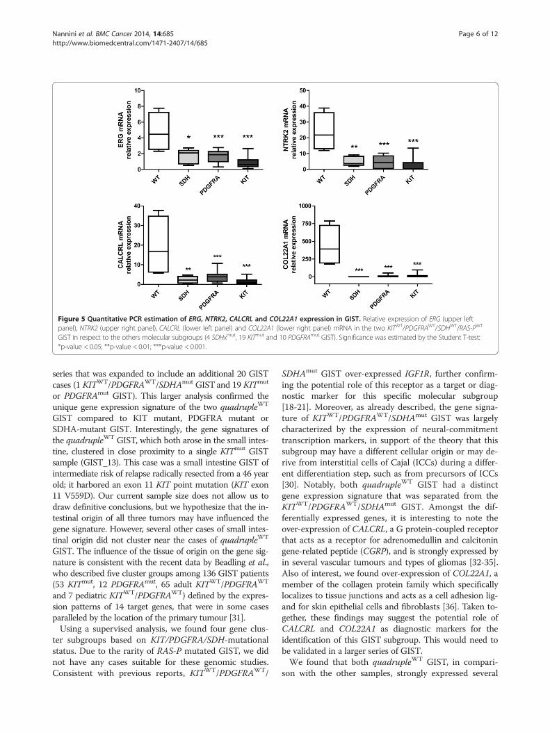
Figure 5 Quantitative PCR estimation of ERG, NTRK2, CALCRL and COL22A1 expression in GIST. Relative expression of ERG (upper leftpanel), NTRK2 (upper right panel), CALCRL (lower left panel) and COL22A1 (lower right panel) mRNA in the two KITWT/PDGFRAWT/SDHWT/RAS-PWT
GIST in respect to the others molecular subgroups (4 SDHxmut, 19 KITmut and 10 PDGFRAmut GIST). Significance was estimated by the Student T-test:*p-value < 0.05; **p-value < 0.01; ***p-value < 0.001.
Nannini et al. BMC Cancer 2014, 14:685 Page 6 of 12http://www.biomedcentral.com/1471-2407/14/685
series that was expanded to include an additional 20 GISTcases (1 KITWT/PDGFRAWT/SDHAmut GISTand 19 KITmut
or PDGFRAmut GIST). This larger analysis confirmed theunique gene expression signature of the two quadrupleWT
GIST compared to KIT mutant, PDGFRA mutant orSDHA-mutant GIST. Interestingly, the gene signatures ofthe quadrupleWT GIST, which both arose in the small intes-tine, clustered in close proximity to a single KITmut GISTsample (GIST_13). This case was a small intestine GIST ofintermediate risk of relapse radically resected from a 46 yearold; it harbored an exon 11 KIT point mutation (KIT exon11 V559D). Our current sample size does not allow us todraw definitive conclusions, but we hypothesize that the in-testinal origin of all three tumors may have influenced thegene signature. However, several other cases of small intes-tinal origin did not cluster near the cases of quadrupleWT
GIST. The influence of the tissue of origin on the gene sig-nature is consistent with the recent data by Beadling et al.,who described five cluster groups among 136 GIST patients(53 KITmut, 12 PDGFRAmut, 65 adult KITWT/PDGFRAWT
and 7 pediatric KITWT/PDGFRAWT) defined by the expres-sion patterns of 14 target genes, that were in some casesparalleled by the location of the primary tumour [31].Using a supervised analysis, we found four gene clus-
ter subgroups based on KIT/PDGFRA/SDH-mutationalstatus. Due to the rarity of RAS-P mutated GIST, we didnot have any cases suitable for these genomic studies.Consistent with previous reports, KITWT/PDGFRAWT/
SDHAmut GIST over-expressed IGF1R, further confirm-ing the potential role of this receptor as a target or diag-nostic marker for this specific molecular subgroup[18-21]. Moreover, as already described, the gene signa-ture of KITWT/PDGFRAWT/SDHAmut GIST was largelycharacterized by the expression of neural-commitmenttranscription markers, in support of the theory that thissubgroup may have a different cellular origin or may de-rive from interstitial cells of Cajal (ICCs) during a differ-ent differentiation step, such as from precursors of ICCs[30]. Notably, both quadrupleWT GIST had a distinctgene expression signature that was separated from theKITWT/PDGFRAWT/SDHAmut GIST. Amongst the dif-ferentially expressed genes, it is interesting to note theover-expression of CALCRL, a G protein-coupled receptorthat acts as a receptor for adrenomedullin and calcitoningene-related peptide (CGRP), and is strongly expressed byin several vascular tumours and types of gliomas [32-35].Also of interest, we found over-expression of COL22A1, amember of the collagen protein family which specificallylocalizes to tissue junctions and acts as a cell adhesion lig-and for skin epithelial cells and fibroblasts [36]. Taken to-gether, these findings may suggest the potential role ofCALCRL and COL22A1 as diagnostic markers for theidentification of this GIST subgroup. This would need tobe validated in a larger series of GIST.We found that both quadrupleWT GIST, in compari-
son with the other samples, strongly expressed several
Nannini et al. BMC Cancer 2014, 14:685 Page 7 of 12http://www.biomedcentral.com/1471-2407/14/685
oncogenes, including ERG and NTRK2 (TrkB). This wasconfirmed by quantitative PCR. ERG is a well-knownmember of the erythroblast transformation-specific (ETS)family of transcription factors, which function as tran-scriptional regulators [37]. ETS proteins are regulated bythe mitogenic (RAS/MAPK) signalling transduction path-way, and play an important role in cell differentiation, pro-liferation, apoptosis and tissue remodelling [38]. There isevidence for an oncogenic role of ERG and the other ETStranscription factors in many human cancers, includingsarcomas, prostate cancer, and acute myeloid leukemia, inmost cases via chromosomal translocations [39-41]. Morerecently, it has been shown that the IHC detection of ERGmay be a useful marker for vascular tumors, prostate car-cinoma and ERG-rearranged Ewing sarcoma [42-44].Over-expression of NTRK2 (TrkB) in quadrupleWT GISTis also of interest, as NTRK2 helps regulated neuronal cellfunction, including synaptic plasticity, differentiation,growth, survival, and motility [45]. It has also been shownthat Trks regulate important processes in non-neuronalcells, contributing to the pathogenesis of several kinds ofcancer, such as medullary thyroid carcinoma, prostatecancer, non-small cell lung cancer, head and neck squa-mous cell carcinoma and pancreatic cancer, in addition totumors of neural origin [46-51]. Given the relevant bio-logical role played by Trks in cancer, different small mol-ecule inhibitors have been developed and evaluated bothin mono-therapy and in combination with chemotherapyin phase 1 and 2 clinical trials [52-58].To our knowledge, the over-expression of ERG and
TrkB in GIST has not been previously reported. How-ever, it is well known that ETV1, another member ofETS family, is highly expressed in GIST and certain sub-sets of ICC. ETV1 expression plays an important role inregulating the growth of KIT mutant GIST cell lines[59]. On the basis of our results, the overexpression ofERG and TrkB seems to be a unique feature of the quad-rupleWT GIST, suggesting that it could play a relevantrole in the pathogenesis of this subset of GIST. To trans-late these observations into clinical practice, the over-expression of both molecules could be investigated asdiagnostic markers of quadrupleWT GIST.
ConclusionsIn conclusion, we report for the first time an integratedgenomic picture of the quadrupleWT GIST, using mas-sively parallel sequencing and gene expression analyses,and have identified a unique subset of GIST among thefamily of the KIT/PDGFRA WT GIST [60]. The fre-quency of this GIST subset amongst the family of GISTwill need to be defined in future studies as well as anyunique clinical-pathological features of this GIST subset,including response to conventional GIST medical ther-apy. In addition, ongoing studies of ICC developmental
biology may help identify the “normal” precursor cellsthat give rise to this unique GIST subgroup.
MethodsThis study was approved by the institutional reviewboard of Azienda Ospedaliero-Universitaria PoliclinicoS.Orsola-Malpighi, Bologna, Italy (approval number 113/2008/U/Tess). All patients provided written informedconsent.
Patients and tumor samplesFresh tissue specimens of GIST from 36 patients werecollected during the surgical operation, snap-frozen in li-quid nitrogen and stored at −80°C until analysis. Pa-tient’s characteristics are listed in Table 1.Whole-Transcriptome Paired-End RNA Sequencing was
performed on 16 GIST, including 2 KITWT/PDGFRAWT
GIST patients without SDH-inactivating mutations (GIST_133 and GIST_127), 2 KITWT/PDGFRAWT GIST patientsharbouring SDHA-mutations (GIST_7 and GIST_10), and12 KIT or PDGFRA mutated GIST patients (7 harbouredexon 11 KIT mutations and 5 harboured exon 18 PDGFRAmutations).Whole-genome gene expression analysis was performed
on 9 of the above 16 GIST and extended to include anadditional 20 GIST: 1 KITWT/PDGFRAWT/SDHAmut GISTand 19 KIT or PDGFRA mutated GIST, of which 13 har-boured KIT mutations (12 in exon 11 and 2 in exon 9),and 5 harboured PDGFRA mutations (2 in exon 12, 1 inexon 14 and 2 in exon 18).
SDH statusSDH protein expression status was evaluated by bothimmunohistochemistry (IHC) of SDHB and SDH sub-units sequencing. IHC was performed on 4-μm sectionsof FFPE GIST tumor samples. Rabbit polyclonal anti-SDHB (HPA002868, Sigma-Aldrich, St Louis, MO, USA,1:800) antibody was used. The sections were deparaffi-nized, rehydrated, and subjected to the appropriate anti-gen retrieval treatment (SDHB: microwave heating incitrate buffer pH 6.0 at 100 1C for 40 min). After coolingat room temperature, the activity of endogenous peroxi-dises was inhibited using methanol/H2O2 (0.5% v/v) for20 min. The sections were then washed in phosphate-buffered saline (PBS, pH 7.2–7.4) and incubated with thespecific primary antibody overnight at room temperature.After that, the sections were washed in PBS and treatedusing the Novolink Polymer Detection System (Novocastra,Newcastle upon Tyne, UK) according to the manufacturer’sinstructions. Liver tissues (for SDHB) were used as positivecontrols. These tissues showed strong granular staining inthe cytoplasm and mitochondria with both of theantibodies.

Table 1 Patient’s characteristic
ID Sex Array RNAseq Age Site Disease status at diagnosis KIT/PDGFRA/SDH mutational status
GIST_133 M X X 57 Duodenum Localized WT
GIST_127 F X X 63 Ileum Localized WT
GIST_07 F X X 27 Stomach Metastatic SDHA exon 9 p.S384X
GIST_10 M X X 29 Stomach Metastatic SDHA exon 2 p.R31X;
SDHA exon 13 p.R589W
GIST_188 F X 57 Duodenum Metastatic KIT exon 11 p.N564-L576 del + KIT exon 17 p.N822K
GIST_174 M X 59 Stomach Metastatic KIT exon 11 p.N564_L576 del + KIT exon 17 p.N822K
GIST_131 M X X 58 Ileum Localized KIT exon 11 p.V569_Y578 del
GIST_11 M X X 65 Stomach Localized KIT exon 11 p.557-558 del
GIST_134 F X X 65 Stomach Localized KIT exon p.V559D
GIST_124 M X X 70 Stomach Localized KIT exon 11 p.1765-1766 ins
GIST_150 F X 55 Stomach Localized KIT exon 11 p.P551_E554 del
GIST_165 M X 50 Stomach Localized PDGFRA exon 18 p.D842V
GIST_136 M X X 76 Stomach Localized PDGFRA exon 18 p.D842V
GIST_140 F X 45 Stomach Localized PDGFRA exon 18 p.D842V
GIST_141 M X 68 Stomach Localized PDGFRA exon 18 p.D842V
GIST_138 F X 75 Stomach Localized PDGFRA exon 18 p.D842V
GIST_02 F X 85 Stomach Localized KIT exon 11 p.V560D
GIST_04 M X 79 Stomach Localized KIT exon 9 p.AY502-503 ins
GIST_05 M X 68 Stomach Localized PDGFRA exon 12 p.SPDGHE566-571RIQ
GIST_08 M X 62 Stomach Localized KIT exon 11 p.V559D
GIST_09 M X 54 Stomach Localized KIT exon 11 TLQPYDHKWEEFP 574–585 ins at P585
GIST_12 F X 66 Stomach Localized PDGFRA exon 14 p.K646E
GIST_13 M X 46 Small intestine Localized KIT exon 11 p.V559D
GIST_14 M X 56 Stomach Localized KIT exon 11 p.WK557-558del
GIST_15 F X 64 Stomach Localized PDGFRA exon 18 DIMH p.842-845 DIMH del
GIST_16 F X 62 Stomach Localized KIT exon 11 p.L576P
GIST_20 M X 38 Small intestine Metastatic KIT exon 11 del MYEQW552-557 Z + KIT exon 18 A829P
GIST_22 F X 76 Stomach NA PDGFRA exon 18 p.D842V
GIST_23 F X 47 Stomach NA KIT exon 11 p.V559D
GIST_24 F X 18 Stomach Metastatic SDHA exon 8 p.L349R fs*11
GIST_26 M X 49 Stomach Localized PDGFRA exon 12 p.V561D
GIST_121 M X 71 Stomach Localized KIT exon 11 p.V559D
GIST_125 F X 48 Stomach Localized KIT exon 11 p.W557R
GIST_129 M X 59 Stomach Localized KIT exon11 p.Y553_V559 delins L
GIST_130 F X 79 Stomach Localized KIT exon 9 p.A502-Y503 ins
GIST_135 F X 61 Stomach Localized KIT exon 11 p.W557-E561 del
Nannini et al. BMC Cancer 2014, 14:685 Page 8 of 12http://www.biomedcentral.com/1471-2407/14/685
SDHA gene exons [1-15], SDHB gene exons [1-8], SDHC(exon 1–6) and SDHD (exon 1–4) were sequenced onfresh-frozen tumor specimens of KITWT/PDGFRAWT
GIST patients by Sanger Sequencing method. DNA wasextracted by the QIAmp DNA Mini kit (Qiagen, Milan,Italy) in accordance with manufacturer’s directions. Eachexon was amplified with Polymerase Chain Reaction (PCR)
amplification using specific primer pairs designed with Pri-mer Express 3.0 Software (Applied Biosystem) to amplifyexons but not SDHA pseudo-genes located on chromo-somes 3 and 5. Then, PCR products were purified with theQiaquick PCR purification kit (Qiagen, Milan, Italy) andsequenced on both strands using the Big Dye Terminatorv1.1 Cycle Sequencing kit (Applied Biosystems). Sanger

Nannini et al. BMC Cancer 2014, 14:685 Page 9 of 12http://www.biomedcentral.com/1471-2407/14/685
sequencing was performed on ABI 3730 Genetic Analyzer(Applied Biosystems).
Whole-transcriptome paired-end RNA sequencingTotal RNA was extracted from tumor specimens withRNeasy Mini Kit (Qiagen, Milan, Italy), then cDNA li-braries were synthesized from 250 ng total RNA withTruSeq RNA Sample Prep Kit v2 (Illumina, San Diego,CA) according to the manufacturer’s recommendations.Sequencing by synthesis was performed on HiScanSQsequencer (Illumina) at 75 bp in paired-end mode.Whole-transcriptome sequencing yielded an average of61 million mapped reads/patient, thus reaching an aver-age coverage of 44X. Two SDHAmut tumor specimenswere previously analyzed by whole transcriptome se-quencing at the Genome Sciences Centre (Vancouver,Canada) [9].
Bioinformatic analysisAfter demultiplexing and FASTQ generation (both stepsperformed with Casava1.8, an application software spe-cifically developed by Illumina), the paired-end readsquality were analyzed with the function fastx_quality_-stats (part of FASTX Toolkit available at http://hannon-lab.cshl.edu/fastx_toolkit/index.html). Based on theseresults we decided to trim each read of each sample at74 bp in order to maximize sequence quality. The paired-end reads were mapped with the pipeline TopHat/Bowtie[61] on human reference genome HG19, collected fromUCSC Genome Browser (http://www.genome.ucsc.edu/).After the alignment procedure the BAM file obtained wasmanipulated with Samtools [62] in order to remove theoptical/PCR duplicate, to sort and to index it.The analysis of gene expression was performed in two
steps: 1) the function htseq-count (Python package HTseq)[63] was adopted to count the number of reads mapped onknown genes, included in the Ensembl release 72 annota-tion features (http://www.ensembl.org); 2) the differentialexpressed genes were evaluated using the R-Bioconductorpackage edger [64]. DeFuse, ChimeraScan and FusionMappackages were used to detect chimeric transcripts fromRNA-seq data.
Gene expression analysisRNA was extracted using RNeasy Mini Kit (Qiagen),quality-controlled and labeled as directed by the Affyme-trix expression technical manual before hybridization toU133Plus 2.0 arrays. Gene expression data were quantifiedby the RMA algorithm, filtered and analyzed with super-vised techniques by Limma modified t-test for the detec-tion of differentially expressed genes. Differential expressedgenes hierarchical clustering and Principal ComponentAnalysis (PCA) were performed with Multiple ArrayViewer (MEV available at http://www.tm4.org/mev.html).
The same software was used to represent the data in theFigure 3 and Figure 4. Gene expression data of KIT/PDGFRA-mutated and SDHA-mutated samples were pre-viously reported [30].
Copy number analysisGenomic DNA was labelled and hybridized to SNP arrayGenome Wide SNP 6.0 (Affymetrix) following manufac-turer’s instructions. Quality control was performed byContrast QC and MAPD calculation. Copy number ana-lysis was performed by Genotyping Console and visual-ized with Chromosome Analysis Suite (ChAS) Software(Affymetrix). Hidden Markov Model algorithm was usedto detect amplified and deleted segments with stringentparameters. To control for hyperfragmentation adjacentsegments separated by < 50 probes were combined intoone single segment, and only segments > 100 probeswere considered.
Quantitative PCR (qPCR)Total RNA was reverse transcribed using TranscriptorFirst Strand cDNA synthesis kit (Roche Applied Science,Monza, Italy) with oligo-dT primers, according to themanufacturer’s guidelines. Gene-specific primers weredesigned with Primer Express 3.0 Software (Applied Bio-systems) and qPCR was performed using FastStart SybrGreen (Roche) on the LightCycler 480 apparatus(Roche). DDCt method was used to quantify gene prod-uct levels relative to the GAPDH and ATP5B house-keeping genes. Significance was estimated by the Student’st test: * p-value < 0.05; ** p-value < 0.01, *** p-value < 0.01.
Western blotProtein expression of NTRK2 was evaluated on 2KITWT/PDGFRAWT/SDHWT/RAS-PWT GIST and 8 KITor PDGFRA or SDH mutant GISTs, of which fresh-frozen tissues were available. Tissue were disrupted inRIPA buffer (Sigma-Aldrich) supplemented with prote-ases inhibitors and lysed for 1 h with gentle agitation at4°C. Lysates were centrifuged at 13,000 × g for 15 min at4°C and supernatants were stored at −80°C. Protein con-centrations were determined with the BCA protein assay(Pierce, Rockford, IL). Twenty micrograms of proteinwere resolved on a 8% SDS-PAGE gel and transferredonto polyvinylidene difluoride (PVDF) membranes. Non-specific binding sites were blocked by incubation inblocking buffer (PBS containing 0.1% Tween-20 with5% w/v milk) for 1 h at room temperature. Membraneswere incubated overnight at 4°C, with the following pri-mary antibodies: rabbit polyclonal TRKB antibody(ab18987 Abcam 1:500), and rabbit polyclonal β-Tubulinantibody (sc-9104 Santa Cruz Biotechnology, Santa Cruz,CA, 1:500). Then, membranes were washed and incubatedwith peroxidase conjugate secondary antibodies for 1 h at
Nannini et al. BMC Cancer 2014, 14:685 Page 10 of 12http://www.biomedcentral.com/1471-2407/14/685
room temperature. Antigens were revealed using EnhancedChemiluminescence Reaction (ECL Select, AmershamPharmacia Biotech, Les Ulis, France).
Nomenclature
KITWT No mutations of KITPDGFRAWT No mutations of PDGFRASDHWT No abnormalities of SDHA/B/C/D proteinexpression and/or gene mutationSDHAIHC – No expression of SDHA proteinSDHAIHC + Normal expression of SDHA proteinSDHBIHC – No expression of SDHB proteinSDHBIHC + Normal expression of SDHB proteinSDHAmut Mutation of SDHA protein (homozygous orcompound heterozygote)SDHBmut Mutation of SDHB protein (homozygous orcompound heterozygote)SDHCmut Mutation of SDHC protein (homozygous orcompound heterozygote)SDHDmut – Mutation of SDHD protein (homozygousor compound heterozygote)
Additional files
Additional file 1: Table S1. NTRK2 protein overexpression in KITWT/PDGFRAWT/SDHWT/RAS-PWT GIST. Western blot immunostaining of NTRK2was perfomed on proteins extracted from two quadrupleWT GIST andfrom eight PDGFRA or KIT or SDH mutated GIST. HL-60 cell line proteinextract was used as positive control.
Additional file 2: Figure S1. NTRK2 protein overexpression in KITWT/PDGFRAWT/SDHWT/RAS-PWT GIST. Western blot immunostaining of NTRK2was perfomed on proteins extracted from two quadrupleWT GIST andfrom eight PDGFRA or KIT or SDH mutated GIST. HL-60 cell line proteinextract was used as positive control.
AbbreviationsCGRP: Calcitonin gene-related peptide; CSS: Carney-Stratakis Syndrome; CT: CarneyTriad; DOG1: Discovered on gastrointestinal stromal tumours 1; ETS: Erythroblasttransformation-specific; GIST: Gastrointestinal stromal tumors; ICCs: Cells of Cajal;IGF1R: Insulin growth factor 1 receptor; IHC: Immunohistochemistry;NF1: Neurofibromatosis type 1; PDGFRA: Platelet-derived growth factor receptoralpha; RAS-P: RAS-pathway; SDH: Succinate dehydrogenase; SNV: Single nucleotidevariant; WT: Wild-type.
Competing interestsThe authors declare that they have no competing interests.
Authors’ contributionsMN: have made substantial contributions to conception and design of thestudy, interpretation of data and drafted the manuscript; AA: carried out themolecular genetic studies, the sequence alignment and have been involvedin drafting the manuscript. MU: carried out the molecular genetic studies,the sequence alignment and have been involved in drafting the manuscript.VI: carried out the bioinformatic analysis and interpretation of data and havebeen involved in drafting the manuscript. DS: carried out the pathologicalanalysis and the collection of samples. MCH: have been involved in revisingthe manuscript critically for important intellectual content and have givenfinal approval of the version to be published. CLC: have been involved inrevising the manuscript critically for important intellectual content and havegiven final approval of the version to be published. MS, AM and CL havehelped to draft and revised the manuscript. GE: carried out the surgical
collection of samples. GB: have been involved in revising the manuscriptcritically for important intellectual content and have given final approval ofthe version to be published. MAP: have made substantial contributions toconception and design of the study, interpretation of data and drafted themanuscript; All authors read and approved the final manuscript.
AcknowledgmentsAll staff of Bologna GIST Study Group: Annalisa Altimari, Claudio Ceccarelli,Paolo Castellucci, Fausto Catena, Monica Di Battista, Massimo Del Gaudio,Valerio Di Scioscio, Stefano Fanti, Michelangelo Fiorentino, Pietro Fusaroli, LidiaGatto, Franco W. Grigioni, Elisa Gruppioni, Alessandra Maleddu, Maria CaterinaPallotti, Antonio Daniele Pinna, Paola Tommasetti, Maurizio Zompatori.
FundingThe present work was done with a financial contribution by NovartisOncology, Italy, and with funds by My First Grant 2013, AIRC 2013.
Author details1Department of Specialized, Experimental and Diagnostic Medicine,Sant’Orsola-Malpighi Hospital, University of Bologna, Via Massarenti 9, 40138Bologna, Italy. 2“Giorgio Prodi” Cancer Research Center, University of Bologna,Bologna, Italy. 3Pathology Unit, S. Orsola-Malpighi Hospital, University ofBologna, Bologna, Italy. 4Portland VA Medical Center and Knight CancerInstitute, and Division of Hematology and Oncology, Oregon Health &Science University Portland, Portland, OR, USA. 5Department of Pathologyand Knight Cancer Institute, Oregon Health & Science University, Portland,OR, USA. 6Transplant, General and Emergency Surgery Department, S.Orsola-Malpighi Hospital, University of Bologna, Bologna, Italy.
Received: 23 January 2014 Accepted: 17 September 2014Published: 20 September 2014
References1. Corless CL, Fletcher JA, Heinrich MC: Biology of gastrointestinal stromal
tumors. J Clin Oncol 2004, 22:3813–3825.2. Janeway KA, Pappo AS: Pediatric gastrointestinal stromal tumor. Hematol
Oncol Clin North Am 2009, 23:15–34.3. Heinrich MC, Corless CL, Demetri GD, Blanke CD, von Mehren M, Joensuu H,
McGreevey LS, Chen CJ, Van den Abbeele AD, Druker BJ, Kiese B, EisenbergB, Roberts PJ, Singer S, Fletcher CD, Silberman S, Dimitrijevic S, Fletcher JA:Kinase mutations and imatinib response in patients with metastaticgastrointestinal stromal tumor. J Clin Oncol 2003, 21:4342–4349.
4. Debiec-Rychter M, Sciot R, Le Cesne A, Schlemmer M, Hohenberger P, vanOosterom AT, Blay JY, Leyvraz S, Stul M, Casali PG, Zalcberg J, Verweij J, VanGlabbeke M, Hagemeijer A, Judson I, EORTC Soft Tissue and Bone SarcomaGroup; Italian Sarcoma Group; Australasian GastroIntestinal Trials Group: KITmutations and dose selection for imatinib in patients with advancedgastrointestinal stromal tumours. Eur J Cancer 2006, 42:1093–1103.
5. Heinrich MC, Owzar K, Corless CL, Hollis D, Borden EC, Fletcher CD, RyanCW, von Mehren M, Blanke CD, Rankin C, Benjamin RS, Bramwell VH,Demetri GD, Bertagnolli MM, Fletcher JA: Correlation of kinase genotypeand clinical outcome in the North American Intergroup Phase III Trial ofimatinib mesylate for treatment of advanced gastrointestinal stromaltumor: CALGB 150105 Study by Cancer and Leukemia Group B andSouthwest Oncology Group. J Clin Oncol 2008, 26:5360–5367.
6. Gill AJ, Chou A, Vilain R, Clarkson A, Lui M, Jin R, Tobias V, Samra J, Goldstein D,Smith C, Sioson L, Parker N, Smith RC, Sywak M, Sidhu SB, Wyatt JM, RobinsonBG, Eckstein RP, Benn DE, Clifton-Bligh RJ: Immunohistochemistry for SDHBdivides gastrointestinal stromal tumors (GISTs) into 2 distinct types. Am JSurg Pathol 2010, 34:636–644.
7. Miettinen M, Wang ZF, Sarlomo-Rikala M, Osuch C, Rutkowski P, Lasota J:Succinate dehydrogenase-deficient GISTs: a clinicopathologic,immunohistochemical, and molecular genetic study of 66 gastric GISTswith predilection to young age. Am J Surg Pathol 2011, 35:1712–1721.
8. Janeway KA, Kim SY, Lodish M, Nosé V, Rustin P, Gaal J, Dahia PL, Liegl B,Ball ER, Raygada M, Lai AH, Kelly L, Hornick JL, NIH Pediatric and Wild-TypeGIST Clinic, O’Sullivan M, de Krijger RR, Dinjens WN, Demetri GD, Antonescu CR,Fletcher JA, Helman L, Stratakis CA: Defects in succinate dehydrogenase ingastrointestinal stromal tumors lacking KIT and PDGFRA mutations. ProcNatl Acad Sci U S A 2011, 108:314–318.
Nannini et al. BMC Cancer 2014, 14:685 Page 11 of 12http://www.biomedcentral.com/1471-2407/14/685
9. Pantaleo MA, Astolfi A, Indio V, Moore R, Thiessen N, Heinrich MC, GnocchiC, Santini D, Catena F, Formica S, Martelli PL, Casadio R, Pession A, Biasco G:SDHA loss-of-function mutations in KIT-PDGFRA wild-type gastrointestinalstromal tumors identified by massively parallel sequencing. J Natl CancerInst 2011, 103:983–987.
10. Pantaleo MA, Nannini M, Astolfi A, Biasco G, GIST Study Group Bologna: Adistinct pediatric-type gastrointestinal stromal tumor in adults: potentialrole of succinate dehydrogenase subunit A mutations. Am J Surg Pathol2011, 35:1750–1752.
11. Italiano A, Chen CL, Sung YS, Singer S, DeMatteo RP, LaQuaglia MP, BesmerP, Socci N, Antonescu CR: SDHA loss of function mutations in a subset ofyoung adult wild-type gastrointestinal stromal tumors. BMC Cancer 2012,12:408.
12. Wagner AJ, Remillard SP, Zhang YX, Doyle LA, George S, Hornick JL: Loss ofexpression of SDHA predicts SDHA mutations in gastrointestinal stromaltumors. Mod Pathol 2013, 26:289–294.
13. Dwight T, Benn DE, Clarkson A, Vilain R, Lipton L, Robinson BG, Clifton-BlighRJ, Gill AJ: Loss of SDHA expression identifies SDHA mutations insuccinate dehydrogenase-deficient gastrointestinal stromal tumors. Am JSurg Pathol 2013, 37:226–233.
14. Oudijk L, Gaal J, Korpershoek E, van Nederveen FH, Kelly L, Schiavon G,Verweij J, Mathijssen RH, den Bakker MA, Oldenburg RA, van Loon RL,O’Sullivan MJ, de Krijger RR, Dinjens WN: SDHA mutations in adult andpediatric wild-type gastrointestinal stromal tumors. Mod Pathol 2013,26:456–463.
15. Miettinen M, Killian JK, Wang ZF, Lasota J, Lau C, Jones L, Walker R, PinedaM, Zhu YJ, Kim SY, Helman L, Meltzer P: Immunohistochemical loss ofsuccinate dehydrogenase subunit A (SDHA) in gastrointestinal stromaltumors (GISTs) signals SDHA germline mutation. Am J Surg Pathol 2013,37:234–240.
16. Pantaleo MA, Astolfi A, Urbini M, Nannini M, Paterini P, Indio V, Saponara M,Formica S, Ceccarelli C, Casadio R, Rossi G, Bertolini F, Santini D, Pirini MG,Fiorentino M, Basso U, Biasco G, on behalf of GIST Study Group, Universityof Bologna, Bologna, Italy: Analysis of all subunits, SDHA, SDHB, SDHC,SDHD, of the succinate dehydrogenase complex in KIT/PDGFRA wild-type GIST. Eur J Hum Genet 2013. Epub online before print.
17. Rege TA, Wagner AJ, Corless CL, Heinrich MC, Hornick JL: “Pediatric-type”gastrointestinal stromal tumors in adults: distinctive histology predictsgenotype and clinical behavior. Am J Surg Pathol 2011, 35:495–504.
18. Chou A, Chen J, Clarkson A, Samra JS, Clifton-Bligh RJ, Hugh TJ, Gill AJ:Succinate dehydrogenase-deficient GISTs are characterized by IGF1Roverexpression. Mod Pathol 2012, 25:1307–1313.
19. Nannini M, Astolfi A, Paterini P, Urbini M, Santini D, Catena F, Indio V,Casadio R, Pinna AD, Biasco G, Pantaleo MA: Expression of IGF-1 receptorin KIT/PDGF receptor-α wild-type gastrointestinal stromal tumors withsuccinate dehydrogenase complex dysfunction. Future Oncol 2013,9:121–126.
20. Belinsky MG, Rink L, Flieder DB, Jahromi MS, Schiffman JD, Godwin AK,Mehren M: Overexpression of insulin-like growth factor 1 receptor andfrequent mutational inactivation of SDHA in wild-type SDHB-negativegastrointestinal stromal tumors. Genes Chromosomes Cancer 2013,52:214–224.
21. Lasota J, Wang Z, Kim SY, Helman L, Miettinen M: Expression of thereceptor for type I insulin-like growth factor (IGF1R) in gastrointestinalstromal tumors: an immunohistochemical study of 1078 cases withdiagnostic and therapeutic implications. Am J Surg Pathol 2013, 37:114–119.
22. McWhinney SR, Pasini B, Stratakis CA: Familial gastrointestinal stromaltumors and germ-line mutations. N Engl J Med 2007, 357:1054–1056.
23. Pasini B, McWhinney SR, Bei T, Matyakhina L, Stergiopoulos S, Muchow M,Boikos SA, Ferrando B, Pacak K, Assie G, Baudin E, Chompret A, Ellison JW,Briere JJ, Rustin P, Gimenez-Roqueplo AP, Eng C, Carney JA, Stratakis CA:Clinical and molecular genetics of patients with the Carney-Stratakissyndrome and germline mutations of the genes coding for the succinatedehydrogenase subunits SDHB, SDHC, and SDHD. Eur J Hum Genet 2008,16:79–88.
24. Zhang L, Smyrk TC, Young WF Jr, Stratakis CA, Carney JA: Gastric stromaltumors in Carney triad are different clinically, pathologically, andbehaviourally from sporadic gastric gastrointestinal stromal tumors:findings in 104 cases. Am J Surg Pathol 2010, 34:53–64.
25. Bajor J: Gastrointestinal stromal tumors in patients with type 1neurofibromatosis. Clin Exp Med J 2009, 3:247–254.
26. Agaram NP, Wong GC, Guo T, Maki RG, Singer S, Dematteo RP, Besmer P,Antonescu CR: Novel V600E BRAF mutations in imatinib-naive andimatinib-resistant gastrointestinal stromal tumors. Genes ChromosomesCancer 2008, 47:853–859.
27. Hostein I, Faur N, Primois C, Boury F, Denard J, Emile JF, Bringuier PP,Scoazec JY, Coindre JM: BRAF mutation status in gastrointestinal stromaltumors. Am J Clin Pathol 2010, 133:141–148.
28. Daniels M, Lurkin I, Pauli R, Erbstösser E, Hildebrandt U, Hellwig K, Zschille U,Lüders P, Krüger G, Knolle J, Stengel B, Prall F, Hertel K, Lobeck H, Popp B,Theissig F, Wünsch P, Zwarthoff E, Agaimy A, Schneider-Stock R: Spectrumof KIT/PDGFRA/BRAF mutations and Phosphatidylinositol-3-Kinasepathway gene alterations in gastrointestinal stromal tumors (GIST).Cancer Lett 2011, 312:43–54.
29. Pantaleo MA, Nannin M, Corless CL, Heinrich MC: Quadruple wild-type (WT)GIST: defining the subset of GISTs that lack abnormalities of KIT,PDGFRA, SDH, and the RAS signalling pathway. Cancer Med 2014. in press.
30. Pantaleo MA, Astolfi A, Nannini M, Ceccarelli C, Formica S, Santini D,Heinrich MC, Corless C, Dei Tos AP, Paterini P, Catena F, Maleddu A,Saponara M, Di Battista M, Biasco G: Differential expression of neuralmarkers in KIT and PDGFRA wild-type gastrointestinal stromal tumours.Histopathology 2011, 59:1071–1080.
31. Beadling C, Patterson J, Justusson E, Nelson D, Pantaleo MA, Hornick JL,Chacón M, Corless CL, Heinrich MC: Gene expression of the IGF pathwayfamily distinguishes subsets of gastrointestinal stromal tumors wild typefor KIT and PDGFRA. Cancer Med 2013, 2:21–31.
32. Muff R, Born W, Fischer JA: Calcitonin, calcitonin gene-related peptide,adrenomedullin and amylin: homologous peptides, separate receptorsand overlapping biological actions. Eur J Endocrinol 1995, 133:17–20.
33. Hagner S, Stahl U, Grimm T, Stürzl M, Lang RE: Expression of calcitoninreceptor-like receptor in human vascular tumours. J Clin Pathol 2006,59:1104–1107.
34. Mennel HD, Hallier-Neelsen M, Hagner S, Benes L: Two novel cell specificreceptor proteins, CRLR and CD 117 in human glial tumors. Clin Neuropathol2006, 25:107–114.
35. Benes L, Kappus C, McGregor GP, Bertalanffy H, Mennel HD, Hagner S: Theimmunohistochemical expression of calcitonin receptor-like receptor(CRLR) in human gliomas. J Clin Pathol 2004, 57:172–176.
36. Koch M, Schulze J, Hansen U, Ashwodt T, Keene DR, Brunken WJ, BurgesonRE, Bruckner P, Bruckner-Tuderman L: A novel marker of tissue junctions,collagen XXII. J Biol Chem 2004, 279:22514–22521.
37. Reddy ES, Rao VN, Papas TS: The erg gene: a human gene related to theets oncogene. Proc Natl Acad Sci U S A 1987, 84:6131–6135.
38. Hart AH, Corrick CM, Tymms MJ, Hertzog PJ, Kola I: Human ERG is aproto-oncogene with mitogenic and transforming activity. Oncogene1995, 10:1423–1430.
39. Sorensen PH, Lessnick SL, Lopez-Terrada D, Liu XF, Triche TJ, Denny CT: Asecond Ewing’s sarcoma translocation, t(21;22), fuses the EWS gene toanother ETS-family transcription factor, ERG. Nat Genet 1994, 6:146–151.
40. Sorensen PH, Lessnick SL, Lopez-Terrada D, Liu XF, Triche TJ, Denny CT:Recurrent fusion of TMPRSS2 and ETS transcription factor genes inprostate cancer. Science 2005, 310:644–648.
41. Martens JH: Acute myeloid leukemia: a central role for the ETS factorERG. Int J Biochem Cell Biol 2011, 43:1413–1416.
42. Miettinen M, Wang ZF, Paetau A, Tan SH, Dobi A, Srivastava S, Sesterhenn I:ERG transcription factor as an immunohistochemical marker for vascularendothelial tumors and prostatic carcinoma. Am J Surg Pathol 2011,35:432–441.
43. Wang WL, Patel NR, Caragea M, Hogendoorn PC, López-Terrada D, HornickJL, Lazar AJ: Expression of ERG, an Ets family transcription factor,identifies ERG-rearranged Ewing sarcoma. Mod Pathol 2012, 25:1378–1383.
44. Miettinen M, Wang Z, Sarlomo-Rikala M, Abdullaev Z, Pack SD, Fetsch JF:ERG expression in epithelioid sarcoma: a diagnostic pitfall. Am J SurgPathol 2013, 37:1580–1585.
45. Thiele CJ, Li Z, McKee AE: On Trk–the TrkB signal transduction pathway isan increasingly important target in cancer biology. Clin Cancer Res 2009,15:5962–5967.
46. Brodeur GM, Nakagawara A, Yamashiro DJ, Ikegaki N, Liu XG, Azar CG, LeeCP, Evans AE: Expression of TrkA, TrkB and TrkC in humanneuroblastomas. J Neurooncol 1997, 31:49–55.
47. McGregor LM, McCune BK, Graff JR, McDowell PR, Romans KE, YancopoulosGD, Ball DW, Baylin SB, Nelkin BD: Roles of trk family neurotrophin
Nannini et al. BMC Cancer 2014, 14:685 Page 12 of 12http://www.biomedcentral.com/1471-2407/14/685
receptors in medullary thyroid carcinoma development and progression.Proc Natl Acad Sci U S A 1999, 96:4540–4545.
48. Satoh F, Mimata H, Nomura T, Fujita Y, Shin T, Sakamoto S, Hamada Y,Nomura Y: Autocrine expression of neurotrophins and their receptors inprostate cancer. Int J Urol 2001, 8:S28–S34.
49. Harada T, Yatabe Y, Takeshita M, Koga T, Yano T, Wang Y, Giaccone G: Roleand relevance of TrkB mutations and expression in non-small cell lungcancer. Clin Cancer Res 2011, 17:2638–2645.
50. Kupferman ME, Jiffar T, El-Naggar A, Yilmaz T, Zhou G, Xie T, Feng L, Wang J,Holsinger FC, Yu D, Myers JN: TrkB induces EMT and has a key role ininvasion of head and neck squamous cell carcinoma. Oncogene 2010,29:2047–2059.
51. Sclabas GM, Fujioka S, Schmidt C, Li Z, Frederick WA, Yang W, Yokoi K, EvansDB, Abbruzzese JL, Hess KR, Zhang W, Fidler IJ, Chiao PJ: Overexpression oftropomysin-related kinase B in metastatic human pancreatic cancer cells.Clin Cancer Res 2005, 11:440–449.
52. Camoratto AM, Jani JP, Angeles TS, Maroney AC, Sanders CY, Murakata C,Neff NT, Vaught JL, Isaacs JT, Dionne CA: CEP-751 inhibits TRK receptortyrosine kinase activity in vitro exhibits anti-tumor activity. Int J Cancer1997, 72:673–679.
53. Miknyoczki SJ, Dionne CA, Klein-Szanto AJ, Ruggeri BA: The novel Trkreceptor tyrosine kinase inhibitor CEP-701 (KT-5555) exhibits antitumorefficacy against human pancreatic carcinoma (Panc1) xenograft growthand in vivo invasiveness. Ann N YAcad Sci 1999, 880:252–262.
54. Evans AE, Kisselbach KD, Yamashiro DJ, Ikegaki N, Camoratto AM, DionneCA, Brodeur GM: Antitumor activity of CEP-751 (KT-6587) on humanneuroblastoma and medulloblastoma xenografts. Clin Cancer Res 1999,5:3594–3602.
55. Strock CJ, Park JI, Rosen M, Dionne C, Ruggeri B, Jones-Bolin S, DenmeadeSR, Ball DW, Nelkin BD: CEP-701 and CEP-751 inhibit constitutivelyactivated RET tyrosine kinase activity and block medullary thyroidcarcinoma cell growth. Cancer Res 2003, 63:5559–5563.
56. Marshall JL, Kindler H, Deeken J, Bhargava P, Vogelzang NJ, Rizvi N, LuhtalaT, Boylan S, Dordal M, Robertson P, Hawkins MJ, Ratain MJ: Phase I trial oforallyadministeredCEP-701, anovelneurotrophin receptor-linked tyrosinekinase inhibitor. Invest New Drugs 2005, 23:31–37.
57. Wang T, Lamb ML, Scott DA, Wang H, Block MH, Lyne PD, Lee JW, DaviesAM, Zhang HJ, Zhu Y, Gu F, Han Y, Wang B, Mohr PJ, Kaus RJ, Josey JA,Hoffmann E, Thress K, Macintyre T, Wang H, Omer CA, Yu D: Identificationof 4-aminopyrazolylpyrimidines as potent inhibitors of Trk kinases. J MedChem 2008, 51:4672–4684.
58. Chan E, Mulkerin D, Rothenberg M, Holen KD, Lockhart AC, Thomas J, BerlinJ: A phase I trial of CEP-701 + gemcitabine in patients with advancedadenocarcinoma of the pancreas. Invest New Drugs 2008, 26:241–247.
59. Chi P, Chen Y, Zhang L, Guo X, Wongvipat J, Shamu T, Fletcher JA, DewellS, Maki RG, Zheng D, Antonescu CR, Allis CD, Sawyers CL: ETV1 is a lineagesurvival factor that cooperates with KIT in gastrointestinal stromaltumours. Nature 2010, 467:849–853.
60. Nannini M, Biasco G, Astolfi A, Pantaleo MA: An overview on molecularbiology of KIT/PDGFRA wild type (WT) gastrointestinal stromal tumours(GIST). J Med Genet 2013, 50:653–661.
61. Trapnell C, Pachter L, Salzberg SL: TopHat: discovering splice junctionswith RNA-Seq. Bioinformatics 2009, 9:1105–1111.
62. Li H, Handsaker B, Wysoker A, Fennell T, Ruan J, Homer N, Marth G, AbecasisG, Durbin R, 1000 Genome Project Data Processing Subgroup: TheSequence Alignment/Map format and SAMtools. Bioinformatics 2009,25:2078–2079.
63. Anders S, Huber W: Differential expression analysis for sequence countdata. Genome Biol 2010, 11:R106.
64. Robinson MD, McCarthy DJ, Smyth GK: edgeR: a Bioconductor package fordifferential expression analysis of digital gene expression data.Bioinformatics 2010, 26:139–140.
doi:10.1186/1471-2407-14-685Cite this article as: Nannini et al.: Integrated genomic study ofquadruple-WT GIST (KIT/PDGFRA/SDH/RAS pathway wild-type GIST).BMC Cancer 2014 14:685.
Submit your next manuscript to BioMed Centraland take full advantage of:
• Convenient online submission
• Thorough peer review
• No space constraints or color figure charges
• Immediate publication on acceptance
• Inclusion in PubMed, CAS, Scopus and Google Scholar
• Research which is freely available for redistribution
Submit your manuscript at www.biomedcentral.com/submit