Embed Size (px)
Citation preview
Journal of Clinical Review & Case Reports
Volume 2 | Issue 2 | 1 of 7J Clin Rev Case Rep, 2017
Bedsore Revitalization by-Laser Therapy (Low Level Laser: LED-Ga-Al- As, 660 nm
Research Article
Nazrul islam1*, Golam Abu Zakaria2, kazi Shamimuzzaman3 and QuamrulAkhter Sanju4
AbstractBackground: In 1967 a few years after the first working laser was invented, EndreMester in Semmelweis University Budapest, Hungary wanted to find out if laser might cause cancer. He took some mice, shaved the hair off their backs, divided them into two groups and gave a laser treatment with a low powered ruby laser to one group. They did not get cancer and to his surprise the hair on the treated group grew back more quickly than the untreated group. That was how “laser biostimulation” effects were discovered.
Purpose of the work: The effects of pulsed monochromatic light, with fixed pulsations and wavelengths, on the healing of pressure ulcers were evaluated in this prospective, randomized, controlled study.
Method: A placebo-controlled, double-blind study using low energy photon therapy (LLLT) was performed in ten patients with bedsore on the back. Treatment was given three times a week for 10 weeks, using monochromatic (red) optical sources; diode 660nm (GaAl-660). The patients who were randomized to placebo treatment received sham therapy from an identical-appearing light source from the same delivery system.
Results: Ten patients with bedsore were randomized to receive LLLT or placebo therapy. At the conclusion of the study, the percentage of the initial ulcer area remaining unhealed in the LLLT and placebo groups was 24.4% and 84.7%, respectively (P = 0.0008). The decrease in ulcer area (compared to baseline) observed in the LLLT and placebo groups was 193.0 mm2 and 14.7 mm2, respectively (P = 0.0002). One patient dropped out of the study, complaining of lack of treatment efficacy; he was found to be randomized to the placebo group. There were no adverse effects.
Conclusions: In this placebo-controlled, double-blind study LLLT was an effective modality for the treatment of bedsore which were resistant to conventional medical management. The results are encouraging as pulsed monochromatic light increased healing rate and shortened healing time. This will positively affect the quality of life in elderly patients with pressure ulcers.
1Head-Biomedical Science and Medical Biotechnology Department, Shaheed Suhrawardy Medical College andHospital, Dhaka-1207. Bangladesh.
2Dept. of Medical Radiation Physics, Kreiskrankenhaus Gummersbach, Teaching Hospital of the University of Cologne, 51643, Germany. And-Dept. of Medical Physics and Biomedical Engineering, Gono - Bishwabidyalay (Gono University), Nayarhat, Savar, Dhaka-1344, Bangladesh.
3Department of Orthopedic and Trauma Surgery, Shaheed Suhrawardy Medical College and Hospital,Dhaka-1207, Bangladesh.
4Department of Surgery, Shaheed Suhrawardy Medical College and Hospital, Dhaka-1207,Bangladesh.
*Corresponding authorNazrulislam, Head-Biomedical Science And Medical Biotechnology Department, Shaheed Suhrawardy Medical College and Hospital,Dhaka-1207. Bangladesh.
Submitted: 29 July 2017; Accepted: 07 Aug 2017; Published: 07 Sep 2017
Keywords: Bedsore Healing, Soft Tissue Healing, Decubitus Ulcer Healing, Wound Healing. Low Level Laser, Laser Therapy.
IntroductionBackgroundIn 1967 a few years after the first working laser was invented, Endre
Mester in Semmelweis University Budapest, Hungary wanted to find out if laser might cause cancer. He took some mice, shaved the hairoff their backs, divided them into two groups and gave a laser treatment with a low powered rubylaser to one group. They did not get cancer and to his surprise the hair on the treated group grew backmore quickly than the untreated group. That was how “laser
J Clin Rev Case Rep, 2017
biostimulation” was discovered[1].
Purpose of the workThe effects of pulsed monochromatic light, with fixed pulsations and wavelengths, on the healing ofpressure ulcers were evaluated in this prospective, randomized, controlled study.
LLLT In soft tissue healing process / Skin lesionsSome of the most common cutaneous wounds include excoriations, burns, surgical incisions,and acute or chronic ulcerations [2,3].Diabetes mellitus is one of the primary predisposingfactors for skin lesion development and one of the most common reasons for patients toseek health care, as it represents an important cause of disability and premature death [4-6]. According to Pedrosa, serious cutaneous foot lesions in diabetic patients are the cause forhospital admission in 51% of patients in endocrinology wards of Brazilian university hospitals [7]. When not properly healed, these lesions represent the main cause of morbidity, immobilityand limb amputation, according to data from the American Diabetes Association [6]. Burn injuries, a clinical condition resulting from direct or indirect action of heat on the humanbody that causes different degrees of skin lesions, are a significant cause of mortality, primarilydue to the infections that can evolve to septicemia. According to the Brazilian Society of BurnInjuries (SociedadeBrasileira de Queimaduras), there are 1 million cases each year in Brazil [8,9]. Skin lesions have a great morbidity potential primarily because of complications in the normalhealing process. To prevent these complications and promote cure, one needs to understandthe normal process of soft tissue repair, as well as the factors that determine its normal healing.
The normal process of soft tissue repair involves the following steps: homeostasis, inflammation (“cleaning”), demolition, proliferation, and maturing [10]. The homeostatic phase occurs immediately after the appearance of the lesion and dependson platelet activity and on blood coagulation process, which includes a complex release ofvasoactive substances, adhesive proteins, and growth factors for the development of otherstages [10,11].Later on, the inflammatory process sets in with the presence of numerous chemicalmediators and inflammatory cells (polymorphonuclear leukocytes, macrophages, andlymphocytes).This phase is responsible for removing necrosed tissue and combating aggressiveagents installed in the wound. Next, tissue proliferation, which is responsible for “closing” thewound, sets in, with re-epithelization, fibroplasia (matrix formation), and angiogenesis, essentialfor the supply of oxygen and nutrients needed for healing. Finally, there is wound contractionfollowed by remodeling, which takes place in the collagen of the region and has the objective ofincreasing tensile force and diminishing the scar size [11,12].
Therefore, tissue healing highlighted as one of the main effects of LLLT, is characterized bythree main factors. First, there is an increment of ATP production, (as laser is considered to raise the production of ATP,) leading to a boost in mitotic activity and to an increase in protein synthesis bymitochondria, resulting in greater tissue regeneration in the repair process [13-16]. Second, there is a stimulus to microcirculation, which increases the delivery of nutritionalelements associated with increased speed of mitosis, facilitating cell multiplication [13,14]. Finally, new vessels are formed from preexisting vessels [13,14,17].
Several factors have a direct influence on tissue healing, altering this
process, making it slower,thus allowing complications associated with wound exposure to the external environment. Thetable below displays the key local and systemic factors that affect tissue wound healing.
Uses of Laser Therapy to Treat BedsoreAllied health professionals regularly care for a variety of skin wounds, such as abrasions, turf burns,surgical incisions, and ulcerations, which are perhaps the most difficult to treat.At present, cutaneous lesions represent a dilemma of global proportions and instigate great clinicalinterest because of the high morbidity associated with changes in the normal healing process.1 Among theclinical aspects involving this issue, we emphasize tissue repair time in an effort to make the processquicker and more harmonious, reduce possible complications in lesion resolution, and allow an adequatechoice of therapy. To do this, familiarity with the pathogenesis of tissue healing is necessary, as well as anunderstanding of the factors affecting the process and the role each one plays in its progress, alwaysseeking a clinical treatment that optimizes skin lesion care. Among the methods currently available, lowlevellaser therapy (LLLT) stands out.
From acute wound management to augmentation of scar tissue remodeling, the clinician seeks tooptimize wound care to promote healing. Experimental in vitro and in vivo studies have been underdevelopment since the 1960s, and in the early 1990s, LLLT was approved by the Food and DrugAdministration (FDA) as an important method for treating healing processes [18-20]. Recent results of a studydemonstrated that LLLT is an effective method to modulate tissue repair, thus significantly contributing toa faster and more organized healing process [21]. Nevertheless, in spite of the large number of studies involving this technique and its wide use in clinicalpractice, the principles of its action in cells and tissues are still not well understood. The objective of thisstudy is to review pathogenetic aspects of soft tissue repair to understand the major complications in skinlesion healing. In addition, it aims at forming a concise compilation of published data from scientificliterature to date to verify whether the use of low-level laser influences wound healing, since itsmechanisms of action are not fully clear yet.
Details ExperimentalMaterials and Parameters-Place of studyShaheed Suhrawardy Medical College Hospital, Ward and Bed: 9/2. Dhaka-1 207 , Bangladesh. Telephone: 88-9130800-19. Period/ Duration of Study: One Year (2009-2010).Patient: Bedsore/ Decubitus Ulcer Patients.
Selection CriteriaSample collection& distributionThe sample was collected randomly from admitted patients inShaheed Suhrawardy Medical College Hospital in the Department of Orthopedics and Traumatology, Dhaka-1207, Bangladesh. A total of 10 patients randomly collected. The patients were briefed aboutthe study and written consent (Informed consent) was obtained from all patients/ medico legalguardian for other patients.
Inclusion Criteria1. Patient suffering from bedsore more than 12 weeks.2. Failed to heal by all means of conventional therapy.3. Male and Female ratio- 50:50.4. Age between 55- 95 years old,
Volume 2 | Issue 2 | 2 of 7
5. Haven’t previously treated with LLLT.
Machine: BioLux MDLLLT, Low Level Laser (LED- Ga-Al- As 660).
Irradiance Parameters• Beam source (Incoherent-Ga-Al-As).• Irradiance dose: 4- 8 J/cm2/min.• Irradiance time: 1- 2 minutes• Mode: Continuous wave• Wavelengths Used: 660 nm.• Total session: 25-35.
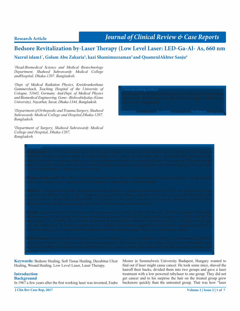
An average power of 5-8 mw was provided through a fiber optic delivery system around the wound margin for about 8-10 min at
each point at a distance of one cm. Since the center of the ulcer was deep, it was decided to give laser therapy concentrating maximum irradiance there (Figure 1).
The optimum distance from probe tip to target surface was 1-4 mm. Probe motion during lasing was a slow, circling movement over each square centimeter of open lesion, timed to permit the suggested dosages. As the lesion is large, i.e., 4-6 cm in diameter, a change in technique is adopted which involves a slow, traversing of the perimeter of the lesion, allowing approximately 90 sec per linear centimeter of the perimeter, at the suggested distances (1-4 cm).
The parameters which have been found to be most effective are in the range of 90 sec/cm2 of open wound surface, with the laser beam set at a pulsed rate of 40-80 pulses per second (PPS), depending upon the chronicity of the lesion. The more chronic, the slower the pulse rate suggested.
This technique apparently provides sufficient exposure to the laser beam to stimulate healing effectively, compared with non-treated areas and previously experienced wound management of a similar nature. Low energy Ga-Al laser provides infrared rays in the wave length of around 660nm by continuous mode.
Table 1: Parameters involved in determining the lllt Irradiation parametersIrradiationparameter
Unit of measurement Comment
Wavelength nm Light is electromagnetic energy which travels in discrete Packets that also have a wave-like property. Wavelength is measure in nanometers (nm) and is visible in the 400-700 nm range.
Irradiance W/cm2 Often called Intensity, or Power Density and is calculated as Irradiance = Power (W)/Area (cm2)
Coherence Coherence length Coherent light produces laser speckle, which has been depends on postulated to play a role in the photobiomodulation Spectral bandwidth interaction with cells and subcellular organelles.
Polarization Linear polarizedor circular identical nonpolarized
Light may have different effects than otherwise light(or even 90-degree rotated polarized polarized light). However, it is Known that polarized light is rapidly scrambled in highly scattering media such as tissue (probably in the first few hundred μm).
Table 2: Parameters involved in determining the lllt “dose” irradiation time orenergy delivered (the dose)Irradiation Unit of
Parameter Measurement CommentEnergy (Joules) J Calculated as: Energy (J) = Power (W) x time (s). This mixes medicine
and dose into a single expression and ignores Irradiance.Using Joules as an expression of dose is potentially unreliable as it assumes reciprocity (the inverse relationship between power and time).
Energy Density J/cm2 Common expression of LLLT “dose” is Energy density. This expression of dose again mixes medicine and dose into a single expression and is potentially unreliable as it assumes a reciprocity relationship between irradiance and time.
Irradiation Time s In our view the safest way to record and prescribe LLLT is to define the four parameters of the medicine (see table 1.) and then define the irradiation time as “dose”.
Treatment Interval Hours, days or weeks The effects of different treatment interval are underexplored at this time though there is sufficient evidence to suggest that this is an important parameter.
J Clin Rev Case Rep, 2017 Volume 2 | Issue 2 | 3 of 7
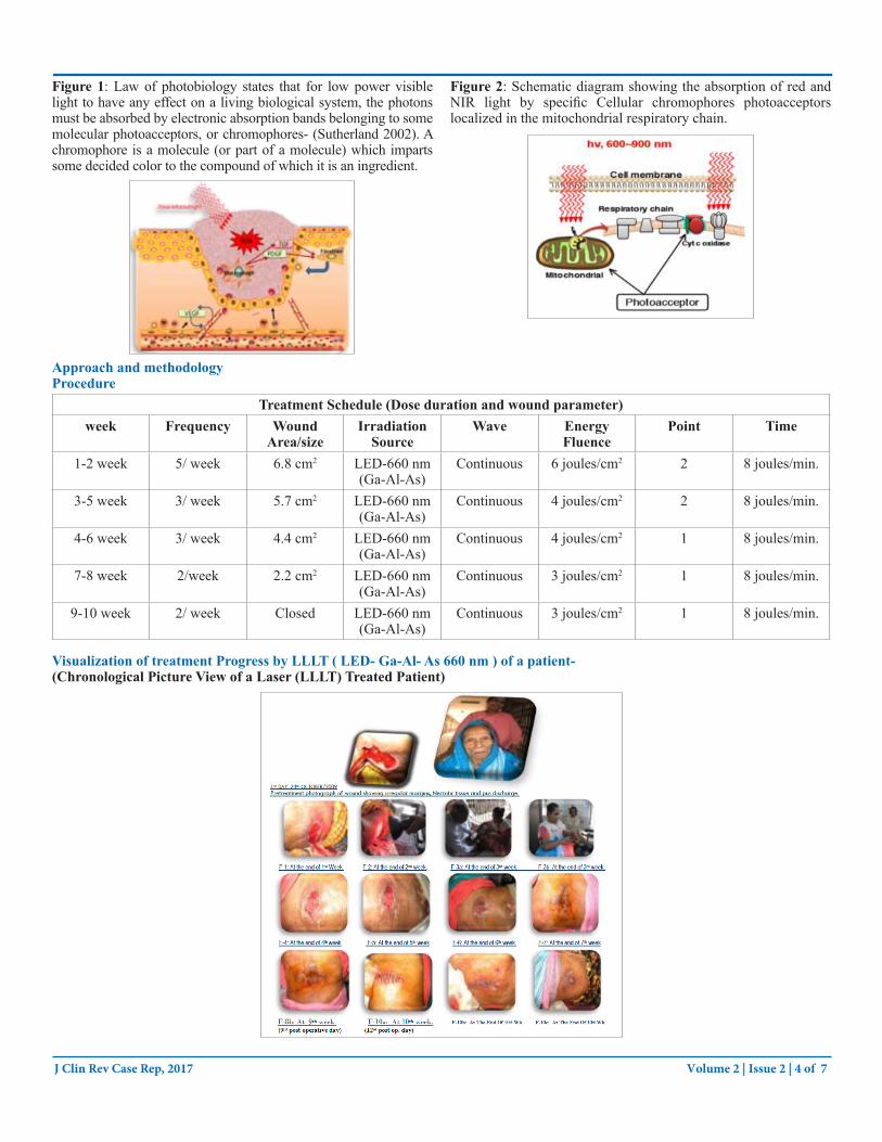
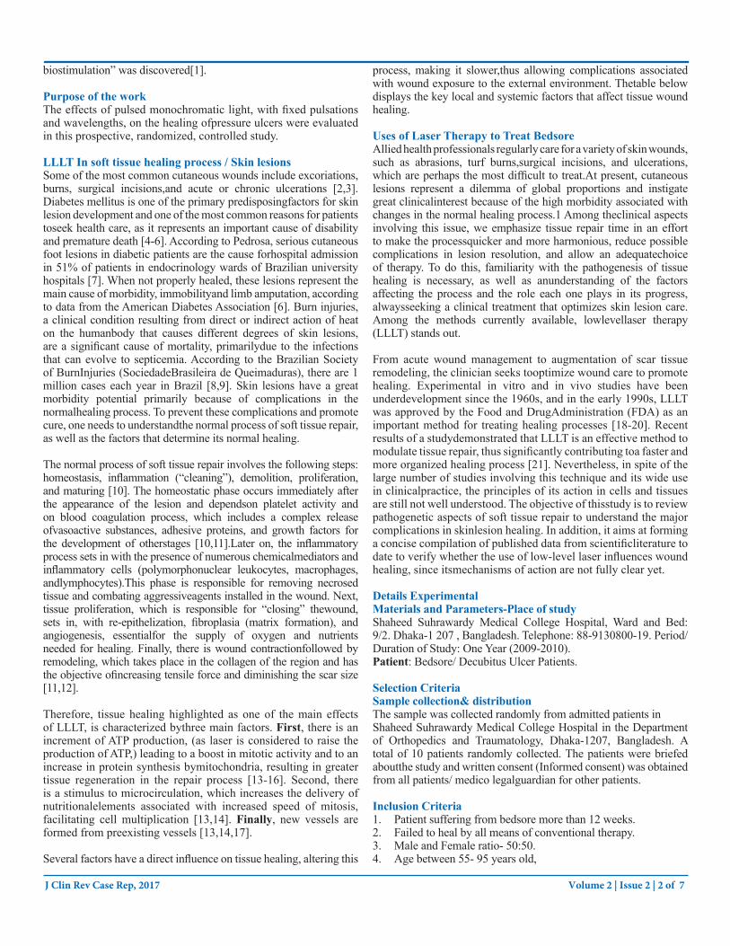
Figure 1: Law of photobiology states that for low power visible light to have any effect on a living biological system, the photons must be absorbed by electronic absorption bands belonging to some molecular photoacceptors, or chromophores- (Sutherland 2002). A chromophore is a molecule (or part of a molecule) which imparts some decided color to the compound of which it is an ingredient.
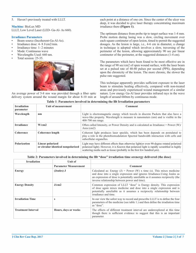
Figure 2: Schematic diagram showing the absorption of red and NIR light by specific Cellular chromophores photoacceptors localized in the mitochondrial respiratory chain.
Approach and methodologyProcedure
Treatment Schedule (Dose duration and wound parameter)week Frequency Wound
Area/sizeIrradiation
SourceWave Energy
FluencePoint Time
1-2 week 5/ week 6.8 cm2 LED-660 nm(Ga-Al-As)
Continuous 6 joules/cm2 2 8 joules/min.
3-5 week 3/ week 5.7 cm2 LED-660 nm(Ga-Al-As)
Continuous 4 joules/cm2 2 8 joules/min.
4-6 week 3/ week 4.4 cm2 LED-660 nm(Ga-Al-As)
Continuous 4 joules/cm2 1 8 joules/min.
7-8 week 2/week 2.2 cm2 LED-660 nm(Ga-Al-As)
Continuous 3 joules/cm2 1 8 joules/min.
9-10 week 2/ week Closed LED-660 nm(Ga-Al-As)
Continuous 3 joules/cm2 1 8 joules/min.
Visualization of treatment Progress by LLLT ( LED- Ga-Al- As 660 nm ) of a patient-(Chronological Picture View of a Laser (LLLT) Treated Patient)
J Clin Rev Case Rep, 2017 Volume 2 | Issue 2 | 4 of 7
Results and DiscussionResults:
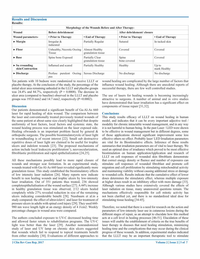
Morphology of the Wounds Before and After Therapy-Wound Before debridement After debridement/ closureWound parameters- ▪ Prior to Therapy ▪ End of Therapy ▪ Prior to Therapy ▪ End of Therapy► Margin Irregular &
induratedPartially Regular Sutured In tacked skin
► Floor Unhealthy, Necrotic Oozing Tissue
Almost Healthygranulation tissue
Covered Covered
► Base Spine bone Exposed Partially Cleargranulation tissue
Spinebone covered
Covered
► Su rroundingskin/Contraction
Inflamed and scared Partially Healthy Healthy Up tomark Healthy
► Discharge Profuse purulent Oozing pus
Serous Discharge No discharge No discharge
Ten patients with 10 bedsore were randomized to receive LLLT or placebo therapy. At the conclusion of the study, the percentage of the initial ulcer area remaining unhealed in the LLLT and placebo groups was 24.4% and 84.7%, respectively (P = 0.0008). The decrease in ulcer area (compared to baseline) observed in the LLLT and placebo groups was 193.0 mm2 and 14.7 mm2, respectively (P =0.0002).
DiscussionOur patients demonstrated a significant benefit of Ga-Al-As 660 laser for rapid healing of skin wound. The comparison between the laser and conventionally treated previously treated wounds of the same patient at about same size clearly highlighted that despite uniformity of host factors, local factors and systemic state, the wound healing process was stimulated on the laser exposed side. Healing ofwounds is an important problem faced by general & orthopedic surgeons. The possible biostimulatoryrole of laser light in woundhealing is of recent interest [22]. Small sub destructive repetitive doses of laser light are claimed to be useful for trophic ulcers and indolent wounds [23]. The proposed mechanisms of action include local leukocyte proliferation^), neovascularization, fibroblastic proliferation and rapid epithelialization [24,25].
All these mechanisms possibly lead to more rapid closure of wounds and stronger scar formation. In an experimental study, wounds treated with Ga-Al-As 660 laser revealed significantly more granulation tissue. This study established the biostimulatory effects of low intensity laser radiation [26]. Many reports now indicate benefit to non healing wounds and trophic ulcers by low-intensity laser irradiation. Out of 351 patients thus treated, 236 showed completeepithelialisation of the wound surface [27]. A 44% increase in healthy granulation tissue was observed, 2/12 ulcers healed completely while 27% revealed reduction in size of the remaining ulcers indicating considerable benefit [28]. Nussbaum et al.in a study compared- the effect of ultraviolet-C and laser for treatment of pressure ulcers in adults with spinal cord injury [29]. They used 660-980 nm wave length light at an energy density of 4 J/cm2. Weekly percentage changes in wound area were compared.
The authors concluded exposure to UV-C decreased healing time and allowed faster return to rehabilitation programs. The UV-C light was better than the laser [29]. Another nonrandomized study of laser and UV lamp on chronic skin ulcers suggested that wounds which fail to respond to topical treatments benefit from either modality [30]. Evaluations of different approaches to
wound healing are complicated by the large number of factors that influence wound healing. Although there are anecdotal reports of successful therapy, there are few well controlled studies.
The use of lasers for healing wounds is becoming increasingly attractive to surgeons. A number of animal and in vitro studies have demonstrated that laser irradiation has a significant effect on components of tissue repair [31,32].
ConclusionsThis study results efficacy of LLLT on wound healing in human model, and indicates that it can be avery important adjective tool /modality for chronic intractable wound management, and in any way it is not harmful to human being. In the past Laser / LED were shown to be effective in wound management but in different degrees, some of those applications showed significant improvement some less effective others no effect. Probably laser/ LED Irradiation parameters are vital for its Biostimulative effects. Inference of those results summaries that irradiation parameters are of vital to laser therapy. We used an optimal dose of irradiance which proved to be most effective biostimulation on human applicationApplication and research of LLLT on cell responses of wounded skin fibroblasts demonstrate that correct energy density or fluence and number of exposures can stimulate cell responses of wounded fibroblast and promote cell migration and cell proliferation by stimulating mitochondrial activity and maintaining viability without causing additional stress or damage to wounded cells. Results indicate that the cumulative effect of lower doses determines the stimulatory effect, whereas multiple exposure at higher doses result in an inhibitory effect with more damage [33].Although various studies have extensively covered the effects of laser radiation on tissue, many unanswered questions remain. The mechanisms effectively responsible for cell mitotic activity has not been clarified yet, and there is no standardized ideal dose for stimulating tissue healing [34-45].
Therefore, we noted that there is a need for research on the action and parameters of low-intensity laser use in cutaneous lesions during the different stages of repair, as an attempt to elucidate how this method acts at a cell level in healing processes [46-51]. Elucidation of these issues will enable the establishment of criteria on the true benefits of laser therapy in diseases that need healing stimulation, minimizing healing time and the complications that may occur during the clinical progress of these wounds. In addition, experimental studies indicated that the LLLT may be an important therapeutic tool to stimulate
J Clin Rev Case Rep, 2017 Volume 2 | Issue 2 | 5 of 7
wound healing in decubitus ulcer patients [52-58].Finally, the present report highlights the possible utility of Galliium-Aluminium laser at 660 wavelengths is as effective as Helium-Neon laser as an adjunctive modality for wound healing in skin/general & orthopedic practices.
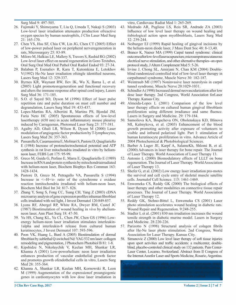
AcknowledgementsI wish to express my deepest gratitude and thanks to Professor Golam Abu Zakaria, for his kinddirections, inspiring guidance, and invaluable discussion throughout the work. Without his advice and encouragement, this work would never be fulfilled. I also express my heartfelt respect to Associate Professor, Dr. P C Debenarh, Department of Orthopedic & Trauma Surgery, Shaheed Suhrawardy Medical College and Hospital.
I would also like to express my profoundest gratitude to my project advisory board / working team members; Associate Prof. Dr Sheikh Abbasuddin Ahmed, Associate Prof Dr. Kazi Shamim uzzaman, Dr. Zia Uddin, consultant, Dr. SubirHossainShuvro, assistant registrar, Dr. Abdul Hannan of Orthopedic &Tramatology Department, Shaheed Suhrawardy Medical College and Hospital.
My deepest admiration and sincerest love to and my laser machine sponsor- Sinha Abu Khalid, technician Mr. Mamun, laser operator Ms. Jannant& Ms. Chewty of LabNucleon, and Md. Abdul Aziz, Mr. Polash, Mr. Abul- kasem, Ms. Fatema, Ms. Farida, Mr. Malek of Orthopedic and Traumatology Department, and KaziMuradHossain of Shaheed SuhrawardyMedical Hospital, Dhaka-1207, Bangladesh for their continuous efforts to successfully complete this work.
References1. Mechanisms of Low Level Laser Therapy. Michael R Hamblin
a, b, c,* and Tatiana N Demidovaa, da) Wellman Center for Photomedicine, Massachusetts General
Hospitalb) Department of Dermatology, Harvard Medical Schoolc) Harvard-MIT Division of Health Sciences andTechnologyd) Graduate Program in Cell Molecular and Developmental
Biology, Sackler School ofGraduate Biomedical Sciences,Tufts University School of Medicine.
2. Pereira AN, Eduardo Cde P, Matson E, Marques MM(2002) Effect of low-power laser irradiation on cell growthandmprocollagen synthesis of cultured fibroblasts, LasersSurg Med 31: 263-267.
3. Sutherland JC (2002) Biological effects of polychromaticlight, PhotochemPhotobiol 76: 164-170.
4. Karu T (1989) Laser biostimulation: a photobiological phenomenon,J Photochem Photobiol B 3: 638-640.
5. KaruTI, Afanas’evaNI (1995) Cytochrome c oxidase as theprimary photoacceptor upon laser exposure of cultured cellstovisible and near IR-range light, Dokl Akad Nauk 342: 693-695.
6. Capaldi RA, Malatesta F Darley-Usmar VM (1983) Structureof cytochrome c oxidase, Biochim Biophys Acta 726: 135-148.
7. Szundi I, Liao GL, Einarsdottir O (2001) Near-infrared time-
resolved optical absorption studies of the reaction of fully reduced cytochrome c oxidase with dioxygen, Biochemistry 40: 2332-2339.
8. Karu TI, Kolyakov SF (2005) Exact action spectra for cellularresponses relevant to phototherapy, Photomed Laser Surg 23:355-361.
9. Yu W, Naim JO, McGowan M, Ippolito K, Lanzafame RJ (1997)Photomodulation of oxidative metabolism and electronchainenzymes in rat liver mitochondria, Photochem Photobiol 66:866-871.
10. Passarella S (1989) He-Ne laser irradiation of isolated mitochondria,J Photochem Photobiol B 3: 642-643.
11. H Friedmann, Lubart R, Laulicht I, Rochkind S (1991) A possible explanation of laser-induced stimulation and damage ofcellcultures, J PhotochemPhotobiol B 11: 87-91.
12. Eichler M, Lavi R, Shainberg A, Lubart R (2005) Flavins aresource of visible-light-induced free radical formation in cells,Lasers Surg Med 37: 314-319.
13. Plaetzer K, Kiesslich T, Krammer B, Hammer lP (2002)Characterization of the cell death modes and the associatedchangesin cellular energy supply in response to AlPcS4-PDT,Photochem Photobiol Sci 1: 172-177.
14. Lubart R, Eichler M, Lavi R, Friedman H, Shainberg A (2005)Low-energy laser irradiation promotes cellular redoxactivity,Photomed Laser Surg 23: 3-9.
15. Duan R, Liu TC, Li Y, Guo H, Yao LB (2001) Signaltransduction pathways involved in low intensity He-Ne laser-inducedrespiratory burst in bovine neutrophils: a potentialmechanism of low intensity laser biostimulation, Lasers SurgMed 29: 174-178.
16. Antunes F, Boveris A, CadenasE (2004) On the mechanismand biology of cytochrome oxidase inhibition by nitric oxide,Proc Natl Acad Sci USA 101: 16774-16779.
17. KaruTI, Pyatibrat LV, Afanasyeva NI (2005) Cellular effectsof low power laser therapy can be mediated by nitric oxide,Lasers Surg Med 36:307-314.
18. Moriyama Y, Moriyama EH, Blackmore K, Akens MK, LilgeL (2005) In Vivo Study of the Inflammatory ModulatingEffectsof Low-level Laser Therapy on iNOS Expression UsingBioluminescence Imaging, Photochem Photobiol 81:1351-1355.
19. Schafer FQ, Buettner GR (2001) Redox environment of the cellas viewed through the redox state of the glutathionedisulfide/glutathione couple, Free Radic Biol Med 30:1191-1212.
20. Liu H, Colavitti R, Rovira (2005) II and T. Finkel, Redox-dependent transcriptional regulation, Circ Res 97: 967-974.
21. Yang M, Nazhat NB, Jiang X, Kelsey SM, Blake DR (1996)Adriamycin stimulates proliferation ofhuman lymphoblasticleukaemic cells via a mechanism of hydrogen peroxide (H2O2)production, Br J Haematol 95: 339-344.
22. Kirlin WG, Cai J, Thompson SA, Diaz D, Kavanagh TJ (1999)Glutathione redox potential in response todifferentiation andenzyme inducers, Free Radic Biol Med 27: 1208-1218.
23. Alaluf S, Muir-Howie H, Hu HL, Evans A, Green MR (2000)Atmospheric oxygen accelerates the induction of a post-mitoticphenotype in human dermal fibroblasts: the key protectiverole of glutathione, Differentiation 66: 147-155.
24. Karu T (1999) Primary and secondary mechanisms of actionof visible to near-IR radiation on cells, J Photochem PhotobiolB 49: 1-17.
25. Young S, Bolton P, Dyson M, Harvey W, Diamantopoulos C(1989) Macrophage responsiveness to light therapy, Lasers
J Clin Rev Case Rep, 2017 Volume 2 | Issue 2 | 6 of 7
Surg Med 9: 497-505.26. Fujimaki Y, Shimoyama T, Liu Q, Umeda T, Nakaji S (2003)
Low-level laser irradiation attenuates production ofreactiveoxygen species by human neutrophils, J Clin Laser Med Surg21: 165-170.
27. Chen YS, Hsu SF, Chiu CW, Lin JG, Chen CT (2005) Effectof low-power pulsed laser on peripheral nerveregeneration inrats, Microsurgery 25: 83-89.
28. Miloro M, Halkias LE, Mallery S, Travers S, Rashid RG (2002)Low-level laser effect on neural regeneration in Gore-Textubes,Oral Surg Oral Med Oral Pathol Oral Radiol Endod 93: 27-34.
29. Balaban P, Esenaliev R, Karu T, Kutomkina E, LetokhovV(1992) He-Ne laser irradiation ofsingle identified neurons,Lasers Surg Med 12: 329-337.
30. Byrnes KR, Waynant RW, Ilev IK, Wu X, Barna L, et al.(2005) Light promotesregeneration and functional recoveryand alters the immune response after spinal cord injury, LasersSurg Med 36: 171-185.
31. S.O. el Sayed SO, Dyson M (1996) Effect of laser pulserepetition rate and pulse duration on mast cell number anddegranulation, Lasers Surg Med 19: 433-437.
32. Lopes-Martins RA, Albertini R, Martins PS, Bjordal JM,Faria Neto HC (2005) Spontaneous effects of low-levellasertherapy (650 nm) in acute inflammatory mouse pleurisyinduced by Carrageenan, Photomed Laser Surg 23: 377-381.
33. Agaiby AD, Ghali LR, Wilson R, Dyson M (2000) Lasermodulation of angiogenic factor production by T-lymphocytes, Lasers Surg Med 26: 357-363.
34. Passarella S, Casamassima E, Molinari S, Pastore D, QuagliarielloE (1984) Increase of protonelectrochemical potential and ATP synthesis in rat liver mitochondria irradiated in vitro by helium-neon laser, FEBS Lett 175: 95-99.
35. Greco M, Guida G, Perlino E, Marra E, Quagliariello E (1989)Increase in RNA and protein synthesis by mitochondriairradiatedwith helium-neon laser, Biochem Biophys Res Commun 163:1428-1434.
36. Pastore D, Greco M, Petragallo VA, Passarella S (1994)Increase in <--H+/e- ratio of the cytochrome c oxidasereaction inmitochondria irradiated with helium-neon laser,Biochem Mol Biol Int 34: 817- 826.
37. Zhang Y, Song S, Fong CC, Tsang CH, Yang Z (2003) cDNA microarray analysis of gene expression profiles inhuman fibroblastcells irradiated with red light, J Invest Dermatol 120:849-857.
38. Lyons RF, Abergel RP, White RA, Dwyer RM, Castel JC(1987) Biostimulation of wound healing in vivo by ahelium-neon laser, Ann Plast Surg 18: 47-50.
39. Yu HS, Chang KL, Yu CL, Chen JW, Chen GS (1996) Low-energy helium-neon laser irradiation stimulates interleukin-1alpha and interleukin-8 release from cultured humankeratinocytes, J Invest Dermatol 107: 593-596.
40. Poon VK, Huang L, Burd A (2005) Biostimulation of dermalfibroblast by sublethal Q-switched Nd: YAG 532 nm laser: collagenremodeling and pigmentation, J Photochem Photobiol B 81: 1-8.
41. Kipshidze N, Nikolaychik V, Keelan MH, Shankar LR,Khanna A (2001) Low-powerhelium: neon laser irradiationenhances production of vascular endothelial growth factorand promotes growth ofendothelial cells in vitro, Lasers SurgMed 28: 355-364.
42. Khanna A, Shankar LR, Keelan MH, Kornowski R, LeonM (1999) Augmentation of the expressionof proangiogenicgenes in cardiomyocytes with low dose laser irradiation in
vitro, Cardiovasc Radiat Med 1: 265-269.43. Medrado AR, Pugliese LS, Reis SR, Andrade ZA (2003)
Influence of low level laser therapy on wound healing anditsbiological action upon myofibroblasts, Lasers Surg Med32: 239-244.
44. Neiburger EJ (1999) Rapid healing of gingival incisions bythe helium-neon diode laser, J Mass Dent Soc 48: 8-13,40.
45. Branco K, Naeser MA (1999) Carpal tunnel syndrome: clinicaloutcome after low-level laser acupuncture, microampstranscutaneouselectrical nerve stimulation, and other alternative therapies--an open protocol study, J Altern Complement Med 5: 5-26.
46. Irvine J, Chong SL, Amirjani N, Chan KM (2004) Double-blind randomized controlled trial of low-level laser therapy incarpaltunnel syndrome, Muscle Nerve 30: 182-187.
47. Weintraub MI (1997) Noninvasive laser neurolysis in carpaltunnel syndrome, Muscle Nerve 20:1029-1031.
48. Schindler A (1998) Increased dermal neovascularization after lowdose laser therapy. 2nd Congress, World Association forLaserTherapy. Kansas City.
49. Almeida-Lopes L (2001) Comparison of the low levellaser therapy effects on cultured human gingival fibroblastsproliferation using different irradiance and same fluence.Lasers in Surgery and Medicine. 29: 179-184.
50. Samoilova KA, Boqacheva ON, Obolenskaya KD, BlinovaMI, Kalmykova, et al. (2003) Enhancement of the bloodgrowth promoting activity after exposure of volunteers tovisible and infrared polarized light. Part I: stimulation ofhuman keratinocyte proliferation in vitro. Advance Article of2004 Photochemical & Photobiological Sciences.
51. Barber A Luger JE, Karpf A, SalameKh, Shlomi B, et al.(2000) Advances in laser therapy for bone repair. The Journalof Laser Therapy. World Association of Laser Therapy 13.
52. Antonio L (2000) Biomodulatory effects of LLLT on boneregeneration. The Journal of Laser Therapy. World Association of Laser Therapy 13.
53. Shefer G, et al. (2002) Low energy laser irradiation pro-motesthe survival and cell cycle entry of skeletal muscle satellitecells. Journalof Cell Science. 115: 1461-1469.
54. Enwemeka CS, Reddy GK (2000) The biological effects oflaser therapy and other modalities on connective tissue repairprocesses. The Journal of Laser Therapy. World Associationof Laser Therapy 12.
55. Reddy GK, Stehno-Bittel L, Enwemeka CS (2001) Laserphoto stimulation accelerates wound healing in diabetic rats.Wound Repair and Regeneration. 9:248-255.
56. Stadler I, et al. (2001) 830 nm irradiation increases the woundtensile strength in diabetic murine model. Lasers in Surgeryand Medicine. 28:220-226.
57. Parizotto N (1998) Structural analysis of colagen fibrilsafter He-Ne laser photo stimulation. 2nd Congress, WorldAssociation for Laser Therapy. Kansas City.
58. Simunovic Z (2000) Low level laser therapy of soft tissue injuriesupon sport activities and traffic accidents: a multicenter, double-blind, placebo-controled clinical study on 132 patients. Pain Center-Laser Center, Locarno, Switzerland. Abstract from II Congress ofthe Internat.Assnfor Laser and Sports Medicine, Rosario, Argentina.
Copyright: ©2017 Nazrul islam, et al. This is an open-access article distributed under the terms of the Creative Commons Attribution License, which permits uwnrestricted use, distribution, and reproduction in any medium, provided the original author and source are credited.
J Clin Rev Case Rep, 2017 Volume 2 | Issue 2 | 7 of 7