Embed Size (px)
Citation preview
Nath et al., J Clin Case Rep 2014, 4:12 DOI: 10.4172/2165-7920.1000466
Volume 4 • Issue 12 • 1000466J Clin Case RepISSN: 2165-7920 JCCR, an open access journal
Open AccessCase Report
Anaesthetic Management of Massive Pulmonary Embolism: Case report and ReviewMridu Paban Nath*, Nitya Nand Kumar, Malavika Barman and Rajib Kr BhattacharyyaDepartment of Anaesthesiology and Critical Care, Gauhati Medical College and Hospital, Guwahati, India
*Corresponding author: Mridu Paban Nath, Department of Anaesthesiology andCritical Care, Gauhati Medical College and Hospital, Guwahati-781 032, Assam,India, Tel: +91-7399003979; E-mail: drmridupaban@yahoo. com
Received May 23, 2014; Accepted December 15, 2014; Published December 30, 2014
Citation: Nath MP, Kumar NN, Barman M, Kr Bhattacharyya R (2014) Anaesthetic Management of Massive Pulmonary Embolism: Case report and Review. J Clin Case Rep 4: 466. doi:10.4172/2165-7920.1000466
Copyright: © 2014 Nath MP, et al. This is an open-access article distributed under the terms of the Creative Commons Attribution License, which permits unrestricted use, distribution, and reproduction in any medium, provided the original author and source are credited.
Keywords: Pulmonary embolism; Anaesthesia; Haemodynamicinstability; CPB; Embolectomy; Inotropes
IntroductionAcute massive Pulmonary Embolism (PE) is a life-threatening
emergency with a very high mortality [1]. The incidence of venous thrombo-embolism in India is 17. 46 per 10,000 hospital admissions and pulmonary embolism is diagnosed in 14. 9% of these patients with a 13. 5% mortality rate [2]. Approximately two third of patients, who die of a pulmonary embolus, die within the first hour after presentation. Rapid evaluation, diagnosis and intervention are necessary to save the life of these patients. Definitive diagnosis is made on the basis of angiography, echocardiography and imaging studies (ventilation-perfusion scanning, Computed Tomographic (CT) pulmonary angiography [3]. Anticoagulation and thrombolysis are the primary methods of treatment of pulmonary embolism along with haemodynamic optimization [1]. Recently, open pulmonary embolectomy has been advocated as a safe and more effective therapy for massive pulmonary embolism with acceptable outcome [4,5].
Case ReportA 34 year old man, presented in our emergency department with
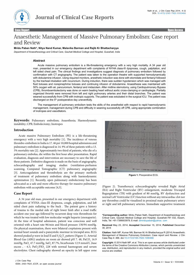
complaints of NYHA class-III dyspnoea, cough, palpitation, and left sided chest pain radiating to the back . The patient gave a history of trauma to the medial side of right lower limb after a road traffic accident one year ago followed by recurrent deep vein thrombosis for which he was treated with low molecular weight heparin (enoxaparin). At the time of hospital admission, patient was conscious and well oriented with a heart rate 96/min and blood pressure 130/94 mmHg. On physical examination, there were bilateral crepitaions present with normal heart sounds and a pansystolic murmur in tricuspid area. ECG showed peaked p wave in lead II and S1Q3T3 pattern (Figure 1). Arterial Blood Gas (ABG) analysis on room air showed pH 7. 48, PaCO2 16.9 mmHg, PaO2 47.7 mmHg, SaO2 87.7%, bicarbonate 12.9 mmol/L ,base excess , - 6.3, PaO2/FIO2 228 with normal haemogram and serum electrolytes. Chest radiography showed an opacity in left upper zone
AbstractAcute massive pulmonary embolism is a life-threatening emergency with a very high mortality. A 34 year old
man, presented in our emergency department with complaints of NYHA class-III dyspnoea, cough, palpitation, and left sided chest pain. The clinical findings and investigations suggest diagnosis of pulmonary embolism followed by confirmation with CT angiography. The patient was taken to the operation theatre with supported hemodynamically with dobutamine infusion. Using required monitors, anesthetic induction was done with etomidate and fentanyl followed by the tracheal intubation with rocuronium. During induction, there was sudden hypotension which was managed with fluid boluses and norepinephrine boluses and continuing infusion of dobutamine. Anaesthesia was maintained with 50% oxygen with air, pancuronium, fentanyl and midazolam. After midline sternotomy, using Cardiopulmonary Bypass (CPB), thromboembolectomy was done on warm beating heart without aortic cross-clamping or cardioplegia. Partially organized thrombi were removed from left and right pulmonary arteries and their distal branches. The patient was weaned successfully from CPB with inotropic supports. The patient was extubated in the surgical ICU. The patient was discharged on the 5th postoperative day uneventfully.
The management of pulmonary embolism tests the skills of the anesthetist with respect to rapid haemodynamic management, management during CPB and difficulty in weaning successfully off CPB, using appropriate combination of inotropes and vasodilators.
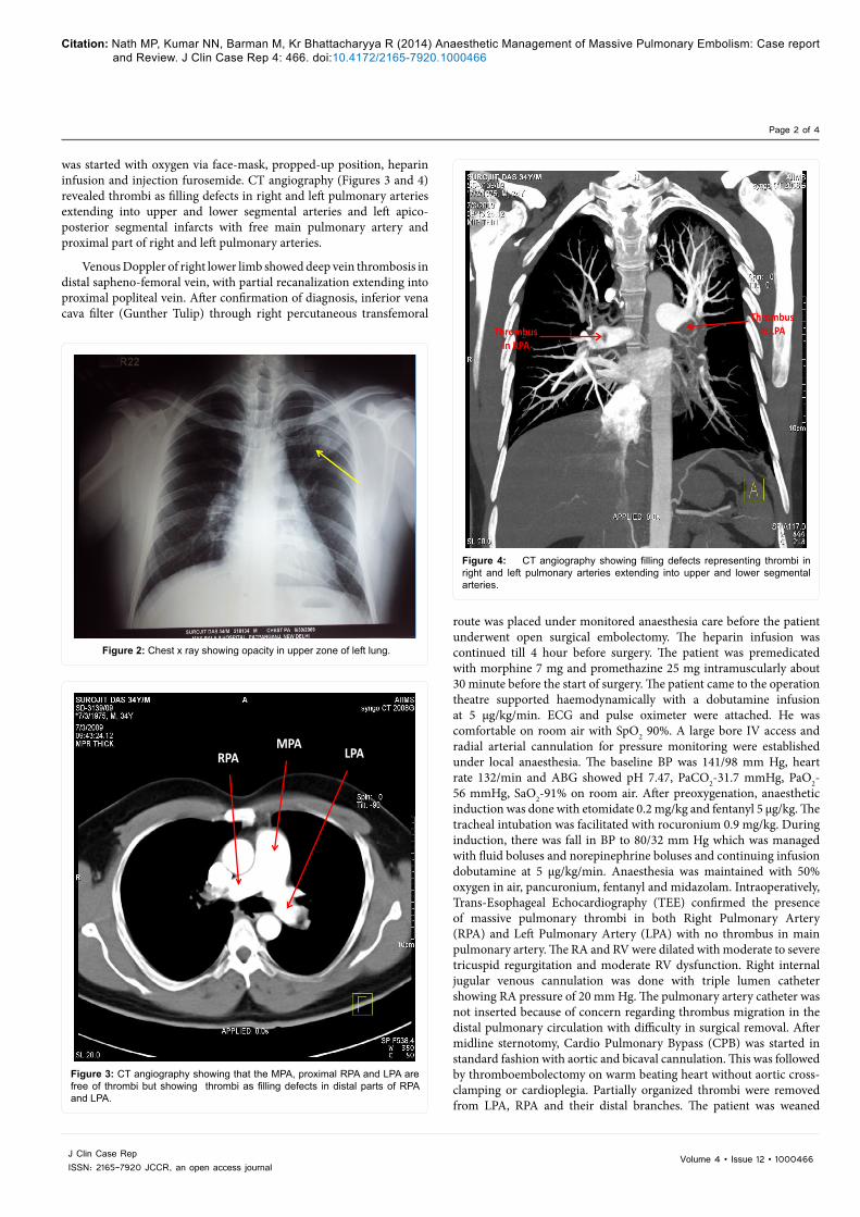
(Figure 2). Transthoracic echocardiography revealed Right Atrial (RA) and Right Ventricular (RV) enlargement, moderate Tricuspid Regurgitation (TR) with gradient of 80 mmHg, RV dysfunction and normal Left Ventricular (LV) function without any intracardiac clot nor any thrombus could be visualized in proximal main pulmonary artery or right and left pulmonary arteries. Immediate supportive treatment
Figure 1: Preoperative ECG.
Journal of Clinical Case ReportsJour
nal o
f Clinical Case Reports
ISSN: 2165-7920
Citation: Nath MP, Kumar NN, Barman M, Kr Bhattacharyya R (2014) Anaesthetic Management of Massive Pulmonary Embolism: Case report and Review. J Clin Case Rep 4: 466. doi:10.4172/2165-7920.1000466
Page 2 of 4
Volume 4 • Issue 12 • 1000466J Clin Case RepISSN: 2165-7920 JCCR, an open access journal
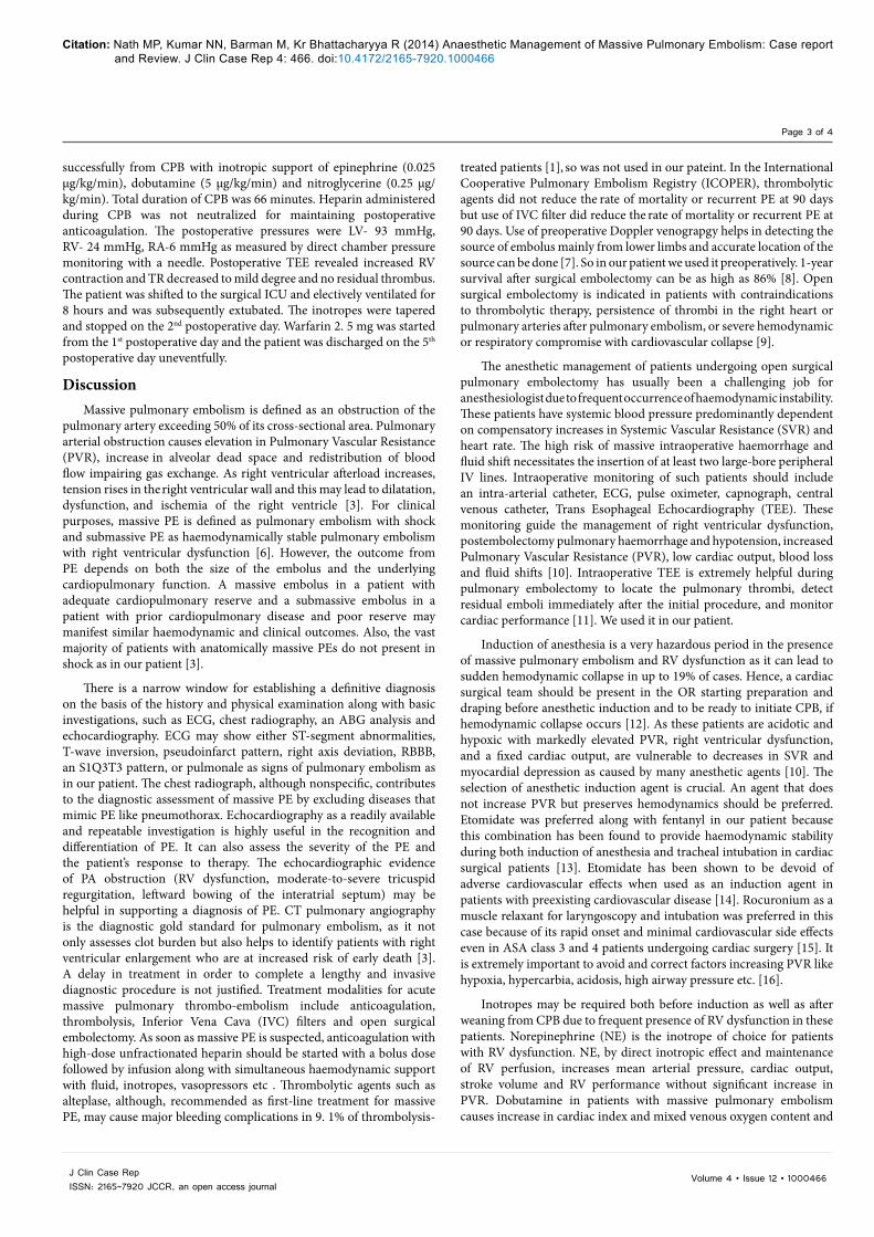
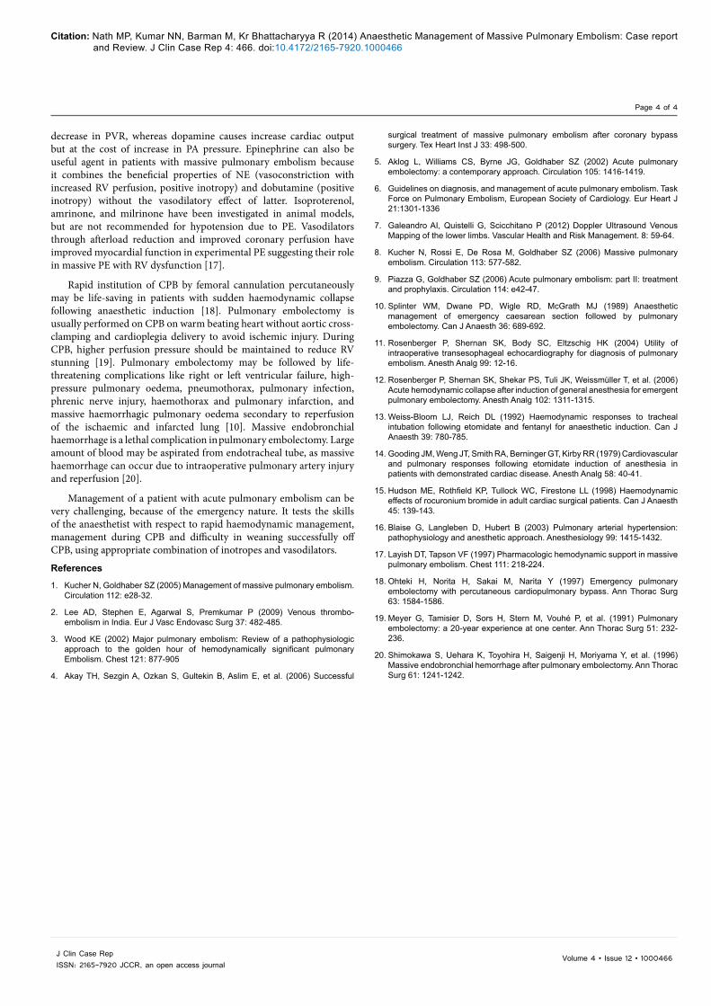
was started with oxygen via face-mask, propped-up position, heparin infusion and injection furosemide. CT angiography (Figures 3 and 4) revealed thrombi as filling defects in right and left pulmonary arteries extending into upper and lower segmental arteries and left apico-posterior segmental infarcts with free main pulmonary artery and proximal part of right and left pulmonary arteries.
Venous Doppler of right lower limb showed deep vein thrombosis in distal sapheno-femoral vein, with partial recanalization extending into proximal popliteal vein. After confirmation of diagnosis, inferior vena cava filter (Gunther Tulip) through right percutaneous transfemoral
route was placed under monitored anaesthesia care before the patient underwent open surgical embolectomy. The heparin infusion was continued till 4 hour before surgery. The patient was premedicated with morphine 7 mg and promethazine 25 mg intramuscularly about 30 minute before the start of surgery. The patient came to the operation theatre supported haemodynamically with a dobutamine infusion at 5 μg/kg/min. ECG and pulse oximeter were attached. He was comfortable on room air with SpO2 90%. A large bore IV access and radial arterial cannulation for pressure monitoring were established under local anaesthesia. The baseline BP was 141/98 mm Hg, heart rate 132/min and ABG showed pH 7.47, PaCO2-31.7 mmHg, PaO2-56 mmHg, SaO2-91% on room air. After preoxygenation, anaesthetic induction was done with etomidate 0.2 mg/kg and fentanyl 5 μg/kg. The tracheal intubation was facilitated with rocuronium 0.9 mg/kg. During induction, there was fall in BP to 80/32 mm Hg which was managed with fluid boluses and norepinephrine boluses and continuing infusion dobutamine at 5 μg/kg/min. Anaesthesia was maintained with 50% oxygen in air, pancuronium, fentanyl and midazolam. Intraoperatively, Trans-Esophageal Echocardiography (TEE) confirmed the presence of massive pulmonary thrombi in both Right Pulmonary Artery (RPA) and Left Pulmonary Artery (LPA) with no thrombus in main pulmonary artery. The RA and RV were dilated with moderate to severe tricuspid regurgitation and moderate RV dysfunction. Right internal jugular venous cannulation was done with triple lumen catheter showing RA pressure of 20 mm Hg. The pulmonary artery catheter was not inserted because of concern regarding thrombus migration in the distal pulmonary circulation with difficulty in surgical removal. After midline sternotomy, Cardio Pulmonary Bypass (CPB) was started in standard fashion with aortic and bicaval cannulation. This was followed by thromboembolectomy on warm beating heart without aortic cross-clamping or cardioplegia. Partially organized thrombi were removed from LPA, RPA and their distal branches. The patient was weaned
Figure 2: Chest x ray showing opacity in upper zone of left lung.
Thrombus in LPA Thrombus
in RPA
Figure 4: CT angiography showing filling defects representing thrombi in right and left pulmonary arteries extending into upper and lower segmental arteries.
MPA RPA LPA
Figure 3: CT angiography showing that the MPA, proximal RPA and LPA are free of thrombi but showing thrombi as filling defects in distal parts of RPA and LPA.
Citation: Nath MP, Kumar NN, Barman M, Kr Bhattacharyya R (2014) Anaesthetic Management of Massive Pulmonary Embolism: Case report and Review. J Clin Case Rep 4: 466. doi:10.4172/2165-7920.1000466
Page 3 of 4
Volume 4 • Issue 12 • 1000466J Clin Case RepISSN: 2165-7920 JCCR, an open access journal
successfully from CPB with inotropic support of epinephrine (0.025 μg/kg/min), dobutamine (5 μg/kg/min) and nitroglycerine (0.25 μg/kg/min). Total duration of CPB was 66 minutes. Heparin administered during CPB was not neutralized for maintaining postoperative anticoagulation. The postoperative pressures were LV- 93 mmHg, RV- 24 mmHg, RA-6 mmHg as measured by direct chamber pressure monitoring with a needle. Postoperative TEE revealed increased RV contraction and TR decreased to mild degree and no residual thrombus. The patient was shifted to the surgical ICU and electively ventilated for 8 hours and was subsequently extubated. The inotropes were tapered and stopped on the 2nd postoperative day. Warfarin 2. 5 mg was started from the 1st postoperative day and the patient was discharged on the 5th postoperative day uneventfully.
Discussion Massive pulmonary embolism is defined as an obstruction of the
pulmonary artery exceeding 50% of its cross-sectional area. Pulmonary arterial obstruction causes elevation in Pulmonary Vascular Resistance (PVR), increase in alveolar dead space and redistribution of blood flow impairing gas exchange. As right ventricular afterload increases, tension rises in the right ventricular wall and this may lead to dilatation, dysfunction, and ischemia of the right ventricle [3]. For clinical purposes, massive PE is defined as pulmonary embolism with shock and submassive PE as haemodynamically stable pulmonary embolism with right ventricular dysfunction [6]. However, the outcome from PE depends on both the size of the embolus and the underlying cardiopulmonary function. A massive embolus in a patient with adequate cardiopulmonary reserve and a submassive embolus in a patient with prior cardiopulmonary disease and poor reserve may manifest similar haemodynamic and clinical outcomes. Also, the vast majority of patients with anatomically massive PEs do not present in shock as in our patient [3].
There is a narrow window for establishing a definitive diagnosis on the basis of the history and physical examination along with basic investigations, such as ECG, chest radiography, an ABG analysis and echocardiography. ECG may show either ST-segment abnormalities, T-wave inversion, pseudoinfarct pattern, right axis deviation, RBBB, an S1Q3T3 pattern, or pulmonale as signs of pulmonary embolism as in our patient. The chest radiograph, although nonspecific, contributes to the diagnostic assessment of massive PE by excluding diseases that mimic PE like pneumothorax. Echocardiography as a readily available and repeatable investigation is highly useful in the recognition and differentiation of PE. It can also assess the severity of the PE and the patient’s response to therapy. The echocardiographic evidence of PA obstruction (RV dysfunction, moderate-to-severe tricuspid regurgitation, leftward bowing of the interatrial septum) may be helpful in supporting a diagnosis of PE. CT pulmonary angiography is the diagnostic gold standard for pulmonary embolism, as it not only assesses clot burden but also helps to identify patients with right ventricular enlargement who are at increased risk of early death [3]. A delay in treatment in order to complete a lengthy and invasive diagnostic procedure is not justified. Treatment modalities for acute massive pulmonary thrombo-embolism include anticoagulation, thrombolysis, Inferior Vena Cava (IVC) filters and open surgical embolectomy. As soon as massive PE is suspected, anticoagulation with high-dose unfractionated heparin should be started with a bolus dose followed by infusion along with simultaneous haemodynamic support with fluid, inotropes, vasopressors etc . Thrombolytic agents such as alteplase, although, recommended as first-line treatment for massive PE, may cause major bleeding complications in 9. 1% of thrombolysis-
treated patients [1], so was not used in our pateint. In the International Cooperative Pulmonary Embolism Registry (ICOPER), thrombolytic agents did not reduce the rate of mortality or recurrent PE at 90 days but use of IVC filter did reduce the rate of mortality or recurrent PE at 90 days. Use of preoperative Doppler venograpgy helps in detecting the source of embolus mainly from lower limbs and accurate location of the source can be done [7]. So in our patient we used it preoperatively. 1-year survival after surgical embolectomy can be as high as 86% [8]. Open surgical embolectomy is indicated in patients with contraindications to thrombolytic therapy, persistence of thrombi in the right heart or pulmonary arteries after pulmonary embolism, or severe hemodynamic or respiratory compromise with cardiovascular collapse [9].
The anesthetic management of patients undergoing open surgical pulmonary embolectomy has usually been a challenging job for anesthesiologist due to frequent occurrence of haemodynamic instability. These patients have systemic blood pressure predominantly dependent on compensatory increases in Systemic Vascular Resistance (SVR) and heart rate. The high risk of massive intraoperative haemorrhage and fluid shift necessitates the insertion of at least two large-bore peripheral IV lines. Intraoperative monitoring of such patients should include an intra-arterial catheter, ECG, pulse oximeter, capnograph, central venous catheter, Trans Esophageal Echocardiography (TEE). These monitoring guide the management of right ventricular dysfunction, postembolectomy pulmonary haemorrhage and hypotension, increased Pulmonary Vascular Resistance (PVR), low cardiac output, blood loss and fluid shifts [10]. Intraoperative TEE is extremely helpful during pulmonary embolectomy to locate the pulmonary thrombi, detect residual emboli immediately after the initial procedure, and monitor cardiac performance [11]. We used it in our patient.
Induction of anesthesia is a very hazardous period in the presence of massive pulmonary embolism and RV dysfunction as it can lead to sudden hemodynamic collapse in up to 19% of cases. Hence, a cardiac surgical team should be present in the OR starting preparation and draping before anesthetic induction and to be ready to initiate CPB, if hemodynamic collapse occurs [12]. As these patients are acidotic and hypoxic with markedly elevated PVR, right ventricular dysfunction, and a fixed cardiac output, are vulnerable to decreases in SVR and myocardial depression as caused by many anesthetic agents [10]. The selection of anesthetic induction agent is crucial. An agent that does not increase PVR but preserves hemodynamics should be preferred. Etomidate was preferred along with fentanyl in our patient because this combination has been found to provide haemodynamic stability during both induction of anesthesia and tracheal intubation in cardiac surgical patients [13]. Etomidate has been shown to be devoid of adverse cardiovascular effects when used as an induction agent in patients with preexisting cardiovascular disease [14]. Rocuronium as a muscle relaxant for laryngoscopy and intubation was preferred in this case because of its rapid onset and minimal cardiovascular side effects even in ASA class 3 and 4 patients undergoing cardiac surgery [15]. It is extremely important to avoid and correct factors increasing PVR like hypoxia, hypercarbia, acidosis, high airway pressure etc. [16].
Inotropes may be required both before induction as well as after weaning from CPB due to frequent presence of RV dysfunction in these patients. Norepinephrine (NE) is the inotrope of choice for patients with RV dysfunction. NE, by direct inotropic effect and maintenance of RV perfusion, increases mean arterial pressure, cardiac output, stroke volume and RV performance without significant increase in PVR. Dobutamine in patients with massive pulmonary embolism causes increase in cardiac index and mixed venous oxygen content and
Citation: Nath MP, Kumar NN, Barman M, Kr Bhattacharyya R (2014) Anaesthetic Management of Massive Pulmonary Embolism: Case report and Review. J Clin Case Rep 4: 466. doi:10.4172/2165-7920.1000466
Page 4 of 4
Volume 4 • Issue 12 • 1000466J Clin Case RepISSN: 2165-7920 JCCR, an open access journal
decrease in PVR, whereas dopamine causes increase cardiac output but at the cost of increase in PA pressure. Epinephrine can also be useful agent in patients with massive pulmonary embolism because it combines the beneficial properties of NE (vasoconstriction with increased RV perfusion, positive inotropy) and dobutamine (positive inotropy) without the vasodilatory effect of latter. Isoproterenol, amrinone, and milrinone have been investigated in animal models, but are not recommended for hypotension due to PE. Vasodilators through afterload reduction and improved coronary perfusion have improved myocardial function in experimental PE suggesting their role in massive PE with RV dysfunction [17].
Rapid institution of CPB by femoral cannulation percutaneously may be life-saving in patients with sudden haemodynamic collapse following anaesthetic induction [18]. Pulmonary embolectomy is usually performed on CPB on warm beating heart without aortic cross-clamping and cardioplegia delivery to avoid ischemic injury. During CPB, higher perfusion pressure should be maintained to reduce RV stunning [19]. Pulmonary embolectomy may be followed by life- threatening complications like right or left ventricular failure, high-pressure pulmonary oedema, pneumothorax, pulmonary infection, phrenic nerve injury, haemothorax and pulmonary infarction, and massive haemorrhagic pulmonary oedema secondary to reperfusion of the ischaemic and infarcted lung [10]. Massive endobronchial haemorrhage is a lethal complication in pulmonary embolectomy. Large amount of blood may be aspirated from endotracheal tube, as massive haemorrhage can occur due to intraoperative pulmonary artery injury and reperfusion [20].
Management of a patient with acute pulmonary embolism can be very challenging, because of the emergency nature. It tests the skills of the anaesthetist with respect to rapid haemodynamic management, management during CPB and difficulty in weaning successfully off CPB, using appropriate combination of inotropes and vasodilators.
References
1. Kucher N, Goldhaber SZ (2005) Management of massive pulmonary embolism. Circulation 112: e28-32.
2. Lee AD, Stephen E, Agarwal S, Premkumar P (2009) Venous thrombo-embolism in India. Eur J Vasc Endovasc Surg 37: 482-485.
3. Wood KE (2002) Major pulmonary embolism: Review of a pathophysiologicapproach to the golden hour of hemodynamically significant pulmonary Embolism. Chest 121: 877-905
4. Akay TH, Sezgin A, Ozkan S, Gultekin B, Aslim E, et al. (2006) Successful
surgical treatment of massive pulmonary embolism after coronary bypass surgery. Tex Heart Inst J 33: 498-500.
5. Aklog L, Williams CS, Byrne JG, Goldhaber SZ (2002) Acute pulmonary embolectomy: a contemporary approach. Circulation 105: 1416-1419.
6. Guidelines on diagnosis, and management of acute pulmonary embolism. Task Force on Pulmonary Embolism, European Society of Cardiology. Eur Heart J21:1301-1336
7. Galeandro AI, Quistelli G, Scicchitano P (2012) Doppler Ultrasound Venous Mapping of the lower limbs. Vascular Health and Risk Management. 8: 59-64.
8. Kucher N, Rossi E, De Rosa M, Goldhaber SZ (2006) Massive pulmonaryembolism. Circulation 113: 577-582.
9. Piazza G, Goldhaber SZ (2006) Acute pulmonary embolism: part II: treatmentand prophylaxis. Circulation 114: e42-47.
10. Splinter WM, Dwane PD, Wigle RD, McGrath MJ (1989) Anaestheticmanagement of emergency caesarean section followed by pulmonaryembolectomy. Can J Anaesth 36: 689-692.
11. Rosenberger P, Shernan SK, Body SC, Eltzschig HK (2004) Utility ofintraoperative transesophageal echocardiography for diagnosis of pulmonaryembolism. Anesth Analg 99: 12-16.
12. Rosenberger P, Shernan SK, Shekar PS, Tuli JK, Weissmüller T, et al. (2006) Acute hemodynamic collapse after induction of general anesthesia for emergent pulmonary embolectomy. Anesth Analg 102: 1311-1315.
13. Weiss-Bloom LJ, Reich DL (1992) Haemodynamic responses to trachealintubation following etomidate and fentanyl for anaesthetic induction. Can JAnaesth 39: 780-785.
14. Gooding JM, Weng JT, Smith RA, Berninger GT, Kirby RR (1979) Cardiovascular and pulmonary responses following etomidate induction of anesthesia inpatients with demonstrated cardiac disease. Anesth Analg 58: 40-41.
15. Hudson ME, Rothfield KP, Tullock WC, Firestone LL (1998) Haemodynamic effects of rocuronium bromide in adult cardiac surgical patients. Can J Anaesth 45: 139-143.
16. Blaise G, Langleben D, Hubert B (2003) Pulmonary arterial hypertension:pathophysiology and anesthetic approach. Anesthesiology 99: 1415-1432.
17. Layish DT, Tapson VF (1997) Pharmacologic hemodynamic support in massive pulmonary embolism. Chest 111: 218-224.
18. Ohteki H, Norita H, Sakai M, Narita Y (1997) Emergency pulmonary embolectomy with percutaneous cardiopulmonary bypass. Ann Thorac Surg63: 1584-1586.
19. Meyer G, Tamisier D, Sors H, Stern M, Vouhé P, et al. (1991) Pulmonary embolectomy: a 20-year experience at one center. Ann Thorac Surg 51: 232-236.
20. Shimokawa S, Uehara K, Toyohira H, Saigenji H, Moriyama Y, et al. (1996) Massive endobronchial hemorrhage after pulmonary embolectomy. Ann Thorac Surg 61: 1241-1242.