Embed Size (px)
Citation preview
RESEARCH ARTICLE
Environmentally Determined Differences inthe Murine Lung Microbiota and TheirRelation to Alveolar ArchitectureYeojun Yun1, Girish Srinivas2,3, Sven Kuenzel2,4, Miriam Linnenbrink2,4,Safa Alnahas5, Kenneth D. Bruce6, Ulrich Steinhoff5, John F. Baines2,4,Ulrich E. Schaible1*
1. Research Center Borstel, Cellular Microbiology Group, Department of Molecular Infection Biology, Borstel,Germany, 2. MPI for Evolutionary Biology, Plon, Germany, 3. Department of Dermatology, University ofLubeck, Lubeck, Germany, 4. Evolutionary Genomics, Institute for Experimental Medicine, Christian-Albrechts-University, Kiel, Germany, 5. Institute for Medical Microbiology and Hospital Hygiene, PhilippsUniversity Marburg, Marburg, Germany, 6. Institute of Pharmaceutical Science, King’s College London,London, United Kingdom
Abstract
Commensal bacteria control the micro-ecology of metazoan epithelial surfaces with
pivotal effect on tissue homeostasis and host defense. In contrast to the upper
respiratory tract, the lower respiratory tract of healthy individuals has largely been
considered free of microorganisms. To understand airway micro-ecology we
studied microbiota of sterilely excised lungs from mice of different origin including
outbred wild mice caught in the natural environment or kept under non-specific-
pathogen-free (SPF) conditions as well as inbred mice maintained in non-SPF, SPF
or germ-free (GF) facilities. High-throughput pyrosequencing of reverse transcribed
16S rRNA revealed metabolically active murine lung microbiota in all but GF mice.
The overall composition across samples was similar at the phylum and family level.
However, species richness was significantly different between lung microbiota from
SPF and non-SPF mice. Non-cultivatable Betaproteobacteria such as Ralstonia
spp. made up the major constituents and were also confirmed by 16S rRNA gene
cloning analysis. Additionally, Pasteurellaceae, Enterobacteria and Firmicutes were
isolated from lungs of non-SPF mice. Bacterial communities were detectable by
fluorescent in situ hybridization (FISH) at alveolar epithelia in the absence of
inflammation. Notably, higher bacterial abundance in non-SPF mice correlated with
more and smaller size alveolae, which was corroborated by transplanting
Lactobacillus spp. lung isolates into GF mice. Our data indicate a common
microbial composition of murine lungs, which is diversified through different
environmental conditions and affects lung architecture. Identification of the
OPEN ACCESS
Citation: Yun Y, Srinivas G, Kuenzel S,Linnenbrink M, Alnahas S, et al. (2014)Environmentally Determined Differences in theMurine Lung Microbiota and Their Relation toAlveolar Architecture. PLoS ONE 9(12): e113466.doi:10.1371/journal.pone.0113466
Editor: Yuanpu Peter Di, University of Pittsburgh,United States of America
Received: May 13, 2014
Accepted: October 27, 2014
Published: December 3, 2014
Copyright: � 2014 Yun et al. This is an open-access article distributed under the terms of theCreative Commons Attribution License, whichpermits unrestricted use, distribution, and repro-duction in any medium, provided the original authorand source are credited.
Data Availability: The authors confirm that all dataunderlying the findings are fully available withoutrestriction. All sequence files are available from theGenBank database (accession KJ729555-KJ729591) and the European NucleotideArchive(accession number PRJEB7007, http://www.ebi.ac.uk/ena/data/view/PRJEB7007).
Funding: This research is supported by ResearchCenter Borstel in house funding. The funders hadno role in study design, data collection andanalysis, decision to publish, or preparation of themanuscript.
Competing Interests: The authors have declaredthat no competing interests exist.
PLOS ONE | DOI:10.1371/journal.pone.0113466 December 3, 2014 1 / 24
microbiota of murine lungs will pave the path to study their influence on pulmonary
immunity to infection and allergens using mouse models.
Introduction
Human-associated microbiota have been characterized at many body sites
including the gut [1] and epithelial surfaces including the skin [2], vagina [3], oral
cavity [4] and upper respiratory tract (RT) [5]. Of these the gut, oral cavity and
upper respiratory tract are particularly heavily colonized by a diverse range of
bacterial species in healthy individuals [6]. There is consensus that the lower
airways can be heavily colonized by microbes in clinical scenarios such as cystic
fibrosis (CF) [7], asthma [8, 9] and chronic obstructive lung disease (COPD)
[8, 10]. In contrast, the lower airways of healthy individuals have been considered
by some authors as sterile [11]. However, evidence for the presence of microbes in
the healthy lung in porcine and murine models has been obtained recently
[12, 13]. Similarly, the presence of ‘‘low levels of bacterial sequences largely
indistinguishable from upper respiratory flora’’ in the healthy human lung has also
been reported [14]. Despite the careful precautions taken in the previous study, a
recurring concern is over the potential for contamination arising from the passage
of lower respiratory tract samples through the heavily colonized regions of the
upper airways and oral cavity.
Excising lung material directly from dead mice offers one means of avoiding
contamination from species in the oral cavity and upper respiratory tract. As such,
this allows the identification of bacterial species present in the lower airways at the
time of sampling, as well as species detectable in lower airways of different mouse
populations. In the present study, this approach was used to identify bacterial
species in lungs from mice, which were either out- or inbred and genetically
defined and kept under different environmental conditions, i.e. caught in the wild,
raised conventionally (non-SPF), under SPF – or under germfree conditions (see
Materials and Methods for details).
To identify and compare the bacterial compositions in lower airway tissues of
excised murine lungs, microbiological culture as well as culture-independent 16S
rRNA gene sequencing was performed [15]. Additionally, identified microbial
species were further characterized by their colonization potential.
Materials and Methods
Mice
Germ-free (GF) C57BL/6 mice were provided by the University of Marburg. GF
animals were kept in plastic isolators (Metall and Plastik, Germany) with
autoclaved food, bedding and water. Sterility of animals was checked biweekly by
Mouse Lung Microbiota
PLOS ONE | DOI:10.1371/journal.pone.0113466 December 3, 2014 2 / 24

culturing feces in thioglycollate medium under aerobic and anaerobic conditions
for at least ten days. All handling procedures for GF mice, including infection
experiments, were conducted in a laminar flow hood under sterile conditions. SPF
raised C57BL/6 mice were purchased from Charles River (Germany). They were
kept for a short term under SPF conditions at the animal facility of the Research
Center Borstel in individually ventilated cages till used. SPF mice were nourished
with sterile chow and regularly tested microbiologically. Non-SPF kept C57BL/6
mice originally also purchased from Charles River (Germany) (termed ‘‘non-SPF
C57BL/6 mice’’) and wild-derived mice originating from natural areas in France
and Germany were bred in a separate facility maintained by the Max Planck
Institute for Evolutionary Biology in Plon, Germany. For organ extraction, mice
were euthanized by CO2 inhalation to avoid mechanical disruption of the lung
and respiratory tract tissue and thus, potential contamination from tracheal
microbiota. The procedure was approved by the Ethics Committee for Animal
Experiments of the Ministry for Agriculture, Environment, and Rural Areas of the
State of Schleswig-Holstein, Germany, i.e. Kommission fur Tierversuche/Ethik-
Kommission des Landes Schleswig-Holstein (V 312-72241.123-34 according to
TierSchG 16 Abs1 Nr.4). All wild-derived mice belong to the Mus musculus
domesticus subspecies and were at least one generation removed from their free-
living progenitors (i.e. were born in the lab). Each non-SPF C57BL/6 and wild-
derived mouse was housed in separated racks with environmental air and fed the
same standard chow.
In addition, wild M. m. domesticus were caught in the Massif Central region of
France (termed ‘‘wild-caught mice’’), sacrificed with CO2 and dissected directly in
the field as described in Linnenbrink et al [16]. Utensils were washed in 70%
ethanol and flame-sterilized before each individual dissection, and a separate set
of instruments was used to remove lungs after opening the thoracic cavity.
Table 1 displays the origin and number of mice that were analyzed in individual
experiments.
DNA/RNA Extraction and cDNA preparation
For the culture-independent analyses, both RNA and DNA were simultaneously
extracted from lung tissue samples frozen in liquid nitrogen or fixed in RNAlater
(Ambion) using the AllPrep DNA/RNA Mini Kit following the manufacturer’s
instructions (Qiagen). Isolated RNA was used as template to generate cDNA
employing the 926r primer and SuperScript III Reverse Transcriptase (Invitrogen)
following the manufacturer’s instructions. 16S rRNA derived cDNAs were used as
templates for 16S rRNA gene pyrosequencing and cloning procedure.
16S rRNA gene pyrosequencing sequencing and analysis
The primer pair 27F-338R flanking the V1 and V2 hypervariable regions of the
bacterial 16S rRNA gene was used for PCR and barcoded pyrosequencing on the
454 GS-FLX with Titanium sequencing chemistry as described by Rausch et al.
Mouse Lung Microbiota
PLOS ONE | DOI:10.1371/journal.pone.0113466 December 3, 2014 3 / 24
[17]. The forward primer and barcode sequences were identified by a Perl script
using a Smith-Waterman alignment allowing no insertions or deletions.
Sequences were required to have a minimum length of 290 nucleotides and quality
score of >20. Chimeric sequences were removed using ChimeraSlayer (http://
microbiomeutil.sourceforge.net/). RDP Multi-Classifier version 1.0 was used to
assign taxonomy with a minimum confidence score of 0.80. Clustering of
sequences into operational taxonomic units (OTUs) at the species-level similarity
threshold (97%) and alpha diversity estimates were performed using QIIME
version 1.7.0 [18]. UCLUST reference based OTU picking was implemented
against a representative dataset (‘‘97_otus.fasta’’ available from Greengenes
database release 13_5; http://greengenes.secondgenome.com/downloads/ data-
base/13_5) to define Operational Taxonomic Unit (OTUs) at 97% sequence
identity and discard singleton OTUs. Reads that did not match the collection of
reference sequences were subsequently clustered as de novo clusters. We used the
Chimeraslayer algorithm using a reference non-chimeric database (http://drive5.
com/uchime/gold.fa) for detecting Chimeric sequences. The raw sequence counts
for samples from the 454 data ranged from 1178 to 9347. All samples were rarefied
to 1000 sequences and the downstream analyses were performed on normalized
counts. Beta diversity analyses were performed using the Vegan R package [19]
including the taxon (OTU)-based Bray-Curtis and Jaccard distances as well as the
phylogenetic-based unweighted and weighted UniFrac distances [20]. Alpha and
beta diversity analyses were performed with a normalized sequence number of
1000 per individual. The entire dataset associated with this study was submitted to
the European Nucleotide Archive under the study accession number PRJEB7007
(http://www.ebi.ac.uk/ena/data/view/PRJEB7007).
Polymerase chain reaction (PCR) and Cloning procedures
cDNA template generated from each GF, SPF, non-SPF C57BL/6 and wild-derived
mouse lung was amplified by 16S rRNA gene PCR primer set: 8f700 (59-AGA GTT
TGA TCC TGG CTC AG-39) and 926r (59-CCG TCA ATT CAT TTG AGT TT-39)
[7]. For the quality control of cDNA, Ralstonia-specific PCR was done using the
primer set: Rp-F (59-ATG ATC TAG CTT GCT AGA TTG AT-39) and Rp-R (59-
ACT GAT CGT CGC CTT GGT G-39) [21]. PCR reactions were perfomed by an
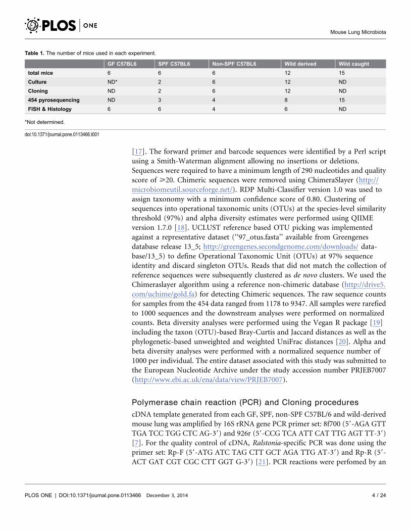
Table 1. The number of mice used in each experiment.
GF C57BL6 SPF C57BL6 Non-SPF C57BL6 Wild derived Wild caught
total mice 6 6 6 12 15
Culture ND* 2 6 12 ND
Cloning ND 2 6 12 ND
454 pyrosequencing ND 3 4 8 15
FISH & Histology 6 6 4 6 ND
*Not determined.
doi:10.1371/journal.pone.0113466.t001
Mouse Lung Microbiota
PLOS ONE | DOI:10.1371/journal.pone.0113466 December 3, 2014 4 / 24

initial denaturation step at 95 C for 5 min followed by 40 cycles of denaturation at
95 C for 30 s, primer annealing at 60 C for 30 s and primer extension at 72 C for
45 s, with final extension step at 72 C for 5 min.
Cloning of 16S rRNA PCR-amplified DNA was performed with the TOPO TA
cloning kit (Invitrogen) according to the manufacturer’s instructions.
Transformation was performed with competent Escherichia coli TOP10 cells
provided by the manufacturer. The transformed cells were then plated onto LB
agar plates supplemented with Ampicillin (100 mg/ml), and the plates were
incubated overnight at 37 C. Correct sizes of the inserts were determined in a
colony PCR with an M13 forward and reverse primer. Prior to sequencing of the
fragments, the PCR-amplified 16S rRNA gene regions were purified with the
GeneJET PCR Purification Kit (Fermentas).
Culture collection from mouse lung
The lungs of all mice, apart from those sampled directly in the wild, were collected
under a clean bench to avoid contamination of extraneous components. The
trachea was removed before lungs were excised carefully without any upper
respiratory parts. One lobe of lung was transferred to a Dispomix (gentleMACS
Dissociator, Miltenyi Biotec) disposable homogenizer with 500 uL sterile
phosphate buffered saline (PBS). The homogenization step with the Dispomix was
performed at 500 g for 20 seconds. After homogenization the centrifuged sample
was gently mixed and transferred to different bacterial culture media: LB plate/
broth, BHI plate/broth (aerobic/anaerobic), anaerobic blood agar plate (BAP),
anaerobic Fluid thioglycolate medium (FTG), and 5% CO2 blood agar (BAP)/
chocolate agar plate (CH). To assess the number of cultivatable bacteria in mouse
lungs, colonies were counted from each BHI and BAP plate. Pure colonies were
isolated and subcultured for further analysis.
DNA extraction and identification of murine lung isolates
DNA was extracted from individual bacterial isolates by the Phenol/chloroform/
isoamyl (PCI) method after treatment with lysozyme and proteinase K and used
as template for 16S rRNA gene PCR. Two universal bacterial primers were used:
8f700 (59-AGA GTT TGA TCC TGG CTC AG-39) and 926r (59-CCG TCA ATT
CAT TTG AGT TT-39) [7].
After sequencing, 11 culture collections of representative taxa were deposited in
Leibniz-Institut DSMZ (Deutsche Sammlung von Mikroorganismen und
Zellkulturen GmbH, which are DSM26538 (Staphylococcus xylosus, G2B1),
DSM26539 (Pasteurella pneumotropica, 3F1B2), DSM26540 (Actinobacillus muris,
G2CH1), DSM26541 (Enterococcus gallinarum, 3B1CH1), DSM26543 (Bacillus
lichenifomis, 3G1B1), DSM26544 (Paenibacillus sp. G3B2), DSM26546
(Lactobacillus sp. 3B2BAP1), DSM26547 (Corynebacterium sp. G1CH1),
DSM26548 (Lactobacillus sp. 3F2BAP4), DSM26549 (Escherichia coli, 3B3BAP1),
and DSM26621 (Streptococcus danieliae 3G1BAP1).
Mouse Lung Microbiota
PLOS ONE | DOI:10.1371/journal.pone.0113466 December 3, 2014 5 / 24
Sequencing analysis
16S rRNA gene sequences from bacterial isolates and cloning were sequenced by
the Sanger method (Seqlab, Germany). Sequences were aligned using Sequence
Match of the ribosomal database project (RDP, http://rdp.cme.msu.edu/) and
compared to NCBI BLAST results for the percentage of sequence identity. The
phylogenetic trees were conducted in MEGA4 software determined by the
Neighbor-Joining method. All positions containing alignment gaps and missing
data were eliminated only in pairwise sequence comparisons. Branches
corresponding to partitions reproduced in less than 50% bootstrap replicates were
collapsed. The percentage of replicate trees in which the associated taxa clustered
together in the bootstrap test (1000 replicates) was shown next to the branches.
The sequences used in Figure S1 in File S1 were stored as GenBank accession
KJ729555-KJ729591.
Organs for histology and fluorescent in situ hybridization (FISH)
GF, SPF and non-SPF C57BL/6 mice between 24 and 25 weeks of age were
compared for pulmonary histological differences. The age of wild–derived mice
varied between 25–45 weeks.
For histopathological and FISH analysis, lungs were sterilely removed. To avoid
alterations and redistribution of lung microbiota, lungs were immediately fixed by
4% paraformaldehyde at 4 C for 24 hours. Organs were further processed for
paraffin embedding.
Histology
Consecutive sections of 8 mm taken at regular intervals were obtained using a
microtome. Sections were stained with either Haematoxylin-Eosin (H&E) or
periodic acid-Schiff (PAS) to reveal mucus production according to standard
procedures.
In short, paraffin sections on slides were deparaffinized and rehydrated. For
H&E staining, slides were stained with Mayer’s Haematoxylin followed by Eosin
after rinsing with tab water. For PAS staining, slides were oxidized in 0.5%
periodic acid solution, and then placed in Schiff reagent followed by counterstain
with Mayer’s Haematoxylin. Both procedures were finalized by dehydration and
mounting.
Morphometry was done by measuring the size of alveolae and counting the
numbers per microscopical field using a 400X magnification. Data represent the
average of 10 microscopical fields. The alveolar area (mm2) of 10 alveolae was
measured using CellB version 3.3 (Olympus Soft Imaging Solution GmbH).
FISH
Hybridization was performed using the probe EUB338 labeled with Cy3, whose
sequence is complementary to the sequence of a region within the 16S rRNA that
Mouse Lung Microbiota
PLOS ONE | DOI:10.1371/journal.pone.0113466 December 3, 2014 6 / 24

is common to all eubacteria as well as the mixture probe, EUB338 I, II, III. The
complementary probe non-EUB338 labeled with Cy3 was used as control. Tissue
sections were prepared using sterile microtome equipment and deparaffinized in
coplin jars by two changes of xylene and dehydrated twice with 99.9% ethanol for
3 min at each step. Slides were air dried and a circle was drawn around the tissue
section using a hydrophobic pen (Dako PAP pen; Glostrup, Denmark). The
hybridization and washing steps were performed as previously reported [22].
Fixed samples were hybridized by the application of 10 ml of hybridization buffer
containing 5 ng of each specific oligonucleotide probe at 45 C for 90 min.
Stringent washing was performed by incubating the slide in washing buffer
(20 mM Tris-HCl pH 7.6, 0.01% sodium dodecyl sulfate, 112 mM NaCl) at 48 C
for 15 min. Finally, the slides were rinsed with water, stained with DAPI, air dried,
and mounted in Citifluor (Citifluor Ltd., London, United Kingdom). Slides were
examined using a Nikon fluorescent microscope equipped with a standard filter
set (Nikon, Germany).
Murine lung microbiota transplants
The following bacterial isolates from C57BL/6 mice raised under non-SPF
conditions were used for colonization experiments: DSM26546 (Lactobacillus sp.
3B2BAP1) and DSM26548 (Lactobacillus sp. 3F2BAP4) were isolated by culture in
Lactobacilli MRS medium (Difco) in a 5% CO2 atmosphere. Isolates were freshly
cultured at 37 C overnight, and washed 3 times in PBS. 56105 CFU in 20 mL PBS
were given intranasally into both nostrils of 10 weeks old GF mice. After 16 weeks,
mouse lungs were isolated and analyzed for lung colonization by Lactobacillus spp.
specific PCR: F(59-AGCAGTAGGGAATCTTCCA-39) and R(59-
CACCGCTACACATGGAG-39). [23] Amplification program included an initial
denaturation step at 95 C for 5 min followed by 30 cycles of denaturation at 95 C
for 30 s, primer annealing at 60 C for 30 s and primer extension at 72 C for 45 s,
with final extension step at 72 C for 5 min. Paraffin embedded lung sections were
analyzed for histological alterations by H&E and PAS stain (see above).
Statistical analysis
Statistical analyses were conducted using GraphPad Prism version 5.01 (GraphPad
software Inc., USA). Statistical significance was determined by one-way analysis of
variation (ANOVA) followed by Tukey’s Multiple comparison test or two tailed
Student’s t-test followed by Mann Whitney test (*p,0.05, **p,0.01,
***p,0.001). Statistical analysis of alpha diversity was performed using the
Wilcoxon rank sum test followed by a correction for multiple testing [24]. The
multivariate ANOVA procedure implemented in analysis of dissimilarity
(‘‘Adonis’’) (included in Vegan R package) was used to test the statistical
significance of beta diversity measurements with respect to the origin of mice with
1000 per mutations.
Mouse Lung Microbiota
PLOS ONE | DOI:10.1371/journal.pone.0113466 December 3, 2014 7 / 24

Results
Murine lung microbiota by 454 pyrosequencing
In this study, nucleic acids were extracted directly from different mouse
populations originating from the wild or from animal suppliers and held under
GF, SPF or non-SPF conditions. Lungs of these mice were compared to those of
outbred Mus musculus domesticus populations derived from the wild, either
housed under non-SPF conditions or sampled directly in the natural environ-
ment. cDNA from RNA extracts was used as template for 454 pyrosequencing
using the ‘926r’ 16S rRNA primer for reverse transcription, since direct 16S rRNA
gene amplification by PCR from lung tissue DNA failed, most likely due to the
low amount of bacterial nucleic acid template in the tissue samples [7]. From
these cDNA templates, a total of 90,662 16S rRNA gene region sequences
averaging 3,022, were generated per sample from 30 excised lungs.
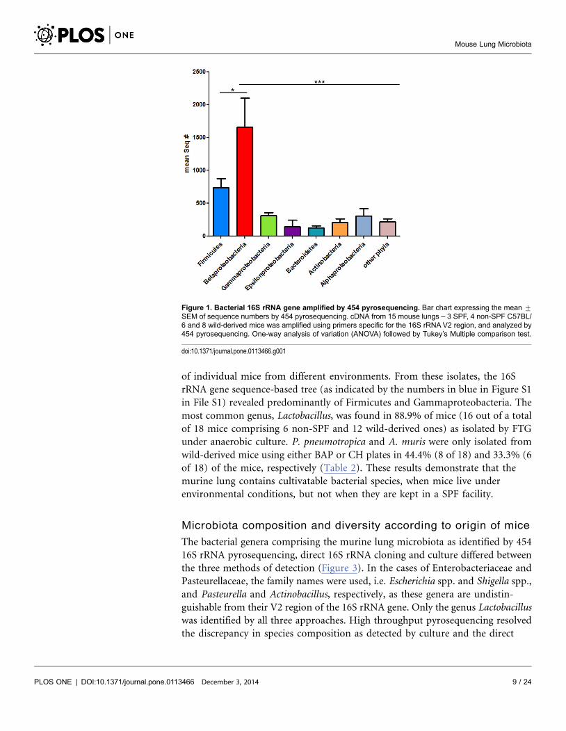
These results revealed distinct bacterial populations in murine lungs, with
Betaproteobacteria dominating over Firmicutes, Gammaproteobacteria,
Epsilonproteobacteria and Actinobacteria (Figure 1). Of note, Bacteroidetes and
Alphaproteobacteria, which cannot be cultured easily, were identified in mouse
lungs by 454 pyrosequencing in similar frequencies to Gammaproteobacteria (
Figure 1).
To confirm and complement 454 pyrosequencing data, lung samples were
additionally subjected to I) direct nucleic acid extraction for culture-independent
16S rRNA gene analysis by generating cDNA libraries using the ‘926r’ reverse
primer for reverse transcription, and II) culture under aerobic, anaerobic or
micro-aerophilic conditions using different growth media. We were able to
amplify 16S rRNA gene from non-SPF as well as SPF, but not from GF mouse
lungs. Cloned sequences of 16S rRNA genes were grouped upon phylogenetic
analysis by the Neighbor-Joining method (shown by the numbers in red in Figure
S1 in File S1). We identified Betaproteobacteria, primarily Ralstonia spp., as well
as Firmicutes and Epsilonproteobacteria (Helicobacter spp.).
Of note, Ralstonia spp. was also found in SPF mouse lungs otherwise relatively
low in microbes. Since Ralstonia is a common laboratory contaminant, the
possibility that its presence in SPF mouse lungs was due to contamination during
the experimental procedure had to be excluded. Therefore quality control of the
samples used for sequencing was performed through Ralstonia-specific PCR of the
16S rRNA gene (Figure 2A) revealing that the experimental procedure employed
avoided bacterial contamination as GF mouse samples generated and processed
simultaneously did not yield PCR products. The presence of Ralstonia spp. was
further confirmed by specific PCR in the lungs from SPF as well as non-SPF, but
not in GF mice, also demonstrating that Ralstonia-specific signals were not due to
contamination during the experimental procedure (Figure 2B).
More importantly, we were able to isolate a total of 128 bacterial colonies from
5/6 non-SPF C57BL6 and 12/12 wild-derived mice but failed to isolate bacteria
from lungs of all GF, SPF as well as 1/6 non-SPF C57BL/6 mice. Figure S2 in File
S1 shows the differences in numbers and diversity of colonies isolated from lungs
Mouse Lung Microbiota
PLOS ONE | DOI:10.1371/journal.pone.0113466 December 3, 2014 8 / 24
of individual mice from different environments. From these isolates, the 16S
rRNA gene sequence-based tree (as indicated by the numbers in blue in Figure S1
in File S1) revealed predominantly of Firmicutes and Gammaproteobacteria. The
most common genus, Lactobacillus, was found in 88.9% of mice (16 out of a total
of 18 mice comprising 6 non-SPF and 12 wild-derived ones) as isolated by FTG
under anaerobic culture. P. pneumotropica and A. muris were only isolated from
wild-derived mice using either BAP or CH plates in 44.4% (8 of 18) and 33.3% (6
of 18) of the mice, respectively (Table 2). These results demonstrate that the
murine lung contains cultivatable bacterial species, when mice live under
environmental conditions, but not when they are kept in a SPF facility.
Microbiota composition and diversity according to origin of mice
The bacterial genera comprising the murine lung microbiota as identified by 454
16S rRNA pyrosequencing, direct 16S rRNA cloning and culture differed between
the three methods of detection (Figure 3). In the cases of Enterobacteriaceae and
Pasteurellaceae, the family names were used, i.e. Escherichia spp. and Shigella spp.,
and Pasteurella and Actinobacillus, respectively, as these genera are undistin-
guishable from their V2 region of the 16S rRNA gene. Only the genus Lactobacillus
was identified by all three approaches. High throughput pyrosequencing resolved
the discrepancy in species composition as detected by culture and the direct
Figure 1. Bacterial 16S rRNA gene amplified by 454 pyrosequencing. Bar chart expressing the mean ¡
SEM of sequence numbers by 454 pyrosequencing. cDNA from 15 mouse lungs – 3 SPF, 4 non-SPF C57BL/6 and 8 wild-derived mice was amplified using primers specific for the 16S rRNA V2 region, and analyzed by454 pyrosequencing. One-way analysis of variation (ANOVA) followed by Tukey’s Multiple comparison test.
doi:10.1371/journal.pone.0113466.g001
Mouse Lung Microbiota
PLOS ONE | DOI:10.1371/journal.pone.0113466 December 3, 2014 9 / 24

cloning approach. All mice, regardless whether they were of SPF or non-SPF
origin, featured the same high ranking genera, i.e. Ralstonia, Lactobacillus,
Enterobacteriaceae, and Sphingomonas. Ralstonia spp. ranked at the top of the list
of all bacterial genera identified in mice caught in the wild, which was comparable
in all mice studied independent of their origin. Interestingly, Helicobacter spp.
(Epsilonproteobacteria) was abundant in wild-caught mice (Figure 3). At the
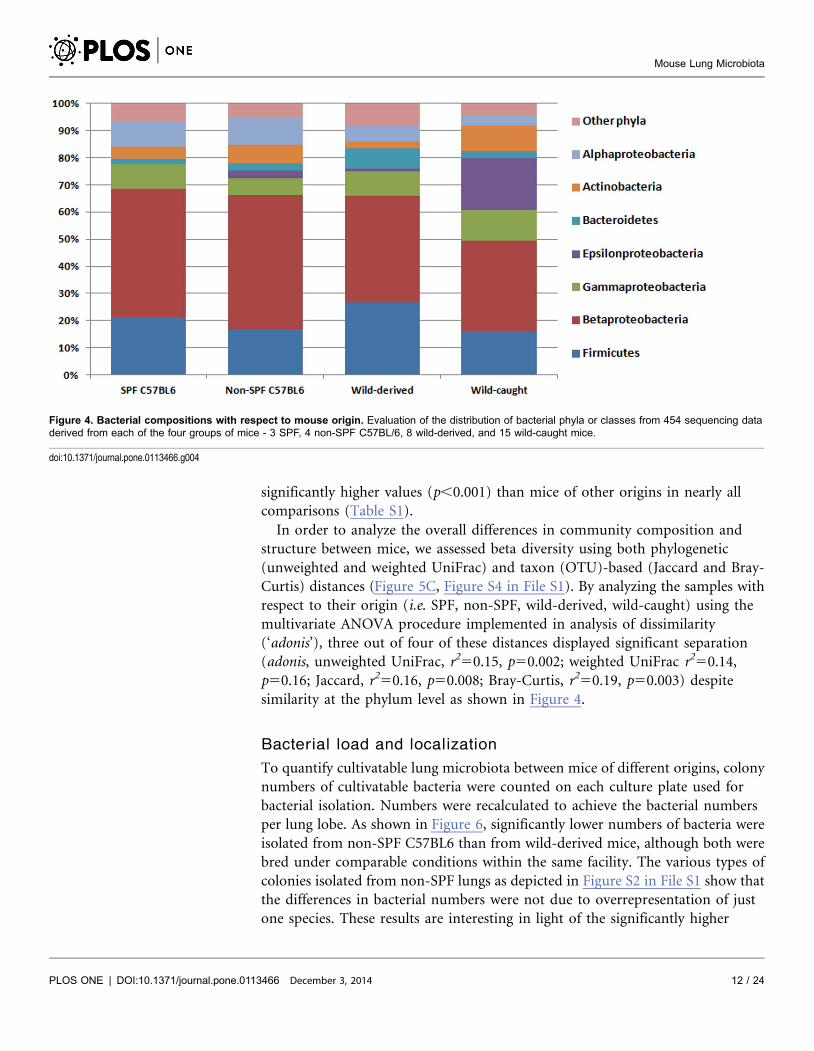
phylum level, we found similar patterns in all mice analyzed, independent of their
origin (Figure 4). In general, however, we observed considerable variability
between mice of different origins at both the genus and family levels (Figure S3 in
File S1). For example, Bacteroides (Firmicutes) was missing only from SPF lungs,
but was abundant in mice caught in the wild (Figure S3B in File S1).
Enterobacteriaceae (Gammaproteobacteria) were equally abundant in all mice
analysed but not in wild-derived mice, which contained predominantly Pasteurella
spp. (Figure S3E in File S1). It has to be noted that these comparisons were
statistically not significant.
To characterize the diversity of bacterial communities within individual mouse
lungs and between individuals, we performed alpha and beta diversity analyses,
respectively. Two different measures of alpha diversity focusing on species
richness (Chao1) and evenness (Shannon) revealed notable differences between
mice of different origins (Figure 5A–B, Table S1). Interestingly, the microbiota
from non-SPF C57BL6 mice showed the highest diversity and richness, displaying
Figure 2. 16S rRNA gene and Ralstonia-specific PCR. 16S rRNA gene PCR (A) and Ralstonia-specificPCR (B) (Abbreviations: GF; Germ Free mouse, SPF1 and SPF2; Specific Pathogen Free C57BL/6, N/C;negative control, N-SPF; non-SPF C57BL/6, WD1 and WD2; wild-derived mice, P/C; positive control, primersused are described in Materials and Methods).
doi:10.1371/journal.pone.0113466.g002
Mouse Lung Microbiota
PLOS ONE | DOI:10.1371/journal.pone.0113466 December 3, 2014 10 / 24
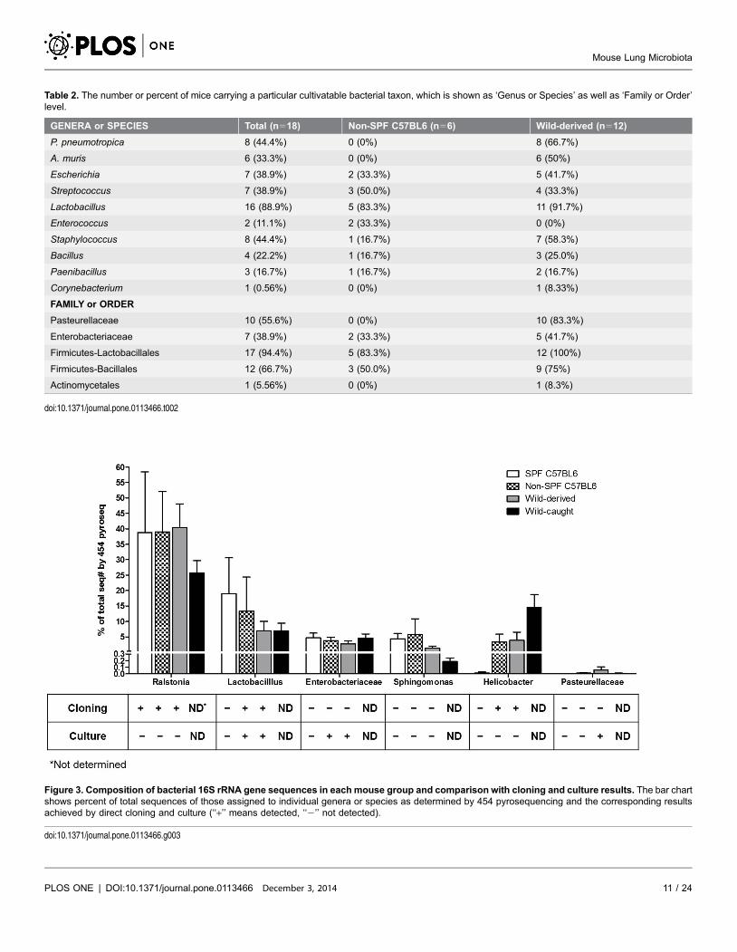
Table 2. The number or percent of mice carrying a particular cultivatable bacterial taxon, which is shown as ‘Genus or Species’ as well as ‘Family or Order’level.
GENERA or SPECIES Total (n518) Non-SPF C57BL6 (n56) Wild-derived (n512)
P. pneumotropica 8 (44.4%) 0 (0%) 8 (66.7%)
A. muris 6 (33.3%) 0 (0%) 6 (50%)
Escherichia 7 (38.9%) 2 (33.3%) 5 (41.7%)
Streptococcus 7 (38.9%) 3 (50.0%) 4 (33.3%)
Lactobacillus 16 (88.9%) 5 (83.3%) 11 (91.7%)
Enterococcus 2 (11.1%) 2 (33.3%) 0 (0%)
Staphylococcus 8 (44.4%) 1 (16.7%) 7 (58.3%)
Bacillus 4 (22.2%) 1 (16.7%) 3 (25.0%)
Paenibacillus 3 (16.7%) 1 (16.7%) 2 (16.7%)
Corynebacterium 1 (0.56%) 0 (0%) 1 (8.33%)
FAMILY or ORDER
Pasteurellaceae 10 (55.6%) 0 (0%) 10 (83.3%)
Enterobacteriaceae 7 (38.9%) 2 (33.3%) 5 (41.7%)
Firmicutes-Lactobacillales 17 (94.4%) 5 (83.3%) 12 (100%)
Firmicutes-Bacillales 12 (66.7%) 3 (50.0%) 9 (75%)
Actinomycetales 1 (5.56%) 0 (0%) 1 (8.3%)
doi:10.1371/journal.pone.0113466.t002
Figure 3. Composition of bacterial 16S rRNA gene sequences in each mouse group and comparison with cloning and culture results. The bar chartshows percent of total sequences of those assigned to individual genera or species as determined by 454 pyrosequencing and the corresponding resultsachieved by direct cloning and culture (‘‘+’’ means detected, ‘‘2’’ not detected).
doi:10.1371/journal.pone.0113466.g003
Mouse Lung Microbiota
PLOS ONE | DOI:10.1371/journal.pone.0113466 December 3, 2014 11 / 24
significantly higher values (p,0.001) than mice of other origins in nearly all
comparisons (Table S1).
In order to analyze the overall differences in community composition and
structure between mice, we assessed beta diversity using both phylogenetic
(unweighted and weighted UniFrac) and taxon (OTU)-based (Jaccard and Bray-
Curtis) distances (Figure 5C, Figure S4 in File S1). By analyzing the samples with
respect to their origin (i.e. SPF, non-SPF, wild-derived, wild-caught) using the
multivariate ANOVA procedure implemented in analysis of dissimilarity
(‘adonis’), three out of four of these distances displayed significant separation
(adonis, unweighted UniFrac, r250.15, p50.002; weighted UniFrac r250.14,
p50.16; Jaccard, r250.16, p50.008; Bray-Curtis, r250.19, p50.003) despite
similarity at the phylum level as shown in Figure 4.
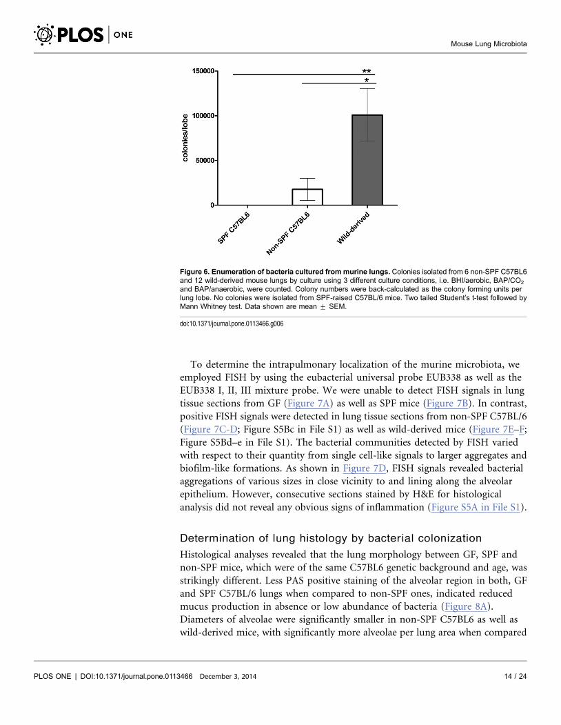
Bacterial load and localization
To quantify cultivatable lung microbiota between mice of different origins, colony
numbers of cultivatable bacteria were counted on each culture plate used for
bacterial isolation. Numbers were recalculated to achieve the bacterial numbers
per lung lobe. As shown in Figure 6, significantly lower numbers of bacteria were
isolated from non-SPF C57BL6 than from wild-derived mice, although both were
bred under comparable conditions within the same facility. The various types of
colonies isolated from non-SPF lungs as depicted in Figure S2 in File S1 show that
the differences in bacterial numbers were not due to overrepresentation of just
one species. These results are interesting in light of the significantly higher
Figure 4. Bacterial compositions with respect to mouse origin. Evaluation of the distribution of bacterial phyla or classes from 454 sequencing dataderived from each of the four groups of mice - 3 SPF, 4 non-SPF C57BL/6, 8 wild-derived, and 15 wild-caught mice.
doi:10.1371/journal.pone.0113466.g004
Mouse Lung Microbiota
PLOS ONE | DOI:10.1371/journal.pone.0113466 December 3, 2014 12 / 24
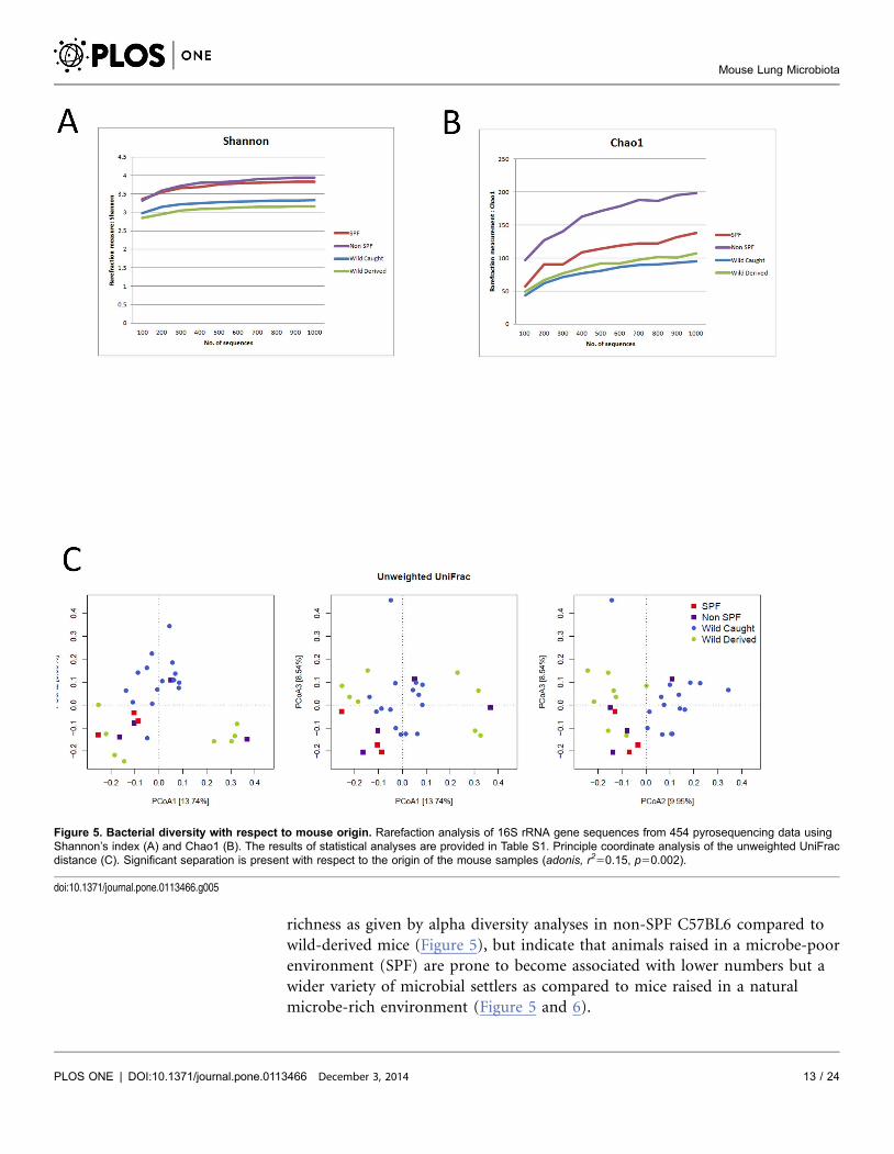
richness as given by alpha diversity analyses in non-SPF C57BL6 compared to
wild-derived mice (Figure 5), but indicate that animals raised in a microbe-poor
environment (SPF) are prone to become associated with lower numbers but a
wider variety of microbial settlers as compared to mice raised in a natural
microbe-rich environment (Figure 5 and 6).
Figure 5. Bacterial diversity with respect to mouse origin. Rarefaction analysis of 16S rRNA gene sequences from 454 pyrosequencing data usingShannon’s index (A) and Chao1 (B). The results of statistical analyses are provided in Table S1. Principle coordinate analysis of the unweighted UniFracdistance (C). Significant separation is present with respect to the origin of the mouse samples (adonis, r250.15, p50.002).
doi:10.1371/journal.pone.0113466.g005
Mouse Lung Microbiota
PLOS ONE | DOI:10.1371/journal.pone.0113466 December 3, 2014 13 / 24
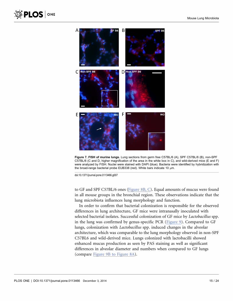
To determine the intrapulmonary localization of the murine microbiota, we
employed FISH by using the eubacterial universal probe EUB338 as well as the
EUB338 I, II, III mixture probe. We were unable to detect FISH signals in lung
tissue sections from GF (Figure 7A) as well as SPF mice (Figure 7B). In contrast,
positive FISH signals were detected in lung tissue sections from non-SPF C57BL/6
(Figure 7C-D; Figure S5Bc in File S1) as well as wild-derived mice (Figure 7E–F;
Figure S5Bd–e in File S1). The bacterial communities detected by FISH varied
with respect to their quantity from single cell-like signals to larger aggregates and
biofilm-like formations. As shown in Figure 7D, FISH signals revealed bacterial
aggregations of various sizes in close vicinity to and lining along the alveolar
epithelium. However, consecutive sections stained by H&E for histological
analysis did not reveal any obvious signs of inflammation (Figure S5A in File S1).
Determination of lung histology by bacterial colonization
Histological analyses revealed that the lung morphology between GF, SPF and
non-SPF mice, which were of the same C57BL6 genetic background and age, was
strikingly different. Less PAS positive staining of the alveolar region in both, GF
and SPF C57BL/6 lungs when compared to non-SPF ones, indicated reduced
mucus production in absence or low abundance of bacteria (Figure 8A).
Diameters of alveolae were significantly smaller in non-SPF C57BL6 as well as
wild-derived mice, with significantly more alveolae per lung area when compared
Figure 6. Enumeration of bacteria cultured from murine lungs. Colonies isolated from 6 non-SPF C57BL6and 12 wild-derived mouse lungs by culture using 3 different culture conditions, i.e. BHI/aerobic, BAP/CO2
and BAP/anaerobic, were counted. Colony numbers were back-calculated as the colony forming units perlung lobe. No colonies were isolated from SPF-raised C57BL/6 mice. Two tailed Student’s t-test followed byMann Whitney test. Data shown are mean ¡ SEM.
doi:10.1371/journal.pone.0113466.g006
Mouse Lung Microbiota
PLOS ONE | DOI:10.1371/journal.pone.0113466 December 3, 2014 14 / 24
to GF and SPF C57BL/6 ones (Figure 8B, C). Equal amounts of mucus were found
in all mouse groups in the bronchial region. These observations indicate that the
lung microbiota influences lung morphology and function.
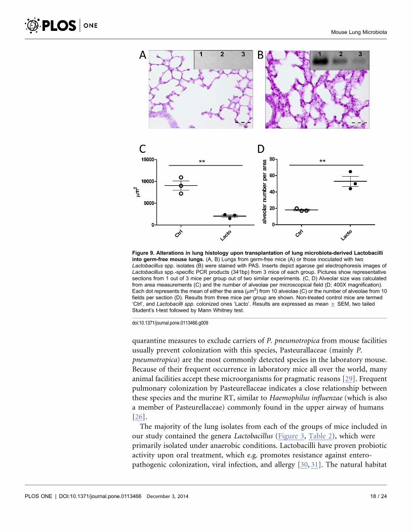
In order to confirm that bacterial colonization is responsible for the observed
differences in lung architecture, GF mice were intranasally inoculated with
selected bacterial isolates. Successful colonization of GF mice by Lactobacillus spp.
in the lung was confirmed by genus-specific PCR (Figure 9). Compared to GF
lungs, colonization with Lactobacillus spp. induced changes in the alveolar
architecture, which was comparable to the lung morphology observed in non-SPF
C57BL6 and wild-derived mice. Lungs colonized with lactobacilli showed
enhanced mucus production as seen by PAS staining as well as significant
differences in alveolar diameter and numbers when compared to GF lungs
(compare Figure 9B to Figure 8A).
Figure 7. FISH of murine lungs. Lung sections from germ free C57BL/6 (A), SPF C57BL/6 (B), non-SPFC57BL/6 (C and D, higher magnification of the area in the white box in C), and wild-derived mice (E and F)were analyzed by FISH. Nuclei were stained with DAPI (blue). Bacteria were identified by hybridization withthe broad-range bacterial probe EUB338 (red). White bars indicate 10 mm.
doi:10.1371/journal.pone.0113466.g007
Mouse Lung Microbiota
PLOS ONE | DOI:10.1371/journal.pone.0113466 December 3, 2014 15 / 24

Mouse Lung Microbiota
PLOS ONE | DOI:10.1371/journal.pone.0113466 December 3, 2014 16 / 24

Discussion
In contrast to the upper RT and other barrier epithelia exposed to the
environment such as the gut, skin and vagina, the lower RT of a healthy human
has been traditionally considered free of microorganisms. Recent analyses in
humans, however, detected bacterial species in samples from the lower RT,
suggesting the existence of a resident microbiota in the mammalian lung.
These studies suggested that, in contrast to the inhabitants of other epithelial
surfaces, the microbiota of human lungs is of lower complexity. Bacterial species
identified in BAL fluid from healthy donors included anaerobes, such as
Prevotella, Veillonella, Propionibacterium [7, 25], indicating anaerobes as common
members of the lower RT microbiota. It has also been suggested that these
anaerobes may have the potential to become pathogenic under certain micro-
ecological conditions, such as those found in CF patients. Together with Prevotella
and Veillonella spp., Proteobacteria including Haemophilus spp. and Neisseria spp.
were identified in bronchoscopy specimens from asthma patients [8].
Pseudomonas spp. represented the most dominant bacterial genus in BAL and
lung explants of COPD patients [10]. Sterile sampling of human lower airway
specimens is however a difficult issue as expectorated sputum and BAL fluid can
easily become contaminated with bacterial residents of the upper airways such as
trachea, oropharynx and sinuses [14, 26].
Our approach to analyze specimens from murine lungs excised and dissected
under sterile conditions avoided upper respiratory tract contamination. In
contrast to lungs from GF mice, those from mice raised under conventional or
SPF conditions as well as those caught in the natural environment contained
bacterial 16S rRNA. Taken together, cultivation as well as genetic identification by
16S rRNA sequencing allowed us to determine the lung microbiota of Mus
musculus. The most dominant lung phyla found were Proteobacteria (64%),
Firmicutes (20%), Actinobacteria (6%), and Bacteroidetes (4%), which shows a
similar phylum distribution as previously reported for lung tissue microbiota
from Balb/c mice [13]. Pasteurellaceae (Gammaproteobacteria) were found in
wild mice derived from the environment, by both 454 sequencing as well as
cultivation (Figure 3). P. pneumotropica is a ubiquitous bacterium that is
frequently isolated from the upper respiratory tract of laboratory rodents
including mice, rats, hamsters, and guinea pigs raised under non-SPF conditions
[27]. Another member of the Pasteurellaceae, Actinobacillus muris can also
colonize the oral cavity of mice and has been considered a commensal, since its
presence could not be associated with pathological alterations [28]. Although
Figure 8. Lung microbiota influences alveolar morphology and mucus production. A. Histology of murine lungs from GF, SPF, non-SPF C57BL6 orwild-derived mice. Sections were stained with periodic acid-Schiff (PAS) and analyzed by light microscopy. Black bars indicate the magnification used(500 mm540X, 200 mm5100X, 50 mm5400X). Pictures show representative sections of 3–6 mice analyzed per group. B–C) Alveolar size as calculatedfrom area measurements and the number of alveolae per microscopical field (400X magnification). Each dot represents the mean of either the area (mm2)from 10 alveolae (B) or the number of alveolae from 10 fields per section (C). Results from three to six mice per group are shown. Results are expressed asmean ¡ SEM, two tailed Student’s t-test followed by Mann Whitney test.
doi:10.1371/journal.pone.0113466.g008
Mouse Lung Microbiota
PLOS ONE | DOI:10.1371/journal.pone.0113466 December 3, 2014 17 / 24
quarantine measures to exclude carriers of P. pneumotropica from mouse facilities
usually prevent colonization with this species, Pasteurallaceae (mainly P.
pneumotropica) are the most commonly detected species in the laboratory mouse.
Because of their frequent occurrence in laboratory mice all over the world, many
animal facilities accept these microorganisms for pragmatic reasons [29]. Frequent
pulmonary colonization by Pasteurellaceae indicates a close relationship between
these species and the murine RT, similar to Haemophilus influenzae (which is also
a member of Pasteurellaceae) commonly found in the upper airway of humans
[26].
The majority of the lung isolates from each of the groups of mice included in
our study contained the genera Lactobacillus (Figure 3, Table 2), which were
primarily isolated under anaerobic conditions. Lactobacilli have proven probiotic
activity upon oral treatment, which e.g. promotes resistance against entero-
pathogenic colonization, viral infection, and allergy [30, 31]. The natural habitat
Figure 9. Alterations in lung histology upon transplantation of lung microbiota-derived Lactobacilliinto germ-free mouse lungs. (A, B) Lungs from germ-free mice (A) or those inoculated with twoLactobacillus spp. isolates (B) were stained with PAS. Inserts depict agarose gel electrophoresis images ofLactobacillus spp.-specific PCR products (341bp) from 3 mice of each group. Pictures show representativesections from 1 out of 3 mice per group out of two similar experiments. (C, D) Alveolar size was calculatedfrom area measurements (C) and the number of alveolae per microscopical field (D; 400X magnification).Each dot represents the mean of either the area (mm2) from 10 alveolae (C) or the number of alveolae from 10fields per section (D). Results from three mice per group are shown. Non-treated control mice are termed‘Ctrl’, and Lactobacilli spp. colonized ones ‘Lacto’. Results are expressed as mean ¡ SEM, two tailedStudent’s t-test followed by Mann Whitney test.
doi:10.1371/journal.pone.0113466.g009
Mouse Lung Microbiota
PLOS ONE | DOI:10.1371/journal.pone.0113466 December 3, 2014 18 / 24
of lactobacilli ranges from fermenting dairy, meat and plant material to oral
cavities and the genital and gastrointestinal tracts of humans and animals [32].
Therefore, it is not surprising to find lactobacilli in murine lungs. Further
characterization of Lactobacillus lung-derived isolates and their function in airway
homeostasis and immunity may reveal additional probiotic roles in respiratory
diseases. Lactococci have been associated with lower incidence rates of atopic
diseases in children who grew up in close association with certain farm animals
[33].
Using RNA samples instead of DNA has the advantage of primarily identifying
metabolically active bacteria, whereas according to Pezzulo et al, the majority of
bacterial DNA from pulmonary material represents dead bacteria [12]. Overall,
the Betaproteobacterium, Ralstonia spp., represents the most abundant bacterial
genus as revealed by non-culture based analysis (Figure 1, 3). Since Ralstonia spp.
is a frequent contaminant in DNA extraction procedures [34], we confirmed that
its presence was not due to contamination during the experimental procedure as
Ralstonia-specific PCR products were absent from GF lungs (Figure 2). Similarly,
Ralstonia species have been isolated not only from the environment, but also from
the upper RT of healthy humans [35].
Together, Firmicutes and Bacteroidetes represent 90–99% of the intestinal
microbiota in humans and mice [36], but make up approximately 20% of the
murine lung microbiota as determined by 454 sequencing (Figure 1). Due to their
anaerobic metabolism, Bacteroidetes species may find only limited niches in the
healthy lung. Bacteroides is a widely studied genus with respect to the immune
function of the gut microbiota [1, 37–39], and represent 10–40% and 20% of the
Bacteroidetes phylum identified in lungs from wild and non-SPF C57BL/6 mice,
respectively, indicating a putative function for this genus also in pulmonary
immune homeostasis. Notably, Bacteroides spp. were absent from the lungs of SPF
mice (Figure S3D in File S1). Interestingly, Sphingomonas dominated the lung
microbiota of C57BL6 mice from both SPF and non-SPF environments.
Sphingomonas is a Gram-negative but lipopolysaccharide (LPS)-free bacterium,
which produce glycosphingolipids, glycolipid antigens recognized by invariant
natural killer T (iNKT) cells [40, 41]. As a commensal, Sphingomonas has been
shown to contribute to immune homeostasis of iNKT cells [42].
Our analysis also revealed quantitative and qualitative changes in the lung
microbiota between mice raised under SPF but transferred to conditions closer to
the natural environment vs. those raised and kept under natural conditions. In the
first group of mice, the number of sequences representing Corynebacteriaceae
(phylum Actinobacteria; Figure S3A in File S1) and Bacteroidetes was high (Figure
S3B in File S1) and the number of those representing Chitinophagaceae (Figure
S3B in File S1) and Moraxellaceae (Figure S3E in File S1) was low. More
importantly, non-SPF C57BL/6 mice displayed the highest species diversity of all
mouse groups studied, even when mice caught in the wild were considered
(Figure 5B). According to the ‘‘Hygiene hypothesis’’, this result suggests that mice
raised under ‘‘clean’’ SPF conditions lack the exposure to a variety of ‘‘natural’’
commensal species, which may cause insufficiently developed lung immunity
Mouse Lung Microbiota
PLOS ONE | DOI:10.1371/journal.pone.0113466 December 3, 2014 19 / 24

rendering those mice more vulnerable to colonization by a wider variety of
bacterial species which may be maintained in the population [43].
The influence of a different environment is also evident in wild mice derived
from a mouse colony the founders thereof originated from the natural
environment before transferred to an indoor animal facility for breeding. Lungs of
these wild-derived mice contained higher numbers of sequences from
Corynebacteriaceae and Microbacteriaceae but less sequences from Actinobacteria
and Firmicutes (Figure 4, Figure S3 in File S1). Although Helicobacter spp. was
abundant in wild mice caught in the environment, this genus was less often found
in wild-derived mice housed indoors (Figure 4). However, apart from these
differences, the overall differences in diversity observed between wild-caught and
wild-derived mice were not as a dramatic as those between SPF and non-SPF mice
(Figure 5B). This phenomenon could be due to colonization resistance, i.e. once
an intact microbiota is formed additional bacterial species only mildly influence
the epithelial ecosystem, as observed in an animal model for enteropathogenic
bacteria colonization [30]. Similar to the lung, the bacterial microbiota of the
stomach is of relatively low complexity, which has been suggested not only to be
determined by niche-specific factors but also by stochastic colonization from the
upstream alimentary tract [44]. Thus, the lung could also in part be comprised of
stochastic colonizers from the upper RT. However, it should be noted that the
colonies identified by FISH indicate specific niches in the lung where bacteria can
build biofilm-like communities (Figure 7C–D). Interestingly, the largest bacterial
aggregations were found in non-SPF C57BL/6 lungs, which also displayed the
highest bacterial species diversity (Figure 5).
Finally and most importantly, the differences in alveolar morphology and
mucus production (Figure 8) in relation to the abundance of bacteria in the lung
are striking observations. This unexpected finding was confirmed by associating
germfree mice with two Lactobacillus isolates from the murine lung (Figure 9),
which lead to similar alterations in alveolar numbers, size and mucus production
as observed in non-SPF lungs. As age and genetic background may influence lung
architecture, we should note that the differences in alveolar size and counts were
observed between GF, SPF and non-SPF mice of the same C57BL/6 background
and age. Taken together, these results strongly suggest that bacteria influence lung
development and barrier functions. Future studies will focus on the stability and
localization of bacterial colonization and its influence on pulmonary immune
responses. Effects of microbiota on organ development have recently been
revealed for a number of tissues including the intestinal crypts, lymphoid tissue
and the blood-brain barrier [45, 46]. Instruction of innate immune responses
including epithelial defense function is also triggered by the presence of
microbiota [31, 47]. Thus, enhanced alveolar mucus production could be due to a
direct effect of alveolar settlers, whereas the bronchial mucus production is rather
influenced by inhaled microbes from the upper respiratory tract. However,
whether the increase in numbers of alveolae and corresponding reduction in
alveolar size requires an indigenous lung community, or general microbial
Mouse Lung Microbiota
PLOS ONE | DOI:10.1371/journal.pone.0113466 December 3, 2014 20 / 24
settlement at other sites such as the gut, will require further colonization
experiments.
Our description of a murine lung microbiota by both cultivation and genetic
methods along with its dependency on the animal’s environmental conditions
opens the path for functional studies on the role of lung commensals in airway
morphogenesis, epithelial homeostasis and immunity. In particular cultivatable
bacteria can be systematically investigated for their colonization and growth
characteristics on lung epithelia, their influence on antibiotic activity, lung
infection and inflammation. This may ultimately enable the discovery of novel
immune modulating or probiotic properties for prophylactic or therapeutic
measures against airway disorders.
Supporting Information
File S1. Figure S1. Phylogenetic tree of isolates and clones from mouse lungs
determined by 16S rRNA gene sequences. The Neighbor-Joining method
implemented in MEGA was applied to 128 taxa from colony isolates and 103 taxa
from cloned library in addition to 33 representative bacterial sequences identified
by BLAST and RDP. Branches corresponding to partitions reproduced in less than
50% bootstrap replicates are collapsed. More than 99% identical reads were given
by number with isolates in blue and clones in red. In addition, strain names with
green background shows deposited strains in DSMZ. Figure S2. Examples of
colonies cultured from mouse lungs under different culture conditions.
Homogenized lung material was plated onto Blood agar (BAP)/chocolate agar
plate (CH) in 5% CO2, Luria Burtani (LB)/Brain Heart Infusion (BHI) and BAP
under aerobic or anaerobic conditions, respectively. Figure S3. Differences at the
genus or family level in each phylum between mouse origins. A. Actinobacteria,
B. Bacteroidetes, C. Firmicutes, D. Alphaproteobacteria, E.
Gammaproteobacteria. Constructed data within each phylum was extracted from
454 pyrosequencing analysis shown in Figure 4. Figure S4. Beta analysis for Bray-
curtis distance measurement based on the taxon abundance revealed significant
community differences among individuals from different categories (Adonis:
R250.18, p50.003). The Jaccard distance measurement based on presence or
absence of taxon also revealed similar trend (Adonis: R250.15, p50.008). Similar
results were obtained when the phylogenetic-based measurements on taxon
abundance was included (i.e. Weighted UniFrac, Adonis: R250.13, p is not
significant). Figure S5. A. Histopathology of murine lungs. Lung sections from
germ-free C57BL/6 (a), SPF C57BL/6 (b), non-SPF C57BL/6 (c) and wild-derived
mice (d and e) were analyzed by H&E staining. Histology reveals no obvious signs
of inflammatory responses. B. FISH of murine lungs. Lung sections from germ-
free C57BL/6 (a), SPF C57BL/6 (b), non-SPF C57BL/6 (c) and wild-derived mice
(d and e) were analyzed using the EUB338 I, II and III mixture probe (red) [48]
and subsequently stained with DAPI (blue) to depict nuclei. The pictures
Mouse Lung Microbiota
PLOS ONE | DOI:10.1371/journal.pone.0113466 December 3, 2014 21 / 24

corresponded to the histological images shown in S5A. The control Non-EUB338
probe is shown in (f). (Magnification 100X).
doi:10.1371/journal.pone.0113466.S002 (PPT)
Table S1. Significance of alpha diversity comparisons.
doi:10.1371/journal.pone.0113466.S001 (DOC)
Acknowledgments
We thank Jacqueline Eich, Kristine Hagens, and Dagmar Meyer for help with lung
isolation, FISH and histological analysis, Prof. Dr. Diethard Tautz and Christine
Pfeifle (Max Planck Institute for Evolutionary Biology, Plon) for wild mouse
supply, Philipp Rausch for help with statistical analysis and Prof. Dr. Rudolf
Amann and Jorg Wulf (Max-Planck-Institute for Marine Microbiology, Bremen)
for teaching us the FISH technique.
Author ContributionsConceived and designed the experiments: YY KDB JFB UES. Performed the
experiments: YY SK SA US. Analyzed the data: YY GS. Contributed reagents/
materials/analysis tools: GS ML US JFB UES. Wrote the paper: YY KDB JFB UES.
References
1. Backhed F, Ley RE, Sonnenburg JL, Peterson DA, Gordon JI (2005) Host-Bacterial Mutualism in theHuman Intestine. Science 307: 1915–1920.
2. Grice EA, Kong HH, Conlan S, Deming CB, Davis J, et al. (2009) Topographical and TemporalDiversity of the Human Skin Microbiome. Science 324: 1190–1192.
3. Fredricks DN, Fiedler TL, Marrazzo JM (2005) Molecular Identification of Bacteria Associated withBacterial Vaginosis. N Engl J Med 353: 1899–1911.
4. Ghannoum MA, Jurevic RJ, Mukherjee PK, Cui F, Sikaroodi M, et al. (2010) Characterization of theOral Fungal Microbiome (Mycobiome) in Healthy Individuals. PLoS Pathogens 6: e1000713.
5. Madan JC, Koestler DC, Stanton BA, Davidson L, Moulton LA, et al. (2012) Serial Analysis of the Gutand Respiratory Microbiome in Cystic Fibrosis in Infancy: Interaction between Intestinal and RespiratoryTracts and Impact of Nutritional Exposures. mBio 3.
6. The Human Microbiome Jumpstart Reference Strains C (2010) A Catalog of Reference Genomesfrom the Human Microbiome. Science 328: 994–999.
7. Rogers G, Carroll M, Serisier D, Hockey P, Kehagia V, et al. (2005) Bacterial activity in cystic fibrosislung infections. Respiratory Research 6: 49.
8. Hilty M, Burke C, Pedro H, Cardenas P, Bush A, et al. (2010) Disordered Microbial Communities inAsthmatic Airways. PLoS ONE 5: e8578.
9. Huang YJ, Nelson CE, Brodie EL, DeSantis TZ, Baek MS, et al. (2011) Airway microbiota andbronchial hyperresponsiveness in patients with suboptimally controlled asthma. The Journal of allergyand clinical immunology 127: 372–381.e373.
10. Erb-Downward JR, Thompson DL, Han MK, Freeman CM, McCloskey L, et al. (2011) Analysis of theLung Microbiome in the ‘‘Healthy’’ Smoker and in COPD. PLoS ONE 6: e16384.
11. Goulding J, Snelgrove R, Saldana J, Didierlaurent A, Cavanagh M, et al. (2007) RespiratoryInfections: Do We Ever Recover? Proc Am Thorac Soc 4: 618–625.
Mouse Lung Microbiota
PLOS ONE | DOI:10.1371/journal.pone.0113466 December 3, 2014 22 / 24
12. Pezzulo AA, Kelly PH, Nassar BS, Rutland CJ, Gansemer ND, et al. (2013) Abundant DNase I-Sensitive Bacterial DNA in Healthy Porcine Lungs and Its Implications for the Lung Microbiome. ApplEnviron Microbiol 79: 5936–5941.
13. Barfod K, Roggenbuck M, Hansen L, Schjorring S, Larsen S, et al. (2013) The murine lungmicrobiome in relation to the intestinal and vaginal bacterial communities. BMC Microbiology 13: 303.
14. Charlson ES, Bittinger K, Haas AR, Fitzgerald AS, Frank I, et al. (2011) Topographical Continuity ofBacterial Populations in the Healthy Human Respiratory Tract. Am J Respir Crit Care Med 184: 957–963.
15. Turnbaugh PJ, Ley RE, Hamady M, Fraser-Liggett CM, Knight R, et al. (2007) The HumanMicrobiome Project. Nature 449: 804–810.
16. Linnenbrink M, Wang J, Hardouin EA, Kunzel S, Metzler D, et al. (2013) The role of biogeography inshaping diversity of the intestinal microbiota in house mice. Molecular Ecology 22: 1904–1916.
17. Rausch P, Rehman A, Kunzel S, Hasler R, Ott SJ, et al. (2011) Colonic mucosa-associated microbiotais influenced by an interaction of Crohn disease and FUT2 (Secretor) genotype. Proc Natl Acad Sci USA108: 19030–19035.
18. Caporaso JG, Kuczynski J, Stombaugh J, Bittinger K, Bushman FD, et al. (2010) QIIME allowsanalysis of high-throughput community sequencing data. Nat Meth 7: 335–336.
19. Dixon P (2003) VEGAN, a package of R functions for community ecology. Journal of Vegetation Science14: 927–930.
20. Lozupone C, Knight R (2005) UniFrac: a New Phylogenetic Method for Comparing MicrobialCommunities. Appl Environ Microbiol 71: 8228–8235.
21. Coenye T, Vandamme P, LiPuma JJ (2002) Infection by Ralstonia Species in Cystic Fibrosis Patients:Identification of R. pickettii and R. mannitolilytica by Polymerase Chain Reaction. Emerg Infect Dis 8:692–696.
22. Amann R, Fuchs BM (2008) Single-cell identification in microbial communities by improvedfluorescence in situ hybridization techniques. Nat Rev Micro 6: 339–348.
23. Rinttila T, Kassinen A, Malinen E, Krogius L, Palva A (2004) Development of an extensive set of 16SrDNA-targeted primers for quantification of pathogenic and indigenous bacteria in faecal samples byreal-time PCR. Journal of Applied Microbiology 97: 1166–1177.
24. Benjamini Y, Hochberg Y (1995) Controlling the False Discovery Rate: A Practical and PowerfulApproach to Multiple Testing. J R Stat Soc B57: 289–300.
25. Tunney MM, Field TR, Moriarty TF, Patrick S, Doering G, et al. (2008) Detection of Anaerobic Bacteriain High Numbers in Sputum from Patients with Cystic Fibrosis. Am J Respir Crit Care Med 177: 995–1001.
26. LiPuma JJ (2010) The Changing Microbial Epidemiology in Cystic Fibrosis. Clin Microbiol Rev 23: 299–323.
27. Sasaki H, Kawamoto E, Ueshiba H, Amao H, Sawada T (2006) Phylogenetic relationship ofPasteurella pneumotropica isolates from laboratory rodents based on 16S rDNA sequence. J Vet MedSci 68: 639–641.
28. Patten CCJ, Myles MH, Franklin CL, Livingston RS (2010) Pertubation in cytokine gene expressionafter inoculation of C57BL/6 mice with Pasteurella pneumotropica. Comparative Medicine 60: 18–24.
29. Dammann P, Hilken G, Hueber B, Kohl W, Bappert MT, et al. (2011) Infectious microorganisms in mice(Mus musculus) purchased from commercial pet shops in Germany. Laboratory Animals 45: 271–275.
30. Stecher B, Hardt W-D (2008) The role of microbiota in infectious disease. Trends in Microbiology 16:107–114.
31. Fujimura KE, Demoor T, Rauch M, Faruqi AA, Jang S, et al. (2014) House dust exposure mediatesgut microbiome Lactobacillus enrichment and airway immune defense against allergens and virusinfection. Proc Natl Acad Sci USA 111: 805–810.
32. Kleerebezem M, Hols P, Bernard E, Rolain T, Zhou M, et al. (2010) The extracellular biology of thelactobacilli. FEMS Microbiology Reviews 34: 199–230.
Mouse Lung Microbiota
PLOS ONE | DOI:10.1371/journal.pone.0113466 December 3, 2014 23 / 24

33. Debarry J, Garn H, Hanuszkiewicz A, Dickgreber N, Blumer N, et al. (2007) Acinetobacter lwoffii andLactococcus lactis strains isolated from farm cowsheds possess strong allergy-protective properties. TheJournal of allergy and clinical immunology 119: 1514–1521.
34. Mohammadi T, Reesink HW, Vandenbroucke-Grauls CMJE, Savelkoul PHM (2005) Removal ofcontaminating DNA from commercial nucleic acid extraction kit reagents. Journal of MicrobiologicalMethods 61: 285–288.
35. Stelzmueller I, Biebl M, Wiesmayr S, Eller M, Hoeller E, et al. (2006) Ralstonia pickettii-innocentbystander or a potential threat? Clinical Microbiology & Infection 12: 99–101.
36. Ley RE, Peterson DA, Gordon JI (2006) Ecological and Evolutionary Forces Shaping MicrobialDiversity in the Human Intestine. Cell 124: 837–848.
37. Cash HL, Whitham CV, Behrendt CL, Hooper LV (2006) Symbiotic Bacteria Direct Expression of anIntestinal Bactericidal Lectin. Science 313: 1126–1130.
38. Hooper LV (2009) Do symbiotic bacteria subvert host immunity? Nat Rev Micro 7: 367–374.
39. Mazmanian SK, Round JL, Kasper DL (2008) A microbial symbiosis factor prevents intestinalinflammatory disease. Nature 453: 620–625.
40. Kinjo Y, Wu D, Kim G, Xing G-W, Poles MA, et al. (2005) Recognition of bacterial glycosphingolipids bynatural killer T cells. Nature 434: 520–525.
41. Krziwon C, Zahringer U, Kawahara K, Weidemann B, Kusumoto S, et al. (1995) Glycosphingolipidsfrom Sphingomonas paucimobilis induce monokine production in human mononuclear cells. InfectImmun 63: 2899–2905.
42. Wei B, Wingender G, Fujiwara D, Chen DY, McPherson M, et al. (2010) Commensal Microbiota andCD8+ T Cells Shape the Formation of Invariant NKT Cells. J Immunol: jimmunol.0902620.
43. McLoughlin RM, Mills KHG (2011) Influence of gastrointestinal commensal bacteria on the immuneresponses that mediate allergy and asthma. Journal of Allergy and Clinical Immunology 127: 1097–1107.
44. Bik EM, Eckburg PB, Gill SR, Nelson KE, Purdom EA, et al. (2006) Molecular analysis of the bacterialmicrobiota in the human stomach. Proc Natl Acad Sci USA 103: 732–737.
45. Falk PG, Hooper LV, Midtvedt T, Gordon JI (1998) Creating and Maintaining the GastrointestinalEcosystem: What We Know and Need To Know from Gnotobiology. Microbiol Mol Biol Rev 62: 1157–1170.
46. Heijtz RD, Wang S, Anuar F, Qian Y, Bjorkholm B, et al. (2011) Normal gut microbiota modulates braindevelopment and behavior. Proc Natl Acad Sci USA 108: 3047–3052.
47. Hooper LV, Littman DR, Macpherson AJ (2012) Interactions Between the Microbiota and the ImmuneSystem. Science 336: 1268–1273.
48. Daims H, Bruhl A, Amann R, Schleifer K-H, Wagner M (1999) The Domain-specific Probe EUB338 isInsufficient for the Detection of all Bacteria: Development and Evaluation of a more ComprehensiveProbe Set. Systematic and Applied Microbiology 22: 434–444.
Mouse Lung Microbiota
PLOS ONE | DOI:10.1371/journal.pone.0113466 December 3, 2014 24 / 24