Embed Size (px)
Citation preview
Hindawi Publishing CorporationBioMed Research InternationalVolume 2013, Article ID 969750, 8 pageshttp://dx.doi.org/10.1155/2013/969750
Research ArticleEffects of Systemic Pesticides Imidacloprid and Metalaxyl onthe Phyllosphere of Pepper Plants
Constantinos Moulas, Christos Petsoulas, Konstantina Rousidou, Chiara Perruchon,Panagiotis Karas, and Dimitrios G. Karpouzas
University of Thessaly, Department of Biochemistry and Biotechnology, Ploutonos 26 and Aiolou Street, 41221 Larisa, Greece
Correspondence should be addressed to Dimitrios G. Karpouzas; [email protected]
Received 18 April 2013; Accepted 22 May 2013
Academic Editor: George Tsiamis
Copyright © 2013 Constantinos Moulas et al. This is an open access article distributed under the Creative Commons AttributionLicense, which permits unrestricted use, distribution, and reproduction in any medium, provided the original work is properlycited.
Microbes inhabiting the phyllosphere of crops are exposed to pesticides applied either directly onto plant foliage or indirectlythrough soil. Although, phyllosphere microbiology has been rapidly evolving, little is still known regarding the impact of pesticideson the epiphyticmicrobial community and especially on fungi.We determined the impact of two systemic pesticides (metalaxyl andimidacloprid), applied either on foliage or through soil, on the epiphytic fungal and bacterial communities via DGGE and cloning.Both pesticides induced mild effects on the fungal and the bacterial communities. The only exception was the foliage applicationof imidacloprid which showed a more prominent effect on the fungal community. Cloning showed that the fungal communitywas dominated by putative plant pathogenic ascomycetes (Erysiphaceae and Cladosporium), while a few basidiomycetes were alsopresent. The former ribotypes were not affected by pesticides application, while selected yeasts (Cryptococcus) were stimulated bythe application of imidacloprid suggesting a potential role in its degradation. A less diverse bacterial community was identifiedin pepper plants. Metalaxyl stimulated an Enterobacteriaceae clone which is an indication of the involvement of members of thisfamily in fungicide degradation. Further studies will focus on the isolation of epiphytic microbes which appear to be stimulated bypesticides application.
1. Introduction
Phyllosphere is the habitat of a diverse microbial communitydominated by bacteria, fungi, and yeasts while archaea are notparticularly abundant [1]. Until recently most studies on themicrobiology of the phyllosphere had focused on the ecologyand interactions of microbial plant pathogens with the plant,whereas little was known regarding the role and ecology ofnonplant pathogenic microorganisms on plant phyllosphere.It is now well documented that epiphytic microorganismscould serve significant functional roles including (a) suppres-sion of plant pathogens in the phyllosphere of agriculturalcrops [2], (b) nitrogen fixation [3], (c) methanol utilization[4], and (d) degradation of organic pollutants [5]. In addi-tion, microbial interactions on plant phyllosphere have beenfound to determine colonization of edible parts of plants byhuman pathogens [6]. This is particularly important for theconsumption of fresh salad, fruits, and vegetables.
In a pioneering study, Yang et al. [7] demonstrated thatthe microbial diversity on plant phyllosphere is much higherthan what had been estimated before based on culture-dependent methods but it is still lower than the microbialdiversity in rhizosphere or even bulk soil [8]. Phyllosphereis an oligotrophic environment with patchy distribution ofC sources where microorganisms are exposed to stress con-ditions including extreme exposure to UV radiation, violentfluctuations of temperature, and limited water availability[9]. In order to survive under these conditions, phyllo-sphere microorganisms have developed various mechanismsincluding pigmentation [10], DNA repair mechanisms [11],production of biosurfactants [12], and extracellular polymericsubstances [13].
Apart from the abiotic and biotic stress conditionsdescribed, microorganisms on the phyllosphere of cultivatedplants are exposed to pesticides.There is a wealth of literatureregarding the impact of pesticides on soil microorganisms
2 BioMed Research International
[14]. This is not surprising considering that soil constituteslargely the final deposit of both foliar and soil-appliedpesticides. However, only a few studies so far have addressedthe impact of pesticides onto nontarget microorganisms onplant phyllosphere. A series of studies by Zhang et al. [15–17]showed that the insecticides cypermethrin and abamectininduced changes in the structure of the bacterial commu-nity in pepper, cucumber, and broccoli phyllosphere. In asimilar study the application of the fungicide enostroburininduced substantial changes in bacterial community in wheatphyllosphere [18]. All those studies have focused on poten-tial effects on the bacterial community after foliar applica-tion of pesticides. However, only limited data are availableregarding pesticides effects on nontarget fungi inhabitingplant phyllosphere. Apart from foliar application, systemicpesticides are commonly applied via soil drenching andthey are translocated through the phloem to the aerial partsof the plants offering protection from pest and pathogens.However, nothing is known regarding the impact of such soilapplications on the epiphytic microbial community.
Imidacloprid is a systemic insecticide which has gainedregistration for 140 uses in 120 countries [19]. It is appliedeither directly on the foliage or via soil drenching forthe control of aphids (Myzus persicae, Myzus nicotianae),white fly (Trialeurodes vaporariorum), and Colorado beetle(Leptinotarsa decemlineata) in fruits crops, vegetables, andpotatoes. Metalaxyl-M is a systemic fungicide which isapplied either on foliage or via soil drenching for the controlof oomycetes such as Phytophthora parasitica (tobacco),Phytopthora infestans (potato), and Pythium sp. [20]. Nothingis known regarding the impact of those pesticides on themicrobial community of plant phyllosphere.
We aimed to investigate the impact of the systemicpesticides imidacloprid and metalaxyl on the fungal andbacterial communities on the phyllosphere of pepper plants.The influence of the mode of application, foliar versussoil application, on the magnitude and type of effects wasalso determined via denaturing gradient gel electrophoresis(DGGE) and cloning.
2. Materials and Methods
2.1. Experimental Setup. Three-week-old pepper plants (Cap-sicum annum L. cv Ozho) (kindly supplied by AgriPlantA.S.) were initially transplanted into 3 L plastic pots whichhad been filled with appropriate amounts of a 1 : 1 mixtureof sand and soil (sandy, pH 7.81, electrical conductivity0.017mmhos cm−1, organic matter content 8.8 g kg−1, P-Olsen 2mg g−1, K 215mg g−1, and Mg 265mg g−1). In total18 pots were prepared and placed randomly in the growthchamber at 22∘Cusing a 16 h light/8 h night period.Theplantswere watered as needed and 30mL of Hoagland solution[21] was applied twice weekly. Pepper plants were grownunder these conditions for a week and then transferred toa commercial greenhouse situated in the area of Velestino,Magnesia, Greece, where the experiment was contacted. Theplants were left in the greenhouse for a period of threeweeks to become acclimatized and allow the development
of a natural phyllosphere microbial community as much aspossible. During the acclimation period the pepper plantswere watered and fertilized as needed.
At the end of the acclimation period the 18 pots withthe pepper plants were divided into 6 groups of three. Thefirst three pots received a foliage treatment with an aqueoussuspension of the insecticide imidachloprid (CONFIDOR,200SL), while the next three pots received a soil drenchingwith an aqueous suspension of the same insecticide. Similarly,the next two groups of pots received a foliar or a soildrenching application of the fungicide metalaxyl (RIDOMILGOLD, 46.52SL). The application rates in both foliar and soilapplications were as suggested for the control of the targetpests and diseases. Finally, the remaining two groups of potsreceived the same volume of water applied through foliar orsoil application of the two pesticides to serve as untreatedcontrols. Five days after pesticide applications 10 leaves perplant were collected and placed into sterile plastic bags andtransported on ice to the laboratory where they were storedat −20∘C until further used.
2.2. DNAExtraction. Themicrobial DNAof the phyllospherewas extracted as described by Yang et al. [7] with slight modi-fications. Briefly, leaf sampleswere transferred aseptically intopolypropylene tubes containing 0.1M potassium phosphatebuffer (pH 7.0) and sonicated for 10min in an ultrasonicbath to dislodge microorganisms from the leaf surface. Theleaf remains were removed by a mild centrifugation step(3min 500 × g) and the clear suspension was subjected tocentrifugation at 7000 × g for 15min. The supernatant wasremoved and themicrobial pellet obtained was used for DNAextraction using the NucleoSpin Tissue kit (Macherey-Nagel,Germany) according to manufacturers’ extraction.
2.3. PCR-DGGE Analysis. For studying the bacterial com-munity, a nested PCR amplification of the 16S rRNA genewas used. In the first PCR round, DNA was amplifiedwith universal bacterial primers 63f-1087r (ca. 1000 bp). Theproduct obtained (1mL) was nested with primers 357f+GCand 534r which amplify a 194 bp fragment of the 16S rRNAgene, including the variable V3 region. A 40 bp GC clampat the 5 end of primer 357f was used [22]. For studying thefungal community, DNA was amplified with primers ITS1F-ITS4 (ca. 600 bp) [23]. The products obtained were used astemplates (1mL) for a second semi nested PCR with theprimers ITS1F + GC and ITS2 (ca. 300 bp). Thermocyclingconditions and the concentrations of the reagents used wereas described elsewhere [24].
DGGE analyses were carried out on an INGENYphorU-2x2 system (Ingeny International BV, The Netherlands).Polyacrylamide gels (8%) in 1 ×TAE buffer (40mMTris base,20mM acetic acid, and 1mM disodium EDTA, pH 8.2) wereprepared. The polyacrylamide gels were made with denatu-rating gradient of 30–55% and 50–60% for DGGE profilingof the fungal and bacterial communities, respectively (where100%denaturant contains 7Murea and 40% formamide).Theelectrophoresis was run for 16 h at 60∘C and 75V and gelswere silver stained. The image was captured using a digital
BioMed Research International 3
camera and subsequent analysis was performed with CrossChecker 2.9 v (Wageningen University, The Netherlands).Binary data for the presence/absence of bands in all sampleswere derived and used for statistical analysis.
2.4. Clone Libraries. Clone libraries for both communitieswere constructed based on the fragments generated by thefirst PCR step. Since the results showed that replicate samplesof the same treatment showed minimum variability, thetriplicate PCRproducts from the same treatment were pooledand purified/concentrated to a final volume of 30 𝜇L usingtheNucleoSpin II PCR clean-up kit (Macherey-Nagel GmbH,Germany). Cloning into the pGEM-T vector (Promega,Madison, USA) was performed as described by Sambrook etal. [25]. Subsequent screening of the clone libraries by PCRand DGGE was carried out as described by Liang et al. [26].Thirty-five white colonies were selected for each treatmentand were subjected to colony PCR using primers 357f+GC-534r and ITS1F+GC-ITS2 for bacterial and fungal libraries,respectively. Positive clones were screened on a DGGE gel todetermine their electrophoretic mobility compared with theband pattern of the original environmental sample. Repre-sentative clones for each band type matching the migrationpattern of bands in the original samples were sequenced.In cases where several clones showed identical migrationpattern with a single DGGE band, three clones or morewere sequenced in order to check for possible comigrationof diverse sequences. For sequencing, plasmid DNA wasextracted and purified from selected colonies using theNucleoSpin Plasmid kit (Macherey-Nagel GmbH, Germany)and sent for sequencing. Sequences were deposited in theEuropean Molecular Biology Laboratory (EMBL) databaseand their accession numbers are HF947094-HF947095 andHF947030-HF947093 for bacterial and fungal clones, respec-tively.
2.5. Statistical Analysis. The binary data matrices obtainedfor each DGGE gel were used for multivariate statisticalanalysis to compare the effect of pesticides and their mode ofapplication on the structure of the microbial communities onphyllosphere. Dendrograms from Jaccard distance matricesusing the group average algorithm were prepared usingthe MultiVariate Statistical Package (MVSP) 3.13v software(http://www.kovcomp.com/).
3. Results
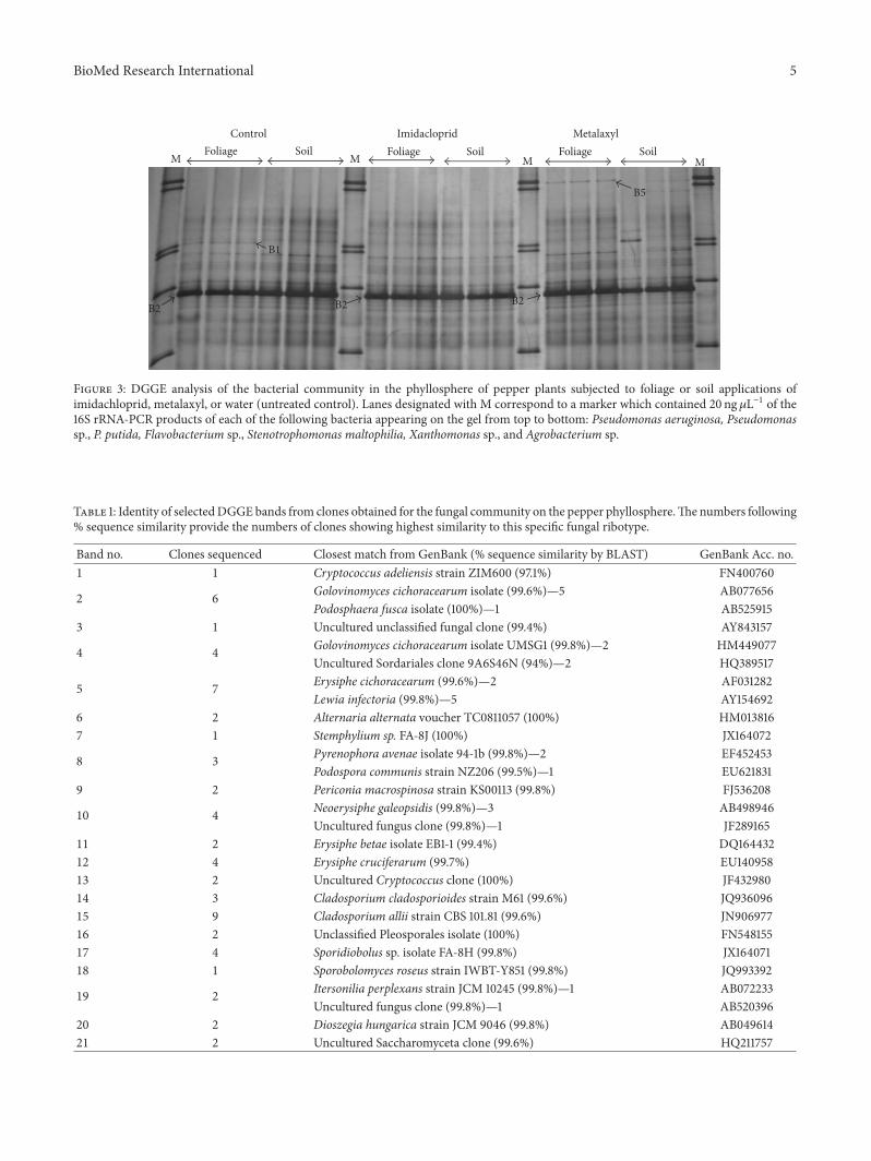
3.1. Effects of Pesticides on the Fungal Community. Thefingerprints produced by the three replicates of the sametreatment were highly similar (Figure 1). Overall, DGGEanalysis of the fungal community provided rather complexbanding patterns with band numbers exceeding 20 in alltreatments. Cluster analysis of the DGGE banding patternsresulted in the development of two main clusters (Figure 2).The first main cluster comprised all control and metalaxyl-treated samples along with the samples which received asoil application of imidacloprid. Samples contained withinthe first cluster shared a similarity of >84%. The second
cluster was composed only by the samples which received asoil application of imidacloprid and showed >70% similaritywith the samples of the first cluster. In the first cluster,samples were further separated according to the pesticideapplied with soil-treated samples of imidacloprid separatedfrom the metalaxyl-treated samples and the controls whichgrouped together. Within the latter subcluster the foliage-treated metalaxyl samples and the control samples clusteredtogether (>90% similarity) while the soil-treated metalaxylsamples were separated.
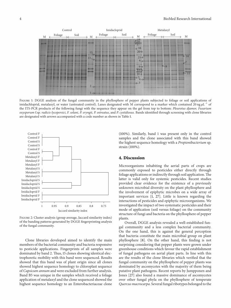
Clone libraries were developed to identify the mainmembers of the fungal community and fungi which wereresponsive to pesticide applications. Overall the phyllospherewas dominated by ascomycetes while a few basidiomycetousyeasts were also present and were represented by bandsappearingmostly in the upper part of the gel belonging to theorders Sporidiobolales (bands 17 and 18), Cystofilobasidiales(band 19), and Filobasidiales (bands 13 and 1) (Table 1).Generally the lower part of the DGGE patterns in all treat-ments, which constitutes the high GC content region, wasdominated by ascomycetes of the order Erysiphales (bands2, 3, 4, 5, 10, 11, and 12). Band 15 was the most dominantband in all treatments and sequencing analysis of the clonesshowed highest homology (99.6%) to a Cladosporium alliistrain (Table 1).
A fewmembers of the fungal community were responsiveto pesticide applications. Therefore, bands 1 and 13 werepresent only in the samples treated with imidacloprid, espe-cially foliage-treated samples (Figure 1). Clones associatedwith those bands showed highest sequence homology to aCryptococcus adeliensis (97.1%) and an uncultured Crypto-coccus clone (100%), respectively. On the contrary, band 3disappeared from the samples which received a foliage appli-cation of imidacloprid. The single clone associated with thisband showed highest sequence homology to an unculturedunclassified fungal clone (99.4%).
Regarding metalaxyl-treated samples, band 21 was onlypresent in the samples treated with the fungicide regardlessof the application mode. Clones associated with this bandshowed highest sequence homology to an uncultured Saccha-romyceta clone (99.6%). In addition, band 9 was stimulatedin the metalaxyl-treated samples. Clones associated withthis band showed highest sequence homology to a Periconiamacrospinosa strain (99.8%).
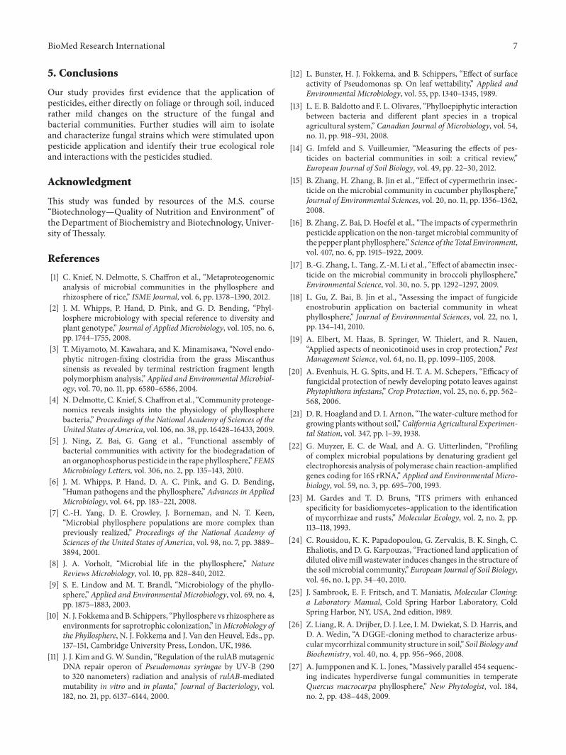
3.2. Effects of Pesticides on the Bacterial Community. DGGEanalysis of the bacterial community showed a less complexbanding pattern, compared to the fungal community finger-prints, with band numbers not exceeding 15 in any of thetreatments (Figure 3). Cluster analysis separated samples intotwo main clusters. The first cluster contained all samplesexcept one of the replicates of the metalaxyl soil application(Figure 4). Within the first cluster samples shared more than90% similarity and were further separated into two sub-clusters: the first one comprising all soil controls and thetwo replicates of the metalaxyl soil application, while thesecond subcluster contained all other control, imidacloprid-and metalaxyl-treated samples.
4 BioMed Research International
Control Imidacloprid MetalaxylFoliage SoilM M M M
15
1
2345
10
171819
6 789
2016
14
1112
13 21
Foliage Soil Foliage Soil
Figure 1: DGGE analysis of the fungal community in the phyllosphere of pepper plants subjected to foliage or soil applications ofimidachloprid, metalaxyl, or water (untreated control). Lanes designated with M correspond to a marker which contained 20 ng𝜇L−1 ofthe ITS-PCR products of the following fungi with the sequence they appear on the gel from top to bottom: Pleurotus djamor, Fusariumoxysporum f.sp. radicis-lycopersici, F. solani, P. eryngii, P. ostreatus, and P. cystidiosus. Bands identified through screening with clone librariesare designated with arrows accompanied with a code number as shown in Table 1.
0.751 0.95 0.9 0.85 0.8Jaccard similarity index
Control FControl F
Control F
Control S
Control S
Control S
Imidacloprid FImidacloprid FImidacloprid F
Imidacloprid S
Imidacloprid SImidacloprid S
Metalaxyl FMetalaxyl FMetalaxyl FMetalaxyl SMetalaxyl SMetalaxyl S
Figure 2: Cluster analysis (group average, Jaccard similarity index)of the banding patterns generated by DGGE fingerprinting analysisof the fungal community.
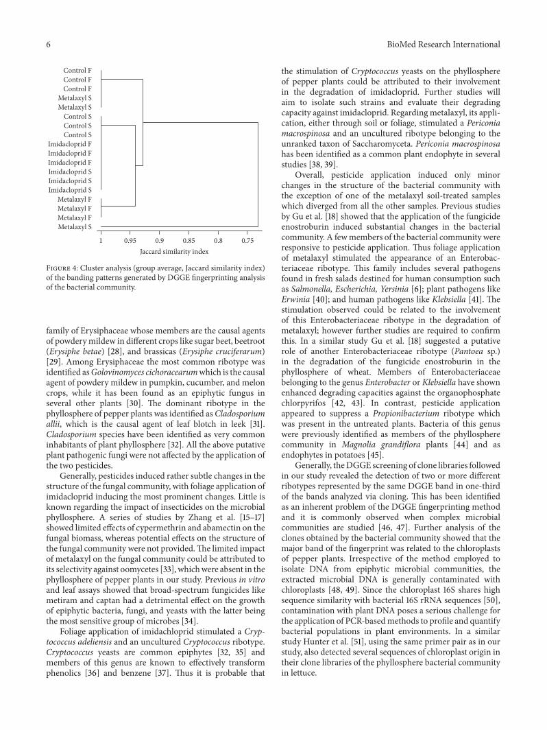
Clone libraries developed aimed to identify the mainmembers of the bacterial community and bacteria responsiveto pesticide applications. Fingerprints of all samples weredominated by band 2. Thus, 15 clones showing identical elec-trophoretic mobility with this band were sequenced. Resultsshowed that this band was of plant origin since all clonesshowed highest sequence homology to chloroplast sequenceofCapsicum annum and were excluded from further analysis.Band B5 was unique in the samples which received a foliageapplication of metalaxyl and the clone sequenced showed thehighest sequence homology to an Enterobacteriaceae clone
(100%). Similarly, band 1 was present only in the controlsamples and the clone associated with this band showedthe highest sequence homology with a Propionibacterium sp.strain (100%).
4. Discussion
Microorganisms inhabiting the aerial parts of crops arecommonly exposed to pesticides either directly throughfoliage applications or indirectly through soil application.Thelatter is valid only for systemic pesticides. Recent studiesprovided clear evidence for the existence of a previouslyunknown microbial diversity on the plant phyllosphere andthe involvement of epiphytic microbes on a wide array ofimportant services [1, 27]. Little is known regarding theinteractions of pesticides and epiphytic microorganisms. Weinvestigated the impact of two systematic pesticides and theirmode of application (soil versus foliage) on the communitystructure of fungi and bacteria on the phyllosphere of pepperplants.
Overall, DGGE analysis revealed a well-established fun-gal community and a less complex bacterial community.On the one hand, this is against the general perceptionthat bacteria constitute the main microbial group on plantphyllosphere [8]. On the other hand, this finding is notsurprising considering that pepper plants were grown undergreenhouse conditions which favour the rapid establishmentof fungal pathogens on aerial plant parts. In line with thisare the results of the clone libraries which verified that thefungal community on the phyllosphere of pepper plants wasdominated by ascomycetes with the majority of them beingputative plant pathogens. Recent reports by Jumpponen andJones [27] also found a massive dominance of ascomycetesover other fungal phyla on the phyllosphere of temperateQuercusmacrocarpa. Several fungal ribotypes belonged to the
BioMed Research International 5
M M M M
Control Imidacloprid Metalaxyl
B1
B2B2B2
B5
Foliage Soil Foliage Soil Foliage Soil
Figure 3: DGGE analysis of the bacterial community in the phyllosphere of pepper plants subjected to foliage or soil applications ofimidachloprid, metalaxyl, or water (untreated control). Lanes designated with M correspond to a marker which contained 20 ng𝜇L−1 of the16S rRNA-PCR products of each of the following bacteria appearing on the gel from top to bottom: Pseudomonas aeruginosa, Pseudomonassp., P. putida, Flavobacterium sp., Stenotrophomonas maltophilia, Xanthomonas sp., and Agrobacterium sp.
Table 1: Identity of selectedDGGEbands from clones obtained for the fungal community on the pepper phyllosphere.The numbers following% sequence similarity provide the numbers of clones showing highest similarity to this specific fungal ribotype.
Band no. Clones sequenced Closest match from GenBank (% sequence similarity by BLAST) GenBank Acc. no.1 1 Cryptococcus adeliensis strain ZIM600 (97.1%) FN400760
2 6
Golovinomyces cichoracearum isolate (99.6%)—5 AB077656Podosphaera fusca isolate (100%)—1 AB525915
3 1 Uncultured unclassified fungal clone (99.4%) AY843157
4 4
Golovinomyces cichoracearum isolate UMSG1 (99.8%)—2 HM449077Uncultured Sordariales clone 9A6S46N (94%)—2 HQ389517
5 7
Erysiphe cichoracearum (99.6%)—2 AF031282Lewia infectoria (99.8%)—5 AY154692
6 2 Alternaria alternata voucher TC0811057 (100%) HM0138167 1 Stemphylium sp. FA-8J (100%) JX164072
8 3
Pyrenophora avenae isolate 94-1b (99.8%)—2 EF452453Podospora communis strain NZ206 (99.5%)—1 EU621831
9 2 Periconia macrospinosa strain KS00113 (99.8%) FJ536208
10 4
Neoerysiphe galeopsidis (99.8%)—3 AB498946Uncultured fungus clone (99.8%)—1 JF289165
11 2 Erysiphe betae isolate EB1-1 (99.4%) DQ16443212 4 Erysiphe cruciferarum (99.7%) EU14095813 2 Uncultured Cryptococcus clone (100%) JF43298014 3 Cladosporium cladosporioides strain M61 (99.6%) JQ93609615 9 Cladosporium allii strain CBS 101.81 (99.6%) JN90697716 2 Unclassified Pleosporales isolate (100%) FN54815517 4 Sporidiobolus sp. isolate FA-8H (99.8%) JX16407118 1 Sporobolomyces roseus strain IWBT-Y851 (99.8%) JQ993392
19 2
Itersonilia perplexans strain JCM 10245 (99.8%)—1 AB072233Uncultured fungus clone (99.8%)—1 AB520396
20 2 Dioszegia hungarica strain JCM 9046 (99.8%) AB04961421 2 Uncultured Saccharomyceta clone (99.6%) HQ211757
6 BioMed Research International
0.751 0.95 0.9 0.85 0.8
Control FControl FControl F
Control SControl S
Control S
Imidacloprid FImidacloprid FImidacloprid FImidacloprid S
Imidacloprid SImidacloprid S
Metalaxyl FMetalaxyl FMetalaxyl F
Metalaxyl SMetalaxyl S
Metalaxyl S
Jaccard similarity index
Figure 4: Cluster analysis (group average, Jaccard similarity index)of the banding patterns generated by DGGE fingerprinting analysisof the bacterial community.
family of Erysiphaceae whose members are the causal agentsof powderymildew in different crops like sugar beet, beetroot(Erysiphe betae) [28], and brassicas (Erysiphe cruciferarum)[29]. Among Erysiphaceae the most common ribotype wasidentified asGolovinomyces cichoracearumwhich is the causalagent of powdery mildew in pumpkin, cucumber, and meloncrops, while it has been found as an epiphytic fungus inseveral other plants [30]. The dominant ribotype in thephyllosphere of pepper plants was identified asCladosporiumallii, which is the causal agent of leaf blotch in leek [31].Cladosporium species have been identified as very commoninhabitants of plant phyllosphere [32]. All the above putativeplant pathogenic fungi were not affected by the application ofthe two pesticides.
Generally, pesticides induced rather subtle changes in thestructure of the fungal community, with foliage application ofimidacloprid inducing the most prominent changes. Little isknown regarding the impact of insecticides on the microbialphyllosphere. A series of studies by Zhang et al. [15–17]showed limited effects of cypermethrin and abamectin on thefungal biomass, whereas potential effects on the structure ofthe fungal community were not provided.The limited impactof metalaxyl on the fungal community could be attributed toits selectivity against oomycetes [33], whichwere absent in thephyllosphere of pepper plants in our study. Previous in vitroand leaf assays showed that broad-spectrum fungicides likemetiram and captan had a detrimental effect on the growthof epiphytic bacteria, fungi, and yeasts with the latter beingthe most sensitive group of microbes [34].
Foliage application of imidachloprid stimulated a Cryp-tococcus adeliensis and an uncultured Cryptococcus ribotype.Cryptococcus yeasts are common epiphytes [32, 35] andmembers of this genus are known to effectively transformphenolics [36] and benzene [37]. Thus it is probable that
the stimulation of Cryptococcus yeasts on the phyllosphereof pepper plants could be attributed to their involvementin the degradation of imidacloprid. Further studies willaim to isolate such strains and evaluate their degradingcapacity against imidacloprid. Regardingmetalaxyl, its appli-cation, either through soil or foliage, stimulated a Periconiamacrospinosa and an uncultured ribotype belonging to theunranked taxon of Saccharomyceta. Periconia macrospinosahas been identified as a common plant endophyte in severalstudies [38, 39].
Overall, pesticide application induced only minorchanges in the structure of the bacterial community withthe exception of one of the metalaxyl soil-treated sampleswhich diverged from all the other samples. Previous studiesby Gu et al. [18] showed that the application of the fungicideenostroburin induced substantial changes in the bacterialcommunity. A fewmembers of the bacterial community wereresponsive to pesticide application. Thus foliage applicationof metalaxyl stimulated the appearance of an Enterobac-teriaceae ribotype. This family includes several pathogensfound in fresh salads destined for human consumption suchas Salmonella, Escherichia, Yersinia [6]; plant pathogens likeErwinia [40]; and human pathogens like Klebsiella [41]. Thestimulation observed could be related to the involvementof this Enterobacteriaceae ribotype in the degradation ofmetalaxyl; however further studies are required to confirmthis. In a similar study Gu et al. [18] suggested a putativerole of another Enterobacteriaceae ribotype (Pantoea sp.)in the degradation of the fungicide enostroburin in thephyllosphere of wheat. Members of Enterobacteriaceaebelonging to the genus Enterobacter or Klebsiella have shownenhanced degrading capacities against the organophosphatechlorpyrifos [42, 43]. In contrast, pesticide applicationappeared to suppress a Propionibacterium ribotype whichwas present in the untreated plants. Bacteria of this genuswere previously identified as members of the phyllospherecommunity in Magnolia grandiflora plants [44] and asendophytes in potatoes [45].
Generally, theDGGE screening of clone libraries followedin our study revealed the detection of two or more differentribotypes represented by the same DGGE band in one-thirdof the bands analyzed via cloning. This has been identifiedas an inherent problem of the DGGE fingerprinting methodand it is commonly observed when complex microbialcommunities are studied [46, 47]. Further analysis of theclones obtained by the bacterial community showed that themajor band of the fingerprint was related to the chloroplastsof pepper plants. Irrespective of the method employed toisolate DNA from epiphytic microbial communities, theextracted microbial DNA is generally contaminated withchloroplasts [48, 49]. Since the chloroplast 16S shares highsequence similarity with bacterial 16S rRNA sequences [50],contamination with plant DNA poses a serious challenge forthe application of PCR-basedmethods to profile and quantifybacterial populations in plant environments. In a similarstudy Hunter et al. [51], using the same primer pair as in ourstudy, also detected several sequences of chloroplast origin intheir clone libraries of the phyllosphere bacterial communityin lettuce.
BioMed Research International 7
5. Conclusions
Our study provides first evidence that the application ofpesticides, either directly on foliage or through soil, inducedrather mild changes on the structure of the fungal andbacterial communities. Further studies will aim to isolateand characterize fungal strains which were stimulated uponpesticide application and identify their true ecological roleand interactions with the pesticides studied.
Acknowledgment
This study was funded by resources of the M.S. course“Biotechnology—Quality of Nutrition and Environment” ofthe Department of Biochemistry and Biotechnology, Univer-sity of Thessaly.
References
[1] C. Knief, N. Delmotte, S. Chaffron et al., “Metaproteogenomicanalysis of microbial communities in the phyllosphere andrhizosphere of rice,” ISME Journal, vol. 6, pp. 1378–1390, 2012.
[2] J. M. Whipps, P. Hand, D. Pink, and G. D. Bending, “Phyl-losphere microbiology with special reference to diversity andplant genotype,” Journal of Applied Microbiology, vol. 105, no. 6,pp. 1744–1755, 2008.
[3] T. Miyamoto, M. Kawahara, and K. Minamisawa, “Novel endo-phytic nitrogen-fixing clostridia from the grass Miscanthussinensis as revealed by terminal restriction fragment lengthpolymorphism analysis,” Applied and Environmental Microbiol-ogy, vol. 70, no. 11, pp. 6580–6586, 2004.
[4] N.Delmotte, C. Knief, S. Chaffron et al., “Community proteoge-nomics reveals insights into the physiology of phyllospherebacteria,” Proceedings of the National Academy of Sciences of theUnited States of America, vol. 106, no. 38, pp. 16428–16433, 2009.
[5] J. Ning, Z. Bai, G. Gang et al., “Functional assembly ofbacterial communities with activity for the biodegradation ofan organophosphorus pesticide in the rape phyllosphere,”FEMSMicrobiology Letters, vol. 306, no. 2, pp. 135–143, 2010.
[6] J. M. Whipps, P. Hand, D. A. C. Pink, and G. D. Bending,“Human pathogens and the phyllosphere,” Advances in AppliedMicrobiology, vol. 64, pp. 183–221, 2008.
[7] C.-H. Yang, D. E. Crowley, J. Borneman, and N. T. Keen,“Microbial phyllosphere populations are more complex thanpreviously realized,” Proceedings of the National Academy ofSciences of the United States of America, vol. 98, no. 7, pp. 3889–3894, 2001.
[8] J. A. Vorholt, “Microbial life in the phyllosphere,” NatureReviews Microbiology, vol. 10, pp. 828–840, 2012.
[9] S. E. Lindow and M. T. Brandl, “Microbiology of the phyllo-sphere,” Applied and Environmental Microbiology, vol. 69, no. 4,pp. 1875–1883, 2003.
[10] N. J. Fokkema and B. Schippers, “Phyllosphere vs rhizosphere asenvironments for saprotrophic colonization,” inMicrobiology ofthe Phyllosphere, N. J. Fokkema and J. Van den Heuvel, Eds., pp.137–151, Cambridge University Press, London, UK, 1986.
[11] J. J. Kim and G.W. Sundin, “Regulation of the rulABmutagenicDNA repair operon of Pseudomonas syringae by UV-B (290to 320 nanometers) radiation and analysis of rulAB-mediatedmutability in vitro and in planta,” Journal of Bacteriology, vol.182, no. 21, pp. 6137–6144, 2000.
[12] L. Bunster, H. J. Fokkema, and B. Schippers, “Effect of surfaceactivity of Pseudomonas sp. On leaf wettability,” Applied andEnvironmental Microbiology, vol. 55, pp. 1340–1345, 1989.
[13] L. E. B. Baldotto and F. L. Olivares, “Phylloepiphytic interactionbetween bacteria and different plant species in a tropicalagricultural system,” Canadian Journal of Microbiology, vol. 54,no. 11, pp. 918–931, 2008.
[14] G. Imfeld and S. Vuilleumier, “Measuring the effects of pes-ticides on bacterial communities in soil: a critical review,”European Journal of Soil Biology, vol. 49, pp. 22–30, 2012.
[15] B. Zhang, H. Zhang, B. Jin et al., “Effect of cypermethrin insec-ticide on the microbial community in cucumber phyllosphere,”Journal of Environmental Sciences, vol. 20, no. 11, pp. 1356–1362,2008.
[16] B. Zhang, Z. Bai, D. Hoefel et al., “The impacts of cypermethrinpesticide application on the non-targetmicrobial community ofthe pepper plant phyllosphere,” Science of the Total Environment,vol. 407, no. 6, pp. 1915–1922, 2009.
[17] B.-G. Zhang, L. Tang, Z.-M. Li et al., “Effect of abamectin insec-ticide on the microbial community in broccoli phyllosphere,”Environmental Science, vol. 30, no. 5, pp. 1292–1297, 2009.
[18] L. Gu, Z. Bai, B. Jin et al., “Assessing the impact of fungicideenostroburin application on bacterial community in wheatphyllosphere,” Journal of Environmental Sciences, vol. 22, no. 1,pp. 134–141, 2010.
[19] A. Elbert, M. Haas, B. Springer, W. Thielert, and R. Nauen,“Applied aspects of neonicotinoid uses in crop protection,” PestManagement Science, vol. 64, no. 11, pp. 1099–1105, 2008.
[20] A. Evenhuis, H. G. Spits, and H. T. A. M. Schepers, “Efficacy offungicidal protection of newly developing potato leaves againstPhytophthora infestans,” Crop Protection, vol. 25, no. 6, pp. 562–568, 2006.
[21] D. R. Hoagland andD. I. Arnon, “Thewater-culture method forgrowing plants without soil,”California Agricultural Experimen-tal Station, vol. 347, pp. 1–39, 1938.
[22] G. Muyzer, E. C. de Waal, and A. G. Uitterlinden, “Profilingof complex microbial populations by denaturing gradient gelelectrophoresis analysis of polymerase chain reaction-amplifiedgenes coding for 16S rRNA,” Applied and Environmental Micro-biology, vol. 59, no. 3, pp. 695–700, 1993.
[23] M. Gardes and T. D. Bruns, “ITS primers with enhancedspecificity for basidiomycetes–application to the identificationof mycorrhizae and rusts,” Molecular Ecology, vol. 2, no. 2, pp.113–118, 1993.
[24] C. Rousidou, K. K. Papadopoulou, G. Zervakis, B. K. Singh, C.Ehaliotis, and D. G. Karpouzas, “Fractioned land application ofdiluted olivemill wastewater induces changes in the structure ofthe soil microbial community,” European Journal of Soil Biology,vol. 46, no. 1, pp. 34–40, 2010.
[25] J. Sambrook, E. F. Fritsch, and T. Maniatis, Molecular Cloning:a Laboratory Manual, Cold Spring Harbor Laboratory, ColdSpring Harbor, NY, USA, 2nd edition, 1989.
[26] Z. Liang, R. A. Drijber, D. J. Lee, I. M. Dwiekat, S. D. Harris, andD. A. Wedin, “A DGGE-cloning method to characterize arbus-cularmycorrhizal community structure in soil,” Soil Biology andBiochemistry, vol. 40, no. 4, pp. 956–966, 2008.
[27] A. Jumpponen andK. L. Jones, “Massively parallel 454 sequenc-ing indicates hyperdiverse fungal communities in temperateQuercus macrocarpa phyllosphere,” New Phytologist, vol. 184,no. 2, pp. 438–448, 2009.
8 BioMed Research International
[28] M. Fernandez-Aparicio, E. Prats, A.A. Emeran, andD.Rubiales,“Characterization of resistancemechanisms to powderymildew(Erysiphe betae) in beet (Beta vulgaris),” Phytopathology, vol. 99,no. 4, pp. 385–389, 2009.
[29] F. Mert-Turk, M. K. Gul, and C. O. Egesel, “Nitrogen and fungi-cide applications against Erysiphe cruciferarum affect qualitycomponents of oilseed rape,”Mycopathologia, vol. 165, no. 1, pp.27–35, 2008.
[30] A. M. R. Almeida, E. Binneck, F. F. Piuga, S. R. R. Marin, P.R. Z. Ribeiro Do Valle, and C. A. Silveira, “Characterizationof powdery mildews strains from soybean, bean, sunflower,and weeds in Brazil using rDNA-ITS sequences,” Tropical PlantPathology, vol. 33, no. 1, pp. 20–26, 2008.
[31] M. M. Jordan, R. T. Burchill, and R. B. Maude, “Epidemiologyof Cladosporium allii and Cladosporium allii-cepae, leaf blotchpathogens of leek and onion. II. Infection of host plants,”Annalsof Applied Biology, vol. 117, no. 2, pp. 327–336, 1990.
[32] Y.-H. He, S. Isono, M. Shibuya et al., “Oligo-DNA cus-tom macroarray for monitoring major pathogenic and non-pathogenic fungi and bacteria in the phyllosphere of appletrees,” PLoS ONE, vol. 7, no. 3, Article ID e34249, 2012.
[33] A. Kerkenaar and A. K. Sijpesteijn, “Antifungal activity ofmetalaxyl and furalaxyl,” Pesticide Biochemistry and Physiology,vol. 15, no. 1, pp. 71–78, 1981.
[34] M. Walter, C. M. Frampton, K. S. H. Boyd-Wilson, P. Harris-Virgin, and N. W. Waipara, “Agrichemical impact on growthand survival of non-target apple phyllospheremicroorganisms,”Canadian Journal of Microbiology, vol. 53, no. 1, pp. 45–55, 2007.
[35] N. Cadez, J. Zupan, and P. Raspor, “The effect of fungicides onyeast communities associated with grape berries,” FEMS YeastResearch, vol. 10, no. 5, pp. 619–630, 2010.
[36] A. Fonseca, G. Scorzetti, and J. W. Fell, “Diversity in theyeast Cryptococcus albidus and related species as revealedby ribosomal DNA sequence analysis,” Canadian Journal ofMicrobiology, vol. 46, no. 1, pp. 7–27, 2000.
[37] W. J. Middelhoven, “Catabolism of benzene compounds byascomycetous and basidiomycetous yeasts and yeastlike fungi.A literature review and an experimental approach,”Antonie vanLeeuwenhoek, vol. 63, no. 2, pp. 125–144, 1993.
[38] K. Mandyam, T. Loughin, and A. Jumpponen, “Isolation andmorphological and metabolic characterization of commonendophytes in annually burned tallgrass prairie,”Mycologia, vol.102, no. 4, pp. 813–821, 2010.
[39] Z.-L. Yuan, C.-L. Zhang, F.-C. Lin, and C. P. Kubicek, “Identity,diversity, and molecular phylogeny of the endophytic myco-biota in the roots of rare wild rice (Oryza granulate) from anature reserve in Yunnan, China,” Applied and EnvironmentalMicrobiology, vol. 76, no. 5, pp. 1642–1652, 2010.
[40] H. H. El-Hendawy, M. E. Osman, and H. A. Ramadan, “Pecticenzymes produced in vitro and in vivo by Erwinia spp. isolatedfrom carrot and pepper in Egypt,” Journal of Phytopathology,vol. 150, no. 8-9, pp. 431–438, 2002.
[41] R. Podschun and U. Ullmann, “Klebsiella spp. as nosoco-mial pathogens: epidemiology, taxonomy, typing methods, andpathogenicity factors,”Clinical Microbiology Reviews, vol. 11, no.4, pp. 589–603, 1998.
[42] B. K. Singh, A. Walker, J. A. W. Morgan, and D. J. Wright,“Biodegradation of chlorpyrifos by Enterobacter strain B-14 andits use in bioremediation of contaminated soils,” Applied andEnvironmentalMicrobiology, vol. 70, no. 8, pp. 4855–4863, 2004.
[43] I. Ghanem, M. Orfi, and M. Shamma, “Biodegradation ofchlorpyrifos by klebsiella sp. isolated from an activated sludge
sample of waste water treatment plant in damascus,” FoliaMicrobiologica, vol. 52, no. 4, pp. 423–427, 2007.
[44] C. R. Jackson andW. C. Denney, “Annual and seasonal variationin the phyllosphere bacterial community associated with leavesof the southern Magnolia (Magnolia grandiflora),” MicrobialEcology, vol. 61, no. 1, pp. 113–122, 2011.
[45] F. Rasche, T. Lueders, M. Schloter et al., “DNA-based stableisotope probing enables the identification of active bacterialendophytes in potatoes,” New Phytologist, vol. 181, no. 4, pp.802–807, 2009.
[46] T. Vallaeys, E. Topp, G. Muyzer et al., “Evaluation of denaturinggradient gel electrophoresis in the detection of 16S rDNAsequence variation in rhizobia and methanotrophs,” FEMSMicrobiology Ecology, vol. 24, no. 3, pp. 279–285, 1997.
[47] C. R. Jackson, E. E. Roden, and E. E. Churchill, “Denaturinggradient gel electrophoresis can fail to separate 16S rDNAfragments with multiple base differences,” Molecular BiologyToday, vol. 1, pp. 49–51, 2000.
[48] L. Sun, F. Qiu, X. Zhang, X. Dai, X. Dong, and W. Song,“Endophytic bacterial diversity in rice (Oryza sativa L.) rootsestimated by 16S rDNA sequence analysis,” Microbial Ecology,vol. 55, no. 3, pp. 415–424, 2008.
[49] G. Rastogi, J. J. Tech, G. L. Coaker, and J. H. J. Leveau, “APCR-based toolbox for the culture-independent quantificationof total bacterial abundances in plant environments,” Journal ofMicrobiological Methods, vol. 83, no. 2, pp. 127–132, 2010.
[50] M. Sakai, A. Matsuka, T. Komura, and S. Kanazawa, “Appli-cation of a new PCR primer for terminal restriction fragmentlength polymorphism analysis of the bacterial communities inplant roots,” Journal of Microbiological Methods, vol. 59, no. 1,pp. 81–89, 2004.
[51] P. J. Hunter, P. Hand, D. Pink, J. M.Whipps, and G. D. Bending,“Both leaf properties and microbe-microbe interactions influ-ence within-species variation in bacterial population diversityand structure in the lettuce (Lactuca species) phyllosphere,”Applied and Environmental Microbiology, vol. 76, no. 24, pp.8117–8125, 2010.

Submit your manuscripts athttp://www.hindawi.com
Hindawi Publishing Corporationhttp://www.hindawi.com Volume 2014
Anatomy Research International
PeptidesInternational Journal of
Hindawi Publishing Corporationhttp://www.hindawi.com Volume 2014
Hindawi Publishing Corporation http://www.hindawi.com
International Journal of
Volume 2014
Zoology
Hindawi Publishing Corporationhttp://www.hindawi.com Volume 2014
Molecular Biology International
GenomicsInternational Journal of
Hindawi Publishing Corporationhttp://www.hindawi.com Volume 2014
The Scientific World JournalHindawi Publishing Corporation http://www.hindawi.com Volume 2014
Hindawi Publishing Corporationhttp://www.hindawi.com Volume 2014
BioinformaticsAdvances in
Marine BiologyJournal of
Hindawi Publishing Corporationhttp://www.hindawi.com Volume 2014
Hindawi Publishing Corporationhttp://www.hindawi.com Volume 2014
Signal TransductionJournal of
Hindawi Publishing Corporationhttp://www.hindawi.com Volume 2014
BioMed Research International
Evolutionary BiologyInternational Journal of
Hindawi Publishing Corporationhttp://www.hindawi.com Volume 2014
Hindawi Publishing Corporationhttp://www.hindawi.com Volume 2014
Biochemistry Research International
ArchaeaHindawi Publishing Corporationhttp://www.hindawi.com Volume 2014
Hindawi Publishing Corporationhttp://www.hindawi.com Volume 2014
Genetics Research International
Hindawi Publishing Corporationhttp://www.hindawi.com Volume 2014
Advances in
Virolog y
Hindawi Publishing Corporationhttp://www.hindawi.com
Nucleic AcidsJournal of
Volume 2014
Stem CellsInternational
Hindawi Publishing Corporationhttp://www.hindawi.com Volume 2014
Hindawi Publishing Corporationhttp://www.hindawi.com Volume 2014
Enzyme Research
Hindawi Publishing Corporationhttp://www.hindawi.com Volume 2014
International Journal of
Microbiology