Embed Size (px)
Citation preview
Multilevel Differential Control of Hormone Gene ExpressionPrograms by hnRNP L and LL in Pituitary Cells
Lei Lei,a Wenguang Cao,a Ling Liu,a Urmi Das,a Yujia Wu,a,b Guodong Liu,a Muhammad Sohail,a Yangjun Chen,b Jiuyong Xiea
aDepartment of Physiology & Pathophysiology, Max Rady College of Medicine, Rady Faculty of HealthSciences, University of Manitoba, Winnipeg, Manitoba, Canada
bDepartment of Applied Computer Science, University of Winnipeg, Winnipeg, Manitoba, Canada
ABSTRACT The pituitary-derived somatolactotrophe GH3 cells secrete both growthhormone (GH) and prolactin (PRL). We have found that the hnRNP L and L-like (LL)paralogs differentially regulate alternative splicing of genes in these cells. Here, weshow that hnRNP L is essential for PRL only, but LL is essential for both PRL and GHproduction. Transcriptome-wide RNA sequencing (RNA-Seq) analysis indicates thatthey differentially control groups of hormone or hormone-related genes involved inhormone production/regulation at total transcript and alternative exon levels. Inter-estingly, hnRNP L also specifically binds and prevents the aberrant usage of a non-conserved CA-rich intron piece of Prl pre-mRNA transcripts, and many others in-volved in endocrine functions, to prevent mostly cryptic last exons and mRNAtruncation. Essential for the full hnRNP L effect on specific exons is a proline-rich re-gion that emerged during evolution in vertebrate hnRNP L only but not LL. To-gether, our data demonstrate that the hnRNP L and its paralog, LL, differentiallycontrol hormone gene expression programs at multiple levels, and hnRNP L in par-ticular is critical for protecting the transcriptome from aberrant usage of intronic se-quences. The multilevel differential control by hnRNPs likely tailors the transcriptometo help refine and safeguard the different gene expression programs for differenthormones.
KEYWORDS hnRNP paralogs, divergence, prolactin, growth hormone, RNA-Seq,cryptic splicing, intron, alternative splicing, hnRNP
Differentiated cells maintain specific programs of precise gene expression for phys-iological functions. Perturbation of functions such as hormone production or
response would have serious consequences. Of the many factors involved, the heter-ogeneous ribonucleoproteins (hnRNPs) are a family of abundant and critical players inthe posttranscriptional processing of pre-mRNA transcripts (1, 2). They have evolved toabout 30 members, including homologs or paralogs (3), for the tens of thousands ofgene transcripts in vertebrates. This number is many more than those in fly, nematode,plant, or yeast (3). However, the role of the different, particularly paralogous, vertebratehnRNPs in relation to specific cell functions and the accompanying gene expressionprograms remains largely unclear.
Among their diverse functions, gene regulation at the transcript and exon levels invertebrate, particularly mammalian, systems has been well known (1, 3–5). Relativelyless explored is the protection of transcriptome integrity from cryptic sites in theexpanded introns, which harbor numerous signals for cryptic splicing and pieces thatcould be aberrantly processed into the mRNA under abnormal or disease conditions (3,6). It has been shown that the exonization of Alu repeats can be prevented by hnRNPC (7), but it is not clear whether its diverged paralog, CL1, has a similar or distinct rolein the process. Similar repression effect on cryptic exons has also been found in the
Received 19 December 2017 Returned formodification 16 January 2018 Accepted 22March 2018
Accepted manuscript posted online 2 April2018
Citation Lei L, Cao W, Liu L, Das U, Wu Y, Liu G,Sohail M, Chen Y, Xie J. 2018. Multileveldifferential control of hormone geneexpression programs by hnRNP L and LL inpituitary cells. Mol Cell Biol 38:e00651-17.https://doi.org/10.1128/MCB.00651-17.
Copyright © 2018 Lei et al. This is an open-access article distributed under the terms ofthe Creative Commons Attribution 4.0International license.
Address correspondence to Jiuyong Xie,[email protected].
L.L., W.C., and L.L. contributed equally to thiswork.
RESEARCH ARTICLE
crossm
June 2018 Volume 38 Issue 12 e00651-17 mcb.asm.org 1Molecular and Cellular Biology
AQ: au
AQ: A
Editor: Section: Designation:Lynch Research Article T
zmb-mcb/zmb01218/zmb1771d18z xppws S�3 4/19/18 6:26 4/Color Fig: 2,3,4,5,6 ArtID: 00651-17 DOI:10.1128/MCB.00651-17open-access CE: SSV
cases of PTBP1 (polypyrimidine tract-binding protein 1, hnRNP I) and PTBP2 as well asTDP-43 (8, 9). The role of the many other hnRNPs, particularly the highly similarparalogs in the transcriptome-wide inhibition of cryptic splicing in relation to cellfunction, remains unclear.
hnRNP paralogs share high levels of sequence similarities but also diverged specificproperties. For example, hnRNP A1/A2 or F/H have partial or nearly complete comple-mentary effects on splicing in RNA interference assays of some exons (10, 11). PTBP1and PTBP2 are both splicing repressors but have differential activities and switchexpression for specific splicing events during neuronal differentiation (12, 13). hnRNPE1, but not E2, reduces the expression of the Rev gene of HIV-1 due to their differentC termini (14). Therefore, elucidating distinct properties and transcript targets of hnRNPparalogs will help reveal the logic of their expansion and orchestration of the process-ing of particular mRNA targets in cell functions/diseases.
HnRNP L and L-like (LL) are paralogs that likely emerged in vertebrates through geneduplication (3, 15). They share approximately 55% overall identity between theirprimary amino acid sequences in humans and rats. They both prefer to bind CA/AC-richmotifs (16, 17) and play important and sometimes distinct roles in the normal processesof gene regulation and cell differentiation (18–26). In particular, hnRNP L preferentiallybinds to introns in the transcriptome and is thought to regulate alternative splicing(27). Moreover, an analysis of microarray data confirmed 11 exons that appeared to bespecifically controlled by hnRNP L but not LL (21). However, comparison data betweenthe paralogs using larger-scale techniques, such as transcriptome-wide RNA sequenc-ing (RNA-Seq), and their effects on cryptic exons and cell functions have not beenreported.
We have found that hnRNP L and LL differentially regulate the alternative splicingof genes involved in hormone production in GH3 rat pituitary cells (28–30). These clonalcells produce two structurally related hormones, prolactin (PRL) and growth hormone(GH) (31). Inside the pituitary, these hormones are produced from the Prl and Gh1 genesin distinct lactotrophes and somatotrophes differentiated from common precursors(32). Deregulation of these genes in humans causes dwarfism (33), infertility, orpuerperal alactogenesis (34–36). The expression of both hormones in the GH3
somatolactotrophes thus provides an ideal system for us to compare the globaleffects of the diverged hnRNP paralogs on the transcriptome and their potentialimpact on two distinct cell functions.
In this report, we show that the diverged hnRNP L and LL paralogs differentiallycontrol the production of PRL and GH hormones, as well as the transcript andalternative exon levels of many other genes involved in endocrine functions. Moreover,hnRNP L not only regulates gene expression but also prevents aberrant splicing of Prland many other transcripts in the transcriptome, distinctively from its paralog, LL. Wefurther show that this paralog-specific effect to protect transcripts requires an evolu-tionarily diverged domain of vertebrate hnRNP L for specific exons.
RESULTSHnRNP L and LL differentially control the production of PRL and GH hormones.
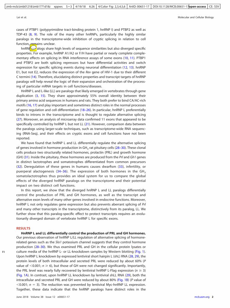
Our previous observation of hnRNP L/LL regulation of alternative splicing of hormone-related genes such as the Slo1 potassium channel suggests that they control hormoneproduction (28–30). We thus examined PRL and GH in the cellular protein lysates orculture media of the hnRNP L- or LL-knockdown samples by Western blotting (Fig. 1).Upon hnRNP L knockdown by expressed lentiviral short hairpin L (shL) RNA (28, 29), theprotein levels of both intracellular and secreted PRL were reduced by about 60% (Pvalue of �0.001; n � 6), but those of GH were not changed significantly. Importantly,the PRL level was nearly fully recovered by lentiviral hnRNP L-Flag expression (n � 3)(Fig. 1A). In contrast, upon hnRNP LL knockdown by lentiviral shLL RNA (29), both theintracellular and secreted PRL and GH were reduced by about 80% (Fig. 1B) (P value of�0.001; n � 3). The reduction was prevented by lentiviral Myc-hnRNP LL expression.Together, these data indicate that the hnRNP paralogs have distinct roles in the
Lei et al. Molecular and Cellular Biology
June 2018 Volume 38 Issue 12 e00651-17 mcb.asm.org 2
F1
zmb-mcb/zmb01218/zmb1771d18z xppws S�3 4/19/18 6:26 4/Color Fig: 2,3,4,5,6 ArtID: 00651-17 DOI:10.1128/MCB.00651-17open-access CE: SSV
production of the two hormones: hnRNP L controls PRL only, while hnRNP LL controlsboth.
We then examined the corresponding hormone transcripts Prl and Gh1 in these cellsby semiquantitative reverse transcription-PCR (RT-PCR). The result confirmed the sametrends of changes of both transcripts (Fig. 1C to E): hnRNP L controls Prl only, but hnRNPLL controls both Prl and Gh1.
HnRNP L and LL differentially control hormone gene expression programs atboth total transcript and alternative exon levels. To compare the transcriptome-wide effect of hnRNP L and LL in their differential control of hormone production, wecarried out RNA-Seq analysis (see Materials and Methods for more details) among threegroups of GH3 samples (mock, shL, and shLL). By edgeR analysis of the changes of totaltranscript levels, we identified 412 shL- and 984 shLL-specific genes that showed less
FIG 1 Control of GH and PRL hormones and transcripts by hnRNP L and hnRNP LL in GH3 pituitary cells.(A and B) Representative Western blots of the cellular and secreted prolactin and growth hormone of theGH3 cells with or without hnRNP L or LL knockdown (shL or shLL)/rescue (shL � L or shLL � LL),respectively, with hnRNP F/H as a loading control. pLKO.1 and pCppt2E, vector controls for shLL andMyc-hnRNP LL, respectively. *, Flag-tagged hnRNP L. In panel B, endogenous (Endo.) and overexpressed(Ovexp.) hnRNP LL are indicated to the right of the gel. (C and D) Agarose gels of the RT-PCR productsof Gh1 and Prl in the GH3 cells shown in panels A and B. �, PCR or RT negative control; Actb, RNA loadingcontrol. (E) PCR linearity test by cycle numbers for the Prl and Gh1 genes. Agarose gels of the productsfrom PCR cycles 25 to 34 and plots of the product intensity versus cycle numbers are shown. Thearrowheads indicate the line, and cycle number taken, for the respective genes.
hnRNP L and LL in Hormone Gene Expression Programs Molecular and Cellular Biology
June 2018 Volume 38 Issue 12 e00651-17 mcb.asm.org 3
zmb-mcb/zmb01218/zmb1771d18z xppws S�3 4/19/18 6:26 4/Color Fig: 2,3,4,5,6 ArtID: 00651-17 DOI:10.1128/MCB.00651-17open-access CE: SSV
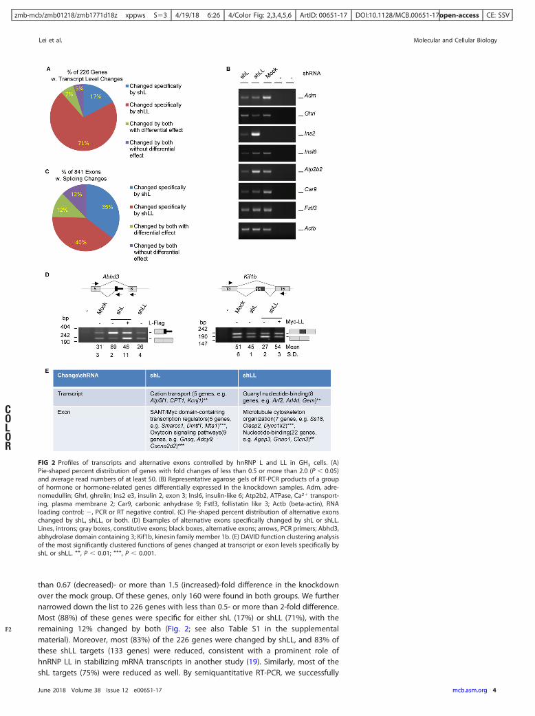
than 0.67 (decreased)- or more than 1.5 (increased)-fold difference in the knockdownover the mock group. Of these genes, only 160 were found in both groups. We furthernarrowed down the list to 226 genes with less than 0.5- or more than 2-fold difference.Most (88%) of these genes were specific for either shL (17%) or shLL (71%), with theremaining 12% changed by both (Fig. 2; see also Table S1 in the supplementalmaterial). Moreover, most (83%) of the 226 genes were changed by shLL, and 83% ofthese shLL targets (133 genes) were reduced, consistent with a prominent role ofhnRNP LL in stabilizing mRNA transcripts in another study (19). Similarly, most of theshL targets (75%) were reduced as well. By semiquantitative RT-PCR, we successfully
FIG 2 Profiles of transcripts and alternative exons controlled by hnRNP L and LL in GH3 cells. (A)Pie-shaped percent distribution of genes with fold changes of less than 0.5 or more than 2.0 (P � 0.05)and average read numbers of at least 50. (B) Representative agarose gels of RT-PCR products of a groupof hormone or hormone-related genes differentially expressed in the knockdown samples. Adm, adre-nomedullin; Ghrl, ghrelin; Ins2 e3, insulin 2, exon 3; Insl6, insulin-like 6; Atp2b2, ATPase, Ca2� transport-ing, plasma membrane 2; Car9, carbonic anhydrase 9; Fstl3, follistatin like 3; Actb (beta-actin), RNAloading control; �, PCR or RT negative control. (C) Pie-shaped percent distribution of alternative exonschanged by shL, shLL, or both. (D) Examples of alternative exons specifically changed by shL or shLL.Lines, introns; gray boxes, constitutive exons; black boxes, alternative exons; arrows, PCR primers; Abhd3,abhydrolase domain containing 3; Kif1b, kinesin family member 1b. (E) DAVID function clustering analysisof the most significantly clustered functions of genes changed at transcript or exon levels specifically byshL or shLL. **, P � 0.01; ***, P � 0.001.
Lei et al. Molecular and Cellular Biology
June 2018 Volume 38 Issue 12 e00651-17 mcb.asm.org 4
F2
COLOR
zmb-mcb/zmb01218/zmb1771d18z xppws S�3 4/19/18 6:26 4/Color Fig: 2,3,4,5,6 ArtID: 00651-17 DOI:10.1128/MCB.00651-17open-access CE: SSV
validated the changes of 25 out of 28 genes examined (Fig. 2B), supporting the accurateprediction by the edgeR analysis. These include the adrenalmedullin (Adm), ghrelin(Ghrl), carbon anhydrase 9 (Car9), and insulin 2 (Ins2) genes, among others.
We also analyzed the changes of alternative exons using DEXSeq (37). We identified841 exons/regions (bins) that changed significantly between at least two pairs of thethree groups of samples (�1.1-fold; P value of �0.05) (Fig. 2C and D and Table S2). Themajority (75%) of the exons were changed specifically by shL (35%) and shLL (40%). Wemanually examined the exons in the Integrative Genomics Viewer (IGV) and verified 31of 33 alternative splicing events by RT-PCR (94%) (Fig. 2D) (unpublished data), sup-porting the accurate prediction of regulated exons by our DEXseq analysis. Of the 453Ensembl genes of the changed exons/bins, only 6 (�1.3%) were also changed at thetranscript level in Fig. 1A, suggesting that the splicing control by hnRNP L or LL ismostly independent of its regulation of transcript levels.
Functional analysis indicates that these shL- or shLL-specifically changed genescluster for different aspects of endocrine functions at both the total transcript and exonlevels (Fig. 2E). Specifically, genes changed at the transcript level by shL cluster forcation transport but those changed by shLL cluster for guanyl nucleotide binding, whilethose changed at the exon level by shL cluster for transcription control and oxytocinsignaling but those changed at the exon level by shLL cluster for microtubule organi-zation and nucleotide binding. These functions are related to cellular excitability,signaling, cytoskeleton rearrangement, hormone vesicle transport/release, and genetranscription, processes that are important in different steps of hormone productionand regulation.
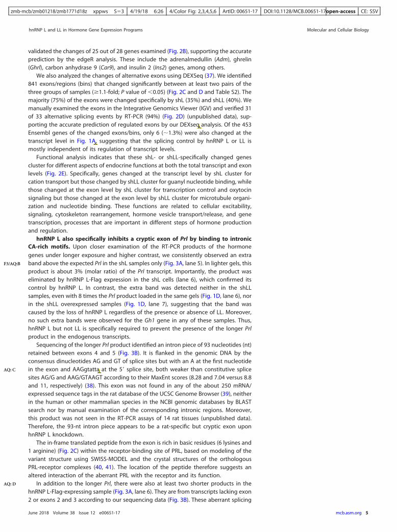
hnRNP L also specifically inhibits a cryptic exon of Prl by binding to intronicCA-rich motifs. Upon closer examination of the RT-PCR products of the hormonegenes under longer exposure and higher contrast, we consistently observed an extraband above the expected Prl in the shL samples only (Fig. 3A, lane 5). In lighter gels, thisproduct is about 3% (molar ratio) of the Prl transcript. Importantly, the product waseliminated by hnRNP L-Flag expression in the shL cells (lane 6), which confirmed itscontrol by hnRNP L. In contrast, the extra band was detected neither in the shLLsamples, even with 8 times the Prl product loaded in the same gels (Fig. 1D, lane 6), norin the shLL overexpressed samples (Fig. 1D, lane 7), suggesting that the band wascaused by the loss of hnRNP L regardless of the presence or absence of LL. Moreover,no such extra bands were observed for the Gh1 gene in any of these samples. Thus,hnRNP L but not LL is specifically required to prevent the presence of the longer Prlproduct in the endogenous transcripts.
Sequencing of the longer Prl product identified an intron piece of 93 nucleotides (nt)retained between exons 4 and 5 (Fig. 3B). It is flanked in the genomic DNA by theconsensus dinucleotides AG and GT of splice sites but with an A at the first nucleotidein the exon and AAGgtatta at the 5= splice site, both weaker than constitutive splicesites AG/G and AAG/GTAAGT according to their MaxEnt scores (8.28 and 7.04 versus 8.8and 11, respectively) (38). This exon was not found in any of the about 250 mRNA/expressed sequence tags in the rat database of the UCSC Genome Browser (39), neitherin the human or other mammalian species in the NCBI genomic databases by BLASTsearch nor by manual examination of the corresponding intronic regions. Moreover,this product was not seen in the RT-PCR assays of 14 rat tissues (unpublished data).Therefore, the 93-nt intron piece appears to be a rat-specific but cryptic exon uponhnRNP L knockdown.
The in-frame translated peptide from the exon is rich in basic residues (6 lysines and1 arginine) (Fig. 2C) within the receptor-binding site of PRL, based on modeling of thevariant structure using SWISS-MODEL and the crystal structures of the orthologousPRL-receptor complexes (40, 41). The location of the peptide therefore suggests analtered interaction of the aberrant PRL with the receptor and its function.
In addition to the longer Prl, there were also at least two shorter products in thehnRNP L-Flag-expressing sample (Fig. 3A, lane 6). They are from transcripts lacking exon2 or exons 2 and 3 according to our sequencing data (Fig. 3B). These aberrant splicing
hnRNP L and LL in Hormone Gene Expression Programs Molecular and Cellular Biology
June 2018 Volume 38 Issue 12 e00651-17 mcb.asm.org 5
F3/AQ:B
AQ: C
AQ: D
zmb-mcb/zmb01218/zmb1771d18z xppws S�3 4/19/18 6:26 4/Color Fig: 2,3,4,5,6 ArtID: 00651-17 DOI:10.1128/MCB.00651-17open-access CE: SSV
FIG 3 hnRNP L-specific effect on the usage of a 93-nt cryptic exon of Prl. (A) A high-contrast, long-exposure image of anagarose gel of the RT-PCR products of Prl in GH3 cells. Identities of the different Prl products are to the right of the gel.Note that this gel highlights the longer Prl product and is not quantitative regarding the Prl level compared to thatdepicted in Fig. 1. (B) Diagram of the Prl variants detected in GH3 cells with different expression levels of hnRNP L. Lines,introns; boxes, exons; narrower boxes, untranslated regions; blue box, the intron piece (cryptic exon) retained in the shLsample. (C) Sequencing chromatogram of the Prl � 93-nt band in the gel shown in panel A. Inclusion of a 93-nt crypticexon between exons 4 and 5 of Prl caused a 31-aa (amino acid) insertion in the PRL protein. (D) Diagram of the Prl splicingreporter minigenes cloned into the vector DUP175. Black bars and filled triangles, CA-rich motifs; horizontal lines, introns;blue box, the 93-nt cryptic exon. Test results of the 93-nt usage in the HEK293T cells are on the right. Arrowheads indicatethe location of PCR primers. (E) hnRNP L specifically inhibits splicing of the 93-nt cryptic exon. Shown is an agarose gel ofthe RT-PCR products of Prl minigenes from HEK293T cells with or without hnRNP L or LL knockdown and rescue. Relativeamounts of the shRNA viruses used for transduction are 30 �l and 90 �l. Identities of the PCR bands are to the right. (F)UV cross-linking assay of the [�-32P]CTP-labeled Prl RNA probes in HeLa nuclear extracts, followed by immunoprecipitation(IP) with anti-hnRNPL. (Top) CA-rich motifs of the Prl RNA probes. (Bottom) Phosphor images of the cross-linked and IPproteins in SDS-PAGE gels. *, uncharacterized protein, likely PTBP1, with increased binding to the mutated sequence motifUCUU, UUCU, or CUCU in M1, as also observed in similar cases by Cao et al. (66). The full 68-nt probe sequence is describedin Materials and Methods. (G) Response of a CA motif mutant Prl-3 minigene splicing reporter (Prl-3mut) to coexpressedhnRNP L-Flag in HEK293T cells. The CA-mutated nucleotides are in boldface and underlined. To make sure the 93-ntincluded product is detected for assessing the inhibitory effect by hnRNP L-Flag, the wild-type Prl-3 reporter, but not themutant, was expressed in hnRNP L-knockdown cells (lanes 4 and 5). The PCR was spiked with a Prl exon-specific reverseprimer at a 5:1 ratio to the downstream vector reverse primer. This helped detection of the exon-included band, which wasconfirmed by PCR with vector primers only.
Lei et al. Molecular and Cellular Biology
June 2018 Volume 38 Issue 12 e00651-17 mcb.asm.org 6
COLOR
zmb-mcb/zmb01218/zmb1771d18z xppws S�3 4/19/18 6:26 4/Color Fig: 2,3,4,5,6 ArtID: 00651-17 DOI:10.1128/MCB.00651-17open-access CE: SSV
events generate premature termination codons (PTCs) that likely cause nonsense-mediated mRNA decay (NMD) of the transcripts based on the rules of NMD (42–44).
Thus, different levels of hnRNP L appears to have an effect on the aberrantsplicing of Prl in three directions: (i) its downregulation induces cryptic exon usage,accompanied by reduced normal Prl mRNA; (ii) its overexpression (at least 2-fold here)causes exon skipping and likely NMD; and (iii) only the endogenous level of hnRNP Lis optimal for efficient proper splicing and expression of Prl. Although the hnRNPL-downregulation-induced cryptic product is not the majority of Prl transcripts and itsrole in the overall reduction of Prl is unknown, its presence suggests a novel role ofhnRNP L to inhibit cryptic exons of endogenous transcripts in cells. Thus, we furtherexamined this phenomenon.
The 93-nt cryptic exon of Prl and its flanking intronic regions contain several CA-richsequences (Fig. 3D to F). Their role in the cryptic splicing was assessed in rat minigenesPrl-1 to Prl-3, containing the 93-nt and different lengths of its upstream intron (Fig. 3D),in the vector DUP175 derived from the human beta globin gene (45, 46). RT-PCRanalysis of their transiently expressed transcripts did not detect any cryptic variant inHEK293T cells, suggesting that the shortest transcript of Prl-3 contains a sequencesufficient for the 93 nt to be inhibited (Fig. 3E). Further tests in shL- or shLL-expressingcells indicated that shL but not shLL allowed usage of the 93 nt (lanes 4 to 8).Importantly, the shL effect was eliminated by hnRNP L-Flag expression (lane 10). Thus,hnRNP L specifically inhibits usage of the 93-nt exon through sequences within the Prl-3transcript.
To identify the CA-rich sequences within Prl-3 essential for hnRNP L binding, wecarried out UV-cross-linking and immunoprecipitation assays of [alpha-32P]CTP-labeledRNA probes containing the 3 CA-rich sequence motifs or their mutants in HeLa nuclearextracts (Fig. 3F). The wild-type (WT) probe mainly cross-linked to a protein of about65 kDa (lanes 1 and 2), which was eliminated by A-to-U mutations of all three motifs(M1; lane 3) but not by those in the first and last ones only (M2; lane 4). Moreover,the 65-kDa protein, but not the proteins between 35 and 48 kDa, was immunopre-cipitated by the hnRNP L-specific antibody 4D11 (lane 5). Thus, the unmutatedACACAUCA and CACACA motifs are critical and sufficient for hnRNP L binding to theRNA transcript.
To determine if the CA-rich motifs are necessary for hnRNP L inhibition of the 93-ntexon, we mutated the motifs in a splicing reporter, Prl3mut (Fig. 3G), and coexpressedit with the hnRNP L-Flag protein in HEK293T cells. As its control, wild-type Prl-3 wasexpressed in shL-expressing cells to help detection of its cryptic product. With animproved PCR scheme (Fig. 3G), we were able to detect the cryptic Prl product at about57%, which was reduced to about 33% by the coexpressed hnRNP L. The mutant hadabout a 63% inclusion, which was reduced to only about 42% by the coexpressedhnRNP L.
hnRNP L specifically inhibits CA-rich cryptic exons in the transcriptome. Todetermine whether exclusion of cryptic exons, as in Prl, is a specific global effect ofhnRNP L, we analyzed the RNA-Seq reads mapped only to introns of the Ensembl-annotated rat genome using edgeR. These introns did not have known annotatedmRNA (exon) sequences in the assembly. We started with a stringent criteria byarbitrarily selecting bins (100 bp/bin) with more than 50 reads on average (among thethree groups) and at least 2-fold changes over the mock samples. This resulted in 892and 1,166 bins for shL and shLL samples (false discovery rate [FDR] of �5%), respec-tively, with only 34 (�5%) changed by both (Fig. 4A). Thus, their intronic target binsmostly (�95%) do not overlap, again consistent with their mainly different targetprofiles of mRNA transcripts.
Most of the bins (�90%) are changed over values for the mock group by 2- to 4-fold.The downregulated ones could be rat exons still to be annotated in the database, andthe upregulated ones could be either unannotated or cryptic exons. Interestingly, adistinct cluster of 78 bins, including the Prl 93-nt exon, showed more than 4-fold higher
hnRNP L and LL in Hormone Gene Expression Programs Molecular and Cellular Biology
June 2018 Volume 38 Issue 12 e00651-17 mcb.asm.org 7
F4
zmb-mcb/zmb01218/zmb1771d18z xppws S�3 4/19/18 6:26 4/Color Fig: 2,3,4,5,6 ArtID: 00651-17 DOI:10.1128/MCB.00651-17open-access CE: SSV
reads in shL than in the mock group (FDR of �2.4E�30) (Fig. 4A, left, yellow oval), withan average read of mostly less than 100 in the mock group. In contrast, no such binswere found in the shLL samples (Fig. 4A, right, yellow oval; the only 3 inside being fromthe shL sample). Thus, like its effect on the 93-nt Prl cryptic exon, hnRNP L knockdown
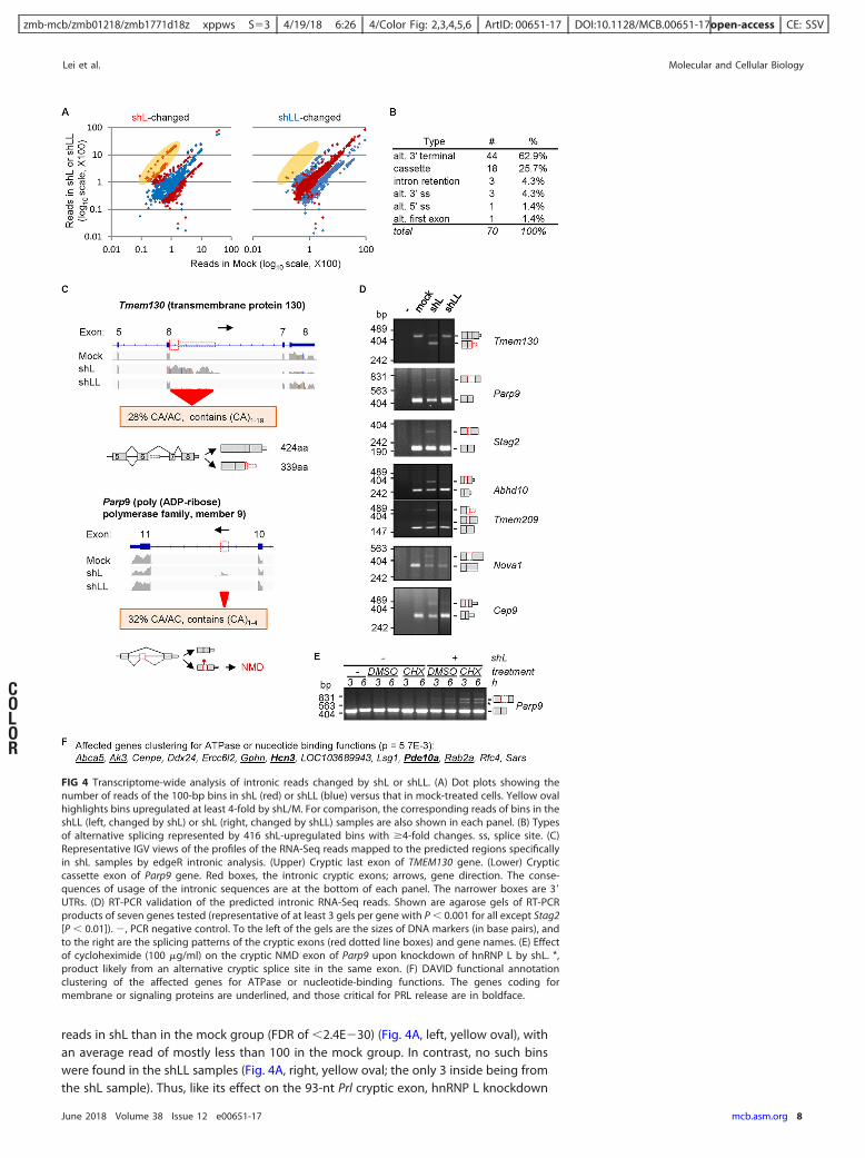
FIG 4 Transcriptome-wide analysis of intronic reads changed by shL or shLL. (A) Dot plots showing thenumber of reads of the 100-bp bins in shL (red) or shLL (blue) versus that in mock-treated cells. Yellow ovalhighlights bins upregulated at least 4-fold by shL/M. For comparison, the corresponding reads of bins in theshLL (left, changed by shL) or shL (right, changed by shLL) samples are also shown in each panel. (B) Typesof alternative splicing represented by 416 shL-upregulated bins with �4-fold changes. ss, splice site. (C)Representative IGV views of the profiles of the RNA-Seq reads mapped to the predicted regions specificallyin shL samples by edgeR intronic analysis. (Upper) Cryptic last exon of TMEM130 gene. (Lower) Crypticcassette exon of Parp9 gene. Red boxes, the intronic cryptic exons; arrows, gene direction. The conse-quences of usage of the intronic sequences are at the bottom of each panel. The narrower boxes are 3=UTRs. (D) RT-PCR validation of the predicted intronic RNA-Seq reads. Shown are agarose gels of RT-PCRproducts of seven genes tested (representative of at least 3 gels per gene with P � 0.001 for all except Stag2[P � 0.01]). �, PCR negative control. To the left of the gels are the sizes of DNA markers (in base pairs), andto the right are the splicing patterns of the cryptic exons (red dotted line boxes) and gene names. (E) Effectof cycloheximide (100 �g/ml) on the cryptic NMD exon of Parp9 upon knockdown of hnRNP L by shL. *,product likely from an alternative cryptic splice site in the same exon. (F) DAVID functional annotationclustering of the affected genes for ATPase or nucleotide-binding functions. The genes coding formembrane or signaling proteins are underlined, and those critical for PRL release are in boldface.
Lei et al. Molecular and Cellular Biology
June 2018 Volume 38 Issue 12 e00651-17 mcb.asm.org 8
COLOR
zmb-mcb/zmb01218/zmb1771d18z xppws S�3 4/19/18 6:26 4/Color Fig: 2,3,4,5,6 ArtID: 00651-17 DOI:10.1128/MCB.00651-17open-access CE: SSV
appears to specifically enhance the usage of a group of cryptic exons that are barelyspliced into mRNA in GH3 cells.
By manually analyzing all bins with at least 4-fold increases (FDR of �1E�4) in IGV,we identified 335 bins (81%) associated with the canonical GT-AG splice sites. Most ofthem (96%) have fewer than 100 reads in the mock group. They belong to 70 splicingevents of 64 genes (Fig. 4B and Table S3), of which 62.9% are alternative last and 25.7%cassette exons. Thus, the majority of them would cause early polyadenylation andshortening of the mRNA transcripts, as observed for U1 snRNP and in cancer or othercells (47–50). In contrast, in the shLL group we identified only 92 such bins and only 7such splicing events of 7 genes. Therefore, the increased usage of cryptic exons ismainly caused by shL.
The two major types of shL-targeting cryptic exons are exemplified in the IGV viewsof the last exon of TMEM130 and the cassette exon of Parp9 genes (Fig. 4C). Reads ofthese CA-rich pieces are barely detectable in the mock and shLL samples but clearlyincreased in shL samples. Their inclusion in the mRNA causes 85-amino-acid (aa)truncation at the COOH terminus of the protein (TMEM130) or a premature stop codonin the transcript (Parp9). Of seven such cryptic exons examined, we confirmed all oftheir specific increases in the shL samples by RT-PCR (Fig. 4D), as well as the furtherincrease of the Parp9 exon upon treatment by cycloheximide, an inhibitor of proteinsynthesis and NMD (51).
The CA/AC dinucleotides of these exons comprise about 30% of their sequences onaverage (n � 25 exons analyzed) (Table S3). Manual examination of 24 cases (6alternative 3=-end and 18 cassette exons) did not identify any of them in the corre-sponding intronic regions of human or mouse genes. Moreover, in the above-described14 rat tissues, none of the 4 examined exons was detected by RT-PCR (unpublisheddata). Furthermore, nine of the 18 (50%) cassette exons introduce premature termina-tion codons. Consistently, higher usage of such cryptic exons (15%, on average, versus5%; P value of 1.6E�06) is correlated with a 2-fold reduction of transcript levels (P valueof 2.1E�05 using edgeR; n � 5) in shL samples. Therefore, these are intronic CA-rich,nonconserved sequences whose usage likely truncates transcripts/proteins or destabi-lizes mRNA transcripts.
A group of 13 of the 64 genes cluster most significantly for ATPase activities ornucleotide binding in DAVID functional analysis (P value of 5.7E�3) (Fig. 4F). Some ofthem, including Pde10a (phosphodiesterase 10A) (52), Rab2a (Ras-related protein Rab-2A) (53), and Hcn3 (hyperpolarization activated cyclic nucleotide gated potassiumchannel 3) (54), are involved in cell signaling, vesicle formation, and electrical firing,which are important for hormone production or regulation. In particular, PDE10a, whichhydrolyzes cyclic AMP (cAMP) (52), and HCN channels, which were found on vesicles,are critical for prolactin release (55, 56).
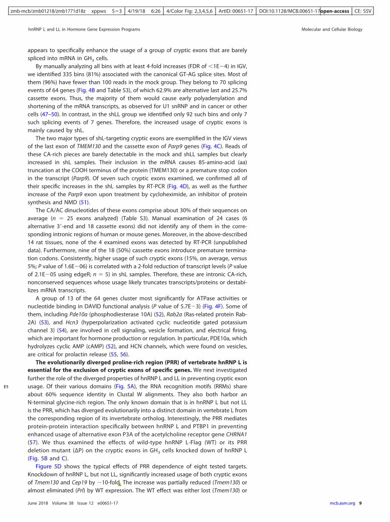
The evolutionarily diverged proline-rich region (PRR) of vertebrate hnRNP L isessential for the exclusion of cryptic exons of specific genes. We next investigatedfurther the role of the diverged properties of hnRNP L and LL in preventing cryptic exonusage. Of their various domains (Fig. 5A), the RNA recognition motifs (RRMs) shareabout 60% sequence identity in Clustal W alignments. They also both harbor anN-terminal glycine-rich region. The only known domain that is in hnRNP L but not LLis the PRR, which has diverged evolutionarily into a distinct domain in vertebrate L fromthe corresponding region of its invertebrate ortholog. Interestingly, the PRR mediatesprotein-protein interaction specifically between hnRNP L and PTBP1 in preventingenhanced usage of alternative exon P3A of the acetylcholine receptor gene CHRNA1(57). We thus examined the effects of wild-type hnRNP L-Flag (WT) or its PRRdeletion mutant (ΔP) on the cryptic exons in GH3 cells knocked down of hnRNP L(Fig. 5B and C).
Figure 5D shows the typical effects of PRR dependence of eight tested targets.Knockdown of hnRNP L, but not LL, significantly increased usage of both cryptic exonsof Tmem130 and Cep19 by �10-fold. The increase was partially reduced (Tmem130) oralmost eliminated (Prl) by WT expression. The WT effect was either lost (Tmem130) or
hnRNP L and LL in Hormone Gene Expression Programs Molecular and Cellular Biology
June 2018 Volume 38 Issue 12 e00651-17 mcb.asm.org 9
F5
zmb-mcb/zmb01218/zmb1771d18z xppws S�3 4/19/18 6:26 4/Color Fig: 2,3,4,5,6 ArtID: 00651-17 DOI:10.1128/MCB.00651-17open-access CE: SSV
FIG 5 Comparison of the evolutionarily diverged PRR of rat hnRNP L with the same region of L and LL of otherrepresentative species (A) and the role of PRR in preventing aberrant splicing by hnRNP L (B to D). (A) Location of the PRRin the structural domains of rat hnRNP L and the aligned corresponding regions of different species using Clustal W. Thediagram is drawn to scale. Amino acids different from the rat ones are shaded. *, invertebrate proteins have significantlyhigher percent identity to vertebrate hnRNP L than LL (39% versus 35%, P � 0.001, n �40 pairs of proteins), using the samespecies as in reference 15; therefore, they are indicated as hnRNP L. The amino acid numbers are according to rat GenBankaccession number EDM07871, as in the hnRNP L-Flag in this study. (B) Domain differences between the exogenous proteinsof wild hnRNP L-Flag, hnRNP L-Flag with PRR deletion, and Myc-hnRNP LL. Above the domains is the deleted sequence ofthe PRR with prolines (P) underlined. Dotted box, Flag tag. (C) Western blots showing expression of the hnRNP L-WT-Flag(WT) and PRR deletion mutant L-ΔP-Flag (ΔP) in shL-transduced GH3 cells. (D) Representative examples of the cryptic exonsin shL cells coexpressing WT or ΔP with a significant difference between the two hnRNP L proteins. Shown are splicingmodes around the cryptic exons (upper) and agarose gels of the RT-PCR products (lower), with the inclusion level of thecryptic exons below each lane. Lines, introns; boxes, exons; dotted red boxes, cryptic exons; arrows, PCR primers. Splicingmodes are depicted with solid (normal) or dotted (cryptic) diagonal lines. Diagrams of the spliced products are to the rightof the gels. The Prl cryptic product was amplified with an additional primer (star) in the 93-nt exon. The product from thisprimer was counted as the cryptic product.
Lei et al. Molecular and Cellular Biology
June 2018 Volume 38 Issue 12 e00651-17 mcb.asm.org 10
COLOR
zmb-mcb/zmb01218/zmb1771d18z xppws S�3 4/19/18 6:26 4/Color Fig: 2,3,4,5,6 ArtID: 00651-17 DOI:10.1128/MCB.00651-17open-access CE: SSV
partially kept (Prl) upon deletion of the PRR domain. Partial reduction of the WT effectsby the deletion were observed for three other cryptic exons (Prl, Abhd3, and Abhd10; Pvalue of � 0.05) but not significantly for the other three (Nova1, Pla2g6, and Tmem209)(unpublished data). Therefore, the diverged PRR domain of hnRNP L is required for it toefficiently inhibit cryptic exons of specific genes.
The remaining hnRNP LL paralog enhances the usage of cryptic exons inhnRNP L knockdown cells. In shL or shLL knockdown cells, the other paralog hadabout 70% (LL) or 30% (L) increase, likely due to their cross-regulation, as reportedpreviously (58). The hnRNP LL did not prevent cryptic exon usage upon hnRNP Lknockdown (Fig. 4 and 5). However, whether hnRNP LL had an enhancing effect on thecryptic exons remains unclear. We thus tested the cryptic exons in both hnRNP L andLL knockdown cells. The double knockdown reduced the hormone or their transcriptlevels similarly to the knockdown of hnRNP LL alone (Fig. 6A and B). More interestingly,the double knockdown did not abolish the cryptic exons; however, it consistentlyreduced the usage of all nine tested exons compared to the corresponding shL samples(P value of �0.005 by paired t test; n � 9) (Fig. 6C and D). We also examined theseexons in the double knockdown of hnRNP L and PTBP1, a binding partner of hnRNP Lthrough the PRR domain (57), using the same lentiviral shRNA as in our previous work(30). This double knockdown did not have a consistent pattern of effects on the ninecryptic exons (Fig. 6C). Together, these indicate that the cryptic exons are inhibited byhnRNP L, and the remaining (increased) hnRNP LL in the shL-expressing cells likely hadan enhancing, instead of inhibiting, effect on the cryptic exon usage.
Taken together, our data indicate that hnRNP L and LL differentially control hor-mone gene expression programs transcriptome-wide at at least three levels: totaltranscript, alternative exon, and beyond these normal targets of gene regulation,(inhibiting) cryptic exons within introns.
DISCUSSION
Of the three levels of the differential control of hormone gene expression programsexamined here in the pituitary cells, the regulation of total transcript level by hnRNP Land LL is likely through their control of mRNA stability, as was found in other reports(19, 59). Their regulation of splicing is well known, as explained in the Introduction. Wethus have focused more on the third level: the hnRNP L inhibition of intronic sequencesfrom aberrant splicing seen in this report. These effects together may play importantroles in not only hormone gene expression but also transcriptome integrity andendocrine functions.
Protection of the transcriptome from cryptic exons in introns by the expandedfamily of vertebrate hnRNP proteins. Vertebrate introns have expanded and containnumerous cryptic exons that could endanger the integrity of many transcripts, but therelationship between transcriptome integrity and hnRNP expansion is not clear. Here,we have found a novel specific function of hnRNP L in protecting transcriptomeintegrity by excluding cryptic exons from mRNA. This effect requires its evolutionarilydiverged domains for specific genes (Fig. 5), suggesting that the divergence of thehnRNP paralogs is necessary for the maintenance of transcriptome integrity besides thedifferential regulation of gene expression.
The difference in the intron targets of the hnRNP L and LL paralogs appears toinvolve intron sequence features, the diverged PRR, as well as other domains, includingthe RRMs. We detected hnRNP L but not LL binding to the Prl cryptic exon in the HeLanuclear extract (Fig. 3E and F), consistent with their differences in preferred targetmotifs, as reported previously (17). However, the hnRNP LL upon hnRNP L knockdownappears to be required to fully promote the cryptic exons, since the double knockdownreduced these exons (Fig. 6C and D). One explanation for this could be that the hnRNPLL interacts with the target CA-rich motifs when hnRNP L is reduced, but it is not asrepressive as when hnRNP L occupies the sites. This is another case that is due to thePRR domain of hnRNP L specifically mediating its interaction with PTBP1, whereashnRNP LL cannot (57, 60), to repress 3= splice site usage (57). The PRR domain is also
hnRNP L and LL in Hormone Gene Expression Programs Molecular and Cellular Biology
June 2018 Volume 38 Issue 12 e00651-17 mcb.asm.org 11
F6
AQ: E
zmb-mcb/zmb01218/zmb1771d18z xppws S�3 4/19/18 6:26 4/Color Fig: 2,3,4,5,6 ArtID: 00651-17 DOI:10.1128/MCB.00651-17open-access CE: SSV
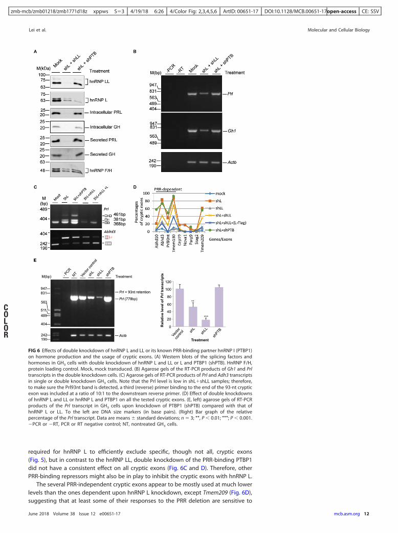
required for hnRNP L to efficiently exclude specific, though not all, cryptic exons(Fig. 5), but in contrast to the hnRNP LL, double knockdown of the PRR-binding PTBP1did not have a consistent effect on all cryptic exons (Fig. 6C and D). Therefore, otherPRR-binding repressors might also be in play to inhibit the cryptic exons with hnRNP L.
The several PRR-independent cryptic exons appear to be mostly used at much lowerlevels than the ones dependent upon hnRNP L knockdown, except Tmem209 (Fig. 6D),suggesting that at least some of their responses to the PRR deletion are sensitive to
FIG 6 Effects of double knockdown of hnRNP L and LL or its known PRR-binding partner hnRNP I (PTBP1)on hormone production and the usage of cryptic exons. (A) Western blots of the splicing factors andhormones in GH3 cells with double knockdown of hnRNP L and LL or L and PTBP1 (shPTB). HnRNP F/H,protein loading control. Mock, mock transduced. (B) Agarose gels of the RT-PCR products of Gh1 and Prltranscripts in the double knockdown cells. (C) Agarose gels of RT-PCR products of Prl and Adh3 transcriptsin single or double knockdown GH3 cells. Note that the Prl level is low in shL�shLL samples; therefore,to make sure the Prl93nt band is detected, a third (reverse) primer binding to the end of the 93-nt crypticexon was included at a ratio of 10:1 to the downstream reverse primer. (D) Effect of double knockdownsof hnRNP L and LL or hnRNP L and PTBP1 on all the tested cryptic exons. (E, left) agarose gels of RT-PCRproducts of the Prl transcript in GH3 cells upon knockdown of PTBP1 (shPTB) compared with that ofhnRNP L or LL. To the left are DNA size markers (in base pairs). (Right) Bar graph of the relativepercentage of the Prl transcript. Data are means � standard deviations; n � 3; **, P � 0.01; ***; P � 0.001.�PCR or �RT, PCR or RT negative control; NT, nontreated GH3 cells.
Lei et al. Molecular and Cellular Biology
June 2018 Volume 38 Issue 12 e00651-17 mcb.asm.org 12
COLOR
zmb-mcb/zmb01218/zmb1771d18z xppws S�3 4/19/18 6:26 4/Color Fig: 2,3,4,5,6 ArtID: 00651-17 DOI:10.1128/MCB.00651-17open-access CE: SSV
detection variations and thus are less likely to be significantly different between thewild type and PRR deletion mutant. We also examined their CA/AC contents, splicingtype (cassette or last exons), and potential PTBP1-binding motifs nearby the CA-richmotifs (found in Prl, Nova1, Cep19, and Pla2g6b; see Table S3 in the supplementalmaterial) and found mostly no correlation with their PRR dependence or independence,except for the Prl exon. The inhibition of the PRR-dependent Prl cryptic exon by hnRNPL could be due to its PRR domain interaction with PTBP1, but the inhibition of thesecryptic exons by hnRNP L and their PRR dependence or independence would needfurther investigation in regard to other features of hnRNP L unique domains (e.g., theNH2-terminal G-rich motif), other hnRNP L partners, more sequences, and preferablymore cryptic exon targets.
Besides the hnRNP L and LL proteins, we also observed differential effects of hnRNPA1 and its paralog, A2B1, in inhibiting cryptic exon sequences enriched in GGG, tripletsin the consensus motif of hnRNP A1 targets (61), from inclusion in the mRNA (unpub-lished data) by analyzing the intronic RNA-Seq reads of shRNA knockdown samplesbased on the raw sequences deposited in the NCBI SRA database by Geissler andcolleagues (62). Moreover, hnRNP C inhibits Alu repeats and PTBP1 and PTBP2 inhibitCU-rich sequences in the introns (7, 8), although their paralog differences on the targetsremain unclear. hnRNP LL also has a similar effect on intron retention (63). Such aninhibitory effect of hnRNP proteins on cryptic intronic sequences has also beensupported by other studies but more commonly in splicing reporters, for example, byhnRNP A1 (64). Together, the current pieces of evidence suggest that at least a groupof hnRNP proteins have diverged to differentially protect transcriptome integritybeyond their roles in the regulation of gene expression. The expanded differentproteins, including paralogs, thus may provide diverse trans-acting factors to recognizethe different sequence motifs (15, 65–67) to guarantee efficient inhibition of crypticexons inside long introns. We anticipate that more diverged hnRNPs or their paralogswith different sequence preferences will be identified to inhibit transcriptome-widecryptic exons in vertebrate cells as more and more cases of intron read mapping areapplied in future studies.
The multilevel control of genes by different hnRNPs in endocrine functions.The transcript level appears to be the major control for growth hormone and prolactinby both hnRNP L and LL. Overall, the genes regulated at this level clustered differen-tially for cation transport and guanyl nucleotide binding by the paralogs. At the exonlevel, the target genes also clustered differentially, for transcription factors and signal-ing by the hormone oxytocin (hnRNP L), and for cytoskeleton and nucleotide binding(hnRNP LL). Inhibition of cryptic exons is mainly an effect by hnRNP L in these cells; thiseffect is critical for the proper expression of many hormone- or hormone-related genes,including the prolactin (Fig. 3 and 4). Therefore, the multilevel differential control ofhormone gene expression programs by hnRNP L and LL covered ranges of molecularprocesses of endocrine functions involving at least growth hormone and prolactinproduction, cell signaling, excitability, vesicle transport, and gene transcription, as wellas the production of other hormones (Fig. 2 and 4). This differential control may serveto tailor and safeguard the transcriptome for specific endocrine functions. It would beinteresting to investigate whether this differential control also plays a role in theproduction of distinct hormones during the differentiation of the common precursorpituitary endocrine cells into lactotrophes and somatotrophes in the pituitary (32).
In summary, our findings demonstrate that the hnRNP L and LL proteins differen-tially control gene expression programs at multiple levels in the production of twophysiologically important hormones. Particularly interesting is their differential roles inglobally inhibiting the inclusion of cryptic intronic sequences in mRNA to protecttranscriptome integrity. The multilevel differential control by hnRNP proteins likelycontributes to the fine-tuning and protection of the gene expression programs and cellphysiology. These effects again emphasize the importance of investigating hnRNPderegulation and aberrant processing of RNA transcripts, particularly intronic se-quences (RNA-Seq reads), in diseases (68–70).
hnRNP L and LL in Hormone Gene Expression Programs Molecular and Cellular Biology
June 2018 Volume 38 Issue 12 e00651-17 mcb.asm.org 13
zmb-mcb/zmb01218/zmb1771d18z xppws S�3 4/19/18 6:26 4/Color Fig: 2,3,4,5,6 ArtID: 00651-17 DOI:10.1128/MCB.00651-17open-access CE: SSV
MATERIALS AND METHODSPlasmid construction. The lentiviral plasmids pFG12-shL (shL) and pLKO.1-shLL (shLL), against
hnRNP L and hnRNP LL, respectively, and pCppt2E-hnRNP L-Flag have been described previously (28, 29).To express hnRNP L-ΔP-Flag, the PRR-encoding fragment was deleted by PCR from hnRNP L-Flag andsubcloned into pCppt2E, as described by Yu et al. (28). Human hnRNP LL cDNA (with an open readingframe from GenBank accession number NM_138394.3; Open Biosystems) was cloned into pCMV-Myc (29)and then subcloned into pCppt2E (29).
A silent mutant of pCppt2E-Myc-hnRNP LL (T342A, G348T, and A351G) was generated by site-directed mutagenesis for rescue experiments based on Fisher’s procedure (71). Briefly, a 25-�l mutagen-esis PCR was carried out with 50 ng of cppt2E-Myc-hnRNP LL plasmid, complementary mutant primers,and 2 U of Phusion high-fidelity DNA polymerase (New England BioLabs, Inc.) for 18 cycles (95°C for 60s, 55°C for 60 s, and 68°C for 20 min [2 min per kilobase of sequence]). The PCR product was digestedwith 10 U of DpnI at 37°C for 1 h and transformed into Escherichia coli DH5�.
For Prl splicing reporter minigene Prl-1, a genomic DNA fragment of the 93-nt cryptic exon withpartial flanking introns (829 bp upstream and 374 bp downstream) was amplified using primers PRL-WT5(5=-CGGGCCCTGCTTTCTGCAATGAGGAAC-3=) and PRL-WT3 (5=-GAGTTGTGACCAAACCAAGTAG-3=) fromrat genomic DNA, digested with ApaI/BglII, and inserted into the DUP175 splicing vector (46). Furtherdeletions (Prl-2 or Prl-3) and mutations were made by PCR based on this template.
All resulting plasmids were confirmed by sequencing.Cell culture and transfection. GH3 cells were maintained in Ham’s F10 nutrient mixture with 10%
horse serum (HS), 2.5% fetal bovine serum (FBS), and 1% penicillin-streptomycin-glutamine solution(PSG). HEK293T human embryonic kidney cells were cultured in Dulbecco’s modified Eagle medium(DMEM) supplemented with 10% newborn calf serum (NCS) and 1% PSG. For minigene assays, thereporter plasmids were transfected with Lipofectamine 3000 (Invitrogen) according to the manufacturer’sprotocol.
Lentivirus transduction. Preparation of lentiviral particles for shRNA, hnRNP L-Flag, L-PRR deletionmutant (ΔP), or Myc-hnRNP LL expression and lentiviral transduction was done as described previously(28, 29).
RT-PCR. Cytoplasmic RNA was fractionated and extracted from GH3 cells with or without lentivirustransduction according to our previous procedures (28, 72). About 400 ng of cytoplasmic RNA was usedfor 10 �l of reverse transcription reaction mixture. PCR for gene expression and splicing analysis wasperformed for 28 to 31 cycles for different genes (28 cycles for Actb and Gh1, 30 cycles for Prl, and 31cycles for Prl splicing analysis). PCR products were resolved in 1.5 to 3.5% agarose gels containingethidium bromide (EtBr) and visualized with a digital camera under UV. The percentages of splice variantswere calculated from band intensities unless otherwise stated.
GH3 total RNA for RNA-Seq was extracted using an RNeasy Plus minikit (Qiagen) by following itsprotocol. Similar RT-PCR procedures were followed for validation, except about 500 ng of total RNA wasused for 10 �l of reverse transcription reaction mix.
Western blot analysis. Western blot analysis was performed according to reference 73, with slightmodifications. For cell lysates, approximately 10 �g of total protein or 5 �g of nuclear protein wasresolved in 10% SDS-PAGE. For secreted hormones, 7.5 �l of fresh medium (12 to 24 h after change) wasresolved in 12% SDS-PAGE. Proteins were transferred overnight to polyvinylidene difluoride (PVDF)membranes at 100 mA at 4°C, blocked with 5% fat-free milk for 1 h at room temperature, incubatedovernight with primary antibody at 4°C, and incubated with secondary antibody at room temperature for1 h. Proteins were detected by applying ECL Western blotting detection reagent and exposed to X-rayfilms. Anti-hnRNP L (4D11) and anti-hnRNP F/H (1G11) were purchased from Santa Cruz BiotechnologyCo., anti-hnRNP LL (4783) from Cell Signaling Technologies, anti-Flag (M2; F1804) from Sigma-Aldrich,anti-PRL (6F11) from Pierce Co., and anti-GH from the National Hormone and Peptide Program (NHPP;Harbor-UCLA Medical Centre).
For equal loading of culture media, Ponceau S staining after protein transfer and hnRNP F/H levelsof the same samples were monitored in experiments.
The index levels of the hormones were normalized to the loading and transduction controls.UV cross-linking and immunoprecipitation. The assay was performed with [�-32P]-CTP labeled RNA
probes and HeLa nuclear extracts as described previously (28). RNA probes were in vitro transcribed usingT7 RNA polymerase from DNA templates (5=-TGTATGTGTGTTTAATCTGATGTGTACAGTTGTctgaaaattacaaaaatatagcatgcatttgcataccctatagtgagtcgtatta-3=, WT, with the reverse complementary sequence of the T7promoter underlined) preannealed with equal molar amounts of T7 promoter oligonucleotide. The finalWT RNA transcript sequence is uaugcaaaugcaugcuauauuuuuguaauuuucagACAACUGUACACAUCAGAUUAAACACACAUACA, with the CA/AC clusters in boldface. Anti-hnRNP L (4D11) was used for immuno-precipitation.
RNA-Seq and bioinformatics analyses. Approximately 3 �g of total RNA from samples of the threegroups, mock (nontransduced; treated with 0.8 ng/�l Polybrene), shL, and shLL (samples in triplicate ineach group), were reverse transcribed using oligo(dT) for cDNA library construction and IlluminaHi-Seq2000 sequencing. RNA quality control, library preparation, and Illumina Hi-Seq 2000 paired-end100-bp sequencing were conducted at the McGill University and Génome Québec Innovation Centre(Montréal, Québec, Canada).
An average of 189 � 42 million paired reads were mapped to genome assembly Rnor_5.0 for 15,169 �247 genes per group. The differential gene expression and aberrant intronic splicing were calculated inthe Bioconductor package edgeR (74, 75). Alternative exon usage was calculated in the package DEXSeq(37), with a validated filter developed in our laboratory. Basically, we filtered out those bins with
Lei et al. Molecular and Cellular Biology
June 2018 Volume 38 Issue 12 e00651-17 mcb.asm.org 14
AQ: F
AQ: G
zmb-mcb/zmb01218/zmb1771d18z xppws S�3 4/19/18 6:26 4/Color Fig: 2,3,4,5,6 ArtID: 00651-17 DOI:10.1128/MCB.00651-17open-access CE: SSV
exonbase means and average exonbase means of less than 50, with a ratio to the highest exonbase mean(after normalized to bin length) of less than 0.002. To visualize the mapped reads of genes/exons, weused the Integrative Genomics Viewer (IGV; developed at the Broad Institute of MIT and Harvard;http://www.broadinstitute.org/igv) (76). The cryptic exons were visually examined in IGV, with thecassette exons characterized by the steep drop of read peaks accompanying the splice site dinucleotidesGT/AG and the alternative 3=-end exons by the long length (mostly �300 nt), with read peaks slowlydeclining to zero or baseline at exon ends, no obvious downstream 5=SS GT dinucleotides, and thepresence of AAUAAA or AUUAAA motifs (77, 78).
For functional annotation and classification of genes, we used the Database for Annotation, Visual-ization, and Integrated Discovery (DAVID; developed at the U.S. National Institute of Allergy andInfectious Diseases; http://david.abcc.ncifcrf.gov) (79).
Image data analysis. Images were quantified with the Image J software (developed at the U.S.National Institutes of Health; http://rsb.info.nih.gov/ij/) (80).
Statistical analysis. Data are presented as means � standard errors of the means for at least threeindependent experiments. Statistical analysis was done with two-tailed unpaired Student’s t test, exceptfor the built-in tests in DAVID (modified Fisher’s exact test) or Bioconductor packages.
SUPPLEMENTAL MATERIAL
Supplemental material for this article may be found at https://doi.org/10.1128/MCB.00651-17.
SUPPLEMENTAL FILE 1, XLSX file, 0.1 MB.SUPPLEMENTAL FILE 2, XLSX file, 0.4 MB.SUPPLEMENTAL FILE 3, XLSX file, 0.1 MB.
ACKNOWLEDGMENTSWe are grateful to Dairong Feng, Peisan Lew, Nan Liang, Hana Vakili, Robert Shiu,
Manli Zhang, Sam Kung, Tooru Mizuno, and Yvonne Myal for technical help or sug-gestions, the Harbor-UCLA Medical Centre for the kind supply of anti-GH, and Ruth Xiefor editing the manuscript. We thank Haihong Shen for critical comments on themanuscript.
The work was supported by the Canadian Institutes of Health Research (FRN106608),a Manitoba Research Chair fund, and in part by National Sciences and EngineeringResearch Council of Canada (NSERC) to J.X.
REFERENCES1. Dreyfuss G, Kim VN, Kataoka N. 2002. Messenger-RNA-binding proteins
and the messages they carry. Nat Rev Mol Cell Biol 3:195–205. https://doi.org/10.1038/nrm760.
2. Dreyfuss G, Matunis MJ, Pinol-Roma S, Burd CG. 1993. hnRNP proteinsand the biogenesis of mRNA. Annu Rev Biochem 62:289 –321. https://doi.org/10.1146/annurev.bi.62.070193.001445.
3. Busch A, Hertel KJ. 2012. Evolution of SR protein and hnRNP splicingregulatory factors. Wiley Interdiscip Rev RNA 3:1–12. https://doi.org/10.1002/wrna.100.
4. Martinez-Contreras R, Cloutier P, Shkreta L, Fisette JF, Revil T, Chabot B.2007. hnRNP proteins and splicing control. Adv Exp Med Biol 623:123–147. https://doi.org/10.1007/978-0-387-77374-2_8.
5. Han SP, Tang YH, Smith R. 2010. Functional diversity of the hnRNPs: past,present and perspectives. Biochem J 430:379 –392. https://doi.org/10.1042/BJ20100396.
6. Pagani F, Baralle FE. 2004. Genomic variants in exons and introns:identifying the splicing spoilers. Nat Rev Genet 5:389 –396. https://doi.org/10.1038/nrg1327.
7. Zarnack K, Konig J, Tajnik M, Martincorena I, Eustermann S, Stevant I,Reyes A, Anders S, Luscombe NM, Ule J. 2013. Direct competitionbetween hnRNP C and U2AF65 protects the transcriptome from theexonization of Alu elements. Cell 152:453– 466. https://doi.org/10.1016/j.cell.2012.12.023.
8. Ling JP, Chhabra R, Merran JD, Schaughency PM, Wheelan SJ, Corden JL,Wong PC. 2016. PTBP1 and PTBP2 repress nonconserved cryptic exons.Cell Rep 17:104 –113. https://doi.org/10.1016/j.celrep.2016.08.071.
9. Ling JP, Pletnikova O, Troncoso JC, Wong PC. 2015. TDP-43 repression ofnonconserved cryptic exons is compromised in ALS-FTD. Science 349:650 – 655. https://doi.org/10.1126/science.aab0983.
10. Clower CV, Chatterjee D, Wang Z, Cantley LC, Vander Heiden MG,Krainer AR. 2010. The alternative splicing repressors hnRNP A1/A2
and PTB influence pyruvate kinase isoform expression and cell me-tabolism. Proc Natl Acad Sci U S A 107:1894 –1899. https://doi.org/10.1073/pnas.0914845107.
11. Garneau D, Revil T, Fisette JF, Chabot B. 2005. Heterogeneous nuclearribonucleoprotein F/H proteins modulate the alternative splicing of theapoptotic mediator Bcl-x. J Biol Chem 280:22641–22650. https://doi.org/10.1074/jbc.M501070200.
12. Makeyev EV, Zhang J, Carrasco MA, Maniatis T. 2007. The microRNAmiR-124 promotes neuronal differentiation by triggering brain-specificalternative pre-mRNA splicing. Mol Cell 27:435– 448. https://doi.org/10.1016/j.molcel.2007.07.015.
13. Boutz PL, Stoilov P, Li Q, Lin CH, Chawla G, Ostrow K, Shiue L, Ares M,Jr, Black DL. 2007. A post-transcriptional regulatory switch in poly-pyrimidine tract-binding proteins reprograms alternative splicing indeveloping neurons. Genes Dev 21:1636 –1652. https://doi.org/10.1101/gad.1558107.
14. Woolaway K, Asai K, Emili A, Cochrane A. 2007. hnRNP E1 and E2 havedistinct roles in modulating HIV-1 gene expression. Retrovirology 4:28.https://doi.org/10.1186/1742-4690-4-28.
15. Xie J. 2014. Differential evolution of signal-responsive RNA elements andupstream factors that control alternative splicing. Cell Mol Life Sci71:4347– 4360. https://doi.org/10.1007/s00018-014-1688-y.
16. Hui J, Hung LH, Heiner M, Schreiner S, Neumuller N, Reither G, Haas SA,Bindereif A. 2005. Intronic CA-repeat and CA-rich elements: a new classof regulators of mammalian alternative splicing. EMBO J 24:1988 –1998.https://doi.org/10.1038/sj.emboj.7600677.
17. Smith SA, Ray D, Cook KB, Mallory MJ, Hughes TR, Lynch KW. 2013.Paralogs hnRNP L and hnRNP LL exhibit overlapping but distinct RNAbinding constraints. PLoS One 8:e80701. https://doi.org/10.1371/journal.pone.0080701.
18. Oberdoerffer S, Moita LF, Neems D, Freitas RP, Hacohen N, Rao A. 2008.
hnRNP L and LL in Hormone Gene Expression Programs Molecular and Cellular Biology
June 2018 Volume 38 Issue 12 e00651-17 mcb.asm.org 15
zmb-mcb/zmb01218/zmb1771d18z xppws S�3 4/19/18 6:26 4/Color Fig: 2,3,4,5,6 ArtID: 00651-17 DOI:10.1128/MCB.00651-17open-access CE: SSV
Regulation of CD45 alternative splicing by heterogeneous ribonucleo-protein, hnRNP LL. Science 321:686 – 691. https://doi.org/10.1126/science.1157610.
19. Chang X, Li B, Rao A. 2015. RNA-binding protein hnRNPLL regulatesmRNA splicing and stability during B-cell to plasma-cell differentiation.Proc Natl Acad Sci U S A 112:E1888 –E1897. https://doi.org/10.1073/pnas.1422490112.
20. Chang X. 2016. RNA-binding protein hnRNPLL as a critical regulator oflymphocyte homeostasis and differentiation. Wiley Interdiscip Rev RNA7:295–302. https://doi.org/10.1002/wrna.1335.
21. Hung LH, Heiner M, Hui J, Schreiner S, Benes V, Bindereif A. 2008. Diverseroles of hnRNP L in mammalian mRNA processing: a combined microar-ray and RNAi analysis. RNA 14:284 –296. https://doi.org/10.1261/rna.725208.
22. Shankarling G, Cole BS, Mallory MJ, Lynch KW. 2014. Transcriptome-wideRNA interaction profiling reveals physical and functional targets ofhnRNP L in human T cells. Mol Cell Biol 34:71– 83. https://doi.org/10.1128/MCB.00740-13.
23. Huang Y, Li W, Yao X, Lin QJ, Yin JW, Liang Y, Heiner M, Tian B, Hui J,Wang G. 2012. Mediator complex regulates alternative mRNA processingvia the MED23 subunit. Mol Cell 45:459 – 469. https://doi.org/10.1016/j.molcel.2011.12.022.
24. Gaudreau MC, Grapton D, Helness A, Vadnais C, Fraszczak J, Shooshta-rizadeh P, Wilhelm B, Robert F, Heyd F, Moroy T. 2016. Heterogeneousnuclear ribonucleoprotein L is required for the survival and functionalintegrity of murine hematopoietic stem cells. Sci Rep 6:27379. https://doi.org/10.1038/srep27379.
25. Gaudreau MC, Heyd F, Bastien R, Wilhelm B, Moroy T. 2012. Alternativesplicing controlled by heterogeneous nuclear ribonucleoprotein L reg-ulates development, proliferation, migration of thymic pre-T cells. JImmunol 188:5377–5388. https://doi.org/10.4049/jimmunol.1103142.
26. Loh TJ, Cho S, Moon H, Jang HN, Williams DR, Jung DW, Kim IC, GhignaC, Biamonti G, Zheng X, Shen H. 2015. hnRNP L inhibits CD44 V10 exonsplicing through interacting with its upstream intron. Biochim BiophysActa 1 849:743–750. https://doi.org/10.1016/j.bbagrm.2015.01.004.
27. Rossbach O, Hung LH, Khrameeva E, Schreiner S, Konig J, Curk T, Zupan B,Ule J, Gelfand MS, Bindereif A. 2014. Crosslinking-immunoprecipitation(iCLIP) analysis reveals global regulatory roles of hnRNP L. RNA Biol 11:146–155. https://doi.org/10.4161/rna.27991.
28. Yu J, Hai Y, Liu G, Fang T, Kung SK, Xie J. 2009. The heterogeneousnuclear ribonucleoprotein L is an essential component in the Ca2�/calmodulin-dependent protein kinase IV-regulated alternative splic-ing through cytidine-adenosine repeats. J Biol Chem 284:1505–1513.https://doi.org/10.1074/jbc.M805113200.
29. Liu G, Razanau A, Hai Y, Yu J, Sohail M, Lobo VG, Chu J, Kung SK, Xie J.2012. A conserved serine of heterogeneous nuclear ribonucleoprotein L(hnRNP L) mediates depolarization-regulated alternative splicing of po-tassium channels. J Biol Chem 287:22709 –22716. https://doi.org/10.1074/jbc.M112.357343.
30. Liu G, Lei L, Yu J, Kung S, Xie J. 2014. Refinement of the spectra of exonusage by combined effects of extracellular stimulus and intracellularfactors. Biochim Biophys Acta 1 839:537–545. https://doi.org/10.1016/j.bbagrm.2014.05.002.
31. Tashjian AH, Jr, Bancroft FC, Levine L. 1970. Production of both prolactinand growth hormone by clonal strains of rat pituitary tumor cells.Differential effects of hydrocortisone and tissue extracts. J Cell Biol47:61–70.
32. Simmons DM, Voss JW, Ingraham HA, Holloway JM, Broide RS, RosenfeldMG, Swanson LW. 1990. Pituitary cell phenotypes involve cell-specificPit-1 mRNA translation and synergistic interactions with other classes oftranscription factors. Genes Dev 4:695–711. https://doi.org/10.1101/gad.4.5.695.
33. Mullis PE. 2010. Genetics of isolated growth hormone deficiency. J ClinRes Pediatr Endocrinol 2:52– 62. https://doi.org/10.4274/jcrpe.v2i2.52.
34. Serri O, Chik CL, Ur E, Ezzat S. 2003. Diagnosis and management ofhyperprolactinemia. CMAJ 169:575–581.
35. Zargar AH, Masoodi SR, Laway BA, Shah NA, Salahudin M. 1997. Familialpuerperal alactogenesis: possibility of a genetically transmitted isolatedprolactin deficiency. Br J Obstet Gynaecol 104:629 – 631. https://doi.org/10.1111/j.1471-0528.1997.tb11548.x.
36. Goffin V, Binart N, Touraine P, Kelly PA. 2002. Prolactin: the new biologyof an old hormone. Annu Rev Physiol 64:47– 67. https://doi.org/10.1146/annurev.physiol.64.081501.131049.
37. Anders S, Reyes A, Huber W. 2012. Detecting differential usage of exons
from RNA-seq data. Genome Res 22:2008 –2017. https://doi.org/10.1101/gr.133744.111.
38. Yeo G, Burge CB. 2004. Maximum entropy modeling of short sequencemotifs with applications to RNA splicing signals. J Comput Biol 11:377–394. https://doi.org/10.1089/1066527041410418.
39. Karolchik D, Barber GP, Casper J, Clawson H, Cline MS, Diekhans M,Dreszer TR, Fujita PA, Guruvadoo L, Haeussler M, Harte RA, Heitner S,Hinrichs AS, Learned K, Lee BT, Li CH, Raney BJ, Rhead B, Rosenbloom KR,Sloan CA, Speir ML, Zweig AS, Haussler D, Kuhn RM, Kent WJ. 2014. TheUCSC Genome Browser database: 2014 update. Nucleic Acids Res 42:D764 –D770. https://doi.org/10.1093/nar/gkt1168.
40. Broutin I, Jomain JB, Tallet E, van Agthoven J, Raynal B, Hoos S, Krage-lund BB, Kelly PA, Ducruix A, England P, Goffin V. 2010. Crystal structureof an affinity-matured prolactin complexed to its dimerized receptorreveals the topology of hormone binding site 2. J Biol Chem 285:8422– 8433. https://doi.org/10.1074/jbc.M109.089128.
41. Biasini M, Bienert S, Waterhouse A, Arnold K, Studer G, Schmidt T, KieferF, Gallo Cassarino T, Bertoni M, Bordoli L, Schwede T. 2014. SWISS-MODEL: modelling protein tertiary and quaternary structure using evo-lutionary information. Nucleic Acids Res 42:W252–W258. https://doi.org/10.1093/nar/gku340.
42. Nagy E, Maquat LE. 1998. A rule for termination-codon position withinintron-containing genes: when nonsense affects RNA abundance. TrendsBiochem Sci 23:198–199. https://doi.org/10.1016/S0968-0004(98)01208-0.
43. Isken O, Maquat LE. 2007. Quality control of eukaryotic mRNA: safe-guarding cells from abnormal mRNA function. Genes Dev 21:1833–1856.https://doi.org/10.1101/gad.1566807.
44. Schoenberg DR, Maquat LE. 2012. Regulation of cytoplasmic mRNAdecay. Nat Rev Genet 13:246 –259. https://doi.org/10.1038/nrg3160.
45. Dominski Z, Kole R. 1991. Selection of splice sites in pre-mRNAs withshort internal exons. Mol Cell Biol 11:6075– 6083. https://doi.org/10.1128/MCB.11.12.6075.
46. Xie JY, Black DL. 2001. A CaMK IV responsive RNA element mediatesdepolarization-induced alternative splicing of ion channels. Nature 410:936 –939. https://doi.org/10.1038/35073593.
47. Berg MG, Singh LN, Younis I, Liu Q, Pinto AM, Kaida D, Zhang Z, Cho S,Sherrill-Mix S, Wan L, Dreyfuss G. 2012. U1 snRNP determines mRNAlength and regulates isoform expression. Cell 150:53– 64. https://doi.org/10.1016/j.cell.2012.05.029.
48. Flavell SW, Kim TK, Gray JM, Harmin DA, Hemberg M, Hong EJ,Markenscoff-Papadimitriou E, Bear DM, Greenberg ME. 2008. Genome-wide analysis of MEF2 transcriptional program reveals synaptic targetgenes and neuronal activity-dependent polyadenylation site selection.Neuron 60:1022–1038. https://doi.org/10.1016/j.neuron.2008.11.029.
49. Mayr C, Bartel DP. 2009. Widespread shortening of 3=UTRs by alternativecleavage and polyadenylation activates oncogenes in cancer cells. Cell138:673– 684. https://doi.org/10.1016/j.cell.2009.06.016.
50. Sandberg R, Neilson JR, Sarma A, Sharp PA, Burge CB. 2008. Proliferatingcells express mRNAs with shortened 3= untranslated regions and fewermicroRNA target sites. Science 320:1643–1647. https://doi.org/10.1126/science.1155390.
51. Ishigaki Y, Li X, Serin G, Maquat LE. 2001. Evidence for a pioneer roundof mRNA translation: mRNAs subject to nonsense-mediated decay inmammalian cells are bound by CBP80 and CBP20. Cell 106:607– 617.https://doi.org/10.1016/S0092-8674(01)00475-5.
52. Fujishige K, Kotera J, Michibata H, Yuasa K, Takebayashi S, Okumura K,Omori K. 1999. Cloning and characterization of a novel human phos-phodiesterase that hydrolyzes both cAMP and cGMP (PDE10A). J BiolChem 274:18438 –18445. https://doi.org/10.1074/jbc.274.26.18438.
53. Tisdale EJ. 2000. Rab2 requires PKC iota/lambda to recruit beta-COP forvesicle formation. Traffic 1:702–712. https://doi.org/10.1034/j.1600-0854.2000.010903.x.
54. Kretschmannova K, Gonzalez-Iglesias AE, Tomic M, Stojilkovic SS. 2006.Dependence of hyperpolarisation-activated cyclic nucleotide-gatedchannel activity on basal cyclic adenosine monophosphate productionin spontaneously firing GH3 cells. J Neuroendocrinol 18:484 – 493.https://doi.org/10.1111/j.1365-2826.2006.01438.x.
55. Calejo AI, Jorgacevski J, Rituper B, Gucek A, Pereira PM, Santos MA,Potokar M, Vardjan N, Kreft M, Goncalves PP, Zorec R. 2014.Hyperpolarization-activated cyclic nucleotide-gated channels andcAMP-dependent modulation of exocytosis in cultured rat lactotrophs. JNeurosci 34:15638 –15647. https://doi.org/10.1523/JNEUROSCI.5290-13.2014.
56. Kucka M, Bjelobaba I, Tomic M, Stojilkovic SS. 2013. The role of cyclic
Lei et al. Molecular and Cellular Biology
June 2018 Volume 38 Issue 12 e00651-17 mcb.asm.org 16
zmb-mcb/zmb01218/zmb1771d18z xppws S�3 4/19/18 6:26 4/Color Fig: 2,3,4,5,6 ArtID: 00651-17 DOI:10.1128/MCB.00651-17open-access CE: SSV
nucleotides in pituitary lactotroph functions. Front Endocrinol (Laus-anne) 4:122.
57. Rahman MA, Masuda A, Ohe K, Ito M, Hutchinson DO, Mayeda A, EngelAG, Ohno K. 2013. hnRNP L and hnRNP LL antagonistically modulatePTB-mediated splicing suppression of CHRNA1 pre-mRNA. Sci Rep3:2931. https://doi.org/10.1038/srep02931.
58. Rossbach O, Hung LH, Schreiner S, Grishina I, Heiner M, Hui J, BindereifA. 2009. Auto- and cross-regulation of the hnRNP L proteins by alterna-tive splicing. Mol Cell Biol 29:1442–1451. https://doi.org/10.1128/MCB.01689-08.
59. Hui J, Reither G, Bindereif A. 2003. Novel functional role of CA repeatsand hnRNP L in RNA stability. RNA 9:931–936. https://doi.org/10.1261/rna.5660803.
60. Hahm B, Cho OH, Kim JE, Kim YK, Kim JH, Oh YL, Jang SK. 1998.Polypyrimidine tract-binding protein interacts with hnRNP L. FEBS Lett425:401– 406. https://doi.org/10.1016/S0014-5793(98)00269-5.
61. Burd CG, Dreyfuss G. 1994. RNA binding specificity of hnRNP A1: signif-icance of hnRNP A1 high-affinity binding sites in pre-mRNA splicing.EMBO J 13:1197–1204.
62. Geissler R, Simkin A, Floss D, Patel R, Fogarty EA, Scheller J, Grimson A.2016. A widespread sequence-specific mRNA decay pathway mediatedby hnRNPs A1 and A2/B1. Genes Dev 30:1070 –1085. https://doi.org/10.1101/gad.277392.116.
63. Cho V, Mei Y, Sanny A, Chan S, Enders A, Bertram EM, Tan A, GoodnowCC, Andrews TD. 2014. The RNA-binding protein hnRNPLL induces a Tcell alternative splicing program delineated by differential intron reten-tion in polyadenylated RNA. Genome Biol 15:R26. https://doi.org/10.1186/gb-2014-15-1-r26.
64. Pastor T, Pagani F. 2011. Interaction of hnRNPA1/A2 and DAZAP1 withan Alu-derived intronic splicing enhancer regulates ATM aberrant splic-ing. PLoS One 6:e23349. https://doi.org/10.1371/journal.pone.0023349.
65. Sohail M, Xie J. 2015. Evolutionary emergence of a novel splice variantwith an opposite effect on the cell cycle. Mol Cell Biol 35:2203–2214.https://doi.org/10.1128/MCB.00190-15.
66. Sohail M, Cao W, Mahmood N, Myschyshyn M, Hong SP, Xie J. 2014.Evolutionarily emerged G tracts between the polypyrimidine tract and 3=AG are splicing silencers enriched in genes involved in cancer. BMCGenomics 15:1143. https://doi.org/10.1186/1471-2164-15-1143.
67. Yeo G, Hoon S, Venkatesh B, Burge CB. 2004. Variation in sequence andorganization of splicing regulatory elements in vertebrate genes. Proc
Natl Acad Sci U S A 101:15700 –15705. https://doi.org/10.1073/pnas.0404901101.
68. Chabot B, Shkreta L. 2016. Defective control of pre-messenger RNAsplicing in human disease. J Cell Biol 212:13–27. https://doi.org/10.1083/jcb.201510032.
69. Zhang J, Manley JL. 2013. Misregulation of pre-mRNA alternative splicingin cancer. Cancer Discov 3:1228 –1237. https://doi.org/10.1158/2159-8290.CD-13-0253.
70. Feng D, Xie J. 2013. Aberrant splicing in neurological diseases. WileyInterdiscip Rev RNA 4:631– 649. https://doi.org/10.1002/wrna.1184.
71. Fisher CL, Pei GK. 1997. Modification of a PCR-based site-directed mu-tagenesis method. Biotechniques 23:570 –574.
72. Ma S, Liu G, Sun Y, Xie J. 2007. Relocalization of the polypyrimidinetract-binding protein during PKA-induced neurite growth. Biochim Bio-phys Acta 1 773:912–923. https://doi.org/10.1016/j.bbamcr.2007.02.006.
73. Xie J, Lee JA, Kress TL, Mowry KL, Black DL. 2003. Protein kinase Aphosphorylation modulates transport of the polypyrimidine tract-binding protein. Proc Natl Acad Sci U S A 100:8776 – 8781. https://doi.org/10.1073/pnas.1432696100.
74. Robinson MD, McCarthy DJ, Smyth GK. 2010. edgeR: a Bioconductor pack-age for differential expression analysis of digital gene expression data.Bioinformatics 26:139–140. https://doi.org/10.1093/bioinformatics/btp616.
75. McCarthy DJ, Chen Y, Smyth GK. 2012. Differential expression analysis ofmultifactor RNA-Seq experiments with respect to biological variation.Nucleic Acids Res 40:4288 – 4297. https://doi.org/10.1093/nar/gks042.
76. Robinson JT, Thorvaldsdottir H, Winckler W, Guttman M, Lander ES, GetzG, Mesirov JP. 2011. Integrative genomics viewer. Nat Biotechnol 29:24 –26. https://doi.org/10.1038/nbt.1754.
77. Colgan DF, Manley JL. 1997. Mechanism and regulation of mRNA poly-adenylation. Genes Dev 11:2755–2766. https://doi.org/10.1101/gad.11.21.2755.
78. Bhat BM, Wold WS. 1985. ATTAAA as well as downstream sequences arerequired for RNA 3=-end formation in the E3 complex transcription unitof adenovirus. Mol Cell Biol 5:3183–3193. https://doi.org/10.1128/MCB.5.11.3183.
79. Huang DW, Sherman BT, Lempicki RA. 2009. Systematic and integrativeanalysis of large gene lists using DAVID bioinformatics resources. NatProtoc 4:44 –57.
80. Schneider CA, Rasband WS, Eliceiri KW. 2012. NIH Image to ImageJ: 25years of image analysis. Nat Methods 9:671– 675. https://doi.org/10.1038/nmeth.2089.
hnRNP L and LL in Hormone Gene Expression Programs Molecular and Cellular Biology
June 2018 Volume 38 Issue 12 e00651-17 mcb.asm.org 17
zmb-mcb/zmb01218/zmb1771d18z xppws S�3 4/19/18 6:26 4/Color Fig: 2,3,4,5,6 ArtID: 00651-17 DOI:10.1128/MCB.00651-17open-access CE: SSV
JOBNAME: AUTHOR QUERIES PAGE: 1 SESS: 2 OUTPUT: Thu Apr 19 06:26:32 2018/rich4/zmb-mcb/zmb-mcb/zmb01218/zmb1771d18z
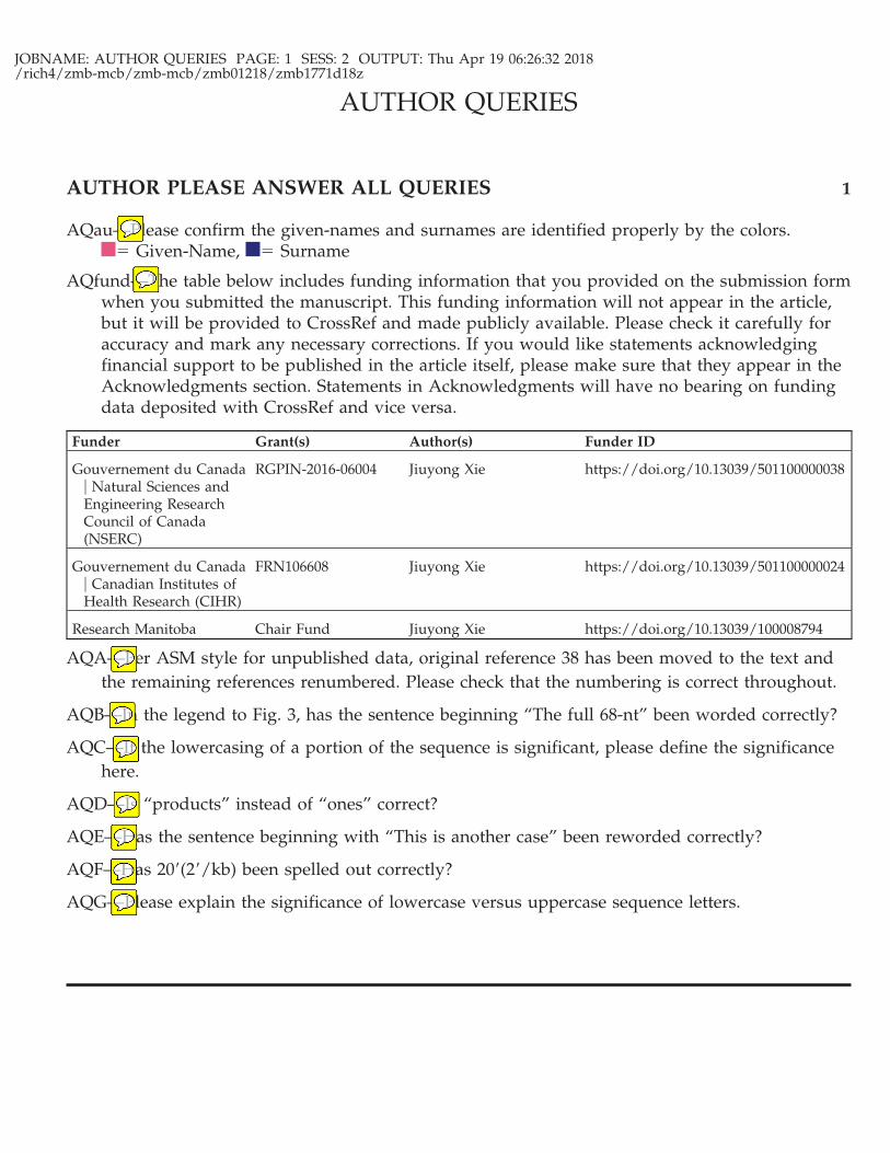
AQau—Please confirm the given-names and surnames are identified properly by the colors.� Given-Name, � Surname
AQfund—The table below includes funding information that you provided on the submission formwhen you submitted the manuscript. This funding information will not appear in the article,but it will be provided to CrossRef and made publicly available. Please check it carefully foraccuracy and mark any necessary corrections. If you would like statements acknowledgingfinancial support to be published in the article itself, please make sure that they appear in theAcknowledgments section. Statements in Acknowledgments will have no bearing on fundingdata deposited with CrossRef and vice versa.
Funder Grant(s) Author(s) Funder ID
Gouvernement du Canada� Natural Sciences andEngineering ResearchCouncil of Canada(NSERC)
RGPIN-2016-06004 Jiuyong Xie https://doi.org/10.13039/501100000038
Gouvernement du Canada� Canadian Institutes ofHealth Research (CIHR)
FRN106608 Jiuyong Xie https://doi.org/10.13039/501100000024
Research Manitoba Chair Fund Jiuyong Xie https://doi.org/10.13039/100008794
AQA—Per ASM style for unpublished data, original reference 38 has been moved to the text andthe remaining references renumbered. Please check that the numbering is correct throughout.
AQB—In the legend to Fig. 3, has the sentence beginning “The full 68-nt” been worded correctly?
AQC—If the lowercasing of a portion of the sequence is significant, please define the significancehere.
AQD—Is “products” instead of “ones” correct?
AQE—Has the sentence beginning with “This is another case” been reworded correctly?
AQF—Has 20=(2=/kb) been spelled out correctly?
AQG—Please explain the significance of lowercase versus uppercase sequence letters.
AUTHOR QUERIES
AUTHOR PLEASE ANSWER ALL QUERIES 1


![A Fitting Model for Feature Selection with Fuzzy Rough Setsyuhuaqian.net/Cms_Data/Contents/SXU_YHQ/Folders/JournalPapers/… · Greedy searching [20], [23], genetic algorithms [32],](https://img.pdfslide.us/doc/110x75/5fdb7db4840ac37d8139a8fe/a-fitting-model-for-feature-selection-with-fuzzy-rough-greedy-searching-20-23.jpg)


![[PPT]PowerPoint Presentation - University of Winnipegion.uwinnipeg.ca/~ychen2/access/ch1.ppt · Web viewExploring Microsoft Access 2003 Chapter 1 Introduction to Microsoft Access:](https://img.pdfslide.us/doc/110x75/5aa21ccb7f8b9ac67a8caf6f/pptpowerpoint-presentation-university-of-ychen2accessch1pptweb-viewexploring.jpg)