Embed Size (px)
Citation preview
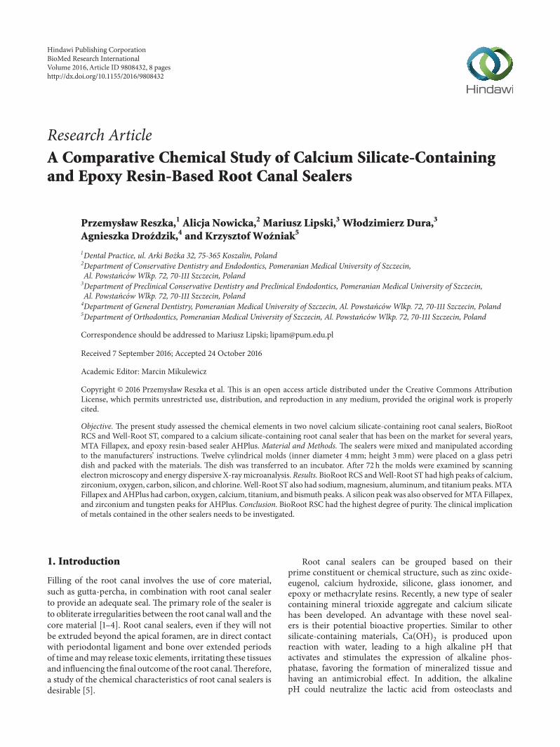
Research ArticleA Comparative Chemical Study of Calcium Silicate-Containingand Epoxy Resin-Based Root Canal Sealers
PrzemysBaw Reszka,1 Alicja Nowicka,2 Mariusz Lipski,3 WBodzimierz Dura,3
Agnieszka Drofdzik,4 and Krzysztof Wofniak5
1Dental Practice, ul. Arki Bozka 32, 75-365 Koszalin, Poland2Department of Conservative Dentistry and Endodontics, Pomeranian Medical University of Szczecin,Al. Powstancow Wlkp. 72, 70-111 Szczecin, Poland3Department of Preclinical Conservative Dentistry and Preclinical Endodontics, Pomeranian Medical University of Szczecin,Al. Powstancow Wlkp. 72, 70-111 Szczecin, Poland4Department of General Dentistry, Pomeranian Medical University of Szczecin, Al. Powstancow Wlkp. 72, 70-111 Szczecin, Poland5Department of Orthodontics, Pomeranian Medical University of Szczecin, Al. Powstancow Wlkp. 72, 70-111 Szczecin, Poland
Correspondence should be addressed to Mariusz Lipski; [email protected]
Received 7 September 2016; Accepted 24 October 2016
Academic Editor: Marcin Mikulewicz
Copyright © 2016 Przemysław Reszka et al. This is an open access article distributed under the Creative Commons AttributionLicense, which permits unrestricted use, distribution, and reproduction in any medium, provided the original work is properlycited.
Objective. The present study assessed the chemical elements in two novel calcium silicate-containing root canal sealers, BioRootRCS and Well-Root ST, compared to a calcium silicate-containing root canal sealer that has been on the market for several years,MTA Fillapex, and epoxy resin-based sealer AHPlus. Material and Methods. The sealers were mixed and manipulated accordingto the manufacturers’ instructions. Twelve cylindrical molds (inner diameter 4mm; height 3mm) were placed on a glass petridish and packed with the materials. The dish was transferred to an incubator. After 72 h the molds were examined by scanningelectronmicroscopy and energy dispersive X-raymicroanalysis.Results.BioRoot RCS andWell-Root ST had high peaks of calcium,zirconium, oxygen, carbon, silicon, and chlorine.Well-Root ST also had sodium,magnesium, aluminum, and titaniumpeaks.MTAFillapex andAHPlus had carbon, oxygen, calcium, titanium, and bismuth peaks. A silicon peakwas also observed forMTAFillapex,and zirconium and tungsten peaks for AHPlus. Conclusion. BioRoot RSC had the highest degree of purity. The clinical implicationof metals contained in the other sealers needs to be investigated.
1. Introduction
Filling of the root canal involves the use of core material,such as gutta-percha, in combination with root canal sealerto provide an adequate seal. The primary role of the sealer isto obliterate irregularities between the root canal wall and thecore material [1–4]. Root canal sealers, even if they will notbe extruded beyond the apical foramen, are in direct contactwith periodontal ligament and bone over extended periodsof time andmay release toxic elements, irritating these tissuesand influencing the final outcome of the root canal.Therefore,a study of the chemical characteristics of root canal sealers isdesirable [5].
Root canal sealers can be grouped based on theirprime constituent or chemical structure, such as zinc oxide-eugenol, calcium hydroxide, silicone, glass ionomer, andepoxy or methacrylate resins. Recently, a new type of sealercontaining mineral trioxide aggregate and calcium silicatehas been developed. An advantage with these novel seal-ers is their potential bioactive properties. Similar to othersilicate-containing materials, Ca(OH)
2is produced upon
reaction with water, leading to a high alkaline pH thatactivates and stimulates the expression of alkaline phos-phatase, favoring the formation of mineralized tissue andhaving an antimicrobial effect. In addition, the alkalinepH could neutralize the lactic acid from osteoclasts and
Hindawi Publishing CorporationBioMed Research InternationalVolume 2016, Article ID 9808432, 8 pageshttp://dx.doi.org/10.1155/2016/9808432
2 BioMed Research International
prevent dissolution of the mineralized components of teeth[6–9].
One of the first mineral trioxide aggregate-containingroot canal sealers introduced on the market was MTAFillapex (Angelus, Londrina, Brazil). Because it has beenavailable for 5 years, it is the most studied MTA-containingroot canal sealer. MTA Fillapex is a paste-catalyst material.Paste A is composed of salicylate resin (methyl salicylate,butylene glycol, and colophony), bismuth oxide, and silica.Paste B includes silicon dioxide, titanium dioxide, and baseresin (pentaerythritol, rosinate, and toluene sulphonamide),and 13.2% set MTA particles as filler. The working time is23min, with a complete set time of approximately 2 h. Severalproperties of this root canal sealer, such as flow and viscosity[10, 11], dimensional change [11], material porosity [12, 13],sealing ability [11], radiopacity, electrical conductivity [14],antibacterial effect [15], biocompatibility [16–20], cytotoxicity[19, 21–23], and genotoxicity [24, 25], have been investigated.
Another recently introduced sealer based on tricalciumsilicate is Well-Root ST (Vericom, Gangwon-Do, Korea).This sealer is a premixed, ready-to-use, injectable bioceramiccement paste developed for permanent obturation of theroot canal. The composition of Well-Root as described bythe manufacturer includes zirconium oxide, calcium silicate,filler, and thickening agents [26]. The material is hydrophilicand usesmoisture in dentinal tubules to initiate and completeits setting reactions. The setting time is 25min, but in rootcanals the setting time can be more than 2.5 h. Accordingto the manufacturer, the Well-Root ST should be usedin conjunction with gutta-percha points. No informationon the chemical composition and physical properties ofthis root canal sealer is available in the current scientificliterature.
A new tricalcium silicate-based root canal sealer wasintroduced recently. BioRoot RCS (Septodont, Saint Maur-des-Fosses, France) consists of a powder and a liquid. Thepowder is composed of tricalcium silicate, zirconium diox-ide, and povidone, and the liquid is composed of water,calcium chloride, and polycarboxylate. The BioRoot RCShas a minimum working time of 10min and a maximumsetting time of 4 h. This silicate-based root canal sealer hasless toxic effects on human periodontal ligament cells thanzinc oxide-eugenol sealer and induces a higher secretionof angiogenic and osteogenic growth factors than ZOE[27]. BioRoot RCS compared to contemporary root canalsealers (AHPlus, Acroseal, EndoRez, RealSeal SE, HybridRoot SEAL, RootSP, and MTA Fillapex) has the lowercytotoxicity and genotoxicity [24]. The sealing properties ofBioRoot RCS combined with gutta-percha are comparable tothose of AHPlus, but microCT has revealed a higher voidvolume for BioRoot RCS than resin-based sealer, possiblydue to the shorter working time and less flow than AHPlus[28].
AHPlus (Dentsply, DeTrey, Konstanz, Germany) is anextensively studied epoxy resin-based sealer and considered agold standard endodontic sealer.Thematerial is composed ofepoxy resin, calcium tungstate, aerosil, iron oxide, adaman-tane amine, N,N-dibenzyl-5-oxanonane, TCD-diamine, cal-cium tungstate, and zirconium oxide.
Because of the good biocompatibility of bioceramiccements, calcium silicate-based root canal sealers are increas-ingly used for permanent root canal filling (BioRoot RCS,Septodont, SaintMaur-des-Fosses, France; Endo-CPMsealer,EGEO, SRL, Buenos Aires, Argentina; Endo Sequence BCSealer, Brasseler Savannah,GA; iRoot, Innovative BioceramixInc., Vancouver, Canada, MTA Fillapex, Angelus, Londrina,Brazil; ProRoot ES Endo Root Canal Sealer, Dentsply Tulsa,Johnson City, TN; Tech Biosealer, Isasan, Rovello Porro,Italy;Well-Root ST,VericomCo., LTD,Gangwon-Do,Korea).However, several studies have shown that some of thesematerialsmay cause cellular degeneration anddelayedwoundhealing of periapical tissues [29, 30]. The cytotoxic effectof these endodontic sealers may be caused by heavy metalsreleased from the set materials [11, 31].
Many studies have evaluated the chemical elements andheavy metals in MTA Fillapex and AHPlus [14, 29, 30] but,to the best of our knowledge, no studies have chemicallyanalyzed the two new calcium silicate-containing root canalsealers, BioRoot RCS and Well-Root ST. The aim of thepresent study was to determine the chemical elements inthese novel calcium silicate-containing root canal sealers.Theresults were compared to a calcium silicate-containing rootcanal sealer that has been on the market for several yearsMTA Fillapex and epoxy resin-based sealer AHPlus.
2. Material and Methods
The following root canal sealers were used in this study:
(1) BioRoot RCS (Septodont, Saint Maur-des-Fosses,France)
(2) Well-Root ST (Vericom, Gangwon-Do, Korea)(3) MTA Fillapex (Angelus, Londrina, Brazil)(4) AHPlus (Dentsply, DeTrey, Konstanz, Germany).
2.1. Sample Preparation. All sealers were mixed and manipu-lated according to the manufacturers’ instructions. For scan-ning electron microscopy (SEM), twenty cylindrical moldswith an inner diameter of 4mm and height of 3mm wereplaced on a glass petri dish and packed with the materials.Five homogeneous specimens were made for each studiedmaterial. The dish was then covered with wet gauze andtransferred to an incubator (37∘C, 95% relative humidity).After 72 h the specimens were ground by progressively finerdiamond discs and pastes using a polishing machine.
2.2. Scanning Electron Microscopy and Energy DispersiveMicroanalysis. The materials were examined using the fol-lowing methods: SEM with a FE-SEM Hitachi SU-70 micro-scope, energy dispersive spectroscopy (EDS) X-ray micro-analysis using NORAN� System 7 UltraDry X-ray detector(Thermo Fisher Scientific). All analyses were performed atan accelerating voltage of 25 kV and electron beam currentof approximately 3 nA. The “PROZA” correction methodwas applied for quantitative EDS analyses. The estimateduncertainty for EDS measurements was 0.1 wt.%. Samples
BioMed Research International 3
(a)
30000250002000015000100005000
00 1 2 3 4 5 6 7 8 9 10
PdC
Ca
Ca
(keV)
O
Si Zr
ClCl
(b)
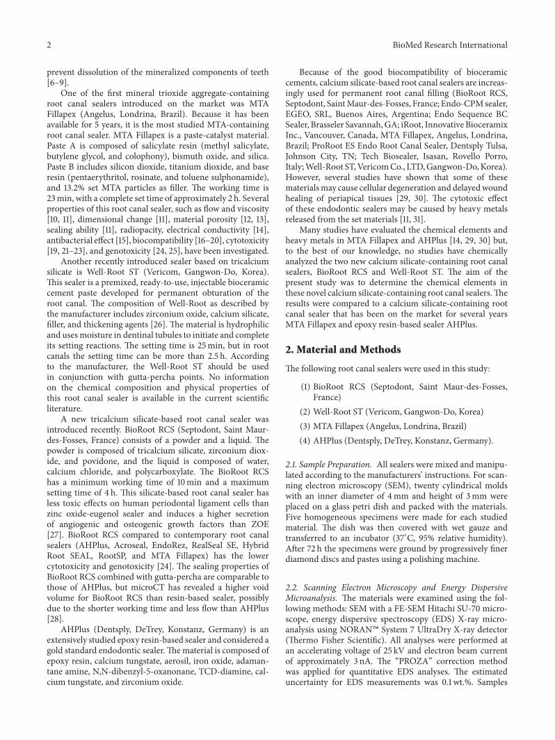
Figure 1: BioRoot RCS: backscatter scanning electron micrographs at 500x magnification (a); EDS X-ray microanalysis (b).
(a)
30000
20000
10000
00 2 4 6 8 10 12 14 16 18 20
Pd
C
Ti
Ca
(keV)
OSi
ZrCl Ti
Zr
Zr
Ti
NaMgAl
(b)
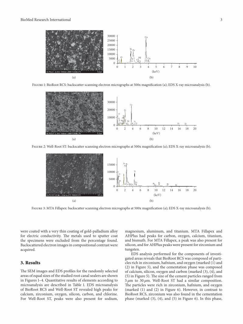
Figure 2: Well-Root ST: backscatter scanning electron micrographs at 500x magnification (a); EDS X-ray microanalysis (b).
(a)
15000
10000
5000
00 2 4 6 8 10 12 14 16 18 20
C Ti
Ca
(keV)
O
Si
BiTiTi
BiBiBiBi
Bi
Bi Au
Au
Ti
(b)
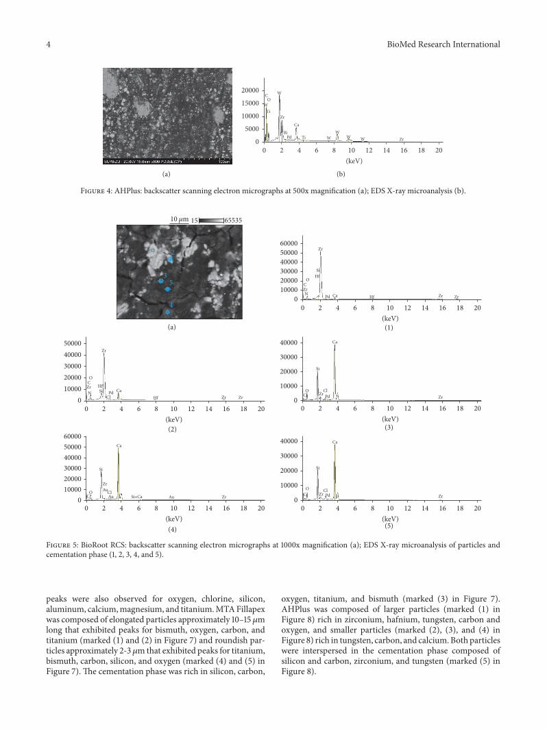
Figure 3: MTA Fillapex: backscatter scanning electron micrographs at 500x magnification (a); EDS X-ray microanalysis (b).
were coated with a very thin coating of gold-palladium alloyfor electric conductivity. The metals used to sputter coatthe specimens were excluded from the percentage found.Backscattered electron images in compositional contrast wereacquired.
3. Results
The SEM images and EDS profiles for the randomly selectedareas of equal sizes of the studied root canal sealers are shownin Figures 1–4. Quantitative results of elements according tomicroanalysis are described in Table 1. EDS microanalysisof BioRoot RCS and Well-Root ST revealed high peaks forcalcium, zirconium, oxygen, silicon, carbon, and chlorine.For Well-Root ST, peaks were also present for sodium,
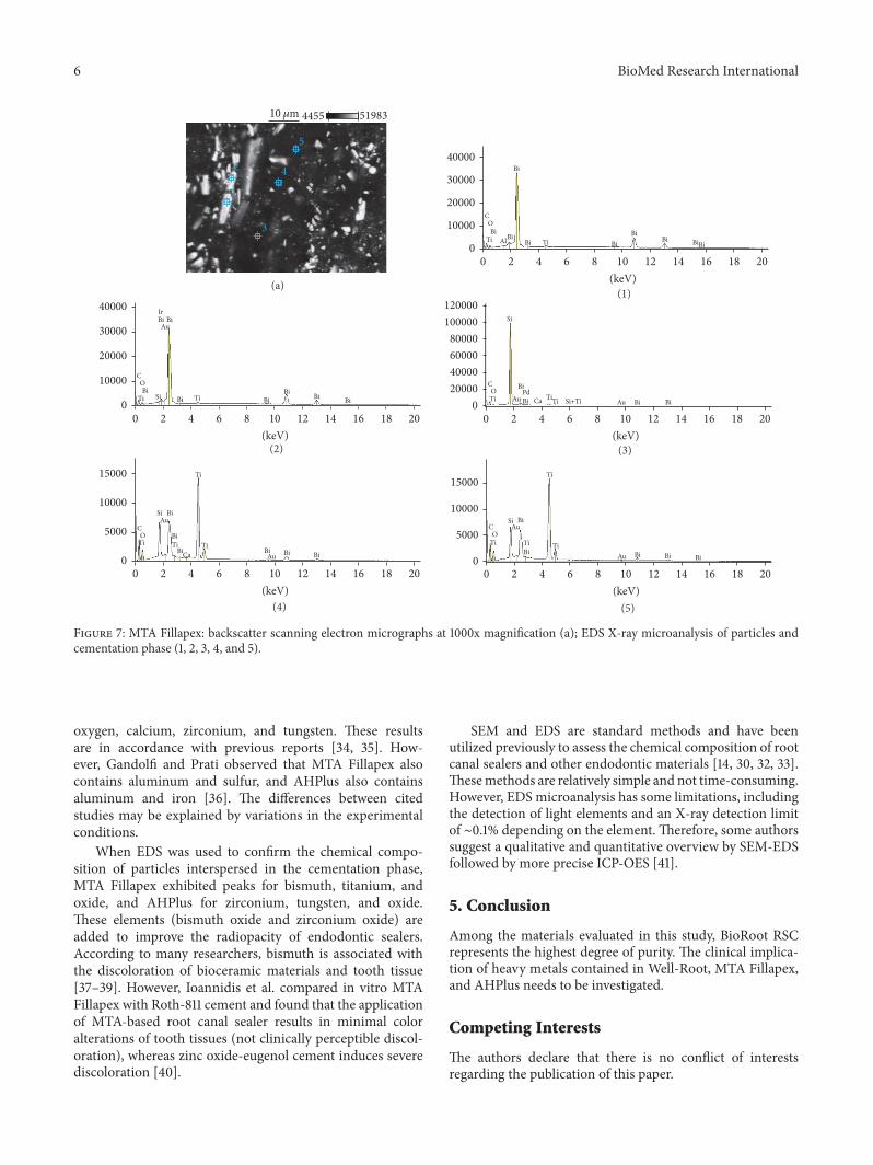
magnesium, aluminum, and titanium. MTA Fillapex andAHPlus had peaks for carbon, oxygen, calcium, titanium,and bismuth. For MTA Fillapex, a peak was also present forsilicon, and for AHPlus peaks were present for zirconium andtungsten.
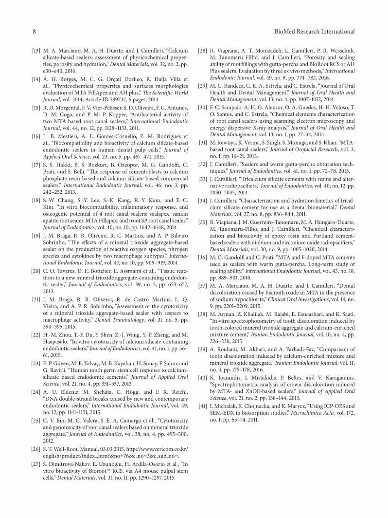
EDS analysis performed for the components of investi-gated areas reveals that BioRoot RCS was composed of parti-cles rich in zirconium, hafnium, and oxygen (marked (1) and(2) in Figure 5), and the cementation phase was composedof calcium, silicon, oxygen and carbon (marked (3), (4), and(5) in Figure 5). The size of the cement particles ranged from5 𝜇m to 30 𝜇m. Well-Root ST had a similar composition.The particles were rich in zirconium, hafnium, and oxygen(marked (1) and (2) in Figure 6). However, in contrast toBioRoot RCS, zirconium was also found in the cementationphase (marked (3), (4), and (5) in Figure 6). In this phase,
4 BioMed Research International
(a)
20000
15000
10000
5000
00 2 4 6 8 10 12 14 16 18 20
C
Ca
(keV)
OW
W
Zr
Zr
TiPd WWW
WBi
Ti
(b)
Figure 4: AHPlus: backscatter scanning electron micrographs at 500x magnification (a); EDS X-ray microanalysis (b).
5000040000300002000010000
0
600005000040000300002000010000
0
40000
30000
20000
10000
0
40000
30000
20000
10000
0
600005000040000300002000010000
00 2 4 6 8 10 12 14 16 18 20
(keV)(a)
(2)
(4)
(3)
(1)
(5)
0 2 4 6 8 10 12 14 16 18 20(keV)
0 2 4 6 8 10 12 14 16 18 20(keV)
0 2 4 6 8 10 12 14 16 18 20(keV)
0 2 4 6 8 10 12 14 16 18 20(keV)
O
O
OO
O
N ZrZr
Zr
Zr
Zr
Zr
ZrAuZr
Zr
Au Au Zr
Zr
Zr
Hf
Hf
Zr
Si+Ca
Cl
ClCl
ClPdN
ZrHf
HfSi
Si
SiSi
Si
Pd
Pd
Pd
12
3
4
5Ca
Ca
CaCa
Ca
C
C
CC
C
655351510 𝜇m
Figure 5: BioRoot RCS: backscatter scanning electron micrographs at 1000x magnification (a); EDS X-ray microanalysis of particles andcementation phase (1, 2, 3, 4, and 5).
peaks were also observed for oxygen, chlorine, silicon,aluminum, calcium,magnesium, and titanium.MTAFillapexwas composed of elongated particles approximately 10–15 𝜇mlong that exhibited peaks for bismuth, oxygen, carbon, andtitanium (marked (1) and (2) in Figure 7) and roundish par-ticles approximately 2-3𝜇mthat exhibited peaks for titanium,bismuth, carbon, silicon, and oxygen (marked (4) and (5) inFigure 7). The cementation phase was rich in silicon, carbon,
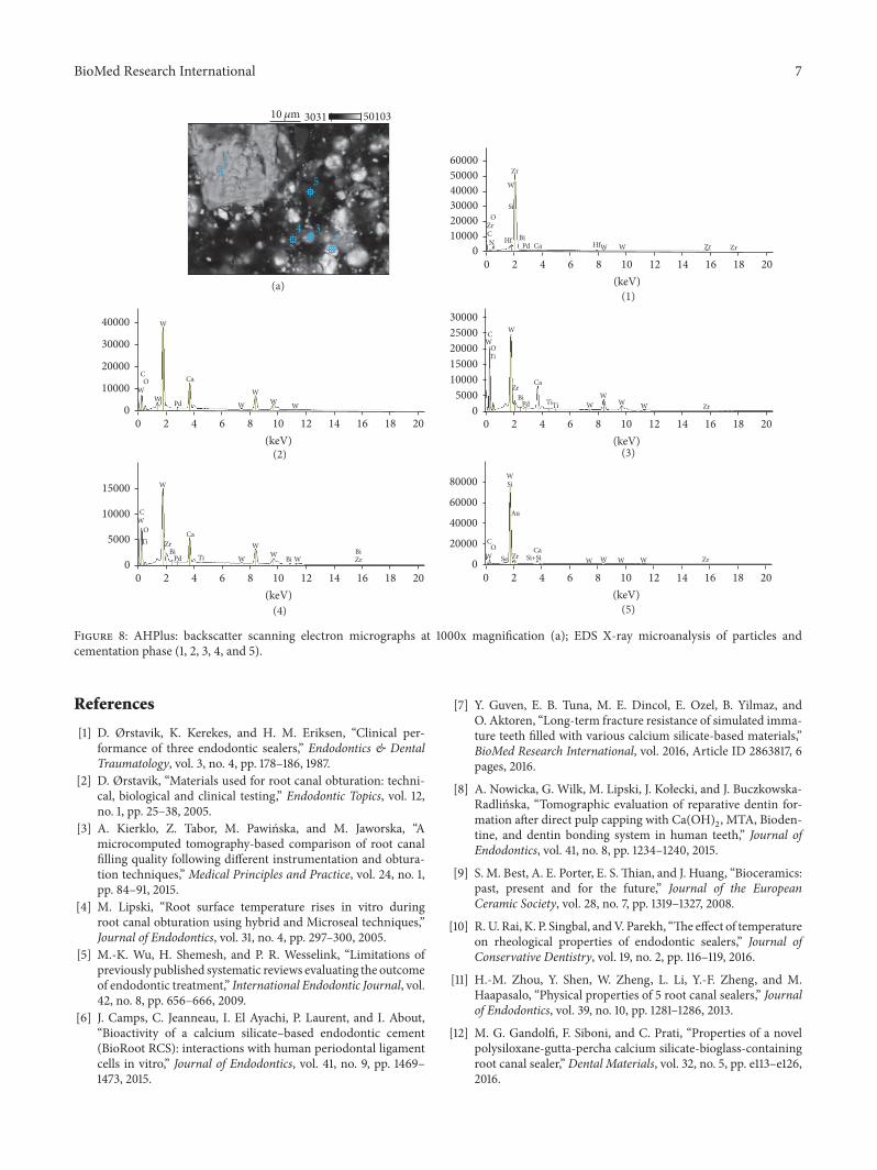
oxygen, titanium, and bismuth (marked (3) in Figure 7).AHPlus was composed of larger particles (marked (1) inFigure 8) rich in zirconium, hafnium, tungsten, carbon andoxygen, and smaller particles (marked (2), (3), and (4) inFigure 8) rich in tungsten, carbon, and calcium.Both particleswere interspersed in the cementation phase composed ofsilicon and carbon, zirconium, and tungsten (marked (5) inFigure 8).

BioMed Research International 5
5000040000300002000010000
0
250002000015000100005000
0
30000
20000
10000
0
20000
15000
10000
5000
0
50000
40000
30000
20000
10000
0
O
O
OO
O
NAl
Al
Al
Mg
Mg
Mg
Na
Na
Na
Zr
Zr
Zr
Zr
Zr
Zr
Zr
Zr
Zr
Zr
Zr
Zr
Zr
Zr
Zr
HfTi
Ti
Ti
Ti
Hf
Hf
Hf
Na
Al
Al
Mg
Mg
Zr Cl
ClS
Cl
Pd
PdS
Ti
Ti
Zr
HfTiTi
Ti
Ti
Ti
Ti
HfTi
Ti
Ti
HfSi
Si
SiSi
Si
Pd
Pd
Pd
12
3 4
5 Ca
Ca
CaCa
Ca
C
C
CC
C
0 2 4 6 8 10 12 14 16 18 20(keV)
0 2 4 6 8 10 12 14 16 18 20(keV)
0 2 4 6 8 10 12 14 16 18 20(keV)
0 2 4 6 8 10 12 14 16 18 20(keV)
0 2 4 6 8 10 12 14 16 18 20(keV)
10 𝜇m
(a)
(2)
(4)
(3)
(1)
(5)
6553515
Figure 6: Well-Root ST: backscatter scanning electron micrographs at 1000x magnification (a); EDS X-ray microanalysis of particles andcementation phase (1, 2, 3, 4, and 5).
Table 1: The percentage (weight%) of elements in the tested rootcanal sealers.
Element Root canal sealerBioRoot Well-Root ST MTA Fillapex AHPlus
C 5.6–6.2 5.4–6.0 20.0–21.9 31.4–34.5O 35.1–37.1 37.9–39.3 21.3–22.8 19.2–21.2Si 8.4–9.4 4.6–5.4 4.5–6.9 —Cl 1.1–1.3 0.4–0.6 — —Ca 25.0–26.6 21.0–22.1 0.1–0.2 4.5–4.9Zr 20.3–22.6 22.2–27.4 — 15.1–18.6Na — 0.3–0.4 — —Mg — 0.5–0.6 — —Al — 1.9–2.5 — —Ti — 1.0–2.1 11.5–15.6 0.1–0.2Bi — — 34.4–38.9 0.3–0.4W — — — 22.5–24.4
4. Discussion
In the current study, the chemical compositions of twonew calcium silicate-containing root canal sealers, BioRoot
RCS and Well-Root ST, were assessed and compared tothe composition of extensively studied materials: calciumsilicate-containing root canal sealer MTA Fillapex and epoxyresin-based sealer AHPlus.
EDS revealed that BioRoot RCS is mostly composed ofcalcium, zirconium, oxygen, carbon, silicon, and chlorine.No heavy metals or other toxic elements were found in thisendodontic sealer.The elements observed in the present studywere biocompatible with those given by manufacturer ofBioRoot RCS and determined by Camilleri in an experimen-tal tricalcium silicate-based endodontic sealer by Septodont[32]. However, the microanalysis revealed that Well-Root STcontained aluminum and titanium in addition to calcium,zirconium, oxygen, carbon, and silicon.
In both new silicate-based root canal sealers, the particlesinterspersed in the cementation phase were composed of zir-conium, hafnium, and oxygen,making up the radiopacifiyingmaterial. Although zirconiumoxide provides a lower contrastthan other radiopacifiers, such as bismuth oxide, it seems tobe more inert [33].
EDS analysis of MTA Fillapex revealed that the outersurface is rich in carbon, calcium, oxygen, silicon, titanium,and bismuth, whereas AHPlus is composed of carbon,
6 BioMed Research International
40000
30000
20000
10000
0
15000
10000
5000
0
12000010000080000600004000020000
0
15000
10000
5000
0
40000
30000
20000
10000
0
O
O
OO
O
Al Bi
Bi
BiBi
BiBi
Bi
Bi Au
Au
Au
Au Si+Ti
BiBi
BiBi
Bi
BiBiBiBi
Bi
BiTi
Au
BiBiBiTi
Ti
Ti
Bi BiBiTi
Ti
TiTi
Ti
Ti
Ti
Ti
Ti
TiTi
Si
SiSi BiAu
Si
IrBi BiAu
BiBi
Bi
Bi
Pd
1
2
3
4
5
Ca
Ca
C
C
CC
C
0 2 4 6 8 10 12 14 16 18 20(keV)
0 2 4 6 8 10 12 14 16 18 20(keV)
0 2 4 6 8 10 12 14 16 18 20(keV)
0 2 4 6 8 10 12 14 16 18 20(keV)
0 2 4 6 8 10 12 14 16 18 20(keV)
10 𝜇m
(a)
(2)
(4)
(3)
(1)
(5)
519834455
Figure 7: MTA Fillapex: backscatter scanning electron micrographs at 1000x magnification (a); EDS X-ray microanalysis of particles andcementation phase (1, 2, 3, 4, and 5).
oxygen, calcium, zirconium, and tungsten. These resultsare in accordance with previous reports [34, 35]. How-ever, Gandolfi and Prati observed that MTA Fillapex alsocontains aluminum and sulfur, and AHPlus also containsaluminum and iron [36]. The differences between citedstudies may be explained by variations in the experimentalconditions.
When EDS was used to confirm the chemical compo-sition of particles interspersed in the cementation phase,MTA Fillapex exhibited peaks for bismuth, titanium, andoxide, and AHPlus for zirconium, tungsten, and oxide.These elements (bismuth oxide and zirconium oxide) areadded to improve the radiopacity of endodontic sealers.According to many researchers, bismuth is associated withthe discoloration of bioceramic materials and tooth tissue[37–39]. However, Ioannidis et al. compared in vitro MTAFillapex with Roth-811 cement and found that the applicationof MTA-based root canal sealer results in minimal coloralterations of tooth tissues (not clinically perceptible discol-oration), whereas zinc oxide-eugenol cement induces severediscoloration [40].
SEM and EDS are standard methods and have beenutilized previously to assess the chemical composition of rootcanal sealers and other endodontic materials [14, 30, 32, 33].Thesemethods are relatively simple and not time-consuming.However, EDS microanalysis has some limitations, includingthe detection of light elements and an X-ray detection limitof ∼0.1% depending on the element. Therefore, some authorssuggest a qualitative and quantitative overview by SEM-EDSfollowed by more precise ICP-OES [41].
5. Conclusion
Among the materials evaluated in this study, BioRoot RSCrepresents the highest degree of purity. The clinical implica-tion of heavy metals contained in Well-Root, MTA Fillapex,and AHPlus needs to be investigated.
Competing Interests
The authors declare that there is no conflict of interestsregarding the publication of this paper.
BioMed Research International 7
40000
30000
20000
10000
0
15000
10000
5000
0
30000250002000015000100005000
0
80000
60000
40000
20000
0
600005000040000300002000010000
0
O
O
O
O
O
HfZrZr
Zr
W
WWW
WBi
Zr
WW
W
WW
Au
ZrSe
HfW
W
ZrBi WWW
W ZrBi
ZrBiPd
WW
WW
Ca
Ti
W WPd
Ti
CaN
Ti
Ti
W
Ti
CaSi+Si
WW
SiW
W
BiPd
Si
Zr
Zr
Pd
1
234
5
Ca
Ca
C
C
C
CW
C
0 2 4 6 8 10 12 14 16 18 20(keV)
0 2 4 6 8 10 12 14 16 18 20(keV)
0 2 4 6 8 10 12 14 16 18 20(keV)
0 2 4 6 8 10 12 14 16 18 20(keV)
0 2 4 6 8 10 12 14 16 18 20(keV)
10 𝜇m
(a)
(2)
(4)
(3)
(1)
(5)
501033031
Figure 8: AHPlus: backscatter scanning electron micrographs at 1000x magnification (a); EDS X-ray microanalysis of particles andcementation phase (1, 2, 3, 4, and 5).
References
[1] D. Ørstavik, K. Kerekes, and H. M. Eriksen, “Clinical per-formance of three endodontic sealers,” Endodontics & DentalTraumatology, vol. 3, no. 4, pp. 178–186, 1987.
[2] D. Ørstavik, “Materials used for root canal obturation: techni-cal, biological and clinical testing,” Endodontic Topics, vol. 12,no. 1, pp. 25–38, 2005.
[3] A. Kierklo, Z. Tabor, M. Pawinska, and M. Jaworska, “Amicrocomputed tomography-based comparison of root canalfilling quality following different instrumentation and obtura-tion techniques,” Medical Principles and Practice, vol. 24, no. 1,pp. 84–91, 2015.
[4] M. Lipski, “Root surface temperature rises in vitro duringroot canal obturation using hybrid and Microseal techniques,”Journal of Endodontics, vol. 31, no. 4, pp. 297–300, 2005.
[5] M.-K. Wu, H. Shemesh, and P. R. Wesselink, “Limitations ofpreviously published systematic reviews evaluating the outcomeof endodontic treatment,” International Endodontic Journal, vol.42, no. 8, pp. 656–666, 2009.
[6] J. Camps, C. Jeanneau, I. El Ayachi, P. Laurent, and I. About,“Bioactivity of a calcium silicate–based endodontic cement(BioRoot RCS): interactions with human periodontal ligamentcells in vitro,” Journal of Endodontics, vol. 41, no. 9, pp. 1469–1473, 2015.
[7] Y. Guven, E. B. Tuna, M. E. Dincol, E. Ozel, B. Yilmaz, andO. Aktoren, “Long-term fracture resistance of simulated imma-ture teeth filled with various calcium silicate-based materials,”BioMed Research International, vol. 2016, Article ID 2863817, 6pages, 2016.
[8] A. Nowicka, G. Wilk, M. Lipski, J. Kołecki, and J. Buczkowska-Radlinska, “Tomographic evaluation of reparative dentin for-mation after direct pulp capping with Ca(OH)
2, MTA, Bioden-
tine, and dentin bonding system in human teeth,” Journal ofEndodontics, vol. 41, no. 8, pp. 1234–1240, 2015.
[9] S. M. Best, A. E. Porter, E. S.Thian, and J. Huang, “Bioceramics:past, present and for the future,” Journal of the EuropeanCeramic Society, vol. 28, no. 7, pp. 1319–1327, 2008.
[10] R.U. Rai, K. P. Singbal, andV. Parekh, “The effect of temperatureon rheological properties of endodontic sealers,” Journal ofConservative Dentistry, vol. 19, no. 2, pp. 116–119, 2016.
[11] H.-M. Zhou, Y. Shen, W. Zheng, L. Li, Y.-F. Zheng, and M.Haapasalo, “Physical properties of 5 root canal sealers,” Journalof Endodontics, vol. 39, no. 10, pp. 1281–1286, 2013.
[12] M. G. Gandolfi, F. Siboni, and C. Prati, “Properties of a novelpolysiloxane-gutta-percha calcium silicate-bioglass-containingroot canal sealer,” Dental Materials, vol. 32, no. 5, pp. e113–e126,2016.
8 BioMed Research International
[13] M. A. Marciano, M. A. H. Duarte, and J. Camilleri, “Calciumsilicate-based sealers: assessment of physicochemical proper-ties, porosity and hydration,”Dental Materials, vol. 32, no. 2, pp.e30–e40, 2016.
[14] A. H. Borges, M. C. G. Orcati Dorileo, R. Dalla Villa etal., “Physicochemical properties and surfaces morphologiesevaluation of MTA FillApex and AH plus,”The Scientific WorldJournal, vol. 2014, Article ID 589732, 6 pages, 2014.
[15] R. D.Morgental, F. V. Vier-Pelisser, S. D. Oliveira, F. C. Antunes,D. M. Cogo, and P. M. P. Kopper, “Antibacterial activity oftwo MTA-based root canal sealers,” International EndodonticJournal, vol. 44, no. 12, pp. 1128–1133, 2011.
[16] L. B. Mestieri, A. L. Gomes-Cornelio, E. M. Rodrigues etal., “Biocompatibility and bioactivity of calcium silicate-basedendodontic sealers in human dental pulp cells,” Journal ofApplied Oral Science, vol. 23, no. 5, pp. 467–471, 2015.
[17] S. S. Hakki, B. S. Bozkurt, B. Ozcopur, M. G. Gandolfi, C.Prati, and S. Belli, “The response of cementoblasts to calciumphosphate resin-based and calcium silicate-based commercialsealers,” International Endodontic Journal, vol. 46, no. 3, pp.242–252, 2013.
[18] S.-W. Chang, S.-Y. Lee, S.-K. Kang, K.-Y. Kum, and E.-C.Kim, “In vitro biocompatibility, inflammatory response, andosteogenic potential of 4 root canal sealers: sealapex, sankinapatite root sealer,MTAFillapex, and iroot SP root canal sealer,”Journal of Endodontics, vol. 40, no. 10, pp. 1642–1648, 2014.
[19] J. M. Braga, R. R. Oliveira, R. C. Martins, and A. P. RibeiroSobrinho, “The effects of a mineral trioxide aggregate-basedsealer on the production of reactive oxygen species, nitrogenspecies and cytokines by two macrophage subtypes,” Interna-tional Endodontic Journal, vol. 47, no. 10, pp. 909–919, 2014.
[20] C. O. Tavares, D. E. Bottcher, E. Assmann et al., “Tissue reac-tions to a new mineral trioxide aggregate-containing endodon-tic sealer,” Journal of Endodontics, vol. 39, no. 5, pp. 653–657,2013.
[21] J. M. Braga, R. R. Oliveira, R. de Castro Martins, L. Q.Vieira, and A. P. R. Sobrinho, “Assessment of the cytotoxicityof a mineral trioxide aggregate-based sealer with respect tomacrophage activity,” Dental Traumatology, vol. 31, no. 5, pp.390–395, 2015.
[22] H.-M. Zhou, T.-F. Du, Y. Shen, Z.-J. Wang, Y.-F. Zheng, and M.Haapasalo, “In vitro cytotoxicity of calcium silicate-containingendodontic sealers,” Journal of Endodontics, vol. 41, no. 1, pp. 56–61, 2015.
[23] E. P. Guven,M. E. Yalvac,M. B. Kayahan,H. Sunay, F. Sahın, andG. Bayirli, “Human tooth germ stem cell response to calcium-silicate based endodontic cements,” Journal of Applied OralScience, vol. 21, no. 4, pp. 351–357, 2013.
[24] A. U. Eldeniz, M. Shehata, C. Hogg, and F. X. Reichl,“DNA double-strand breaks caused by new and contemporaryendodontic sealers,” International Endodontic Journal, vol. 49,no. 12, pp. 1141–1151, 2015.
[25] C. V. Bin, M. C. Valera, S. E. A. Camargo et al., “Cytotoxicityand genotoxicity of root canal sealers based onmineral trioxideaggregate,” Journal of Endodontics, vol. 38, no. 4, pp. 495–500,2012.
[26] S. T.Well-Root, Manual, 03.03.2015, http://www.vericom.co.kr/english/product/index .htm?&no=76&c no=3&c sub no=.
[27] S. Dimitrova-Nakov, E. Uzunoglu, H. Ardila-Osorio et al., “Invitro bioactivity of Bioroot� RCS, via A4 mouse pulpal stemcells,” Dental Materials, vol. 31, no. 11, pp. 1290–1297, 2015.
[28] R. Viapiana, A. T. Moinzadeh, L. Camilleri, P. R. Wesselink,M. Tanomaru Filho, and J. Camilleri, “Porosity and sealingability of root fillingswith gutta-percha andBioRoot RCS orAHPlus sealers. Evaluation by three ex vivomethods,” InternationalEndodontic Journal, vol. 49, no. 8, pp. 774–782, 2016.
[29] M. C. Bandeca, C. R. A. Estrela, and C. Estrela, “Journal of OralHealth and Dental Management,” Journal of Oral Health andDental Management, vol. 13, no. 4, pp. 1007–1012, 2014.
[30] F. C. Sampaio, A. H. G. Alencar, O. A. Guedes, H. H. Veloso, T.O. Santos, and C. Estrela, “Chemical elements characterizationof root canal sealers using scanning electron microscopy andenergy dispersive X-ray analysis,” Journal of Oral Health andDental Management, vol. 13, no. 1, pp. 27–34, 2014.
[31] M.Rawtiya, K.Verma, S. Singh, S.Munuga, and S. Khan, “MTA-based root canal sealers,” Journal of Orofacial Research, vol. 3,no. 1, pp. 16–21, 2013.
[32] J. Camilleri, “Sealers and warm gutta-percha obturation tech-niques,” Journal of Endodontics, vol. 41, no. 1, pp. 72–78, 2015.
[33] J. Camilleri, “Tricalcium silicate cements with resins and alter-native radiopacifiers,” Journal of Endodontics, vol. 40, no. 12, pp.2030–2035, 2014.
[34] J. Camilleri, “Characterization and hydration kinetics of trical-cium silicate cement for use as a dental biomaterial,” DentalMaterials, vol. 27, no. 8, pp. 836–844, 2011.
[35] R.Viapiana, J.M.Guerreiro-Tanomaru,M.A.Hungaro-Duarte,M. Tanomaru-Filho, and J. Camilleri, “Chemical characteri-zation and bioactivity of epoxy resin and Portland cement-based sealerswith niobiumand zirconiumoxide radiopacifiers,”Dental Materials, vol. 30, no. 9, pp. 1005–1020, 2014.
[36] M. G. Gandolfi and C. Prati, “MTA and F-doped MTA cementsused as sealers with warm gutta-percha. Long-term study ofsealing ability,” International Endodontic Journal, vol. 43, no. 10,pp. 889–901, 2010.
[37] M. A. Marciano, M. A. H. Duarte, and J. Camilleri, “Dentaldiscoloration caused by bismuth oxide in MTA in the presenceof sodium hypochlorite,” Clinical Oral Investigations, vol. 19, no.9, pp. 2201–2209, 2015.
[38] M. Arman, Z. Khalilak, M. Rajabi, E. Esnaashari, and K. Saati,“In vitro spectrophotometry of tooth discoloration induced bytooth-coloredmineral trioxide aggregate and calcium-enrichedmixture cement,” Iranian Endodontic Journal, vol. 10, no. 4, pp.226–230, 2015.
[39] A. Rouhani, M. Akbari, and A. Farhadi-Faz, “Comparison oftooth discoloration induced by calcium-enriched mixture andmineral trioxide aggregate,” Iranian Endodontic Journal, vol. 11,no. 3, pp. 175–178, 2016.
[40] K. Ioannidis, I. Mistakidis, P. Beltes, and V. Karagiannis,“Spectrophotometric analysis of crown discoloration inducedby MTA- and ZnOE-based sealers,” Journal of Applied OralScience, vol. 21, no. 2, pp. 138–144, 2013.
[41] I.Michalak, K. Chojnacka, andK.Marycz, “Using ICP-OES andSEM-EDX in biosorption studies,” Microchimica Acta, vol. 172,no. 1, pp. 65–74, 2011.

Submit your manuscripts athttp://www.hindawi.com
Hindawi Publishing Corporationhttp://www.hindawi.com Volume 2014
Anatomy Research International
PeptidesInternational Journal of
Hindawi Publishing Corporationhttp://www.hindawi.com Volume 2014
Hindawi Publishing Corporation http://www.hindawi.com
International Journal of
Volume 2014
Zoology
Hindawi Publishing Corporationhttp://www.hindawi.com Volume 2014
Molecular Biology International
GenomicsInternational Journal of
Hindawi Publishing Corporationhttp://www.hindawi.com Volume 2014
The Scientific World JournalHindawi Publishing Corporation http://www.hindawi.com Volume 2014
Hindawi Publishing Corporationhttp://www.hindawi.com Volume 2014
BioinformaticsAdvances in
Marine BiologyJournal of
Hindawi Publishing Corporationhttp://www.hindawi.com Volume 2014
Hindawi Publishing Corporationhttp://www.hindawi.com Volume 2014
Signal TransductionJournal of
Hindawi Publishing Corporationhttp://www.hindawi.com Volume 2014
BioMed Research International
Evolutionary BiologyInternational Journal of
Hindawi Publishing Corporationhttp://www.hindawi.com Volume 2014
Hindawi Publishing Corporationhttp://www.hindawi.com Volume 2014
Biochemistry Research International
ArchaeaHindawi Publishing Corporationhttp://www.hindawi.com Volume 2014
Hindawi Publishing Corporationhttp://www.hindawi.com Volume 2014
Genetics Research International
Hindawi Publishing Corporationhttp://www.hindawi.com Volume 2014
Advances in
Virolog y
Hindawi Publishing Corporationhttp://www.hindawi.com
Nucleic AcidsJournal of
Volume 2014
Stem CellsInternational
Hindawi Publishing Corporationhttp://www.hindawi.com Volume 2014
Hindawi Publishing Corporationhttp://www.hindawi.com Volume 2014
Enzyme Research
Hindawi Publishing Corporationhttp://www.hindawi.com Volume 2014
International Journal of
Microbiology