Embed Size (px)
Citation preview
Requirement for DNA Ligase IV during Embryonic Neuronal Development
Article (Published Version)
http://sro.sussex.ac.uk
Gatz, Susanne A, Ju, Limei, Gruber, Ralph, Hoffmann, Eva, Carr, Antony M, Wang, Zhao-Qi, Liu, Cong and Jeggo, Penny A (2011) Requirement for DNA Ligase IV during Embryonic Neuronal Development. Journal of Neuroscience, 31. pp. 10088-100100.
This version is available from Sussex Research Online: http://sro.sussex.ac.uk/22156/
This document is made available in accordance with publisher policies and may differ from the published version or from the version of record. If you wish to cite this item you are advised to consult the publisher’s version. Please see the URL above for details on accessing the published version.
Copyright and reuse: Sussex Research Online is a digital repository of the research output of the University.
Copyright and all moral rights to the version of the paper presented here belong to the individual author(s) and/or other copyright owners. To the extent reasonable and practicable, the material made available in SRO has been checked for eligibility before being made available.
Copies of full text items generally can be reproduced, displayed or performed and given to third parties in any format or medium for personal research or study, educational, or not-for-profit purposes without prior permission or charge, provided that the authors, title and full bibliographic details are credited, a hyperlink and/or URL is given for the original metadata page and the content is not changed in any way.
Development/Plasticity/Repair
Requirement for DNA Ligase IV during Embryonic NeuronalDevelopment
Susanne A. Gatz,1,2* Limei Ju,2 Ralph Gruber,3 Eva Hoffmann,2 Antony M. Carr,2 Zhao-Qi Wang,3,4 Cong Liu,1,2*and Penny A. Jeggo2
1Development and Stem Cell Institute, Key Laboratory of Ministry of Education, Department of Paediatrics, West China Second University Hospital,
Sichuan University, Chengdu, China 610041, 2Genome Damage and Stability Centre, University of Sussex, East Sussex, BN1 9RQ, United Kingdom, 3Leibniz
Institute for Age Research, Fritz Lipmann Institute, D-07745 Jena, Germany, and 4Faculty of Biology–Pharmacy, Friedrich Schiller University of Jena,
D-07745 Jena, Germany
The embryonic ventricular and subventricular zones (VZ/SVZ) contain the neuronal stem and progenitor cells and undergo rapid
proliferation. The intermediate zone (IZ) contains nonreplicating, differentiated cells. The VZ/SVZ is hypersensitive to radiation-induced
apoptosis. Ablation of DNA non-homologous end-joining (NHEJ) proteins, XRCC4 or DNA ligase IV (LigIV), confers ataxia telangiectasia
mutated (ATM)-dependent apoptosis predominantly in the IZ. We examine the mechanistic basis underlying these distinct sensitivities
using a viable LigIV (Lig4Y288C) mouse, which permits an examination of the DNA damage responses in the embryonic and adult brain.
Via combined analysis of DNA breakage, apoptosis, and cell-cycle checkpoint control in tissues, we show that apoptosis in the VZ/SVZ
and IZ is activated by low numbers of DNA double-strand breaks (DSBs). Unexpectedly, high sensitivity in the VZ/SVZ arises from
sensitive activation of ATM-dependent apoptosis plus an ATM-independent process. In contrast, the IZ appears to be hypersensitive to
persistent DSBs. NHEJ functions efficiently in both compartments. The VZ/SVZ and IZ regions incur high endogenous DNA breakage,
which correlates with VZ proliferation. We demonstrate a functional G2 /M checkpoint in VZ/SVZ cells and show that it is not activated by
low numbers of DSBs, allowing damaged VZ/SVZ cells to transit into the IZ. We propose a novel model in which microcephaly in LIG4
syndrome arises from sensitive apoptotic induction from persisting DSBs in the IZ, which arise from high endogenous breakage in the
VZ/SVZ and transit of damaged cells to the IZ. The VZ/SVZ, in contrast, is highly sensitive to acute radiation-induced DSB formation.
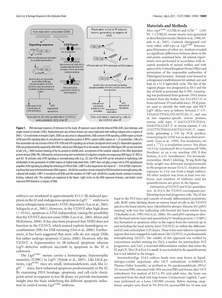
IntroductionDNA double-strand breaks (DSBs) arise from oxidative damage,replication, and exogenous sources, including ionizing radiation(IR). DSBs undergo repair and activate a signaling transductionprocess. The IR-induced damage responses relevant to this studyare overviewed in Figure 1. Importantly, XRCC4 and DNA ligaseIV (LigIV) are essential for DNA non-homologous end-joining(NHEJ), the major DSB repair mechanism, and their loss is em-bryonic lethal (Barnes et al., 1998; Frank et al., 1998; Gao et al.,1998).
LIG4 syndrome is a human disorder conferred by hypomor-phic LigIV mutations (O’Driscoll et al., 2001). Patients showimmunodeficiency, because NHEJ functions in V(D)J recombi-
nation, and microcephaly, which is observed at birth but is notprogressive, suggesting a specific requirement for NHEJ duringembryonic neuronal development.
Pioneering studies analyzing the neocortical ventricular/sub-ventricular zones (VZ/SVZ) were made using the rat brain (Bayeret al., 1991). The VZ/SVZ, which lies adjacent to the ventricle,encompasses neuronal stem/progenitor cells. The murine VZ/SVZ cells replicate rapidly between embryonic day 8 (E8) and E17generating the intermediate zone (IZ), a predominantly nonrep-licating, differentiated postmitotic layer (Pontious et al., 2008;Mitsuhashi and Takahashi, 2009). The IZ cells stain positively forTuj1; the intermediate precursors of the SVZ, present predomi-nantly at the basal layer of the VZ/SVZ close to the IZ, are Tbr2-positive (Tbr2�) studies on LigIV�/� mouse embryos that haveshown high neuronal cell death from E11.5 to E14.5, declining atE16.5 (Barnes et al., 1998; Frank et al., 2000). Most apoptosis inLigIV�/� embryos occurs in IZ neurons (Gao et al., 1998; Orii etal., 2006; Shull et al., 2009). Collectively, studies with XRCC4/LigIV null mice suggest that the IZ may suffer high DNA break-age. [Note that we use the recently accepted nomenclature forVZ/SVZ/IZ (Pontious et al., 2008)].
The embryonic brain is hyper-radiosensitive, with lowdoses (e.g., 0.125 Gy) generating apoptosis in rodent embryos(Hoshino and Kameyama, 1988; Hoshino et al., 1991). IR-induced apoptosis in mouse embryonic brain occurs predomi-nantly in the VZ/SVZ as early as 6 h after IR and is maximal when
Received March 15, 2011; revised May 9, 2011; accepted May 13, 2011.
Author contributions: S.A.G., C.L., and P.A.J. designed research; S.A.G., C.L., L.J., and R.G. performed research;
S.A.G., C.L., E.H., A.M.C., Z.-Q.W., and P.A.J. analyzed data; S.A.G. and P.A.J. wrote the paper.
The Jeggo laboratory is funded by the Medical Research Council (United Kingdom), Association for International
Cancer Research, the Wellcome Research Fund, the Department of Health (United Kingdom), and European Union
Integrated Projects DNA-Repair Grant LSHG-CT-2005-512113 and Risc-Rad Grant FI6R-CT-2003-508842. C.L. was
funded by the European Union Integrated Project DNA-Repair, and S.A.G. was funded by the Wellcome Research
Fund. Z.-Q.W. and R.G. are funded by Deutsche Forschungsgesellschaft.
*S.A.G. and C.L. contributed equally to this work.
The authors declare no competing financial interests.
Correspondence should be addressed to P. A. Jeggo, Genome Damage and Stability Centre, University of Sussex,
Science Park Road, East Sussex, BN1 9RQ, UK. E-mail: [email protected].
DOI:10.1523/JNEUROSCI.1324-11.2011
Copyright © 2011 the authors 0270-6474/11/3110088-13$15.00/0
10088 • The Journal of Neuroscience, July 6, 2011 • 31(27):10088 –10100
embryos are irradiated at approximately E13.5. IR-induced apo-ptosis in the IZ and endogenous apoptosis in LigIV�/� embryos isataxia telangiectasia mutated (ATM) dependent (Lee et al., 2001;Sekiguchi et al., 2001). However, in the VZ/SVZ after high doses(�10 Gy), apoptosis is ATM independent, raising the possibilitythat the VZ/SVZ does not sense DSBs (Lee et al., 2001; Abner andMcKinnon, 2004). It has also been proposed that NHEJ does notfunction in the VZ/SVZ, which instead exploits homologous re-combination (HR) for DSB rejoining (Orii et al., 2006). Further-more, it has been suggested that stem cells do not repair DSBsbut rather undergo apoptosis (Cairns, 2006). However, why theVZ/SVZ is hypersensitive to IR-induced apoptosis whereasLigIV-defective embryos succumb to apoptosis in the IZ isunknown.
The Lig4Y288C mouse carries a homozygous, hypomorphicmutation (Y288C) in LigIV (Nijnik et al., 2007). Like LIG4 pa-tients, Lig4Y288C mice are small, immunodeficient, and, like Li-gIV�/� mice, have enhanced apoptosis predominantly in the IZ.By examining DNA breakage, apoptosis, and cell-cycle check-point arrest in response to low IR doses, we provide mechanisticinsight into the basis underlying the different apoptotic induc-tion in control versus Lig4Y288C embryos.
Materials and MethodsMice. Lig4Y288C (C57BL/6) and ATM�/� (129/SV � C57BL/6) mouse strains were generatedas described previously (Barlow et al., 1996; Ni-jnik et al., 2007). Controls (designated ctrl)were either wild-type or Lig4Y288C heterozy-gous littermates of either sex. Analysis revealedno significant differences between them in theend points examined here. All animal experi-ments were performed in accordance with ac-cepted standards of animal welfare and withapproval by United Kingdom Home Office andpermission of the responsible authorities ofThuringen/Germany. Animals were housed ina designated establishment for animal care andkept in a 12 h light/dark cycle. The day of thevaginal plaque was designated as E0.5 and theday of birth as postnatal day 0 (P0). Genotyp-ing was performed from genomic DNA lysatesisolated from the bodies (for E14.5/E17.5) orfrom tail tissue (P5 and adult mice). PCR prim-ers used to identify the wild-type and MUTLigIV alleles were as follows: forward, 5�-CT-TAAAGCTTGGCATCAGTCAG-3�; and oneof two sequence-specific reverse primers,reverse wild type, 5�-AACCGTTTCTGGA-GAAGTACCGAT-3� or reverse mutant, 5�-AACCGTTTCTGGAGAAGTACCGAC-3�, respec-tively, generating a 310 bp PCR product.Genotyping for ATM was as described previ-ously (Liao et al., 1999). For irradiation, weused a 137Cs �-irradiation source (for doses�0.5 Gy; Gammacell 40 or Gammacell 1000;Nordion) or an x-ray machine (HS X-RaySystem; A.G.O. Installation). For bromode-oxyuridine (BrdU) labeling, 50 mg BrdU/kgbody weight was delivered intraperitoneallyto the pregnant mouse (E14.5). Analysis afterexposure to 3 Gy was from a single embryo.All other analysis was from at least two em-bryos, and numbers of embryos used forquantifications are given in the figures.
Delineation of VZ/SVZ and IZ for quantifica-
tion. At E14.5, the VZ/SVZ encompasses pro-
liferating stem and progenitor cells. The IZ lies
basal to the SVZ layer and consists of mostly differentiated postmitotic
cells. BrdU pulse labeling shows an intense band of cells in the VZ/SVZ
apical to the basal mitotic layer [identified by phospo-Histone H3 (pH3)
staining] with very few replicating cells beyond this basal mitotic layer
(Takahashi et al., 1995; Orii et al., 2006). We used pH3 staining to iden-
tify the basal mitotic layer and quantified p53-binding protein 1 (53BP1)
foci formation or apoptosis either in the region from the ventricle up to
and including the basal mitotic layer (VZ/SVZ) or within the differenti-
ated IZ and cortical plate (CP) layers. These zones were taken to represent
regions that were engaged in replication (the VZ/SVZ) or predominantly
nonreplicating (IZ/CP). The validity of this approach was verified by
concomitant marker staining for Tbr2, a marker for intermediate SVZ
progenitors, and Tuj1, a neuronal differentiation marker that stains the
IZ and CP. The CP at E14.5 is small. It was included in the quantifications
but is not labeled in the images.
Immunohistology. E14.5 embryo heads were snap frozen in liquid-
nitrogen-cooled isopentane after OCT embedment (LAMB/OCT;
Thermo Fisher Scientific) or prefixed in 3% paraformaldehyde (PFA)/
2% sucrose/PBS, saturated with 30% sucrose/PBS and frozen after OCT
embedment. For analysis of E17.5, P5, and adult mice, the brain was
separated from the skull and snap frozen. Cryosections (sagittal, 7 �m)
were performed on a Leica CM1900 cryostat. Before staining, snap-
frozen samples were fixed in 3% PFA/2% sucrose/PBS for 10 min and
Figure 1. DNA damage responses of relevance to this study. IR exposure causes directly induced DSBs (left), base damage, and
single-strand (ss) breaks (SSBs). Replication past any of these lesions can cause replication fork stalling/collapse and ss regions of
DNA (�25 nucleotides in length) (right). DSBs can also arise at collapsed forks. DSBs activate ATM signaling; ssDNA regions activate
(ATM and ATR) signaling after its recruitment to replication protein A (RPA)-coated ssDNA regions of �25 nucleotides. SSBs of a
few nucleotides do not activate ATM or ATR. ATM and ATR signaling can activate checkpoint arrest and p53-dependent apoptosis.
DSBs are predominantly repaired by DNA NHEJ, which uses DNA ligase IV as the double-strand (ds) DNA ligase (HR can also function
in late S/G2 ). NHEJ involves binding of the Ku protein to dsDNA ends, recruitment of the catalytic subunit of the DNA-dependent
protein kinase (DNA-PK), followed by end processing and recruitment of a ligation complex encompassing DNA ligase IV, XRCC4,
and XLF. IR activates only ATM signaling in nonreplicating cells (e.g., IZ), but ATM and ATR can be activated in replicating cells
attributable to the generation of ssDNA regions at stalled replication forks. 53BP1 does not play a major role in ATR signaling but
amplifies ATM signaling by aiding the tethering of ATM at DSBs. 53BP1 is also required for the repair of �15% of DSBs (represent-
ing those that arise in heterochromatic DNA regions). �H2AX foci numbers closely respond to DSB formation in nonreplicating cells,
cultured cell models; 53BP1 is recruited to all DSBs and the numbers of 53BP1 and �H2AX foci usually closely correlate in nonrep-
licating, cultured cells. The symbols are explained in the figure. Light circles on the DNA represent histones, and darker circles
represent RPA bound to ss regions of DNA.
Gatz et al. • Damage Response in the Embryonic Brain J. Neurosci., July 6, 2011 • 31(27):10088 –10100 • 10089
lysed with 0.2% Triton X-100/PBS for 5 min. Antigen retrieval (10 mM
citrate buffer and 0.05% Tween 20, pH 6.0, for 40 min at 95°C) was usedfor prefixed samples. All samples were blocked in 10% normal goat se-rum in PBS for 1–3 h (Sigma) (Santa Cruz Biotechnology for �H2AXstaining in prefixed embryonic tissues). Immunofluorescence (IF) anal-ysis of tissues was performed using the following primary antibodies:anti-53BP1 rabbit polyclonal (1:1000; A300-272A; Bethyl), anti-phospho-S139 H2AX mouse monoclonal (1:1000; ab18311; Abcam),anti-BrdU rat monoclonal (1:150; ab6326; Abcam), anti-Tbr2 rabbitpolyclonal (1:200 –1:500; ab23345; Abcam), anti-�-Tubulin III rabbit(1:200; T2200; Sigma-Aldrich), anti-phospho-S10 Histone H3 mousemonoclonal (1:200; catalog #9706S; Cell Signaling Technology), anti-phospho-S10 Histone H3 rabbit polyclonal (1:200; 06-570; Millipore),and anti-Histone H3 (phospho-S10) mouse monoclonal (1:2000;ab14955; Abcam). Anti-pH3 antibody from Cell Signaling Technologyonly detects mitotic cells, whereas the anti-pH3 antibodies from Milli-pore and Abcam detect mitotic cells (bright pan-nuclear staining) andalso G2-phase cells (speckled nuclear staining). Incubation of slides wasdone in a humidified chamber for 30 min at 37°C or 60 min at roomtemperature (RT). Secondary antibodies used were anti-rabbit Cy3 (1:500) and anti-rat TRITC (1:150; both from Sigma), anti-mouse AlexaFluor 488, anti-rabbit Alexa Fluor 488, and anti-mouse Alexa Fluor 555(all 1:500; all from Invitrogen), and anti-rat TRITC and anti-rabbit Cy5(1:200; Jackson ImmunoResearch). Sections were counterstained with4�,6-diamidino-2-phenylindole (DAPI) (0.05– 0.1 �g/ml) and mountedwith Vectashield (Vector Laboratories).
A Carl Zeiss Axioplan or Nikon eclipse microscope was used for imagecapturing (Simple PCI software). Quantifications were done by scrollingthrough the entire section depth at 100� magnification. ConcomitantpH3 staining was used to define the basal mitotic layer between SVZ andIZ regions that coincides with the basal layer of Tbr2 � cells (see Fig. 2).For quantification of 53BP1 foci, snap-frozen or prefixed samples wereused with comparable results. For all quantification (apoptosis, 53BP1foci, mitotic index), the combined results for all sections derived from asingle embryo were estimated, and the given results represent the meanand SD of the mean between embryos. p values are estimated using theStudent’s unpaired t test. DeltaVision Core and personalDV microscopes(Olympus) were used to visualize Cy5 staining and obtain images forFigure 7; imaging and deconvolution were performed with SoftWorxsoftware (version 3.7.1), and images were further edited using Omero(version beta 4.1.1) and Photoshop (Adobe Systems).
TUNEL/neuronal marker/BrdU staining. Snap-frozen samples wereused. TUNEL staining (In Situ Cell Death Detection kit, Fluorescein;Roche) was performed according to the instructions of the manufac-turer. After fixation and permeabilization, the reaction mixture wasadded and samples were incubated for 1 h at 37°C in a darkened humid-ified chamber. TUNEL staining was performed after the IF staining (pH3or 53BP1 analysis) when performed in conjunction. For concomitantneuronal marker/TUNEL/BrdU staining, the neuronal marker stainingwas performed first as described above and then TUNEL staining wasperformed according to the instruction manual. After this, sections wererefixed in 3% PFA/2% sucrose in PBS for 10 min. After three washes,sections were denatured in 2.5 M HCl for 20 min at RT, washed for 10 minwith PBS, and subjected to standard IF staining. For neuronal marker/BrdU staining, after fixation and permeabilization, the neuronal markerstaining was performed using appropriate first and secondary antibodies,followed by refixation for 10 min and denaturation with 2.5 M HCl for 30min, followed by BrdU staining and counterstaining with DAPI.
Mitotic index. pH3 staining gave comparable results in snap-frozenand prefixed samples. Prefixed sections were used for quantification be-cause they optimally conserved cytological structure, and pH3-positivecells (antibodies from Cell Signaling Technology) at the ventricular sur-face were enumerated by scrolling through the entire section depth. Toadjust for changes in shape and size of the ventricular surface betweensamples, we used a method to estimate the length of the ventricularsurface: the length was defined by an estimation of the number ofaverage-sized DAPI nuclei that can be accommodated in the mitotic layerat the ventricular surface in one focal plane. The mitotic index was de-fined as the number of quantified mitotic cells divided by the estimated
length of the mitotic border. This was normalized to 100% for untreatedctrl samples.
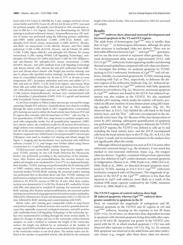
ResultsLig4Y288C embryos show abnormal neuronal development andincreased apoptosis in the VZ and SVZ regionsThe adult brain of homozygous Lig4Y288C mice is smaller thanthat of Lig4�/� or heterozygous littermates, although the grossbrain structure is unchanged (data not shown). There was nodetectable difference between Lig4�/� and Lig4Y288C/� genotypes,and we use the term control to represent either genotype. Neu-ronal developmental delay starts at approximately E14.5, withE15.5 Lig4Y288C embryonic brains appearing smaller and showingthinned neural epithelium compared with ctrls (data not shown).To investigate this further, we analyzed apoptosis by TUNELstaining in E14.5 embryonic forebrains of Lig4Y288C and ctrl em-bryos. Initially, we examined apoptosis by TUNEL staining usingcostaining with Tuj1 or Tbr2, respectively, to delineate the dis-tinct regions of the embryonic forebrain (Fig. 2a). Enhanced ap-optosis was observed in Lig4Y288C embryos with little apoptosispresent in ctrl embryos (Fig. 2a). Moreover, maximum apoptosisin Lig4Y288C embryos was found in the IZ/CP, but enhanced ap-optosis was also evident in the VZ/SVZ. We also examinedwhether pH3 staining, which marks the mitotic cell layers, pro-vided an efficient monitor of zone demarcation using pH3 stain-ing together with the Tuj1 or Tbr2 markers (Fig. 2b). Weobserved that, at E14.5, Tuj1 labeled the cells basal to the basalmitotic layer, and the Tbr2� cells were encompassed in the mi-totically active layer (Fig. 2b). Because of the clear demarcation ofzones by pH3 staining, subsequent quantification of apoptosiswas performed using only pH3 staining to delineate the VZ/SVZand IZ/CP regions; the VZ/SVZ was defined as cells up to andincluding the basal mitotic layer, and the IZ/CP encompassedcells from the basal mitotic layer to the CP (Fig. 2b). At E.14.5, theCP layer is small, and its inclusion/exclusion in the analysis doesnot significantly affect the results.
Although enhanced apoptosis was seen at E14.5 in some otherembryonic neuronal tissues (e.g., the striatum), it was much lessmarked in non-neuronal embryonic tissue (e.g., the tongue)(data not shown). Together, consistent with previous reports thatgerm-line deletion of LigIV confers dramatic neuronal apoptosisin midgestation (Barnes et al., 1998; Frank et al., 2000; Orii et al.,2006; Shull et al., 2009), we detected elevated apoptosis moni-tored by TUNEL staining in E14.5–E15.5 Lig4Y288C embryonicforebrains compared with ctrl littermates. The magnitude of ap-optosis in the IZ/CP in the Lig4Y288C embryos is less than thatreported in LigIV null embryos, most likely as a result of theresidual DSB repair capacity conferred by the Y288C mutation(Orii et al., 2006; Shull et al., 2009).
The VZ/SVZ regions of control embryos show highIR-induced apoptosis, whereas Lig4Y288C embryos showgreater sensitivity to apoptosis in the IZNext, we examined the magnitude of endogenous and IR-induced apoptosis in the VZ/SVZ and IZ regions in ctrl andLig4Y288C embryos, focusing on the response to low IR doses(0.05–3 Gy). In ctrl mice, we observed a dose-dependent increasein apoptosis with elevated apoptosis being detectable after expo-sure to 50 mGy IR. Apoptosis was greater in the VZ/SVZ com-pared with the IZ, in which marked apoptosis (�1%) was onlyobserved after exposure to doses �0.5 Gy (Fig. 2c). In contrast,little apoptosis was observed in the adult brain and other embry-onic tissues (e.g., tongue) after these doses (data not shown).
10090 • J. Neurosci., July 6, 2011 • 31(27):10088 –10100 Gatz et al. • Damage Response in the Embryonic Brain
Figure 2. Endogenous and IR-induced apoptosis in ctrl and Lig4Y288C embryonic forebrains. a, At E14.5, no significant endogenous apoptosis (TUNEL staining; green) is observed in control
forebrains (ctrl), whereas Lig4Y288C embryos show increased apoptosis in the VZ/SVZ and IZ/CP with greater apoptosis being observed in the IZ/CP; representative sections of two Lig4Y288C embryonic
forebrains from different litters are shown (I, II). Tuj1 (red) and Tbr2 (red) antibodies are used to visualize the differentiated IZ or the intermediate precursors of the SVZ, respectively. b, Costaining
of the G2/M-marker pH3 (green, antibody from Abcam) with Tuj1 or Tbr2 (red) in ctrl and Lig4Y288C samples. Tuj1 labels the postmitotic IZ/CP compartment, whereas the intermediate precursors of
the SVZ are contained in the mitotically active compartment. c, A dose-dependent increase in apoptosis is observed in the VZ/SVZ and IZ/CP of the forebrains of ctrl embryos 6 h after IR at E14.5.
Greater apoptosis is observed in the VZ/SVZ compared with the IZ/CP. Analysis after exposure to 3 Gy was from a single embryo. All other analysis, here and elsewhere, was from at least two embryos.
d, Quantification of apoptosis in the VZ/SVZ and IZ/CP of forebrains either untreated (�IR) or 6 h after 0.1 Gy (�IR) at E14.5. The percentage apoptosis represents (Figure legend continues.)
Gatz et al. • Damage Response in the Embryonic Brain J. Neurosci., July 6, 2011 • 31(27):10088 –10100 • 10091
Strikingly, both the Lig4Y288C embryonicVZ/SVZ and IZ/CP regions showed en-hanced apoptosis after 0.1 Gy comparedwith ctrl littermates (Fig. 2d,e). IR-induced apoptosis (i.e., subtracting thebackground level) was similar in the VZ/SVZ and IZ/CP regions, suggesting thatNHEJ functions efficiently in both com-partments (Fig. 2d,e). Endogenous apo-ptosis in the IZ of Lig4Y288C embryos iscomparable with that seen 6 h after 0.5 Gyin ctrl embryos (Fig. 2a,c). Previous stud-ies have shown that the peak of apoptosisafter doses below 0.5 Gy occurs �6 h afterIR (Hoshino and Kameyama, 1988;Hoshino et al., 1991). In ctrl embryos, ap-optosis was reduced 14 h after 0.1 Gy butwas maintained in the Lig4Y288C VZ/SVZand enhanced in the IZ/CP (Fig. 2f).Thus, the Lig4Y288C VZ/SVZ and IZ/CP re-gions are more sensitive to apoptosis thanevident from the 6 h analysis. BecauseDSBs in Lig4Y288C mice are more persis-tent than in ctrl mice as a result of theLig4Y288C DSB repair defect, these findingssuggest that the IZ/CP could be hypersen-sitive to persistent DSBs.
In summary, although NHEJ func-tions efficiently to rejoin DSBs in the VZ/SVZ, apoptosis is stillreadily activated by a low threshold number of DSBs. Apoptosis isless sensitively activated in the IZ/CP region in control mice afterradiation exposure. However, in untreated Lig4Y288C mice, apo-ptosis is sensitively activated by a low number of DSBs in theIZ/CP.
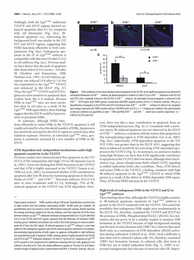
ATM-dependent and -independent mechanisms confer highapoptotic sensitivity in the VZ/SVZPrevious studies have demonstrated that apoptosis in the VZ/SVZ is ATM-independent after high (14 Gy) IR exposure (Lee etal., 2001). Given our finding that NHEJ functions in the VZ/SVZand that ATM is highly expressed in the VZ/SVZ (Soares et al.,1998; Lee et al., 2001), we examined whether ATM contributes toapoptosis after low IR doses by examining apoptosis in the fore-brains of ATM�/� and ATM�/� littermate embryos (E14.5) 6 hafter in utero irradiation with 0.5 Gy. Strikingly, 75% of IR-induced apoptosis in the VZ/SVZ was ATM dependent. How-
ever, there was also a clear contribution to apoptosis from anATM-independent process (Fig. 3a–c). Consistent with a previ-ous report, IR-induced apoptosis was not observed in the IZ/CPof ATM�/� embryos, consistent with the notion that apoptosis inthis nonreplicating region is ATM dependent (Lee et al., 2001)(Fig. 3b,c). Importantly, ATM-dependent apoptosis in the VZ/SVZ (19%) was greater than in the IZ/CP (6%), suggesting thatthere is enhanced sensitivity for activating ATM-dependent apo-ptosis in the VZ/SVZ (Fig. 3c). In summary, in contrast to studiesusing high IR doses, we show that ATM significantly contributesto apoptosis in the VZ/SVZ after low doses, although other mech-anisms [e.g., ataxia telangiectasia Rad3-related (ATR) signalingat stalled replication forks] also contribute. Thus, ATM detectsand senses DSBs in the VZ/SVZ, a finding consistent with highIR-induced apoptosis in the Lig4Y288C VZ/SVZ in which DSBspersist as a result of the defect in NHEJ-dependent DSB repair.Thus, ATM and NHEJ function in the VZ/SVZ.
High levels of endogenous DSBs in the VZ/SVZ and IZ inLig4Y288C embryosThese findings show that, although the VZ/SVZ is highly sensitiveto IR-induced apoptosis, apoptosis in Lig4Y288C embryos isgreater in the IZ/CP compared with the VZ/SVZ. This raised thepossibility that endogenous DSBs might arise predominantly inthe IZ/CP. We, therefore, examined the VZ/SVZ and IZ/CP forthe presence of DSBs. Phosphorylated H2AX (�H2AX) foci for-mation did not prove to be a suitable marker to monitor DSBformation in the VZ/SVZ because of high background stainingand the lack of colocalization with 53BP1 foci, features that mostlikely arise as a consequence of ATR-dependent �H2AX activa-tion during replication (Lobrich et al., 2010). Such a difficulty isalso observed in cultured S-phase cells. We, therefore, examined53BP1 foci formation because, in cultured cells, they form atDSBs but not at stalled replication forks (Fig. 1). 53BP1 is ex-pressed endogenously but relocates to form foci after IR. Impor-
4
(Figure legend continued.) TUNEL-positive cells per 100 nuclei. Quantification was from four
to eight sections from two embryos representing 20,000 – 40,000 nuclei per condition. All
quantification given was based on zones differentiated using pH3 staining. Results represent
the average percentage apoptosis for the two embryos; error bars represent SD of the mean
between embryos. e, Lig4 Y288C embryonic forebrains are hypersensitive to 0.1 Gy (6 h after IR at
E14.5) in the VZ/SVZ and IZ/CP regions compared with the littermate ctrl forebrain (TUNEL
staining; green). Additional concomitant Tuj1 (red) or Tbr2 (red) stainings are shown to distin-
guish the IZ from the SVZ regions. IR-induced apoptosis in Lig4Y288C embryos is more than
additive of the endogenous apoptosis plus the IR-induced apoptosis observed in ctrl embryos,
demonstrating hypersensitivity of both regions to apoptosis attributable to LigIV deficiency
(see quantification in d). f, Apoptosis (TUNEL staining; green) is highly elevated in the VZ/SVZ
and IZ/CP of Lig4Y288C embryonic forebrains 14 h after 0.1 Gy IR at E14.5. Apoptosis in Lig4Y288C
IZ/CP is greater at 14 h compared to 6 h (additional costaining with Tuj1; red); apoptosis in ctrl
embryos is less than at 6 h. Thus, the relative difference is greater at 14 versus 6 h. a–f, Repre-
sentative images of sagittal sections counterstained with DAPI. V, Ventricle. Scale bar, 100 �m.
Figure 3. ATM contributes to low-dose radiation-induced apoptosis in the VZ/SVZ. a, No significant apoptosis was observed in
unirradiated forebrains of ATM�/� embryos. b, Marked apoptosis is evident 6 h after 0.5 Gy in ATM�/� forebrains in the VZ/SVZ
and IZ/CP and is markedly reduced in the VZ/SVZ of ATM�/� embryos. No detectable increased apoptosis is observed in the
ATM�/� IZ/CP region. a, b, TUNEL (green), counterstain with DAPI, sagittal sections, at E14.5. V, Ventricle. Scale bar, 100 �m. c,
Quantification of apoptosis in the VZ/SVZ and IZ/CP of forebrains from ATM�/� or ATM�/� embryos 6 h after 0.5 Gy. Apoptosis
(percentage) represents the TUNEL-positive cells per 100 DAPI nuclei; at E14.5, n � 3 embryos per condition. One representative
section per condition was quantified. In total �7500 and 9500 ATM�/� and ATM�/� nuclei were counted, respectively. *p �
0.005 for VZ/SVZ and IZ/CP.
10092 • J. Neurosci., July 6, 2011 • 31(27):10088 –10100 Gatz et al. • Damage Response in the Embryonic Brain
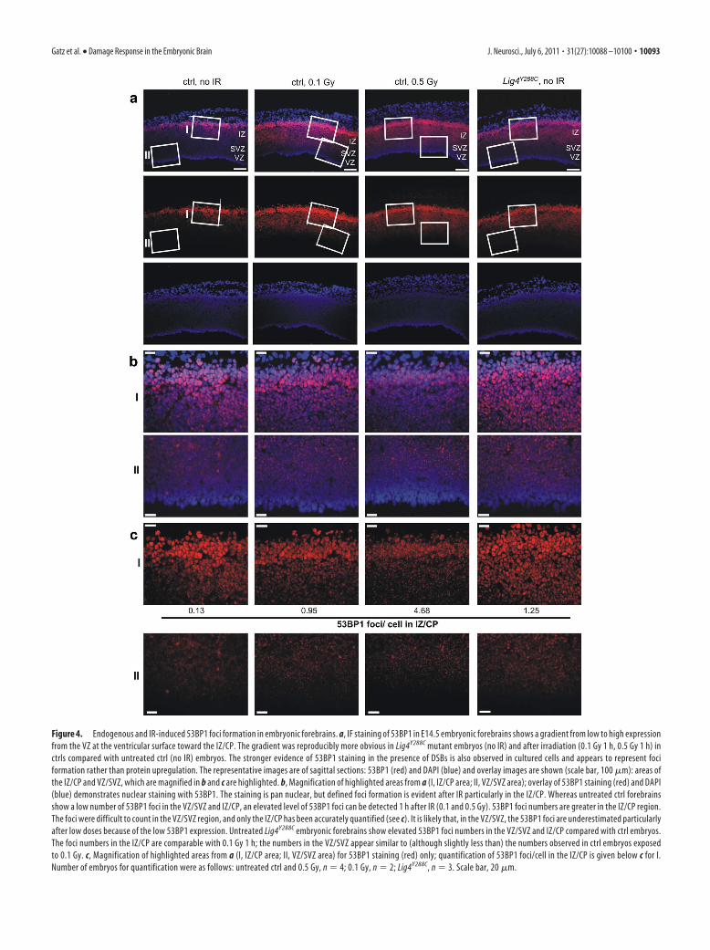
Figure 4. Endogenous and IR-induced 53BP1 foci formation in embryonic forebrains. a, IF staining of 53BP1 in E14.5 embryonic forebrains shows a gradient from low to high expression
from the VZ at the ventricular surface toward the IZ/CP. The gradient was reproducibly more obvious in Lig4Y288C mutant embryos (no IR) and after irradiation (0.1 Gy 1 h, 0.5 Gy 1 h) in
ctrls compared with untreated ctrl (no IR) embryos. The stronger evidence of 53BP1 staining in the presence of DSBs is also observed in cultured cells and appears to represent foci
formation rather than protein upregulation. The representative images are of sagittal sections: 53BP1 (red) and DAPI (blue) and overlay images are shown (scale bar, 100 �m): areas of
the IZ/CP and VZ/SVZ, which are magnified in b and c are highlighted. b, Magnification of highlighted areas from a (I, IZ/CP area; II, VZ/SVZ area); overlay of 53BP1 staining (red) and DAPI
(blue) demonstrates nuclear staining with 53BP1. The staining is pan nuclear, but defined foci formation is evident after IR particularly in the IZ/CP. Whereas untreated ctrl forebrains
show a low number of 53BP1 foci in the VZ/SVZ and IZ/CP, an elevated level of 53BP1 foci can be detected 1 h after IR (0.1 and 0.5 Gy). 53BP1 foci numbers are greater in the IZ/CP region.
The foci were difficult to count in the VZ/SVZ region, and only the IZ/CP has been accurately quantified (see c). It is likely that, in the VZ/SVZ, the 53BP1 foci are underestimated particularly
after low doses because of the low 53BP1 expression. Untreated Lig4Y288C embryonic forebrains show elevated 53BP1 foci numbers in the VZ/SVZ and IZ/CP compared with ctrl embryos.
The foci numbers in the IZ/CP are comparable with 0.1 Gy 1 h; the numbers in the VZ/SVZ appear similar to (although slightly less than) the numbers observed in ctrl embryos exposed
to 0.1 Gy. c, Magnification of highlighted areas from a (I, IZ/CP area; II, VZ/SVZ area) for 53BP1 staining (red) only; quantification of 53BP1 foci/cell in the IZ/CP is given below c for I.
Number of embryos for quantification were as follows: untreated ctrl and 0.5 Gy, n � 4; 0.1 Gy, n � 2; Lig4Y288C, n � 3. Scale bar, 20 �m.
Gatz et al. • Damage Response in the Embryonic Brain J. Neurosci., July 6, 2011 • 31(27):10088 –10100 • 10093
tantly, although not all �H2AX foci in theVZ/SVZ were associated with 53BP1 foci,all 53BP1 foci coassociated with �H2AX(data not shown). Strikingly, at E14.5 weobserved a marked gradient in 53BP1 ex-pression extending from low expressionin the VZ/SVZ to medium expression inthe IZ and high in the incipient CP (Fig.4a). Furthermore, expression in the VZ/SVZ compartment was not homogenous.In untreated ctrl embryos, we observedfew 53BP1 foci in the VZ/SVZ and the IZwith elevated foci evident in both regions1 h after 0.1 Gy IR (Fig. 4b,c). Although53BP1 foci were prominent and readilyquantified in the IZ/CP, they were lesswell defined and less numerous in the VZ/SVZ. Quantification in the VZ/SVZ washampered by low 53BP1 expression, non-homogenous expression, and a high celldensity. Furthermore, although 53BP1foci numbers were dose dependent in theIZ (Fig. 4b,c, I boxes), the response did notappear to be as obviously linear with dosein the VZ/SVZ (Fig. 4b,c, II boxes). Thefindings suggest that we may underesti-mate DSBs in the VZ/SVZ, particularly athigh doses. Because we would expect sim-ilar IR-induced DSB induction levels inthe VZ/SVZ and IZ [and possibly greaterDSB numbers in the VZ/SVZ as a result ofa higher percentage of 4n cells (i.e., cells inlate S/G2 phase with double the 2N DNAcontent)], we consider that the reduced53BP1 foci numbers in the VZ/SVZ arelikely a consequence of an impaired abilityto detect DSBs (attributable to low 53BP1expression). Nonetheless, 53BP1 foci aredetectable in the VZ/SVZ in unirradiatedLig4Y288C embryos, demonstrating thatDSBs can arise in this zone. DSB detectionin the IZ is approximately twofold tothreefold less than observed in culturedcells, although it is consistent with other analyses of foci induc-tion levels in vivo (Rube et al., 2008).
Despite the difficulty in quantification of 53BP1 foci in theVZ/SVZ, there were clearly elevated 53BP1 foci present in theuntreated Lig4Y288C VZ/SVZ with levels similar to (althoughslightly less than) that in ctrl embryos after 0.1 Gy IR (Fig. 4b,c, IIboxes), Similarly in the IZ region, there were elevated 53BP1 fociin untreated Lig4Y288C embryos, and quantification revealed thatthis was similar to the level in the control IZ region after exposureto 0.1 Gy (Fig. 4b,c, I boxes).
Concomitant 53BP1–�H2AX analysis in the IZ region endog-enously and in irradiated samples showed colocalization consis-tent with the suggestion that 53BP1 foci represent DSBs (data notshown).
Although the VZ/SVZ quantification was limited, we clearlyobserved elevated endogenous DSBs in the VZ/SVZ and IZ ofLig4Y288C embryos, with the steady-state level being similar to thatinduced by 0.1 Gy IR, which from physical studies is estimated tobe two to three DSBs per cell (Rydberg, 2000). Interestingly, al-though 53BP1 foci numbers in the Lig4Y288C IZ were similar to
that induced by 0.1 Gy IR, endogenous apoptosis corresponded
to that induced by 0.5 Gy IR (Fig. 2). This suggests that endoge-
nous DSBs in the Lig4Y288C IZ have a greater capacity to signal to
apoptosis compared with IR-induced DSBs in ctrl mice. This is
likely attributable to the persistence of the DSBs in Lig4Y288C cells
and suggests that the IZ sensitively activates apoptosis from per-
sistent DSBs.
DSB formation and apoptosis in the IZ correlate with
VZ/SVZ proliferation
High apoptosis in LigIV�/� and XRCC4�/� embryos correlates
temporally with neuronal generation, i.e., with neuronal stem/
progenitor cell replicative activity (Gao et al., 1998). To gain in-
sight into whether DSB formation and apoptosis in the IZ
correlates with VZ/SVZ proliferation, we quantified 53BP1 foci
and apoptosis in the IZ/CP at E14.5, in the developing cortex at
E17.5, and at P5 in ctrl and Lig4Y288C littermates (Fig. 5a–c). E17.5
represents a time when the VZ/SVZ has just ceased division and
by P5 replication has fully ceased (Mitsuhashi and Takahashi,
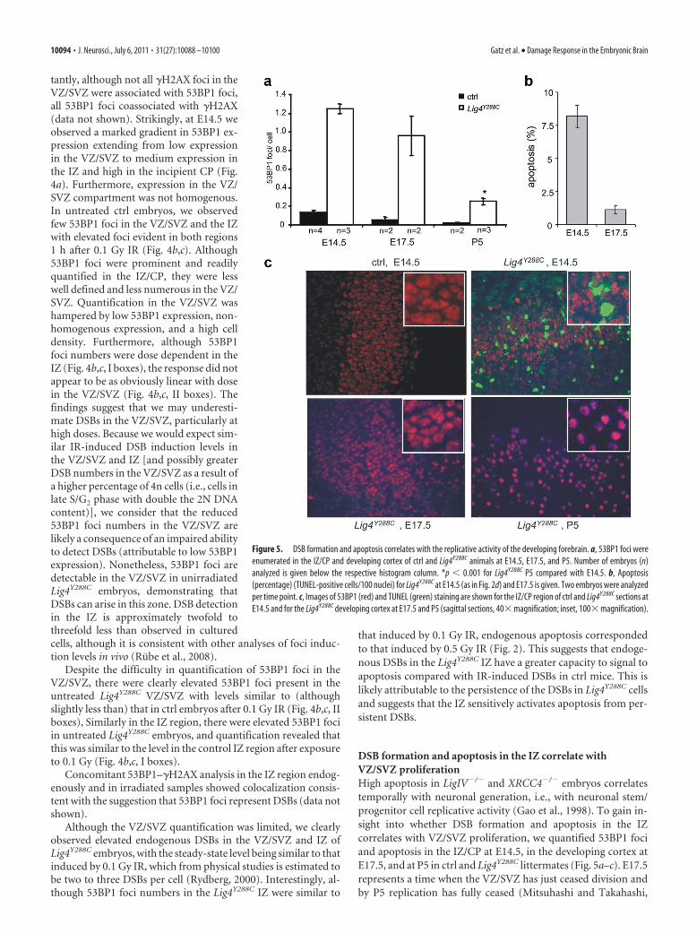
Figure 5. DSB formation and apoptosis correlates with the replicative activity of the developing forebrain. a, 53BP1 foci were
enumerated in the IZ/CP and developing cortex of ctrl and Lig4Y288C animals at E14.5, E17.5, and P5. Number of embryos (n)
analyzed is given below the respective histogram column. *p � 0.001 for Lig4Y288C P5 compared with E14.5. b, Apoptosis
(percentage) (TUNEL-positive cells/100 nuclei) for Lig4Y288C at E14.5 (as in Fig. 2d) and E17.5 is given. Two embryos were analyzed
per time point. c, Images of 53BP1 (red) and TUNEL (green) staining are shown for the IZ/CP region of ctrl and Lig4Y288C sections at
E14.5 and for the Lig4Y288C developing cortex at E17.5 and P5 (sagittal sections, 40� magnification; inset, 100� magnification).
10094 • J. Neurosci., July 6, 2011 • 31(27):10088 –10100 Gatz et al. • Damage Response in the Embryonic Brain
2009). Significantly, in Lig4Y288C embryos, 53BP1 foci numbersare modestly reduced at E17.5 and substantially decreased by P5(Fig. 5a). This rate of decrease is consistent with the rate of DSBrepair observed in Lig4Y288C mouse embryonic fibroblasts (Nijniket al., 2007) (data not shown). Thus, the decrease in DSB num-bers is consistent with high DSB generation primarily in the VZ/SVZ/IZ and slow DSB repair in Lig4Y288C embryos. In contrast,apoptosis diminishes more rapidly than 53BP1 foci numbers (i.e.,at E17.5 apoptosis is low despite persisting DSBs) (Fig. 5b,c),suggesting that the IZ/CP at E14.5 is hypersensitive to apoptosiscompared with the developing cortex at later stages. Consistentwith these findings, apoptosis in the developing forebrain inXRCC4�/� embryos was previously reported to diminish afterE15 (Gao et al., 1998).
To further explore the differential tissue sensitivity for DSBinduction and apoptosis, we examined DSBs and apoptosis inirradiated 3-month-old mice. Strikingly, although endogenous53BP1 foci numbers and apoptosis were low in Lig4Y288C mice(demonstrating low endogenous breakage), radiation causedhigh persisting DSBs but no significant apoptosis (Table 1). Thus,the presence of endogenously generated, persistent DSBs in theLig4Y288C embryonic brain plus hypersensitivity to activate apo-ptosis at persistent DSBs underlies the high level of apoptosis.
In contrast, there is a much lower level of formation of DSBs inthe neuronal cortex of 3-month-old Lig4Y288C mice as well as aninsensitivity to activate apoptosis.
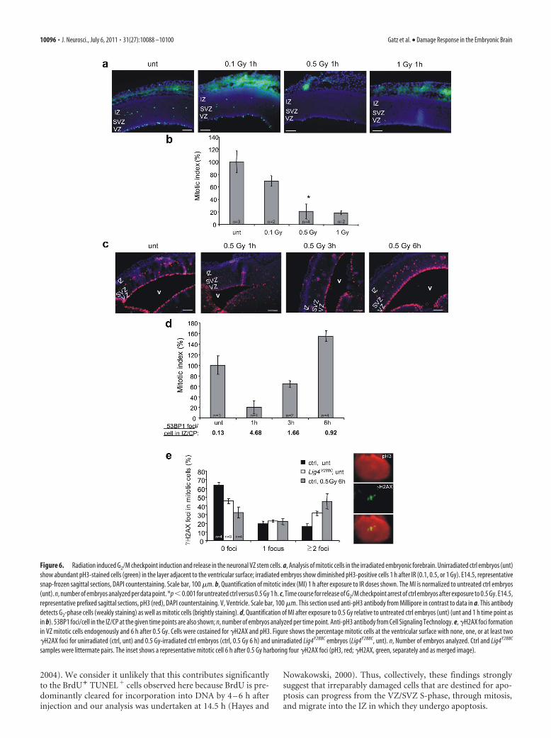
Insensitive G2 /M checkpoint arrest in the VZ/SVZIn cultured cells, the G2/M checkpoint, a damage response thatrestricts the proliferation of damaged cells, has a defined sensitiv-ity and is not initiated by low doses nor maintained until thecompletion of DSB repair (Deckbar et al., 2007). Consequently,mitotic breaks arise predominantly in cells released from check-point arrest. Using anti-pH3 staining to identify mitotic cells, weobserved the expected mitotic zone in the VZ adjacent to theventricular surface (plus the basal mitotic layer) (Fig. 6a). Weobserved that G2/M checkpoint arrest is fully activated 1 h after0.5 Gy IR but is only partially activated after 0.1 Gy (Fig. 6a,b).The 0.1 Gy dose induces four to six DSBs in G2 cells, suggestingthat, as in cultured cells, the G2/M checkpoint has a defined sen-sitivity in vivo (Deckbar et al., 2007). Examination of the durationof arrest showed that mitotic entry occurs 3 h after 0.5 Gy, and fullrecovery is observed after 6 h (Fig. 6c,d). The increased mitoticindex at 6 h compared with unirradiated samples most likelyrepresents the accumulation of G2-phase cells during checkpointarrest. Because we could not quantify DSBs in G2-phase VZ cellsattributable to very low 53BP1 expression, they were enumeratedin the IZ 3 and 6 h after 0.5 Gy (Fig. 6d). Assuming a similar rateof DSB repair in the VZ/SVZ and IZ/CP, the presence of DSBs at3– 6 h strongly suggests that the checkpoint is released before thecompletion of DSB repair.
To investigate this further, we examined �H2AX foci in mi-totic cells after checkpoint release using �H2AX analysis. 53BP1was not used because it dissociates from chromatin during mito-sis (Nakamura et al., 2010). A total of 36% of mitotic cells inuntreated embryos harbor �H2AX foci (Fig. 6e). A previousstudy also observed endogenous �H2AX foci in undamaged mi-totic cells (McManus and Hendzel, 2005). However, 6 h after 0.5Gy, when checkpoint release has occurred, 67% of mitotic cellsharbor �H2AX, with 45% having at least two foci versus 17% inthe ctrl (Fig. 6e). Interestingly, untreated Lig4Y288C mitotic cellsshow an intermediate phenotype consistent with persisting DSBs.These findings strikingly demonstrate that checkpoint arrest inthe VZ cells has a defined sensitivity that allows progression ofcells with �H2AX foci into mitosis. This finding is consistent witha similar mitotic index in Lig4Y288C and ctrl embryos despite ele-vated DSBs in Lig4Y288C cells (data not shown). This quantifica-tion of �H2AX foci was performed on the apical mitotic cells ofthe VZ. The number of �H2AX foci in the basal mitotic layer ofthe SVZ was similar to that found in the VZ mitotic cells, dem-onstrating that the G2/M checkpoint in the SVZ is also insensitiveand that the intermediate precursor cells of the SVZ can similarlyprogress through mitosis before DSB repair is completed.
Irradiated VZ/SVZ cells can progress to the IZ andundergo apoptosisAlthough the presence of �H2AX on mitotic chromosomes sug-gests that they harbor DSBs, it is also possible that H2AX dephos-phorylation lags behind DSB repair. We, therefore, examineddirectly whether damaged VZ/SVZ cells can transit to the IZ andundergo apoptosis. We injected a standard dose of BrdU intopregnant mice to label S-phase cells and, 30 min later, mock orIR-treated the mice with 0.5 Gy. Embryos were collected 1 or14.5 h after IR because the estimated cell cycle time in the VZ atE14.5 is 15 h (Mitsuhashi and Takahashi, 2009). The 0.5 Gy dosewas chosen as a dose that induced apoptosis but not prolongedcheckpoint arrest. Previous and our own pilot experiments withembryos exposed to BrdU for 30 min to 2 h at E14.5 showed asingle broad band of BrdU staining covering the proliferativeareas of the VZ/SVZ (Fig. 7a) (Hayes and Nowakowski, 2000;Orii et al., 2006; Sunabori et al., 2008). Importantly, analysis at 1 hafter 0.5 Gy showed that most BrdU-labeled cells in the SVZcoexpress Tbr2 (Fig. 7a), consistent with previous findings(Englund et al., 2005). At 15 h after BrdU injection, staining ofunirradiated embryos for Tbr2 and BrdU revealed that someBrdU-positive cells were basal to the Tbr2-expressing cell layer(Fig. 7b) and were present in the Tuj1-positive differentiated IZlayer (Fig. 7c). These cells predominantly no longer express Tbr2.These findings are consistent with the presence of S-phase-labeled cells that have progressed into the IZ and differentiated(Fig. 7b,c). Triple-staining for BrdU, TUNEL, and Tuj1 demon-strated that BrdU treatment alone did not induce apoptosis (Fig.7c). Importantly, after exposure to 0.5 Gy and analysis 14.5 hlater, although the number of BrdU-positive cells is reduced in allregions probably as a result of delayed replication and cell cycleprogression, and cell loss attributable to apoptosis, �10 –20% ofthe BrdU-positive cells present in the IZ were TUNEL positive(Fig. 7d,e). BrdU-negative TUNEL-positive cells were also ob-served, likely representing irradiated IZ cells and/or unlabeledVZ/SVZ-irradiated cells. Studies have suggested that apoptosis inpostmitotic neurons occurs via cell cycle reentry and can result inBrdU incorporation (Becker and Guarente, 1991; Kruman et al.,
Table 1. 53BP1 foci and apoptosis in the neuronal cortex of adult mice
ctrl Lig4Y288C
53BP1foci/cell Apoptosis
53BP1foci/cell Apoptosis
Untreated (n � 3) 0.02 �1.0% 0.09 �1.0%3 Gy 15 h (n � 1) 1.66 �1.0% 19.15 �1.0%10 Gy 15 h (n � 1) 4.49 �1.0% ��20.00(notquantified) �1.0%
Quantification of 53BP1 foci/cell and apoptotis (percentage, measured by TUNEL staining) for ctrl and Lig4Y288C
neuronal cortex regions of animals at 3–5 months of age. All pairs were sex matched and (except 1 untreated pair)were littermate pairs. n represents the number of animals analyzed per condition.
Gatz et al. • Damage Response in the Embryonic Brain J. Neurosci., July 6, 2011 • 31(27):10088 –10100 • 10095
2004). We consider it unlikely that this contributes significantlyto the BrdU� TUNEL� cells observed here because BrdU is pre-dominantly cleared for incorporation into DNA by 4 – 6 h afterinjection and our analysis was undertaken at 14.5 h (Hayes and
Nowakowski, 2000). Thus, collectively, these findings stronglysuggest that irreparably damaged cells that are destined for apo-ptosis can progress from the VZ/SVZ S-phase, through mitosis,and migrate into the IZ in which they undergo apoptosis.
Figure 6. Radiation induced G2/M checkpoint induction and release in the neuronal VZ stem cells. a, Analysis of mitotic cells in the irradiated embryonic forebrain. Unirradiated ctrl embryos (unt)
show abundant pH3-stained cells (green) in the layer adjacent to the ventricular surface; irradiated embryos show diminished pH3-positive cells 1 h after IR (0.1, 0.5, or 1 Gy). E14.5, representative
snap-frozen sagittal sections, DAPI counterstaining. Scale bar, 100 �m. b, Quantification of mitotic index (MI) 1 h after exposure to IR doses shown. The MI is normalized to untreated ctrl embryos
(unt). n, number of embryos analyzed per data point. *p�0.001 for untreated ctrl versus 0.5 Gy 1 h. c, Time course for release of G2/M checkpoint arrest of ctrl embryos after exposure to 0.5 Gy. E14.5,
representative prefixed sagittal sections, pH3 (red), DAPI counterstaining. V, Ventricle. Scale bar, 100 �m. This section used anti-pH3 antibody from Millipore in contrast to data in a. This antibody
detects G2-phase cells (weakly staining) as well as mitotic cells (brightly staining). d, Quantification of MI after exposure to 0.5 Gy relative to untreated ctrl embryos (unt) (unt and 1 h time point as
in b). 53BP1 foci/cell in the IZ/CP at the given time points are also shown; n, number of embryos analyzed per time point. Anti-pH3 antibody from Cell Signaling Technology. e, �H2AX foci formation
in VZ mitotic cells endogenously and 6 h after 0.5 Gy. Cells were costained for �H2AX and pH3. Figure shows the percentage mitotic cells at the ventricular surface with none, one, or at least two
�H2AX foci for unirradiated (ctrl, unt) and 0.5 Gy-irradiated ctrl embryos (ctrl, 0.5 Gy 6 h) and unirradiated Lig4Y288C embryos (Lig4Y288C, unt). n, Number of embryos analyzed. Ctrl and Lig4Y288C
samples were littermate pairs. The inset shows a representative mitotic cell 6 h after 0.5 Gy harboring four �H2AX foci (pH3, red; �H2AX, green, separately and as merged image).
10096 • J. Neurosci., July 6, 2011 • 31(27):10088 –10100 Gatz et al. • Damage Response in the Embryonic Brain
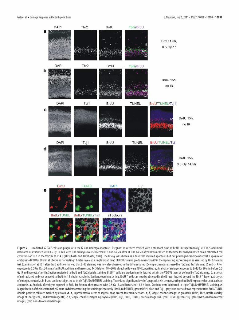
Figure 7. Irradiated VZ/SVZ cells can progress to the IZ and undergo apoptosis. Pregnant mice were treated with a standard dose of BrdU (intraperitoneally) at E14.5 and mock
irradiated or irradiated with 0.5 Gy 30 min later. The embryos were collected at 1 and 14.5 h after IR. The 14.5 h after IR was chosen as the time for analysis based on an estimated cell
cycle time of 15 h in the VZ/SVZ at E14.5 (Mitsuhashi and Takahashi, 2009). The 0.5 Gy was chosen as a dose that induced apoptosis but not prolonged checkpoint arrest. Exposure of
embryos to BrdU for 30 min at E14.5 and harvesting 1 h later revealed a single broad band of BrdU staining predominantly within the replicating VZ/SVZ region as assessed by Tbr2 staining
(a). Examination at 15 h after BrdU addition showed that BrdU staining was now also observed in the differentiated IZ compartment as assessed by Tbr2 and Tuj1 staining (b and c). After
exposure to 0.5 Gy IR at 30 min after BrdU addition and harvesting 14.5 h later, 10 –20% of such cells were TUNEL positive. a, Analysis of embryos exposed to BrdU for 30 min before 0.5
Gy IR and harvest after 1 h. Section subjected to BrdU and Tbr2 double staining. BrdU � cells are predominantly located within the VZ/SVZ layer as defined by Tbr2 staining; b, analysis
of unirradiated embryos exposed to BrdU for 15 h before analysis. Sections examined as in a. BrdU � cells can now be observed in the IZ layer located beyond the Tbr2 � layer. c, Analysis
of embryos treated as in b and sections subjected to triple Tuj1/BrdU/TUNEL staining. There is no significant level of apoptotic cells demonstrating that BrdU exposure does not activate
apoptosis. d, Analysis of embryos exposed to BrdU for 30 min, then treated with 0.5 Gy IR, and harvested 14.5 h later. Sections were subjected to triple Tuj1/BrdU/TUNEL staining. e,
Magnification of the inset from the IZ zone in d demonstrating the stainings separately (BrdU, red; TUNEL, green; DAPI, blue; and Tuj1, gray) and overlaid; two representative BrdU/TUNEL
double-positive cells are marked by arrows. a–d, Representative areas of sagittal snap-frozen forebrain sections. a, b, Single-channel images in grayscale (DAPI, Tbr2, BrdU), overlay
image of Tbr2 (green), and BrdU (magenta). c, d, Single-channel images in grayscale (DAPI, Tuj1, BrdU, TUNEL), overlay image BrdU (red)/TUNEL (green)/Tuj1 (blue) (a/b/e) deconvolved
images, (c/d) non-deconvolved images.
Gatz et al. • Damage Response in the Embryonic Brain J. Neurosci., July 6, 2011 • 31(27):10088 –10100 • 10097
DiscussionMicrocephaly is a marked feature of LIG4 syndrome and XLF-deficiency, suggestingthatNHEJplaysacritical roleduringembryogen-esis (O’Driscoll et al., 2001; Buck et al., 2006). Although these featureshave been demonstrated previously, our aim here is to gain mechanistic
insight into the underlying cause, which we achieve by examining DNA
breakage, apoptosis, and cell cycle checkpoint arrest in parallel. Further-
more, we focus on the use of physiologically relevant low doses.
Previous studies have shown that the rapidly dividing neuro-nal VZ/SVZ stem/progenitor cells are highly radiation sensitivebetween E13.5 and E16.5, when rapid proliferation occurs(Hoshino and Kameyama, 1988; Hoshino et al., 1991). We sub-stantiate these findings and show that apoptosis in the VZ/SVZcan be activated by two to five DSBs because 100 mGy introducestwo to three DSBs per cell (Rydberg, 2000; Lobrich et al., 2010).Previous studies using high IR doses have suggested that apopto-sis in the VZ/SVZ is ATM independent, although being entirelyATM dependent in the IZ (Lee et al., 2001). However, a morerecent study showed diminished endogenous apoptosis in theVZ/SVZ of ATM�/� mice conditionally inactivated for LigIVcompared with ATM�/� Lig4Nes-Cre mice (Shull et al., 2009).Consistent with the latter finding, we observe that, after 0.5 Gy,apoptosis in the VZ/SVZ occurs via ATM-dependent and ATM-independent processes, with 75% apoptosis being ATM depen-dent. Consistent with previous findings, apoptosis in the IZ isentirely (or at least predominantly) ATM dependent. Thus, wesuggest that two processes contribute to the high apoptotic sen-sitivity in the VZ/SVZ; hypersensitivity to ATM-dependent apo-ptosis at DSBs [which is greater than in the IZ (Fig. 3b)] and anATM-independent process. The latter could represent ATR-activation at stalled replication or a stress response process (e.g.,p38 signaling). Furthermore, we demonstrate that the thresholdsensitivity for activating apoptosis diminishes during develop-ment from the VZ/SVZ to the IZ/CP, the developing cortex, andis very low in the cortex of 3-month-old mice. We show thatNHEJ functions efficiently in the VZ/SVZ and IZ/CP after IR.This is important because repair mechanisms have been sug-gested to be downregulated in stem cells to preclude the survivalof damaged cells (Stambrook, 2007). Although we could notmonitor the rate of DSB repair in the VZ/SVZ, the similar mag-nitude of IR-induced apoptosis in the VZ/SVZ and IZ/CP inLig4Y288C mice suggests that NHEJ functions similarly in bothcompartments.
We show a functional G2/M checkpoint in the VZ/SVZ but, asin cultured cells, the process is insensitive to low DSB numbers(Deckbar et al., 2007). It is not fully activated after 0.1 Gy and isreleased before the completion of DSB repair after higher doses.Importantly, the checkpoint does not prevent irradiated VZ/SVZcells progressing into the IZ to undergo apoptosis. This repre-sents the first examination of the efficacy of checkpoint arrest invivo and shows that damaged cells can transit from the stem/progenitor zone to the differentiated, postmitotic compartment.We conclude that NHEJ and G2/M checkpoint arrest function inthe VZ/SVZ and that hyper-radiosensitivity is caused by exqui-sitely sensitive signaling to apoptosis.
Although Lig4Y288C embryos show high apoptosis in the IZ/CPas observed in previous studies using LigIV�/� mice (Barnes etal., 1998; Frank et al., 2000; Orii et al., 2006; Shull et al., 2009),apoptosis is also significantly elevated in the VZ/SVZ. The muta-tion in Lig4Y288C mice allows slow DSB repair. Thus, the observedDSBs represent steady-state levels between induction and repair.Elevated DSBs in the Lig4Y288C VZ/SVZ/IZ/CP diminish tempo-
rally with the arrest of VZ proliferation, suggesting that VZ pro-liferation causes high DSB formation. Although quantification inthe VZ is difficult, 53BP1 foci numbers in the Lig4Y288C IZ/CPwere similar to that induced by 0.1 Gy IR, suggesting a steady-state level of approximately two to three DSBs. A question arisingfrom this work is the origin of the DSBs in the VZ/SVZ/IZ/CP.One possibility is that DSBs originate in the VZ/SVZ because ofthe rapid replication and that cells with DSBs transit to the IZ. Weprovide strong evidence to support this model. However, it is alsopossible that high DSB formation arises in the IZ as a conse-quence of high metabolic activity in the VZ/SVZ, e.g., IZ cells maymaintain high oxidative stress levels. Regardless of their origin,such high DSB formation is not observed in non-neuronalLig4Y288C embryonic tissues (e.g., the tongue) nor in the brain of3-month-old Lig4Y288C mice.
The combined analysis of 53BP1 foci numbers and apoptosisin the embryonic, newborn, and adult neuronal tissues providesinsight into the distinct tissue responses to unrepaired DSBs andstrongly suggests that the high sensitivity to DSB-induced apo-
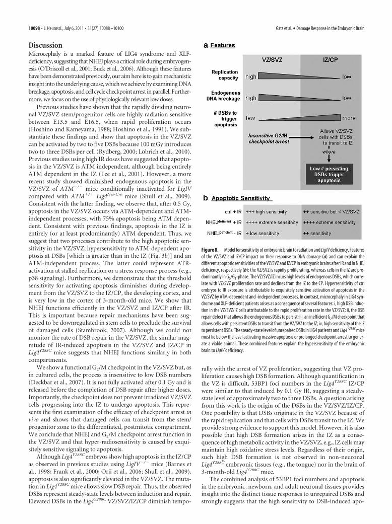
Figure 8. Model for sensitivity of embryonic brain to radiation and LigIV deficiency. Features
of the VZ/SVZ and IZ/CP impact on their response to DNA damage (a) and can explain the
different apoptotic sensitivities of the VZ/SVZ and IZ/CP in embryonic brains after IR and in NHEJ
deficiency, respectively (b): the VZ/SVZ is rapidly proliferating, whereas cells in the IZ are pre-
dominantly in G0/G1-phase. The VZ/SVZ/IZ incurs high levels of endogenous DSBs, which corre-
late with VZ/SVZ proliferation rate and declines from the IZ to the CP. Hypersensitivity of ctrl
embryos to IR exposure is attributable to exquisitely sensitive activation of apoptosis in the
VZ/SVZ by ATM-dependent and -independent processes. In contrast, microcephaly in LIG4 syn-
drome and XLF-deficient patients arises as a consequence of several features: i, high DSB induc-
tion in the VZ/SVZ/IZ cells attributable to the rapid proliferation rate in the VZ/SVZ; ii, the DSB
repair defect that allows the endogenous DSBs to persist; iii, an inefficient G2/M checkpoint that
allows cells with persistent DSBs to transit from the VZ/SVZ to the IZ; iv, high sensitivity of the IZ
to persistent DSBs. The steady-state level of unrepaired DSBs in LIG4 patients and Lig4Y288C mice
must lie below the level activating massive apoptosis or prolonged checkpoint arrest to gener-
ate a viable animal. These combined features explain the hypersensitivity of the embryonic
brain to LigIV deficiency.
10098 • J. Neurosci., July 6, 2011 • 31(27):10088 –10100 Gatz et al. • Damage Response in the Embryonic Brain
ptosis diminishes during neuronal development and represents aunique feature of the VZ/SVZ and IZ/CP cells.
Why is apoptosis in Lig4Y288C embryos greater in the IZ/CPthan the VZ/SVZ, which is not expected if DSBs originate in theVZ/SVZ? Although similar DSB numbers are observed in theLig4Y288C VZ/SVZ/IZ/CP and 0.1 Gy irradiated ctrl mice, 0.1 Gyinduces little apoptosis in the ctrl IZ/CP. DSBs in Lig4Y288C cellsdiffer from those in ctrl cells because they are persistent as a resultof the impact of the Lig4Y288C mutation on DSB repair. One pos-sibility is that HR can effect DSB repair in late S/G2-phase VZ/SVZ cells, which does not occur in nonreplicating IZ/CP cells.However, analysis of G2-phase DSB repair in cultured cells fromLIG4 syndrome patients exposed a pronounced DSB repair de-fect, suggesting that HR cannot fully compensate for loss of LigIV(Beucher et al., 2009). Significantly, the steady-state number ofDSBs (two to three) in Lig4Y288C mice is lower than the thresholdinducing extensive VZ/SVZ apoptosis. Furthermore, two to threeDSBs do not activate prolonged G2/M checkpoint arrest, andLig4Y288C and ctrl mice have a similar mitotic index. Moreover,our findings suggest that the IZ/CP may be hypersensitive topersisting DSBs because IR-induced apoptosis in the Lig4Y288C
VZ/SVZ and IZ/CP regions were similar, in contrast to muchlower apoptosis in the IR-treated ctrl IZ/CP, in which DSBs arerapidly repaired. Thus, we propose that, although the VZ/SVZhas exquisite sensitivity to acute DSB formation, the IZ/CP ishypersensitive to persisting DSBs. Interestingly, we observed agradient in 53BP1 expression with low to higher expression in theVZ/SVZ/IZ/CP. 53BP1 has been reported to amplify ATM signal-ing and specifically functions in signaling from persistent DSBs(Fernandez-Capetillo et al., 2002; Shibata et al., 2010). However,despite the low 53BP1 levels, ATM-dependent apoptosis is moresensitively activated in the VZ/SVZ compared with the IZ region(Fig. 3c). Thus, this finding would suggest that, although 53BP1binds to p53 and is reported to function as a p53 transcriptionalcoactivator, its reduced expression levels in the VZ/SVZ does notprevent the activation of apoptosis (Iwabuchi et al., 1998). Asdiscussed above, however, there is evidence that 53BP1 mightspecifically enhances signaling from a low level of persistingDSBs.
Based on these findings, we propose a novel model to explainthe distinct sensitivities caused by irradiation and LigIV defi-ciency (Fig. 8). We suggest that microcephaly in LIG4 syndromeand XLF patients arises as a consequence of two features: highDSB induction in the VZ/SVZ/IZ cells as a consequence of rapidVZ/SVZ replication coupled with a low threshold for undergoingapoptosis in the VZ/SVZ and IZ/CP. Although the VZ/SVZ re-gion is highly sensitive to acute DSB formation, the IZ/CP ap-pears to be sensitive to a low level of persisting DSBs. Because ofthe insensitivity of the G2/M checkpoint, damaged cells with lowDSBs can transit into the IZ and undergo apoptosis. The sensi-tivity of the IZ/CP to persistent DSBs may provide a mechanismto eliminate such damaged cells and in LIG4 syndromes contrib-utes to neuronal cell loss.
These studies provide insight into the interplay between DNAdamage responses in the developing brain and their roles in repairingendogenously arising DSBs that can be significant during embryonicdevelopment. Given the increased usage of CT scanning and fre-quency of flying, it is important to understand the magnitude of, andbasis underlying, embryonic neuronal sensitivity.
ReferencesAbner CW, McKinnon PJ (2004) The DNA double-strand break response in
the nervous system. DNA Repair (Amst) 3:1141–1147.
Barlow C, Hirotsune S, Paylor R, Liyanage M, Eckhaus M, Collins F, Shiloh Y,
Crawley JN, Ried T, Tagle D, Wynshaw-Boris A (1996) Atm-deficient
mice: a paradigm of ataxia telangiectasia. Cell 86:159 –171.
Barnes DE, Stamp G, Rosewell I, Denzel A, Lindahl T (1998) Targeted dis-
ruption of the gene encoding DNA ligase IV leads to lethality in embry-
onic mice. Curr Biol 8:1395–1398.
Bayer SA, Altman J, Dai XF, Humphreys L (1991) Planar differences in nu-
clear area and orientation in the subventricular and intermediate zones of
the rat embryonic neocortex. J Comp Neurol 307:487– 498.
Becker DM, Guarente L (1991) High efficiency transformation of yeast by
electroporation. Methods Enzymol 194:182–187.
Beucher A, Birraux J, Tchouandong L, Barton O, Shibata A, Conrad S, Goo-
darzi AA, Krempler A, Jeggo PA, Lobrich M (2009) ATM and Artemis
promote homologous recombination of radiation-induced DNA double-
strand breaks in G2. EMBO J 28:3413–3427
Buck D, Malivert L, de Chasseval R, Barraud A, Fondaneche MC, Sanal O,
Plebani A, Stephan JL, Hufnagel M, le Deist F, Fischer A, Durandy A, de
Villartay JP, Revy P (2006) Cernunnos, a novel nonhomologous end-
joining factor, is mutated in human immunodeficiency with microceph-
aly. Cell 124:287–299.
Cairns J (2006) Cancer and the immortal strand hypothesis. Genetics
174:1069 –1072.
Deckbar D, Birraux J, Krempler A, Tchouandong L, Beucher A, Walker S, Stiff
T, Jeggo P, Lobrich M (2007) Chromosome breakage after G2 check-
point release. J Cell Biol 176:749 –755.
Englund C, Fink A, Lau C, Pham D, Daza RA, Bulfone A, Kowalczyk T,
Hevner RF (2005) Pax6, Tbr2, and Tbr1 are expressed sequentially by
radial glia, intermediate progenitor cells, and postmitotic neurons in de-
veloping neocortex. J Neurosci 25:247–251.
Fernandez-Capetillo O, Chen HT, Celeste A, Ward I, Romanienko PJ, Mo-
rales JC, Naka K, Xia Z, Camerini-Otero RD, Motoyama N, Carpenter PB,
Bonner WM, Chen J, Nussenzweig A (2002) DNA damage-induced
G2-M checkpoint activation by histone H2AX and 53BP1. Nat Cell Biol
4:993–997.
Frank KM, Sekiguchi JM, Seidl KJ, Swat W, Rathbun GA, Cheng HL, David-
son L, Kangaloo L, Alt FW (1998) Late embryonic lethality and impaired
V(D)J recombination in mice lacking DNA ligase IV. Nature
396:173–177.
Frank KM, Sharpless NE, Gao Y, Sekiguchi JM, Ferguson DO, Zhu C, Manis
JP, Horner J, DePinho RA, Alt FW (2000) DNA ligase IV deficiency in
mice leads to defective neurogenesis and embryonic lethality via the p53
pathway. Mol Cell 5:993–1002.
Gao Y, Sun Y, Frank KM, Dikkes P, Fujiwara Y, Seidl KJ, Sekiguchi JM,
Rathbun GA, Swat W, Wang J, Bronson RT, Malynn BA, Bryans M, Zhu
C, Chaudhuri J, Davidson L, Ferrini R, Stamato T, Orkin SH, Greenberg
ME, Alt FW (1998) A critical role for DNA end-joining proteins in both
lymphogenesis and neurogenesis. Cell 95:891–902.
Hayes NL, Nowakowski RS (2000) Exploiting the dynamics of S-phase trac-
ers in developing brain: interkinetic nuclear migration for cells entering
versus leaving the S-phase. Dev Neurosci 22:44 –55.
Hoshino K, Kameyama Y (1988) Developmental-stage-dependent radio-
sensitivity of neural cells in the ventricular zone of telencephalon in
mouse and rat fetuses. Teratology 37:257–262.
Hoshino K, Kameyama Y, Inouye M (1991) Split-dose effect of
X-irradiation on the induction of cell death in the fetal mouse brain.
J Radiat Res (Tokyo) 32:23–27.
Iwabuchi K, Li B, Massa HF, Trask BJ, Date T, Fields S (1998) Stimulation of
p53-mediated transcriptional activation by the p53-binding proteins,
53BP1 and 53BP2. J Biol Chem 273:26061–26068.
Kruman II, Wersto RP, Cardozo-Pelaez F, Smilenov L, Chan SL, Chrest FJ,
Emokpae R, Jr., Gorospe M, Mattson MP (2004) Cell cycle activation
linked to neuronal cell death initiated by DNA damage. Neuron
41:549 –561.
Lee Y, Chong MJ, McKinnon PJ (2001) Ataxia telangiectasia mutated-
dependent apoptosis after genotoxic stress in the developing nervous
system is determined by cellular differentiation status. J Neurosci
21:6687– 6693.
Liao MJ, Yin C, Barlow C, Wynshaw-Boris A, van Dyke T (1999) Atm is
dispensable for p53 apoptosis and tumor suppression triggered by cell
cycle dysfunction. Mol Cell Biol 19:3095–3102.
Lobrich M, Shibata A, Beucher A, Fisher A, Ensminger M, Goodarzi AA,
Barton O, Jeggo PA (2010) gamma H2AX foci analysis for monitoring
Gatz et al. • Damage Response in the Embryonic Brain J. Neurosci., July 6, 2011 • 31(27):10088 –10100 • 10099
DNA double-strand break repair: Strengths, limitations and optimiza-tion. Cell Cycle 9:662– 669.
McManus KJ, Hendzel MJ (2005) ATM-dependent DNA damage-independent mitotic phosphorylation of H2AX in normally growingmammalian cells. Mol Biol Cell 16:5013–5025.
Mitsuhashi T, Takahashi T (2009) Genetic regulation of proliferation/dif-ferentiation characteristics of neural progenitor cells in the developingneocortex. Brain Dev 31:553–557.
Nakamura AJ, Rao VA, Pommier Y, Bonner WM (2010) The complexity ofphosphorylated H2AX foci formation and DNA repair assembly at DNAdouble-strand breaks. Cell Cycle 9:389 –397.
Nijnik A, Woodbine L, Marchetti C, Dawson S, Lambe T, Liu C, RodriguesNP, Crockford TL, Cabuy E, Vindigni A, Enver T, Bell JI, Slijepcevic P,Goodnow CC, Jeggo PA, Cornall RJ (2007) DNA repair is limiting forhaematopoietic stem cells during ageing. Nature 447:686 – 690.
O’Driscoll M, Cerosaletti KM, Girard PM, Dai Y, Stumm M, Kysela B, HirschB, Gennery A, Palmer SE, Seidel J, Gatti RA, Varon R, Oettinger MA,Neitzel H, Jeggo PA, Concannon P (2001) DNA Ligase IV mutationsidentified in patients exhibiting development delay and immunodefi-ciency. Mol Cell 8:1175–1185.
Orii KE, Lee Y, Kondo N, McKinnon PJ (2006) Selective utilization of non-homologous end-joining and homologous recombination DNA repairpathways during nervous system development. Proc Natl Acad Sci U S A103:10017–10022.
Pontious A, Kowalczyk T, Englund C, Hevner RF (2008) Role of intermedi-ate progenitor cells in cerebral cortex development. Dev Neurosci30:24 –32.
Rube CE, Grudzenski S, Kuhne M, Dong X, Rief N, Lobrich M, Rube C(2008) DNA double-strand break repair of blood lymphocytes and nor-
mal tissues analysed in a preclinical mouse model: implications for radi-
osensitivity testing. Clin Cancer Res 14:6546 – 6555.
Rydberg B (2000) Radiation-induced heat-labile sites that convert into
DNA double-strand breaks. Radiat Res 153:805– 812.
Sekiguchi J, Ferguson DO, Chen HT, Yang EM, Earle J, Frank K, Whitlow S,
Gu Y, Xu Y, Nussenzweig A, Alt FW (2001) Genetic interactions be-
tween ATM and the nonhomologous end-joining factors in genomic sta-
bility and development. Proc Natl Acad Sci U S A 98:3243–3248.
Shibata A, Barton O, Noon AT, Dahm K, Deckbar D, Goodarzi AA, Lobrich
M, Jeggo PA (2010) Role of ATM and the damage response mediator
proteins 53BP1 and MDC1 in the maintenance of G(2)/M checkpoint
arrest. Mol Cell Biol 30:3371–3383.
Shull ER, Lee Y, Nakane H, Stracker TH, Zhao J, Russell HR, Petrini JH,
McKinnon PJ (2009) Differential DNA damage signaling accounts for
distinct neural apoptotic responses in ATLD and NBS. Genes Dev
23:171–180.
Soares HD, Morgan JI, McKinnon PJ (1998) Atm expression patterns sug-
gest a contribution from the peripheral nervous system to the phenotype
of ataxia-telangiectasia. Neuroscience 86:1045–1054.
Stambrook PJ (2007) An ageing question: do embryonic stem cells protect
their genomes? Mech Ageing Dev 128:31–35.
Sunabori T, Tokunaga A, Nagai T, Sawamoto K, Okabe M, Miyawaki A,
Matsuzaki Y, Miyata T, Okano H (2008) Cell-cycle-specific nestin ex-
pression coordinates with morphological changes in embryonic cortical
neural progenitors. J Cell Sci 121:1204 –1212.
Takahashi T, Nowakowski RS, Caviness VS Jr (1995) Early ontogeny of the
secondary proliferative population of the embryonic murine cerebral
wall. J Neurosci 15:6058 – 6068.
10100 • J. Neurosci., July 6, 2011 • 31(27):10088 –10100 Gatz et al. • Damage Response in the Embryonic Brain