Embed Size (px)
Citation preview
Reproductive constraint is a developmentalmechanism that maintains social harmonyin advanced ant societiesAbderrahman Khila1 and Ehab Abouheif1
Department of Biology, McGill University, 1205 Avenue Docteur Penfield, Montreal, QC, Canada H3A 1B1
Edited by Gene E. Robinson, University of Illinois, Urbana, IL, and approved October 7, 2008 (received for review July 29, 2008)
A hallmark of eusociality in ants is the reproductive division oflabor between queens and workers. Yet, nothing is known aboutthe molecular mechanisms underlying reproduction in this group.We therefore compared the developmental genetic capacity ofqueens and workers to reproduce in several eusocially advancedspecies from the two largest subfamilies of ants, the Myrmicinaeand Formicinae. In flies, the asymmetric localization of maternallyencoded determinants (mRNAs and proteins) during oogenesisestablishes oocyte polarity and subsequently ensures proper em-bryonic development. Vasa and nanos, two key maternal deter-minants, are properly localized in the posterior of queen oocytes,but their localization is impaired in those of the workers. Thismislocalization leads to severe embryonic defects in worker prog-eny, and therefore, represents a constraint on worker reproductionthat we call ‘reproductive constraint.’ We show that reproductiveconstraint is phylogenetically widespread, and is at high levels inmost species tested. Reproductive constraint can simultaneouslyreduce or eliminate the workers’ ability to produce viable eggs forreproduction, while preserving their ability to produce trophic eggsfor nutrition, and thus, may have been the basis for the evolutionaryretention of worker ovaries in the majority of ants. We propose thathigh levels of reproductive constraint has most likely evolved as aconsequence of selection at the colony level to reduce or eliminateany potential conflict over worker reproduction, therefore maintain-ing harmony and colony efficiency in advanced ant societies.
conflict � oogenesis � Vasa/nanos � worker reproduction
The reproductive division of labor in ants is a key feature of theireusocial organization. Workers in the vast majority of ant
species, however, have retained ovaries and are thought to havesignificant reproductive potential (1–3). A major and widespreadconflict will potentially arise in ant societies if workers selfishlyengage in reproduction at the expense of colony tasks, such asforaging and brood rearing (4). An important challenge for evo-lutionary biology is to understand how highly social groups canreduce or prevent such costly conflicts from occurring. Althoughthere exist a large body of theory to understand cooperation andconflict over reproduction in ants (5–11), little is known about thedevelopmental and molecular mechanisms that control reproduc-tion in queens and workers.
Reproduction in ants follows a haplo-diploid sex determinationmechanism, whereby fertilized eggs develop into females, andunfertilized eggs develop into males (12). Workers in advanced antsocieties cannot mate and have lost the spermatheca, and thus, theycan only produce unfertilized eggs that develop either into males forreproduction (1–3) or trophic eggs for nutrition (12, 13). Hamilton’srule, which is based on both the genetic relatedness betweenindividuals and the ecological benefits and costs these individualsincur, has been used to predict the potential for conflict over maleproduction in ant societies (5). Because ecological costs and ben-efits are difficult to assess, relatedness alone has mainly been usedto predict the potential for this conflict (3, 14). Relatedness predictsthat in colonies headed by one queen that is singly mated, workersare more related to other workers’ sons (nephews) than to the
queen’s sons (brothers), and therefore should rear their nephewsover their brothers. Conversely, in colonies headed by multiplequeens or a multiply mated queen, workers are more related to theirbrothers and therefore should refrain (through ‘‘self-restraint’’) orprevent other workers (through ‘‘policing’’) from reproducing.Empirical data, however, show that the actual levels of conflict overworker production of males in nature are much lower than expected(3, 14), as worker reproduction is usually reduced or eliminated inthe majority of ant species. Because it is generally assumed thatworker-produced eggs successfully develop into males (1, 2), thegap between actual and potential levels of this conflict has mainlybeen explained by behavioral control, such as self-restraint (15, 16)or policing (17–20).
Here, we tested the basic assumption that workers in advancedant societies have a significant capacity to successfully producemales in three focal ant species: Aphaenogaster rudis, Myrmicaamericana, and Lasius niger. We examined the developmentalgenetic capacity of the workers in these species to perform oogen-esis, as well as the capacity of their eggs to undergo properembryonic development, both in the presence and in the absenceof the queen. We uncovered a phylogenetically widespread devel-opmental mechanism that can simultaneously reduce or eliminatethe ability of the workers to produce viable eggs, while maintainingtheir ability to produce trophic eggs for nutrition.
ResultsWe first asked whether the process of oogenesis is generallyconserved between ant queens and the fruit fly, Drosophila mela-nogaster. In Drosophila, oogenesis begins by the division of germ-line stem cells (GSCs), which give rise to cysts and then to eggchambers (21). The oocyte develops within the egg chamber, whereit acquires its polarity through the precise localization of mRNAsand proteins known as maternal determinants (22, 23). Maternaldeterminants are produced by the nurse cells, and then transportedto the oocyte where they are asymmetrically localized to the poles.Vasa and nanos, two highly conserved maternal determinants, arelocalized to the posterior pole of the oocyte. These molecules arenecessary for patterning the posterior compartment of early em-bryos, and for specifying germ-line formation and differentiation(24, 25). Vasa mutant embryos lack the specialized posterior poleplasm and show impaired polarity (25), whereas nanos mutantembryos fail to form most of the abdomen altogether (24). Wefound that the cellular organization (Fig. 1 A and B) and the
Author contributions: A.K. and E.A. designed research; A.K. performed research; A.K. andE.A. analyzed data; and A.K. and E.A. wrote the paper.
The authors declare no conflict of interest.
This article is a PNAS Direct Submission.
Data deposition: The sequences reported in this paper have been deposited in the GenBankdatabase (accession nos. EU272791, EU272792, and EU272793).
1To whom correspondence may be addressed. E-mail: [email protected] [email protected].
This article contains supporting information online at www.pnas.org/cgi/content/full/0807351105/DCSupplemental.
© 2008 by The National Academy of Sciences of the USA
17884–17889 � PNAS � November 18, 2008 � vol. 105 � no. 46 www.pnas.org�cgi�doi�10.1073�pnas.0807351105
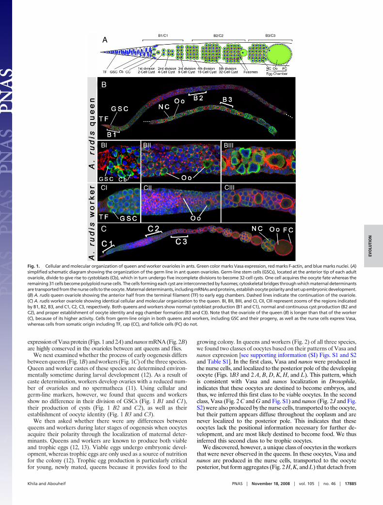
expression of Vasa protein (Figs. 1 and 2A) and nanos mRNA (Fig. 2B)are highly conserved in the ovarioles between ant queens and flies.
We next examined whether the process of early oogenesis differsbetween queens (Fig. 1B) and workers (Fig. 1C) of the three species.Queen and worker castes of these species are determined environ-mentally sometime during larval development (12). As a result ofcaste determination, workers develop ovaries with a reduced num-ber of ovarioles and no spermatheca (11). Using cellular andgerm-line markers, however, we found that queens and workersshow no difference in their division of GSCs (Fig. 1 B1 and C1),their production of cysts (Fig. 1 B2 and C2), as well as theirestablishment of oocyte identity (Fig. 1 B3 and C3).
We then asked whether there were any differences betweenqueens and workers during later stages of oogenesis when oocytesacquire their polarity through the localization of maternal deter-minants. Queens and workers are known to produce both viableand trophic eggs (12, 13). Viable eggs undergo embryonic devel-opment, whereas trophic eggs are only used as a source of nutritionfor the colony (12). Trophic egg production is particularly criticalfor young, newly mated, queens because it provides food to the
growing colony. In queens and workers (Fig. 2) of all three species,we found two classes of oocytes based on their patterns of Vasa andnanos expression [see supporting information (SI) Figs. S1 and S2and Table S1]. In the first class, Vasa and nanos were produced inthe nurse cells, and localized to the posterior pole of the developingoocyte (Figs. 1B3 and 2 A, B, D, K, H, and L). This pattern, whichis consistent with Vasa and nanos localization in Drosophila,indicates that these oocytes are destined to become embryos, andthus, we inferred this first class to be viable oocytes. In the secondclass, Vasa (Fig. 2 C and G and Fig. S1) and nanos (Fig. 2 J and Fig.S2) were also produced by the nurse cells, transported to the oocyte,but their pattern appears diffuse throughout the ooplasm and arenever localized to the posterior pole. This indicates that theseoocytes lack the positional information necessary for further de-velopment, and are most likely destined to become food. We thusinferred this second class to be trophic oocytes.
We discovered, however, a unique class of oocytes in the workersthat were never observed in the queens. In these oocytes, Vasa andnanos are produced in the nurse cells, transported to the oocyteposterior, but form aggregates (Fig. 2 H, K, and L) that detach from
A
B
C
BI BII BIII
CI CII CIII
B1/C1 B2/C2 B3/C3
Fig. 1. Cellular and molecular organization of queen and worker ovarioles in ants. Green color marks Vasa expression, red marks F-actin, and blue marks nuclei. (A)simplified schematic diagram showing the organization of the germ line in ant queen ovarioles. Germ-line stem cells (GSCs), located at the anterior tip of each adultovariole, divide to give rise to cystoblasts (Cb), which in turn undergo five incomplete divisions to become 32-cell cysts. One cell acquires the oocyte fate whereas theremaining 31 cells become polyploid nurse cells. The cells forming each cyst are interconnected by fusomes; cytoskeletal bridges through which maternal determinantsare transported from the nurse cells to the oocyte. Maternal determinants, including mRNAs and proteins, establish oocyte polarity and set up embryonic development.(B) A. rudis queen ovariole showing the anterior half from the terminal filament (TF) to early egg chambers. Dashed lines indicate the continuation of the ovariole.(C) A. rudis worker ovariole showing identical cellular and molecular organization to the queen. BI, BII, BIII, and CI, CII, CIII represent zooms of the regions indicatedby B1, B2, B3, and C1, C2, C3, respectively. Both queens and workers show normal cystoblast production (B1 and C1), normal and continuous cyst production (B2 andC2), and proper establishment of oocyte identity and egg chamber formation (B3 and C3). Note that the ovariole of the queen (B) is longer than that of the worker(C), because of its higher activity. Cells from germ-line origin in both queens and workers, including GSC and their progeny, as well as the nurse cells express Vasa,whereas cells from somatic origin including TF, cap (CC), and follicle cells (FC) do not.
Khila and Abouheif PNAS � November 18, 2008 � vol. 105 � no. 46 � 17885
EVO
LUTI
ON
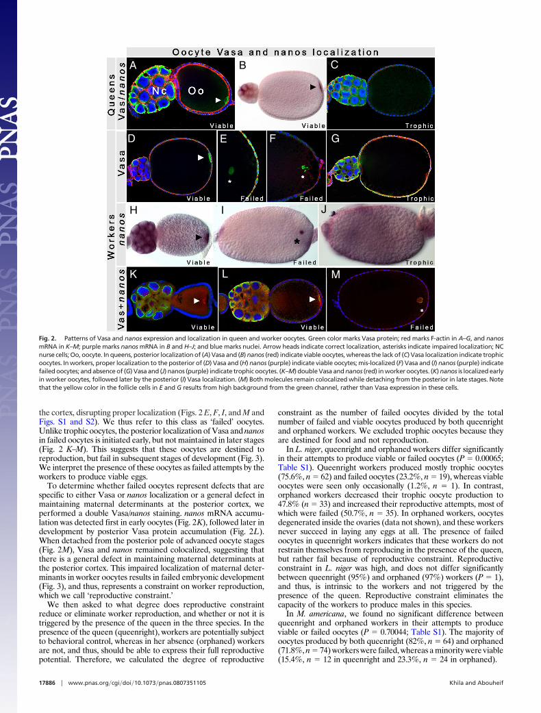
the cortex, disrupting proper localization (Figs. 2 E, F, I, and M andFigs. S1 and S2). We thus refer to this class as ‘failed’ oocytes.Unlike trophic oocytes, the posterior localization of Vasa and nanosin failed oocytes is initiated early, but not maintained in later stages(Fig. 2 K–M). This suggests that these oocytes are destined toreproduction, but fail in subsequent stages of development (Fig. 3).We interpret the presence of these oocytes as failed attempts by theworkers to produce viable eggs.
To determine whether failed oocytes represent defects that arespecific to either Vasa or nanos localization or a general defect inmaintaining maternal determinants at the posterior cortex, weperformed a double Vasa/nanos staining. nanos mRNA accumu-lation was detected first in early oocytes (Fig. 2K), followed later indevelopment by posterior Vasa protein accumulation (Fig. 2L).When detached from the posterior pole of advanced oocyte stages(Fig. 2M), Vasa and nanos remained colocalized, suggesting thatthere is a general defect in maintaining maternal determinants atthe posterior cortex. This impaired localization of maternal deter-minants in worker oocytes results in failed embryonic development(Fig. 3), and thus, represents a constraint on worker reproduction,which we call ‘reproductive constraint.’
We then asked to what degree does reproductive constraintreduce or eliminate worker reproduction, and whether or not it istriggered by the presence of the queen in the three species. In thepresence of the queen (queenright), workers are potentially subjectto behavioral control, whereas in her absence (orphaned) workersare not, and thus, should be able to express their full reproductivepotential. Therefore, we calculated the degree of reproductive
constraint as the number of failed oocytes divided by the totalnumber of failed and viable oocytes produced by both queenrightand orphaned workers. We excluded trophic oocytes because theyare destined for food and not reproduction.
In L. niger, queenright and orphaned workers differ significantlyin their attempts to produce viable or failed oocytes (P � 0.00065;Table S1). Queenright workers produced mostly trophic oocytes(75.6%, n � 62) and failed oocytes (23.2%, n � 19), whereas viableoocytes were seen only occasionally (1.2%, n � 1). In contrast,orphaned workers decreased their trophic oocyte production to47.8% (n � 33) and increased their reproductive attempts, most ofwhich were failed (50.7%, n � 35). In orphaned workers, oocytesdegenerated inside the ovaries (data not shown), and these workersnever succeed in laying any eggs at all. The presence of failedoocytes in queenright workers indicates that these workers do notrestrain themselves from reproducing in the presence of the queen,but rather fail because of reproductive constraint. Reproductiveconstraint in L. niger was high, and does not differ significantlybetween queenright (95%) and orphaned (97%) workers (P � 1),and thus, is intrinsic to the workers and not triggered by thepresence of the queen. Reproductive constraint eliminates thecapacity of the workers to produce males in this species.
In M. americana, we found no significant difference betweenqueenright and orphaned workers in their attempts to produceviable or failed oocytes (P � 0.70044; Table S1). The majority ofoocytes produced by both queenright (82%, n � 64) and orphaned(71.8%, n � 74) workers were failed, whereas a minority were viable(15.4%, n � 12 in queenright and 23.3%, n � 24 in orphaned).
A B C
D E F G
H I J
K L M
Fig. 2. Patterns of Vasa and nanos expression and localization in queen and worker oocytes. Green color marks Vasa protein; red marks F-actin in A–G, and nanosmRNA in K–M; purple marks nanos mRNA in B and H–J; and blue marks nuclei. Arrow heads indicate correct localization, asterisks indicate impaired localization; NCnurse cells; Oo, oocyte. In queens, posterior localization of (A) Vasa and (B) nanos (red) indicate viable oocytes, whereas the lack of (C) Vasa localization indicate trophicoocytes. In workers, proper localization to the posterior of (D) Vasa and (H) nanos (purple) indicate viable oocytes; mis-localized (F) Vasa and (I) nanos (purple) indicatefailed oocytes; and absence of (G) Vasa and (J) nanos (purple) indicate trophic oocytes. (K–M) double Vasa and nanos (red) in worker oocytes. (K) nanos is localized earlyin worker oocytes, followed later by the posterior (I) Vasa localization. (M) Both molecules remain colocalized while detaching from the posterior in late stages. Notethat the yellow color in the follicle cells in E and G results from high background from the green channel, rather than Vasa expression in these cells.
17886 � www.pnas.org�cgi�doi�10.1073�pnas.0807351105 Khila and Abouheif
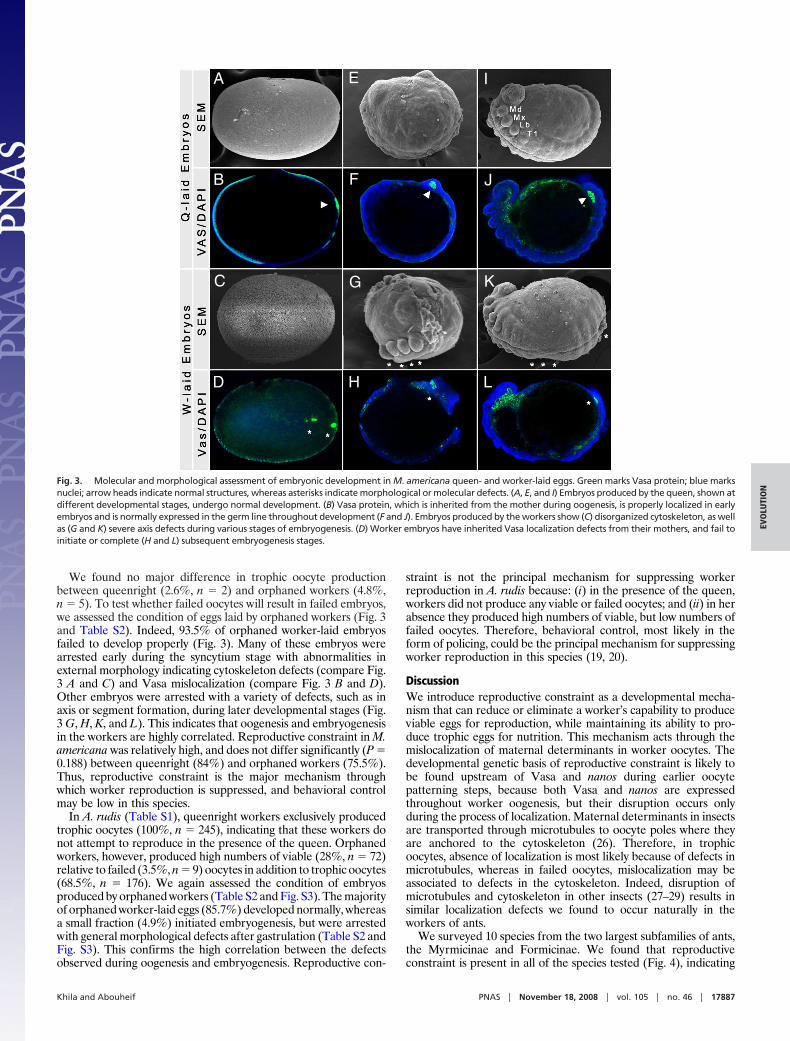
We found no major difference in trophic oocyte productionbetween queenright (2.6%, n � 2) and orphaned workers (4.8%,n � 5). To test whether failed oocytes will result in failed embryos,we assessed the condition of eggs laid by orphaned workers (Fig. 3and Table S2). Indeed, 93.5% of orphaned worker-laid embryosfailed to develop properly (Fig. 3). Many of these embryos werearrested early during the syncytium stage with abnormalities inexternal morphology indicating cytoskeleton defects (compare Fig.3 A and C) and Vasa mislocalization (compare Fig. 3 B and D).Other embryos were arrested with a variety of defects, such as inaxis or segment formation, during later developmental stages (Fig.3 G, H, K, and L). This indicates that oogenesis and embryogenesisin the workers are highly correlated. Reproductive constraint in M.americana was relatively high, and does not differ significantly (P �0.188) between queenright (84%) and orphaned workers (75.5%).Thus, reproductive constraint is the major mechanism throughwhich worker reproduction is suppressed, and behavioral controlmay be low in this species.
In A. rudis (Table S1), queenright workers exclusively producedtrophic oocytes (100%, n � 245), indicating that these workers donot attempt to reproduce in the presence of the queen. Orphanedworkers, however, produced high numbers of viable (28%, n � 72)relative to failed (3.5%, n � 9) oocytes in addition to trophic oocytes(68.5%, n � 176). We again assessed the condition of embryosproduced by orphaned workers (Table S2 and Fig. S3). The majorityof orphaned worker-laid eggs (85.7%) developed normally, whereasa small fraction (4.9%) initiated embryogenesis, but were arrestedwith general morphological defects after gastrulation (Table S2 andFig. S3). This confirms the high correlation between the defectsobserved during oogenesis and embryogenesis. Reproductive con-
straint is not the principal mechanism for suppressing workerreproduction in A. rudis because: (i) in the presence of the queen,workers did not produce any viable or failed oocytes; and (ii) in herabsence they produced high numbers of viable, but low numbers offailed oocytes. Therefore, behavioral control, most likely in theform of policing, could be the principal mechanism for suppressingworker reproduction in this species (19, 20).
DiscussionWe introduce reproductive constraint as a developmental mecha-nism that can reduce or eliminate a worker’s capability to produceviable eggs for reproduction, while maintaining its ability to pro-duce trophic eggs for nutrition. This mechanism acts through themislocalization of maternal determinants in worker oocytes. Thedevelopmental genetic basis of reproductive constraint is likely tobe found upstream of Vasa and nanos during earlier oocytepatterning steps, because both Vasa and nanos are expressedthroughout worker oogenesis, but their disruption occurs onlyduring the process of localization. Maternal determinants in insectsare transported through microtubules to oocyte poles where theyare anchored to the cytoskeleton (26). Therefore, in trophicoocytes, absence of localization is most likely because of defects inmicrotubules, whereas in failed oocytes, mislocalization may beassociated to defects in the cytoskeleton. Indeed, disruption ofmicrotubules and cytoskeleton in other insects (27–29) results insimilar localization defects we found to occur naturally in theworkers of ants.
We surveyed 10 species from the two largest subfamilies of ants,the Myrmicinae and Formicinae. We found that reproductiveconstraint is present in all of the species tested (Fig. 4), indicating
A
B
C
D
E
F
G
H
I
J
K
L
Fig. 3. Molecular and morphological assessment of embryonic development in M. americana queen- and worker-laid eggs. Green marks Vasa protein; blue marksnuclei; arrow heads indicate normal structures, whereas asterisks indicate morphological or molecular defects. (A, E, and I) Embryos produced by the queen, shown atdifferent developmental stages, undergo normal development. (B) Vasa protein, which is inherited from the mother during oogenesis, is properly localized in earlyembryos and is normally expressed in the germ line throughout development (F and J). Embryos produced by the workers show (C) disorganized cytoskeleton, as wellas (G and K) severe axis defects during various stages of embryogenesis. (D) Worker embryos have inherited Vasa localization defects from their mothers, and fail toinitiate or complete (H and L) subsequent embryogenesis stages.
Khila and Abouheif PNAS � November 18, 2008 � vol. 105 � no. 46 � 17887
EVO
LUTI
ON
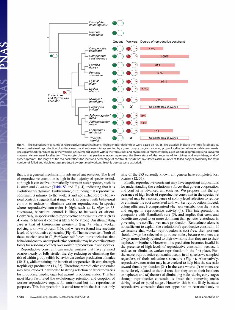
that it is a general mechanism in advanced ant societies. The levelof reproductive constraint is high in the majority of species tested,although it can evolve dramatically between sister species, such asL. niger and L. alienus (Table S3 and Fig. 4), indicating that it isevolutionarily dynamic. Furthermore, our finding that reproductiveconstraint is intrinsic to the workers and not influenced by behav-ioral control, suggests that it may work in concert with behavioralcontrol to reduce or eliminate worker reproduction. In specieswhere reproductive constraint is high, such as L. niger or M.americana, behavioral control is likely to be weak or absent.Conversely, in species where reproductive constraint is low, such asA. rudis, behavioral control is likely to be strong. An illuminatingcase is that of Camponotus floridanus (Fig. 4), where workerpolicing is known to occur (16), and where we found intermediatelevels of reproductive constraint (Fig. 4). The occurrence of both ofthese mechanisms in C. floridanus reinforces our conclusion thatbehavioral control and reproductive constraint may be complimentaryforces for resolving conflicts over worker reproduction in ant societies.
Reproductive constraint can render workers that have retainedovaries nearly or fully sterile, thereby reducing or eliminating therisk of within group selfish behavior via worker production of males(30, 31), while retaining the benefit of cooperative sib-care throughtrophic egg production (13, 32). Therefore, reproductive constraintmay have evolved in response to strong selection on worker ovariesfor producing trophic eggs but against producing males. This hasmost likely facilitated the evolutionary retention and cooption ofworker reproductive organs for nutritional but not reproductivepurposes. This interpretation is consistent with the fact that only
nine of the 283 currently known ant genera have completely lostovaries (12, 33).
Finally, reproductive constraint may have important implicationsfor understanding the evolutionary forces that govern cooperationand conflict in advanced ant societies. We propose that the ap-pearance of high levels of reproductive constraint in the species wesampled may be a consequence of colony-level selection to reduceor eliminate the cost associated with worker reproduction. Indeed,colony efficiency is compromised when workers abandon their tasksand engage in reproductive activity (4). This interpretation iscompatible with Hamilton’s rule (5), and implies that costs andbenefits are equal to, or more dominant than genetic relatedness ingoverning the conflict over male production. Relatedness alone isnot sufficient to explain the evolution of reproductive constraint. Ifwe assume that worker reproduction is cost-free, then workersshould always be selected to produce males, because workers arealways more closely related to their own sons than they are to theirnephews or brothers. However, this prediction becomes invalid inthe presence of high levels of reproductive constraint, because itreduces or eliminates worker reproduction in the first place. Fur-thermore, reproductive constraint occurs in all species we sampledregardless of their relatedness structure (Fig. 4). Alternatively,reproductive constraint may have evolved to help bias the sex ratiotoward female production (34) in the case where: (i) workers aremore closely related to their sisters than they are to their brothersor nephews; and (ii) the cost of eliminating males during early stagesthrough reproductive constraint is lower than removing malesduring larval or pupal stages. However, this is not likely becausereproductive constraint does not appear to be restricted only to
Fig. 4. The evolutionary dynamic of reproductive constraint in ants. Phylogenetic relationships were based on ref. 36. The asterisks indicate the three focal species.The unconstrained reproduction of solitary insects and ant queens is represented by a green oocyte diagram showing proper localization of maternal determinants.The constrained reproduction in the workers of several ant species within the formicines and myrmicines is represented by a red oocyte diagram showing impairedmaternal determinant localization. The oocyte diagram at particular nodes represents the likely state of the ancestor of formicines and myrmicines, and ofhymenopterans. The length of the red bars reflects the level and percentage of constraint, which was calculated as the number of failed oocytes divided by the totalnumber of failed and viable oocytes produced by orphaned workers. Trophic oocytes were excluded.
17888 � www.pnas.org�cgi�doi�10.1073�pnas.0807351105 Khila and Abouheif
species that may follow this particular case (Fig. 4). A furtherunderstanding of both sociogenetic and ecological conditions thatfavor the evolution of reproductive constraint will require testingfor reproductive constraint in both ancestral and derived specieswhere relatedness, behavioral control mechanisms, and colony-sizehave been well documented.
Materials and MethodsAnts. A. rudis and L. niger colonies were collected at Montreal, Canada and M.americana colonies were collected in Medford, NY. The rest of the species in Fig.4 were collected in Arizona, Florida, and New York. Colonies were maintained at27°C,70%humidityanda12:12-hday:nightcycle,andwerefedonacombinationof Crickets and Bachtar-Whitcomb diet. Three replicates of orphaned workersfrom each species were isolated for two to six months, and their egg productionmonitored. Orphaned A. rudis and M. americana workers were able to produceeggs, larvae, and adult males, but orphaned L. niger workers were not. Queen-right and orphaned worker ovaries were then dissected and the number ofviable, failed and trophic oocytes (as determined by Vasa localization) wascounted.Allpercentagespresented intheresult sectionarethemeanpercentagefor each three replicates.
Statistical Analysis. We performed a Fisher’s exact test (35) to determine: (i) ifthere is a significant difference in reproductive attempts between queenrightand orphaned workers by comparing the frequency of trophic versus non trophicoocytes (viable plus failed) produced by these workers; and (ii) if there is asignificant difference in reproductive constraint between queenright and or-phaned workers by comparing the frequency of failed versus viable oocytesproduced by these workers. We could not perform the test for A. rudis becausequeenright workers produced no reproductive oocytes (Table S1).
Nanos Cloning. The nanos gene was cloned using degenerate and RACE PCR(Invitrogen) using the following primers: forward 5�-TGCGTWTTCTG-YARAAATAA-3� and reverse 5�-GGRCAATACTTKAYCGTRTGAG-3� for degener-ate PCR and Ant�nos Forward: 5�-TGATGGAAGAGTTTCGACACAATGG-3� forRACE PCR. GenBank accession numbers for nanos sequences: A. rudis nanos,EU272791; L. niger nanos, EU272792; M. americana nanos, EU272793.
Ovary and Embryo Fixation. Ovaries were dissected in PTW (1� PBS; 0.05%Tween-20) and kept on ice during the dissection process. The peritoneal sheetcovering each ovariole was removed by using fine forceps. The ovarioles werefixed in 4% paraformaldehyde (200 �l) supplemented with 10% DMSO (20 �l),and heptane (600 �l) for 20 min at room temperature. Fixed ovarioles were thenwashed three times in PBT (1� PBS; 0.3% Triton X-100) and processed forsubsequent staining.
Embryos are boiled for 45 seconds in PBT, and then quickly placed on ice.The chorion and vitelline membrane were removed manually by using fineforceps. Embryos were then fixed in 4% formaldehyde and heptane for 20 minand then washed three times in PBT.
Vasa Staining. A cross-reacting anti-Vasa antibody, which was raised againstDrosophila Vasa (25), was used to detect the Vasa protein in ants. Fixed ovariolesor embryos were permeabilized in PBT (1� PBS; 1% Triton X-100) for 1 h at roomtemperature. Permeabilization is followed by blocking step in PAT (1� PBS; 1%Triton X-100 and 1% bovine serum albumin) for 1 h at room temperature.Ovarioles/embryos were then incubated with a rabbit anti-Vasa antibody at a1:100 dilution in PTW overnight at 4°C. Ovarioles/embryos were washed fromexcess antibody five times 10 min at room temperature in PBT (0.3% TritonX-100), then blocked again in PAT for one hour. A secondary, goat Cy2-conjugated anti-rabbit antibody (Jackson ImmunoResearch) was used to detectthe rabbit anti-Vasa antibody at a 1:300 dilution in PTW. Ovarioles/embryos wereincubated with the secondary antibody for two hours at room temperature inPTW. DAPI and Phalloidin were added at the same time as the secondary anti-body. The ovarioles were finally washed five times 10 min in PTW then inincreasing concentrations of glycerol in PBS and mounted in glycerol/1� PBS(80%/20%).
Nanos in situ hybridization. Nanos staining was performed by using DIG-labeled nanos RNA probes (Roche). Fixed ovarioles were incubated one hourat 58°C in a hybridization solution (50% formamide, 5� SSC pH 6.5, 50 �g/mlsalmon sperm DNA, 50 �g/ml heparin, 0.1% Tween-20, and 0.3% SDS). Ova-rioles were then hybridized over-night at 58°C with the nanos DIG-labeledprobe in the hybridization solution. Ovarioles were washed five times in PBT,blocked one hour in PAT then incubated two hours at room temperature withthe anti-DIG antibody, conjugate with alkaline phosphatase. nanos expressionwas revealed by using NBT/BCIP (purple, Roche) or Fast-red (red, Sigma).
Image capture was performed by using either 510 confocal or Axiovert Zeissmicroscopes. Large samples, which do not fit in a 40� or 20� objective fieldwere assembled from multiple files and processed by using Adobe Photoshop.
ACKNOWLEDGMENTS. We thank P. Lasko (McGill University) for providing theanti-Vasa antibody; A. Francoeur (Universite du Quebec a Chicoutimi, Saguenay,QC, Canada), R. Johnson (Arizona State University, Tempe, AZ), R. Sanwald(Medford, NY), and W. Tschinkel (Florida State University, Tallahassee, FL) for antidentification or collection; J.J. Boomsma, C. Desplan, M.B. Dijkstra, J. Heinze, B.Holldobler, L. Nilson, D. Schoen, A. Vincent, E.O. Wilson, the members of theAbouheif, Nilson, Shoek, and Lasko labs, the Behavioral Ecology and Drosophilagroups at McGill, and two anonymous reviewers for discussion or critical readingof the manuscript; and McGill University for permitting us to work on the GaultNature Reserve. This work was supported by Natural Sciences and EngineeringResearch Council and Alfred P. Sloan funding (to E.A.).
1. Bourke AFG (1988) Worker reproduction in the higher eusocial hymenoptera. Q Rev Biol291–311.
2. Ratnieks FLW, Foster KR, Wenseleers T (2006) Conflict resolution in insect societies. AnnuRev Entomol 51:581–608.
3. Wenseleers T, Ratnieks FL (2006) Comparative analysis of worker reproduction and polic-ing in eusocial hymenoptera supports relatedness theory. Am Nat 168:E163–179.
4. Wenseleers T, Helantera H, Hart A, Ratnieks FL (2004) Worker reproduction and policingin insect societies: An ESS analysis. J Evol Biol 17:1035–1047.
5. Bourke AFG, Franks NR (1995) Social Evolution in Ants (Princeton Univ Press, Princeton).6. Hamilton WD (1964) The genetical evolution of social behaviour. J Theor Biol 7:1–16.7. Korb J, Heinze J (2004) Multilevel selection and social evolution of insect societies. Natur-
wissenschaften 91:291–304.8. LehmannL,KellerL,WestS,RozeD(2007)Groupselectionandkin selection:Twoconcepts
but one process. Proc Natl Acad Sci USA 104:6736–6739.9. Reeve HK, Holldobler B (2007) The emergence of a superorganism through intergroup
competition. Proc Natl Acad Sci USA 104:9736–9740.10. Wilson EO (2008) One giant leap: How insects achieved altruism and colonial life. Bio-
Science 58:17–25.11. Wilson EO, Holldobler B (2005) Eusociality: Origin and consequences. Proc Natl Acad Sci
USA 102:13367–13371.12. Holldobler B, Wilson EO (1990) The Ants (Belknap Press of Harvard Univ Press, Cambridge,
Mass.).13. Gobin B, Ito F (2000) Queens and major workers of Acanthomyrmex ferox redistribute
nutrients with trophic eggs. Naturwissenschaften 87:323–326.14. Hammond RL, Keller L (2004) Conflict over male parentage in social insects. PLoS Biol
2:E248.15. Dijkstra MB, Nash DR, Boomsma JJ (2005) Self-restraint and sterility in workers of Acro-
myrmex and Atta leafcutter ants. Insect Soc 52:67–76.16. Endler A, et al. (2004) Surface hydrocarbons of queen eggs regulate worker reproduction
in a social insect. Proc Natl Acad Sci USA 101:2945–2950.17. D’Ettorre P, Heinze J, Ratnieks FL (2004) Worker policing by egg eating in the ponerine ant
Pachycondyla inversa. Proc Biol Sci 271:1427–1434.18. Foster KR, Ratnieks FL (2000) Facultative worker policing in a wasp. Nature 407:692–693.19. Holldobler B, Carlin NF (1989) Colony founding, queen control, and worker reproduction
in the ant Aphaenogaster (�Novomessor) cockerelli (Hymenoptera: Formicidae). Psyche96:131–151.
20. Iwanishi S, Hasegawa E, Ohkawara K (2003) Worker oviposition and policing behaviour inthe myrmicine ant Aphaenogaster smythiesi japonica Forel. Anim Behav 66:513–519.
21. van Eeden F, St Johnston D (1999) The polarisation of the anterior–posterior and dorsal-ventral axes during Drosophila oogenesis. Curr Opin Genet Dev 9:396–404.
22. Nusslein-Volhard C, Frohnhofer HG, Lehmann R (1987) Determination of anteroposteriorpolarity in Drosophila. Science 238:1675–1681.
23. Riechmann V, Ephrussi A (2001) Axis formation during Drosophila oogenesis. Curr OpinGenet Dev 11:374–383.
24. Lehmann R, Nusslein-Volhard C (1991) The maternal gene nanos has a central role inposterior pattern formation of the Drosophila embryo. Development 112:679–691.
25. Styhler S, Nakamura A, Swan A, Suter B, Lasko P (1998) vasa is required for GURKENaccumulation in the oocyte, and is involved in oocyte differentiation and germline cystdevelopment. Development 125:1569–1578.
26. Ephrussi A, Lehmann R (1992) Induction of germ cell formation by oskar. Nature 358:387–392.
27. Huynh JR, St Johnston D (2004) The origin of asymmetry: Early polarisation of the Dro-sophila germline cyst and oocyte. Curr Biol 14:R438–449.
28. Olesnicky EC, Desplan C (2007) Distinct mechanisms for mRNA localization during embry-onic axis specification in the wasp Nasonia. Dev Biol 306:134–142.
29. Steinhauer J, Kalderon D (2006) Microtubule polarity and axis formation in the Drosophilaoocyte. Dev Dyn 235:1455–1468.
30. Clutton-Brock TH, Vincent AC (1991) Sexual selection and the potential reproductive ratesof males and females. Nature 351:58–60.
31. Dijkstra MB, Boomsma JJ (2007) The economy of worker reproduction in Acromyrmexleafcutter ants. Animal Behaviour 47:519–529.
32. Wradlaw JC, Elmes GW (1995) Trophic eggs laid by fertile Myrmica queens (Hymenoptera:Formicidae). Insect Soc 42:303–308.
33. Oster GF, Wilson EO (1978) Caste and ecology in the social insects. Monogr Popul Biol12:1–352.
34. FosterKR,RatnieksFL (2001)Convergentevolutionofworkerpolicingbyeggeating in thehoneybee and common wasp. Proc Biol Sci 268:169–174.
35. Sokal RR, Rohlf FJ (1995) Biometry (W. H. Freeman and Company, New York).36. Brady SG, Schultz TR, Fisher BL, Ward PS (2006) Evaluating alternative hypotheses for the
early evolution and diversification of ants. Proc Natl Acad Sci USA 103:18172–18177.
Khila and Abouheif PNAS � November 18, 2008 � vol. 105 � no. 46 � 17889
EVO
LUTI
ON