Embed Size (px)
Citation preview
10/15/2008
1
Caring for Patients with
Common Health Problems of
the Renal System
Celeste Armenta RNNP
Nursing 210
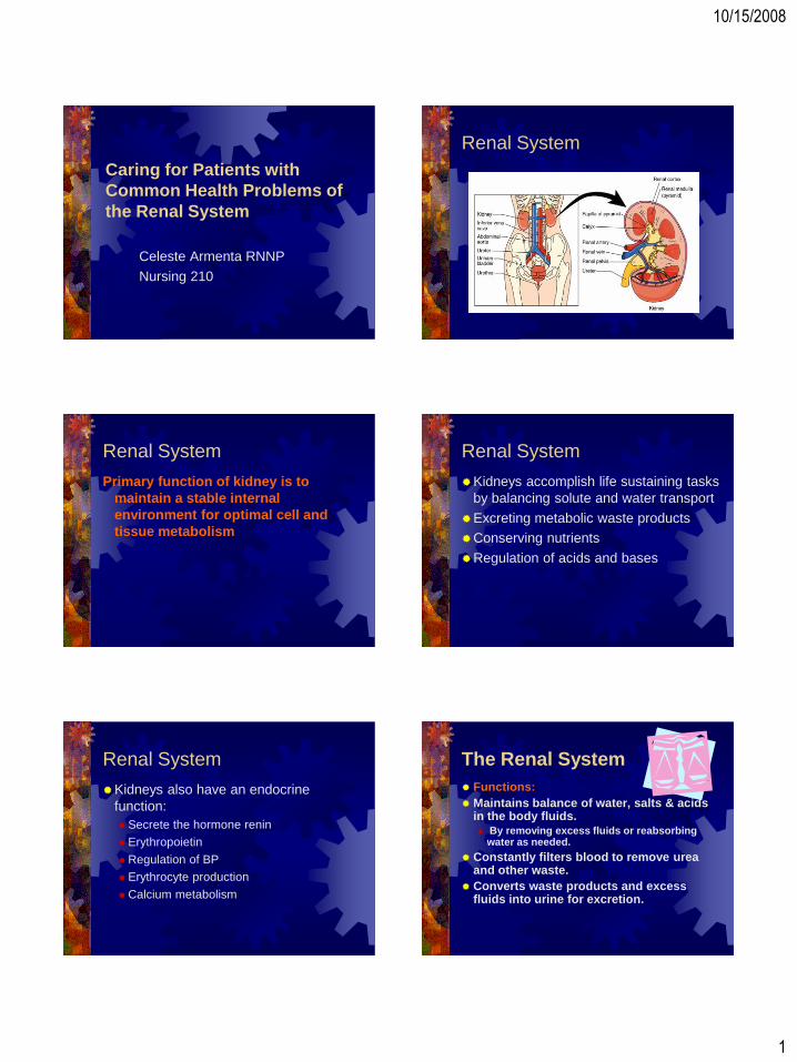
Renal System
Primary function of kidney is to
maintain a stable internal
environment for optimal cell and
tissue metabolism
Renal System Renal System
Kidneys accomplish life sustaining tasks
by balancing solute and water transport
Excreting metabolic waste products
Conserving nutrients
Regulation of acids and bases
Renal System
Kidneys also have an endocrine
function:
Secrete the hormone renin
Erythropoietin
Regulation of BP
Erythrocyte production
Calcium metabolism
The Renal System
Functions:
Maintains balance of water, salts & acids in the body fluids. By removing excess fluids or reabsorbing
water as needed.
Constantly filters blood to remove urea and other waste.
Converts waste products and excess fluids into urine for excretion.
10/15/2008
2
Renal System
Formation of urine is achieved by
process called filtration, reabsorption
and secretion by the glomeruli and
tubules within the kidney.
Bladder stores the urine that it receives
from the kidney by way of ureters. Urine
is then removed from the body through
the urethra.
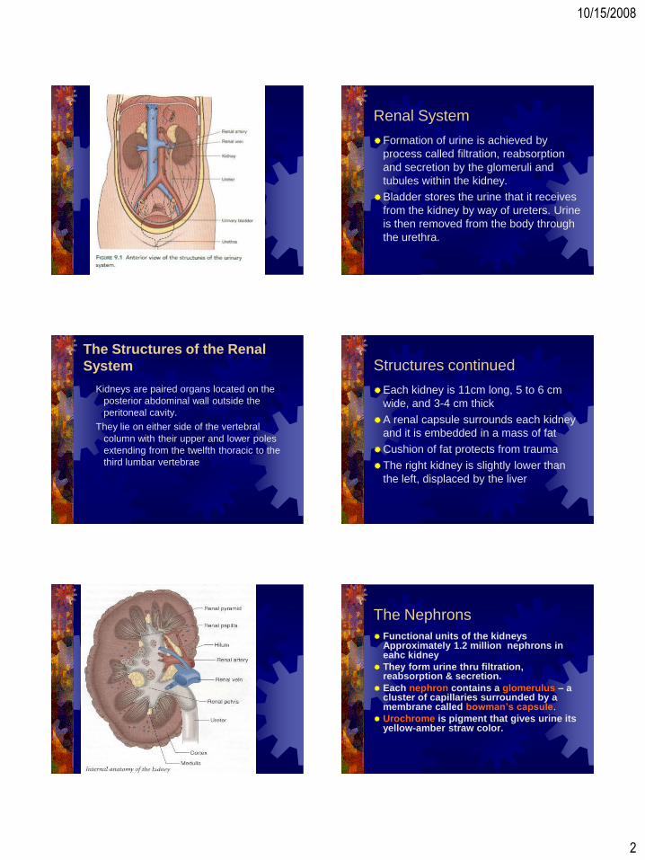
The Structures of the Renal
System
Kidneys are paired organs located on the
posterior abdominal wall outside the
peritoneal cavity.
They lie on either side of the vertebral
column with their upper and lower poles
extending from the twelfth thoracic to the
third lumbar vertebrae
Structures continued
Each kidney is 11cm long, 5 to 6 cm
wide, and 3-4 cm thick
A renal capsule surrounds each kidney
and it is embedded in a mass of fat
Cushion of fat protects from trauma
The right kidney is slightly lower than
the left, displaced by the liver
The Nephrons
Functional units of the kidneys Approximately 1.2 million nephrons in eahc kidney
They form urine thru filtration, reabsorption & secretion.
Each nephron contains a glomerulus – a cluster of capillaries surrounded by a membrane called bowman’s capsule.
Urochrome is pigment that gives urine its yellow-amber straw color.
10/15/2008
3
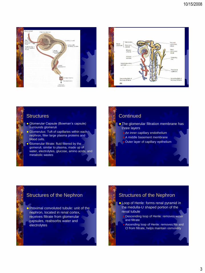
Structures
Glomerular Capsule (Bowman’s capsule)
surrounds glomeruli
Glomerulus: Tuft of capillaries within each
nephron, filter large plasma proteins and
blood cells.
Glomerular filtrate: fluid filtered by the
gomeruli, similar to plasma, made up of
water, electrolytes, glucose, amino acids, and
metabolic wastes
Continued
The glomerular filtration membrane has
three layers
An inner capillary endothelium
A middle basement membrane
Outer layer of capillary epithelium
Structures of the Nephron
Proximal convoluted tubule: unit of the
nephron, located in renal cortex,
receives filtrate from glomerular
capsules, reabsorbs water and
electrolytes
Structures of the Nephron
Loop of Henle: forms renal pyramid in
the medulla-U shaped portion of the
renal tubule
Descending loop of Henle: removes water
and filtrate
Ascending loop of Henle: removes Na and
Cl from filtrate, helps maintain osmolality
10/15/2008
4
Structures of the Nephron
Distal Convoluted tubule: Convoluted
portion of the tubule beyond loop of
Henle, located in renal cortex, removes
more Na and H20.
Structures of the Nephron
Distal Tubules: reabsorb NA by active and passive transport in smaller amounts than proximal tubules
Collecting Ducts: prevent water from leaving the filtrate use active and passive reabsorption
Tubular Secretion: movement out of the blood into the tubular fluid, tubule cells secrete certain substances in addition to performing reabsorption
Renal Blood Flow
Kidneys highly vascular organs receive 1000 to 1200ml of blood per minute, or about 20% to 25% of the cardiac output.
From renal plasma flow, 20% (approximately 120 to 140ml/min) is filtered at the glomerulus and passes into Bowmans capsule.
Filtration of plasma per unit of time is glomerular filtration rate (GFR), related to perfusion pressure in the glomerular capillaries
Autoregulation
In the kidney a local mechanism tends to
keep the rate of blood flow and therefore the
GFR fairly constant
Changes in afferent arteriolar resistance and
arteriolar pressure occur in the same
direction EX: As systemic blood pressure
increases, the afferent arterioles constrict,
preventing an increase in glomerular blood
flow and filtration pressure.
Neural Regulation
When systemic arterial pressure
decreases, increased renal sympathetic
nerve activity is mediated reflexively
through the carotid sinus and the
baroreceptors of the aortic arch. This
stimulates renal arteriolar
vasoconstriction and decreases both
RBF and GFR.
Ureters
10 – 12 inch tube that carry urine
from the kidneys to the bladder.
10/15/2008
5
Urinary Bladder
Hollow muscular organ
Reservoir for urine
Stores about 1 pint of urine
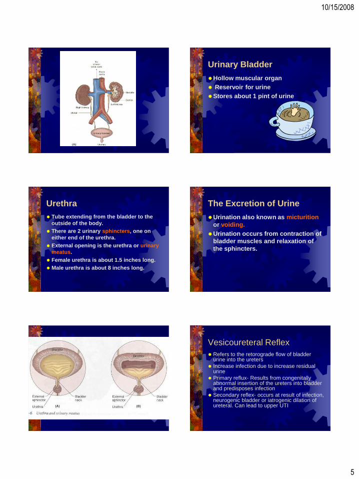
Urethra
Tube extending from the bladder to the
outside of the body.
There are 2 urinary sphincters, one on
either end of the urethra.
External opening is the urethra or urinary
meatus.
Female urethra is about 1.5 inches long.
Male urethra is about 8 inches long.
The Excretion of Urine
Urination also known as micturition
or voiding.
Urination occurs from contraction of
bladder muscles and relaxation of
the sphincters.
Vesicoureteral Reflex
Refers to the retorograde flow of bladder urine into the ureters
Increase infection due to increase residual urine
Primary reflux- Results from congenitally abnormal insertion of the ureters into bladder and predisposes infection
Secondary reflex- occurs at result of infection, neurogenic bladder or iatrogenic dilation of ureteral. Can lead to upper UTI
10/15/2008
6
Management
Continous low dose antibacterial therapy with frequent urine cultures
Surgical Interventions if:
Significant anatomic abnormality
Recurrent UTI
High Grades of VUR
Noncompliance with medical therapy
Nursing: Encourage compliance
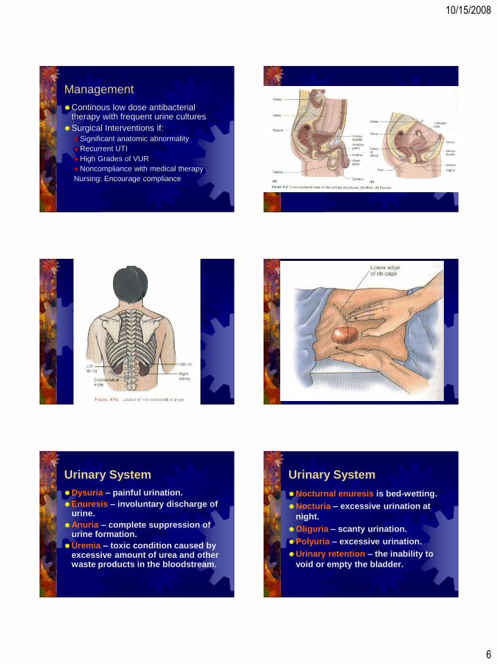
Urinary System
Dysuria – painful urination.
Enuresis – involuntary discharge of urine.
Anuria – complete suppression of urine formation.
Uremia – toxic condition caused by excessive amount of urea and other waste products in the bloodstream.
Urinary System
Nocturnal enuresis is bed-wetting.
Nocturia – excessive urination at
night.
Oliguria – scanty urination.
Polyuria – excessive urination.
Urinary retention – the inability to
void or empty the bladder.
10/15/2008
7
Urinary Tract Infection
No bacteria except the distal 1/3 of
urethra
Site of infection difficult to determine
with accuracy
Child peak incidence of UTI 2-6 years
(not structural anomalies) females 10-
30% Greater risk
Classification of UTI
Bacteruria- growth of bacteria in uncontaminated urine
A symptomatic bacteriuria-significant bacteriuria with no clinical symptoms
Symptomatic-significant bacteriuria with physical symptoms
Recurrent UTI-Repeated UTI
Relapse of UTI- Persistance of the same organism despite therapy
Continued
Urethritis- Inflammation of the urethra
Cystitis- Inflammation of the bladder
Ureteritis- Inflammation of the ureter
Pylonephritis – Inflammation of kidney
and upper tract
Continued
Female short urethra and lack of
prostatic fluid that provides protecton
Infancy infection has incidence of renal
scar
Mechanisms- Stasis of urine
Symptoms
Over 2 years encounter enuresis or daytime incontinence
Fever
Strong- foul smelling urine
Increased frequency in urination
Dysuria
Urgency/ABD Pain/ Flank Pain/ Hematuria
Pylonephritis
Admit and IV antibiotics
Increase fluid intake 3-4 liters
Nurses: Evaluate for UTI, will see
incontinence in toilet trained child
Strong smelling urine
Frequency and or urgency
10/15/2008
8
Prevention
Complete emptying of bladder (prevent
urinary stasis)
Teach symptoms of UTI
Need for prompt medical attention
Continue drugs even though symptoms
abate, follow up care
Maintenance of fluid intake of 3-4 liters
Pyelonephritis
Bacterial infectio of kidney tissue
Usually begings as lower UTI and ascends to kidney Ecoli. Most common organism
Associated with Cystitis, Pregnancy, Obstruction, risk factors-septicemia
Chronic health problems or analgesic, polycystic kidney
Signs and Symptoms
Inflammation/Chills/ Fever/ malaise
Flank pain/ costovertebral tenderness
Leukocytosis
WBC, casts, bacteria, BUN. Creat, Pyuria
Treatment: Check culture and sensitivity, start broad spectrum antibiotics
Continued
Most common cause of acute bacterial sepsis in older 65 year olds
Structural abnormalities, neurogenic bladder due to strokes
Autonomic neuropathy in diabetic patients
In absence of estrogen, in post menopausal women suseptible to colonization increase adherence of bacteria to vagina and urethra. Estrogen therapy helps with vaginal PH
Nephrotic Syndrome
Most common presentation of
glomerular injury
Massive proteinuria,
hypoalbuminemia, hyperlipidemia
and edema
Sequence of Events in Nephrotic
Syndrome
Renal glomerular damage, leads to proteinuria (massive), leads to hypoproteinemia which increases hepatic synthesis of proteins and lipids causes hyperlipidemia.
Hypoproteinemia causes decreased oncotic pressure leading to hypvolemia which decreases renal blood flow, renin is released vasoconstriciton occurs, increased hydrostatic pressure and end result edema
10/15/2008
9
Types of Nephrotic Syndrome
Primary-Restricted to glomerular injury
Secondary- When it develops as part of a systemic illness, idiopathic, hypersensitivity reaction
Minimal Change Nephritic Syndrome –most common preschool 2-7 yrs
Secondary Nephritis- cause glomerular damage in acute or chronic glomerulonephritis
Continued
Patho- Glomerular membrane becomes
permeable to proteins, especially
albumin also immunoglobulins patient
susceptible to infection.
Continued
Decrease in colloidal osmotic pressure,
and hydrostatic pressure exceeds, fluid
accumulates in extravascular spaces
(ascites) leads to hypovolemia, renin
stimualted, vasoconstriciton, secretion
of ADH and aldosterone, increase in NA
and water reabsorption to increase
intravascular volume
Clinical Manifestations
Well child gains wt over days or weeks
Puffiness in face, especially around eyes
Swelling worst in am’s and subsides during the day (clothes fit tight)=fluid shifts to abdomen and lower extremities
Anasarca- severe generalized edema
Diarrhea- edema of intestinal mucosa
Loss of appetite, poor intestinal absorption
Urine volume decreases, appears darkly opalescent, frothy
Continued
Pale with easy skin breakdown
Irritable easily fatigued or lethargic
BP WNL or low
Child susceptible to infection
Continued
Diagnosis based on history and
symptoms/ Renal Biopsy
Edema
Proteinuria 10g/24hrs
Hypoalbuminemia
Hypercholestremia
10/15/2008
10
Management
Reduce the excretion of urinary protein
Prevent or treat any acute infection
Control edema
Establishment of good nutrition
Correct metabolic process
Med Management
ACE inhibitors with diuretics/ Salt poor albumin
Diuretics especially loop (Lasix) control edema or hypertension. Aldactone to supress aldosterone and conserve K.
Antineoplastic agents (Cytoxan)
NA decrease liberal K, to assist in NA/K pump mechanism, reduction of edema
Biologic proteins ( dairy products, eggs, meats) decrease in saturated fats.
Treatment
Bedrest at child level of tolerance
No added salt
Corticosteroid therapy
Prednisone (safest and least expensive)
Complications with steroids
Cataracts/obesity/bone demineralization
Infection/ Hyperglycemia/ GI bleed
Acute Glomerulonephritis
Immunologic mechanisms are primarily
responsible for glomerular disease. The
onset may be sudden or insidous with
HTN, edema, elevated BUN
Can be assymptomatic detected through
presence of hematuria routine urinalysis.
Most definite indication obtained by renal
biopsy
Continued
Antigen (group A beta hemolytic streptococcus)
Antigen antibody product
Deposition of antigen antibody in glomerulus
Increased production of epithelial cells lining the glomerulus
Leukocytes infiltrate the glomerulus
Thickening of the glomerular filtration membrane
Scarring and loss of glomerular filtration membrane
Decreased GFR rate
Common Manifestations
Oliguria
Hypertension
Hematuria (Primary presenting feature)
Proteinuria
Symptoms occur 7-21 days (7-10 days
post infection)
10/15/2008
11
More Manifestations
Puffiness of face (periorbital edema)
feet and ankles dependent edema
Anorexia
Pass dark colored urine (smoky brown,
tea colored
Decrease in output
Pale irritable and lethargic
Continued
Acute Edematous Phase
Listless, anorexic, and apathetic
Wt fluctuates, urine remains thick and
smoky brown
Blood pressure may increase
Continued
Prognosis- OK, death can occur due to
complications
Complications- HTN encephalopathy-
headache, dizziness, vomiting, discomfort
Acute cardiac decompensation-
hypervolemia, edema
Acute renal failure- Persistent oliguria or
anuria
Diagnosis
Urine Sp. Gr. Seldom exceeds 1.020
Proteinuria +3/+4
Gross discoloration- RBC, WBC, Cell Casts
BUN, Creatinine levels- Level and severity of disease related
ASO, ESR, C-reactive protein reflect acute inflammatory process
Therapy
General supportive measures and early
recognition and TX
Normal BP and satisfactory UO-TX at
home
Substantial edema, HTN, Gross
hematuria and significant oliguria-
Hospitalization
General
Can ambulate if not lethargic
Fluid restriction only with UO decreased
Lasix only with significant edema and
fluid overload
Digitalis- for CHF
Diet- High in carbs to provide energy
and decrease protein catabolism
10/15/2008
12
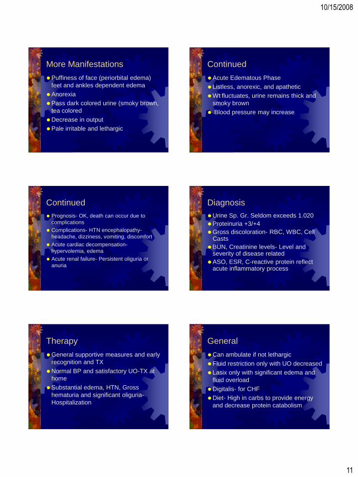
Chronic/ Progressive
Glomerulonephritis
Advanced glomerulonephritis- describes
advanced glomerular disease, causing
progression of renal function and rapid
deterioration
Clinical Manifestations
Nephritic Syndrome
HTN
Edema
Proteinuria
Cardiac failure
Dyspnea
Osteodystrophy
Anemia
Kidneys
Pyelitis – inflammation of the renal
pelvis.
Pyelonephritis – inflammation of the
renal pelvis and of the kidney.
Renal colic – acute pain in kidney
area caused by blockage during
passage of a kidney stone.
10/15/2008
13
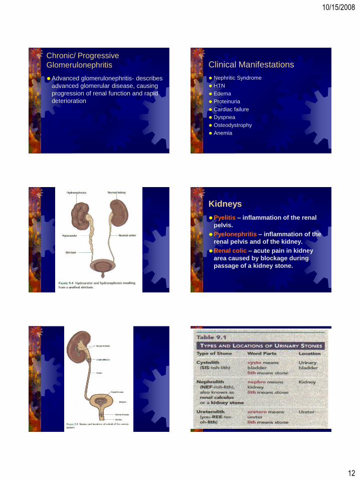
Urolithiasis
Most common urologic problem
Most stones formed in kidneys, but bladder stones are common in clients with catheters or inability to empty bladder completely
Can be single or multiple, large calculi can cause pressure necrosis and lead to obstruction
Risk Factors
Dehydration
Infection- change in PH provide and
environment for calculi
Obstruction- urine stasis allows for solid
material to collect
Metabolic factors-increase in uric acid,
vitD, calcium
Signs and Symptoms
Pain, renal colic, fever, chills, abdominal distention, N/V
Diagnosis- UA, strain all urine, crystal fragments, pyuria, hematuria, KUB, IVP ultrasound, CT scan
Stones that are too large more than 5mm diameter multiple stones, require surgical intervention.
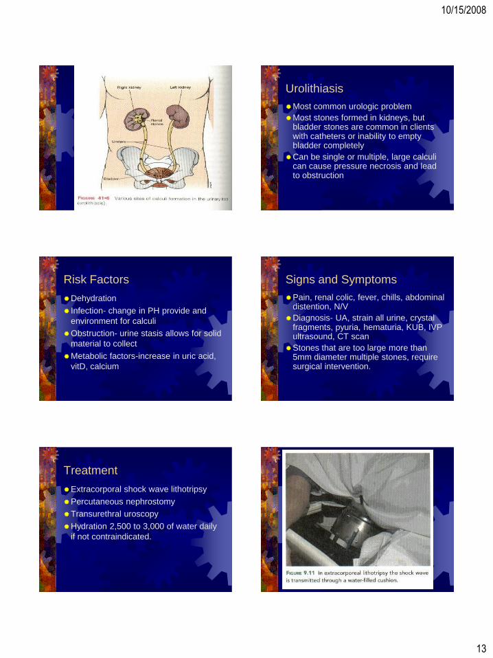
Treatment
Extracorporal shock wave lithotripsy
Percutaneous nephrostomy
Transurethral uroscopy
Hydration 2,500 to 3,000 of water daily
if not contraindicated.
10/15/2008
14
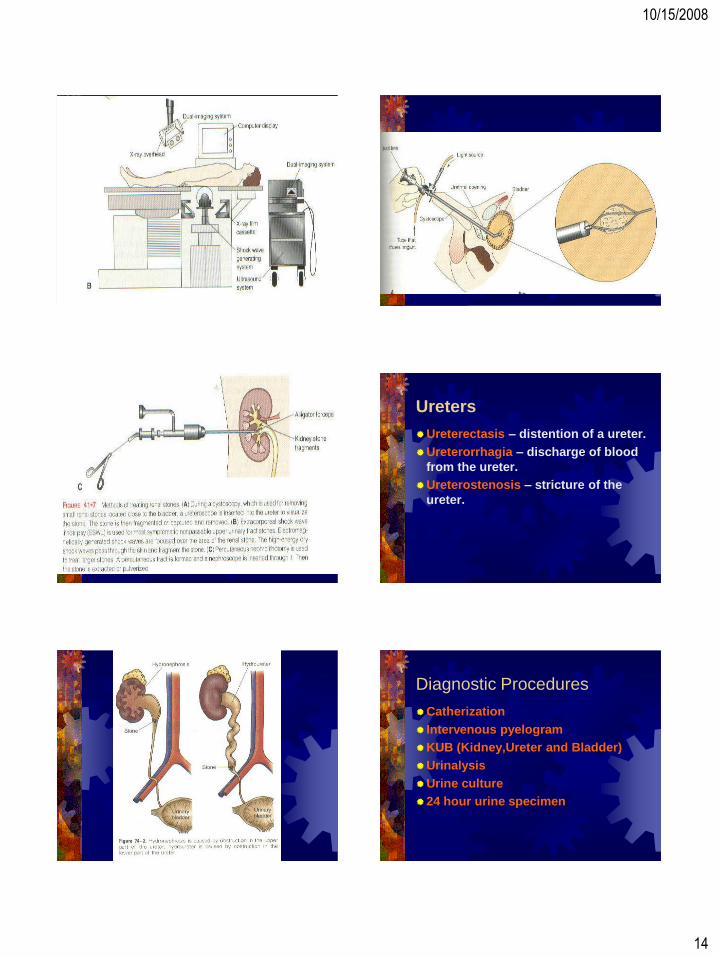
Ureters
Ureterectasis – distention of a ureter.
Ureterorrhagia – discharge of blood
from the ureter.
Ureterostenosis – stricture of the
ureter.
Diagnostic Procedures
Catherization
Intervenous pyelogram
KUB (Kidney,Ureter and Bladder)
Urinalysis
Urine culture
24 hour urine specimen
10/15/2008
15
Catherization
Intervenous
pyelogram
Bladder
cystocele – hernia of the bladder
through the vaginal wall.
Urinary tract infections (UTI’s)..
Incontinence – loss of bladder
control.
10/15/2008
16
Bladder
Cystitis – inflammation of the bladder.
Interstitial cystitis – inflammation within the bladder wall. This is a chronic condition.
Vesicovaginal fissure – an opening between bladder and vagina.
Urethra
Epispadias – urethral opening of the male is on the dorsal (upper surface) of the penis.
Hypospadias - opening is on the undersurface of the penis. In female the urethra opens into the vagina.
Reflux is a back up of urine into the bladder from blockage of the urethra
Paraspadias – congenital abnormality in males in which uretheral opening is on one side of the penis.
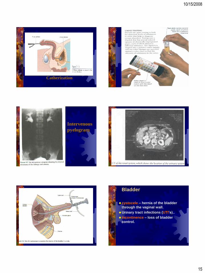
Signs and Symptoms
Abdominal or flank pain
Hematuria
Palpable kidneys
Enlarged kidneys
Recurrent UTI with chills and fever
Intravenous Pylography to confirm
diagnosis
10/15/2008
17
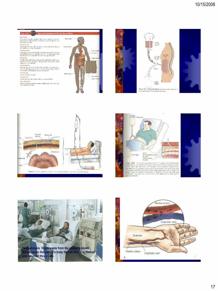
Hemodialysis filters waste from the patient’s blood.
Blood leaves the patient’s body thru an artery, is filtered
and returned thru a vein.
10/15/2008
18
10/15/2008
19
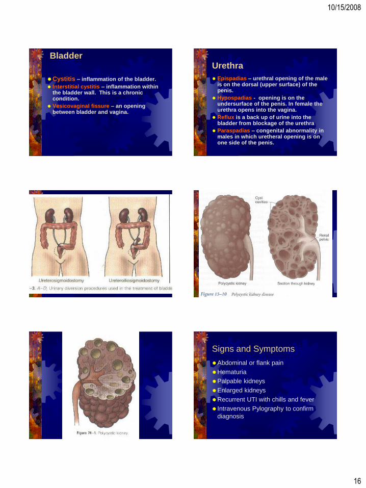
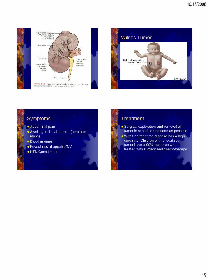
Wilm’s Tumor
Symptoms
Abdominal pain
Swelling in the abdomen (hernia or
mass)
Blood in urine
Fever/Loss of appetite/NV
HTN/Constipation
Treatment
Surgical exploration and removal of
tumor is scheduled as soon as possible
With treatment the disease has a high
cure rate, Children with a localized
tumor have a 90% cure rate when
treated with surgery and chemotherapy.