Embed Size (px)
Citation preview

Renal failure/disease and sepsis
Cherelle Fitzclarence
November 2009

Overview
• Definition• At risk patients• Recognition• Cases

Definition
• Sepsis– condition in which the body is fighting a severe
infection that has spread via the bloodstream. If a patient becomes "septic," they will likely be in a state of low blood pressure termed “shock."
– Inflammatory response– Release of endotoxins
http://www.emedicinehealth.com/sepsis_blood_infection/page2_em.htm

At risk patients• Chemotherapy/or other immunosuppressive therapy• patients undergoing radiotherapy• Chronic disease eg AIDS, diabetes, chronic kidney disease• Very young• Very old• Patients without a spleen• Black patients – black men have highest rates• Long term steroid use• People who have ‘foreign bodies’ eg hickman catheter, AV
graft, catheter• Someone who has very large burns or severe injuries• People with infections such as the following:
• Pneumonia • Meningitis• Cellulitis• UTI• Ruptured viscus
http://www.emedicinehealth.com/sepsis_blood_infection/page2_em.htm

Symptoms• Nothing• Fever• Fatigue• Weakness• Nausea, vomiting• Abdominal pain• Diarrhoea• Tachycardia• Lethargy• Rigors• Confusion• Dizziness• Rash• Epigastric pain• Arthralgias, myalgias

Sepsis – 3 stages
• Sepsis To diagnose sepsis, there must be at least two of the following symptoms:
• Fever above 38C (37.5) or below 36C • Heart rate higher than 90 beats a minute • Respiratory rate higher than 20 breaths a
minute • WCC > 12,000 or < 4000• Probable or confirmed infection
http://www.mayoclinic.com/health/sepsis/DS01004/DSECTION=symptoms

Severe Sepsis
• The diagnosis will be upgraded to severe sepsis if there is also at least one of the following signs and symptoms, which indicate organ dysfunction:
• Areas of mottled skin • Significantly decreased urine output • Abrupt change in mental status • Decrease in platelet count • Difficulty breathing • Abnormal heart function

Septic Shock
• In addition to the above– all the symptoms of severe sepsis and – Hypotension– Multiorgan failure– Cardiovascular failure – hypotension– Renal failure – oliguria– Respiratory failure- hypoxaemia– Haematologic failure - coagulopathy

Pathophysiology
• An inflammatory stimulus (eg, a bacterial toxin) triggers production of proinflammatory mediators, including tumor necrosis factor and IL-1.
• Cytokines cause neutrophil-endothelial cell adhesion, activate the clotting mechanism, and generate microthrombi. They also release numerous other mediators, including leukotrienes, lipoxygenase, histamine, bradykinin, serotonin, and IL-2.
• Cytokines are opposed by anti-inflammatory mediators, such as IL-4 and IL-10, resulting in a negative feedback mechanism.
http://www.merck.com/mmpe/sec06/ch068/ch068a.html

Pathophysiology
• Initially, arteries and arterioles dilate, decreasing peripheral arterial resistance;
• Cardiac output typically increases. This stage has been referred to as “warm shock.”
• Later, cardiac output may decrease, BP falls (with or without an increase in peripheral resistance), and typical features of shock appear.
http://www.merck.com/mmpe/sec06/ch068/ch068a.html

Pathophysiology
• Even in the stage of increased cardiac output, vasoactive mediators cause blood flow to bypass capillary exchange vessels (a distributive defect).
• Poor capillary flow from this shunting, along with capillary obstruction by microthrombi, decreases delivery of O2 and impairs removal of CO2 and waste products.
• Decreased perfusion causes dysfunction and sometimes failure of one or more organs, including the kidneys, lungs, liver, brain, and heart.
• Coagulopathy may develop because of intravascular coagulation with consumption of major clotting factors, excessive fibrinolysis
http://www.merck.com/mmpe/sec06/ch068/ch068a.html

Signs
• Fever• Tachycardia• Tachypnea• BP remains normal to start with – then decreases.• Other signs of the causative infection are generally
present.• Confusion or decreased alertness. • Skin is paradoxically warm• Oliguria (< 0.5 mL/kg/h) • Extremities become cool and pale with peripheral
cyanosis and mottling• Organ failure causes additional symptoms and
signs specific to the organ involved.
http://www.merck.com/mmpe/sec06/ch068/ch068a.html

Diagnosis
• High index of suspicion• History• Examination• Septic screen
– MSU – m/c/s
– Serial blood cultures – m/c/s
– FBC
– UECB
– LFT
– CRP
– CXR
– ECG
– Drug levels

Kidney in sepsis
• Noradrenaline vasoconstricts the afferent arterioles dropping the transglomerular perfusion pressure leading to a low GFR and Na retention
• High systemic Nitrous oxide levels leads to down regulation of intra renal nitrous oxide production, altering renal blood flow further, especially for the metabolically vulnerable outer medulla
• Inflammatory cells produce oxidants and proteases that injure renal epithelium
• Local coagulopathy leads to intraglomerular thrombus formation

Case study
• 65 yo female• Long standing history of diabetes,
hypertension, mild obesity, diabetic retinopathy, peripheral neuropathy, near end stage kidney failure – pre dialysis, anaemia
• Important person of high standing in her community

Case study 1
• Social worker notifies GP that patient is not her normal self
• GP gets patient in• Nothing revealed on history and nothing to
find on examination except patient did not recognise GP whom she knew well
• Sent to hospital with a provisional diagnosis of sepsis, with a written admission, medication chart and form for blood tests

Case Study 1
• Seen by nurse in A and E• Decision made that there was nothing
wrong with her• Bloods not collected• Patient sent home• GP not notified and worse, sarcastic
comments about the GP referral in notes

Case study 1
• Next day GP rang to check on patient • Discovered events• Home visit to patient
• Patient at daughter’s house• In bed• GCS of 8• Vomiting• Probable aspiration

Case Study 1
• Patient transported to hospital via ambulance
• Decreased LOC, RR 28, P 120, T 39C, BP 70/30
• BSL 11, WCC 28 with 95% neutrophilia, CRP 250, HCO3 12, Creat 600, Urea 23
• MSU - EColi• Rx?

Case study 1• DRABC always first• oxygen• Broad spectrum antibiotics in first instance, as
didn’t have E Coli result• Narrow the antibiotics when source of sepsis
identified• IV fluid resuscitation with 0.9% normal saline• Abscesses drained, necrotic tissue excised• Blood glucose levels normalized• Replacement-dose corticosteroids
• This patient spent a month in hospital and did not truly recover her cognitive function nor her physical well being

Case 2
• 45 yo male• Diabetes, hypertension, alcoholic, end
stage kidney disease, peritoneal dialysis• From Bidyadanga

Case 2
• Presents on a Saturday afternoon to hospital with diarrhoea and abdo pain
• Stated to doctor that pd bags were clear, had some ‘dodgy’ kangaroo the night before
• No vomiting or nausea nor any other complaints

Case 2
• Afebrile, BP 120/80, P 70• Diarrhoea while in Cas• Abdo soft and mildly tender in the lower
half• No rebound or guarding
• Dx gastroenteritis• Rx IV fluid, admit to ward
• Thoughts?
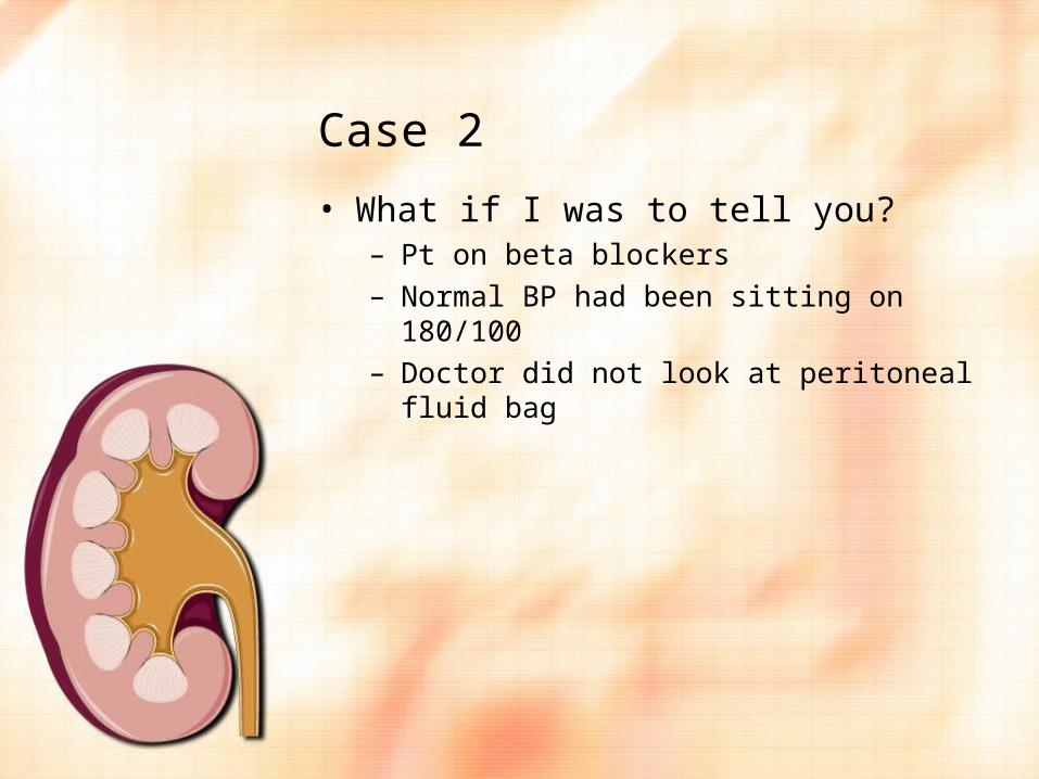
Case 2
• What if I was to tell you?– Pt on beta blockers– Normal BP had been sitting on 180/100– Doctor did not look at peritoneal fluid bag

Case 2
• The following morning patient was reviewed– Noted to have BP of 70/40, PR of 100, T 35– GCS of 9– BSL of 3
– ‘Pea soup’ bag

Case 2
• This patient is septic• This patient always had peritonitis – from
the second he walked in this should have been the presumed diagnosis until proved otherwise
• This non recognition, cost this man his PD tube and required him to be transferred to Perth and started on Haemo
• It very nearly cost his life

Case 2
• Correct management should have been;– Insist on viewing a CAPD bag– FBC, UECB, LFTG, CRP, Blood cultures, send
the peritoneal fluid bag to the lab for urgent microscopy and culture, MSU-m/c/s
– Vancomycin 2.5g and gentamicin 200mg loaded into a PD bag and leave for 6 hour dwell
– This could well have avoided the subsequent scenario
– If pt presents how this man ended up, it would be worthwhile getting a broad spectrum antibiotic in to the pt system intravenously
– He may now also require inotropic support if fluid resuscitation is not successful

Pearls
• Combination of pyrexia, hypotension, tachycardia and increased WCC is consistent with sepsis
• Hypotension is due to peripheral vasodilatation secondary to altered metabolic auto regulation by vasoactive mediators produced during sepsis
• Severe sepsis responds to intravenous fluids while shock requires inotropic support

Pearls
• Sepsis often associated with warm peripheries due to vasodilation
• Hyperglycaemia and insulin resistance is common in septic patients due to a combination of cytokine release and counter regulatory hormones
• Be very aware of transplant patients• Risk of infections in solid organ transplant
recipients is determined by the net state of immunosuppression and level of exposure to a pathogen

Pearls
• Be aware that mycophenolate and valganciclovir can cause neutropenia
• Prednisolone is standard therapy in transplant medication and depresses the immune system

Case 3
• 65 yo lady presents for haemodialysis• Hx of haemo for 7 years• Walked in ok and seemed ok• 1 hour into dialysis started screaming in language• T 36, P 120, BP 200/100• Chest clear, abdo soft, no obvious source of skin
sepsis• No focus of infection identified• More distressed and quite aggressive – trying to
pull needles out• Other patients getting very distressed
Case 3
• Seen by GP and assumed to have sepsis• Given vancomycin and gentamicin
intravenously after collection of basic bloods and 2 sets of blood cultures
• Patient remained agitated and distressed• Dialysis ceased and sent to hospital

Case 3
• Path– Hb 90, WCC4, plats 150– LFT’s – normal except GGT 200 consistent with
her known alcoholism– Ca 1.9 corrected– PO4 3.2, PTH 156
– CRP 290– Blood cultures 2 days later E Coli in both bottles

Reference
• As per body of talk • Clinical cases in kidney disease – Harris et
al• Oxford handbook of nephrology and
hypertension