Embed Size (px)
Citation preview

Relatório Final de Estágio Mestrado Integrado em Medicina Veterinária
Seasonal dynamics in cellular and humoral immune parameters in farmed rainbow trout (Oncorhynchus mykiss)
Paulo Joaquim da Silva Santos
Orientador: Professor Doutor Paulo Manuel Rodrigues Vaz Pires Co-orientador: Doutor Benjamin Costas Refojos
Porto 2017
i
Agradecimentos
A realização deste estágio e consequente relatório marcam o final de uma importante etapa da
minha vida. Como tal quero agradecer a todos aqueles que, de alguma forma, me ajudaram
neste percurso, tendo um papel decisivo para a sua concretização.
Ao meu orientador, Professor Doutor Paulo Vaz Pires por, num curso com tantas oportunidades
de trabalho e espécies por explorar, despertar em mim o gosto pela ictiologia, por ter embarcado
comigo nesta aventura sempre com um espírito positivo e arrojado, por todos os conselhos
iniciais e por estar sempre preocupado em acompanhar a evolução deste relatório, ajudando-me
e incentivando-me a pesquisar e aprender sempre mais.
Ao Doutor Benjamím Costas, co-orientador, por me ter permitido colaborar com a sua incrível
equipa, proporcionando-me um ótimo ambiente no CIIMAR durante todo o estágio e facultando-
me todos meios que precisava para desenvolver este relatório, acreditando no meu trabalho
mesmo sabendo que tinha um longo caminho a percorrer, orientando-me e despertando em mim
um interesse pela investigação. Estou-lhe muito grato por tudo o que me ensinou, pela paciência
e por me ajudar a realizar uma dissertação da qual me orgulho.
Também gostava de agradecer à Dra. Manuela Castro-Cunha e Dr. José Calheiros, proprietária
e biólogo responsáveis pela Quinta do Salmão, por me permitirem aceder às vossas instalações
e recolher amostras dos vossos animais que foram fulcrais para que este trabalho fosse levado
a cabo.
Não me posso esquecer também dos meus colegas de laboratório, que tanto me ajudaram num
ambiente ao qual não estava acostumado e me fizeram sentir em casa. Um agradecimento
especial à Filipa, por me orientares e me motivares sempre que as coisas não corriam como eu
desejava, alargando-o a todos os outros integrantes desta fantástica equipa: Professor Doutor
António Afonso, Bruno, Carlota, Fran, Helena, Lourenço, Joana, Marina, Pilar, Rita, Sérgio e
Tiago. Obrigado por estarem sempre disponíveis a ajudar, pela boa disposição, por todos os
ensinamentos e companheirismo e por criarem um ambiente tão acolhedor que permitiu que
durante estes ultimos 4 meses acordasse sempre contente e motivado para ir trabalhar.
Quero ainda agradecer a alguns professores que marcaram o meu percurso como estudante e
pelos quais nutro muita admiração quer pelo seu conhecimento quer pela sua simplicidade e
amizade: Professor Armando Lemos, Professor Augusto Faustino, Professora Margarida Araújo,
Professora Maria João Moreira, Professora Marta Santos, Professor Pablo Payo Puente,
Professora Paula Proença e Professor Ricardo Marcos.
Aos amigos da faculdade, que me aturaram ao longo destes últimos anos e que estiveram
sempre a incentivar-me e permitiram que esta passagem pelo ICBAS fosse tão divertida, mas ao

ii
mesmo tempo tão veloz: Filipe Pinto, José Pedro Guimarães, João Ribeiro, Miguel Carvalho,
Catarina Silva, Cláudia Gonçalves, Helena Ferreira e Mariana Meireles…. Vocês são pessoas
muito especiais e ser vosso amigo é um privilégio para mim. Muito obrigado por todos os bons
momentos e pelo vosso apoio incondicional.
Ao meu clube, aos meus atletas e a todos os meus amigos da patinagem, obrigado por aturarem
o meu mau feitio dos últimos meses, por estarem sempre lá para mim e me ajudarem a nunca
desistir dos meus objetivos. Todos os dias me surpreendo com a vossa persistência e com o que
aprendo convosco.
E por último, mas não menos importante, gostaria de dedicar este trabalho á minha família por
todo o esforço, investimento e carinho que me transmitiram ao longo destes anos todos e a ti,
Giulia, por todo o teu apoio, cooperação, compreensão e paciência, ajudando-me a crescer
cientificamente e pessoalmente.
A todos o meu MUITO OBRIGADO!
This work was supported by Project ALISSA (reference ALG-01-0247-FEDER-3520), financed by Portugal and the European Union through FEDER, COMPETE 2020 and CRESC Algarve 2020, in the framework of Portugal 2020.

iii
Abstract
Seasonal variations in both light duration and water temperature are known to be important factors
that may affect fish immunity and disease resistance. The present study was thus conceived to
evaluate the rainbow trout (Oncorhynchus mykiss) immunological status under farming conditions
throughout a year. Blood samples were collected at Quinta do Salmão (Pisões, Portugal) from
three different groups (diploids, triploids, and female diploids of bigger size) in nine time points.
Ten animals from each diploid and triploid group were evaluated at each time point, whereas in
the group of fish close to commercial size only five animals were sampled per time. Then, at
CIIMAR facilities, blood smears were performed, air dried and the rest of the the plasma collected
for humoral parameters quantification. Immune cells were identified and a differential count of
neutrophils, monocytes, lymphocytes and thrombocytes was made. Humoral parameters on
plasma such as peroxidase, lysozyme and antiprotease activity were measured. Results showed
significant differences in all groups with temperature variation, being that fish haematological
values showed highest levels of lymphocyte and thrombocyte cells at cooler temperatures and
phagocytic cells on warmer waters. Instead humoral parameters showed increased activity on
summer months for peroxidase and winter months for lysozyme and antiproteases. Although
some of these variations may be caused by fish normal development, genetic or environmental
interactions, the evaluation of more immune parameters, including IgM levels in plasma could
clarify our hypothesis.
Keywords: rainbow trout, temperature, leukocytes, immune status, peroxidase, lysozyme,
antiproteases.

iv
Resumo
As variações sazonais no fotoperiodo e na temperatura da água são conhecidos por serem
fatores importantes que podem afetar a imunidade e a resistência a doenças nos peixes. O
presente estudo foi assim concebido para avaliar o estado imunológico da truta arco íris
(Oncorhynchus mykiss) em condições de cultivo ao longo de um ano. As amostras de sangue
foram recolhidas na Quinta do Salmão (Pisões, Portugal) de três grupos diferentes (diploides,
triploides e diploides femininos de tamanho maior) em nove períodos temporais. Foram avaliados
dez animais de cada grupo diploide e triploide em cada amostragem, enquanto que no grupo de
peixes perto do tamanho comercial apenas cinco animais foram amostrados por tempo.
Seguidamente, nas instalações de CIIMAR, foram realizados esfregaços de sangue, secos ao ar
e o resto do plasma recolhido para quantificação de parâmetros humorais. Os leucócitos foram
identificados e uma contagem diferencial de neutrófilos, monócitos, linfócitos e trombócitos.
Foram medidos parâmetros humorais no plasma, tais como atividade da peroxidase, lisozima e
antiprotease. Os resultados mostraram diferenças significativas em todos os grupos com a
variação da temperatura, sendo que os valores hematológicos de peixes apresentaram níveis
mais altos de linfócitos e trombócitos a temperaturas mais baixas e células fagocíticas em águas
mais quentes. Em vez disso, os parâmetros humorais mostraram aumento da atividade nos
meses de verão para a peroxidase e nos meses de inverno para a lisozima e as antiproteases.
Embora algumas dessas variações possam ser causadas pelo desenvolvimento normal dos
peixes ou por interações genéticas ou ambientais, a avaliação de mais parâmetros imunes,
incluindo níveis de IgM no plasma, poderiam esclarecer a nossa hipótese.
Palavras-chave: truta-arco-íris, temperatura, leucócitos, sistema imune, peroxidase, lisozima,
antiproteases.

v
This thesis also includes one scientific poster with the title “Seasonal Blood Cell Dynamics in
Farmed Rainbow Trout: A Comparative Study Between Diploid and Triploid Fish”, presented at
the 2nd AquaImprove, Aquaculture Research Workshop, that took place on 17th March 2017 at
CIIMAR (Interdisciplinary Centre of Marine and Environmental Research) facilities: Terminal de
Cruzeiros do Porto de Leixões, Avenida General Norton de Matos, S/N, 4450-208 Matosinhos,
Portugal (Appendix).

vi
INDEX
1. Introduction 1
1.1 Evolution of World and European Aquaculture 1
1.2 Characterization of the species 3
1.3 Difficulties of aquaculture production 4
1.4 Haematology 5
1.5 Immune system and inflammation 7
1.6 Water biotic and abiotic factors in fish haematology and immunity 9
1.6.1 Fish growth and maturation and genetical improvements 9
1.6.2 Stress and parasitism 10
1.6.3 Water dissolved oxygen and pH 11
1.6.4 Water Temperature 12
1.7 Scope of study 13
2. Materials and Methods 14
2.1 Sampling 14
2.2 Haematological analysis 15
2.3 Humoral parameters analytical procedures 15
2.4 Statistical analysis 18
3. Results 19
4. Discussion 23
5. Conclusions 25
6. Bibliography 26
7. Appendix 31
1
1. Introduction
1.1 Evolution of World and European Aquaculture
World’s fish market has been rising over the last 50 years, following population growth and
food consumption, being that fish constitutes an important source of nutrients and proteins for
several people all over the globe (FAO 2012). In 2010, fish consumption represented about 17
percent of animal protein ingestion and 6.5 percent of total protein ingested (FAO 2014). Already
in 2014, World per capita apparent fish consumption reached a new record, fixed in 20.2 kg
compared to 9.9 kg in 1960s, and is estimated that till 2025 it hits 21.8 kg, corresponding to a
growth of 7.8 percent. At the same time, the supply of fish has accompanied this growth from the
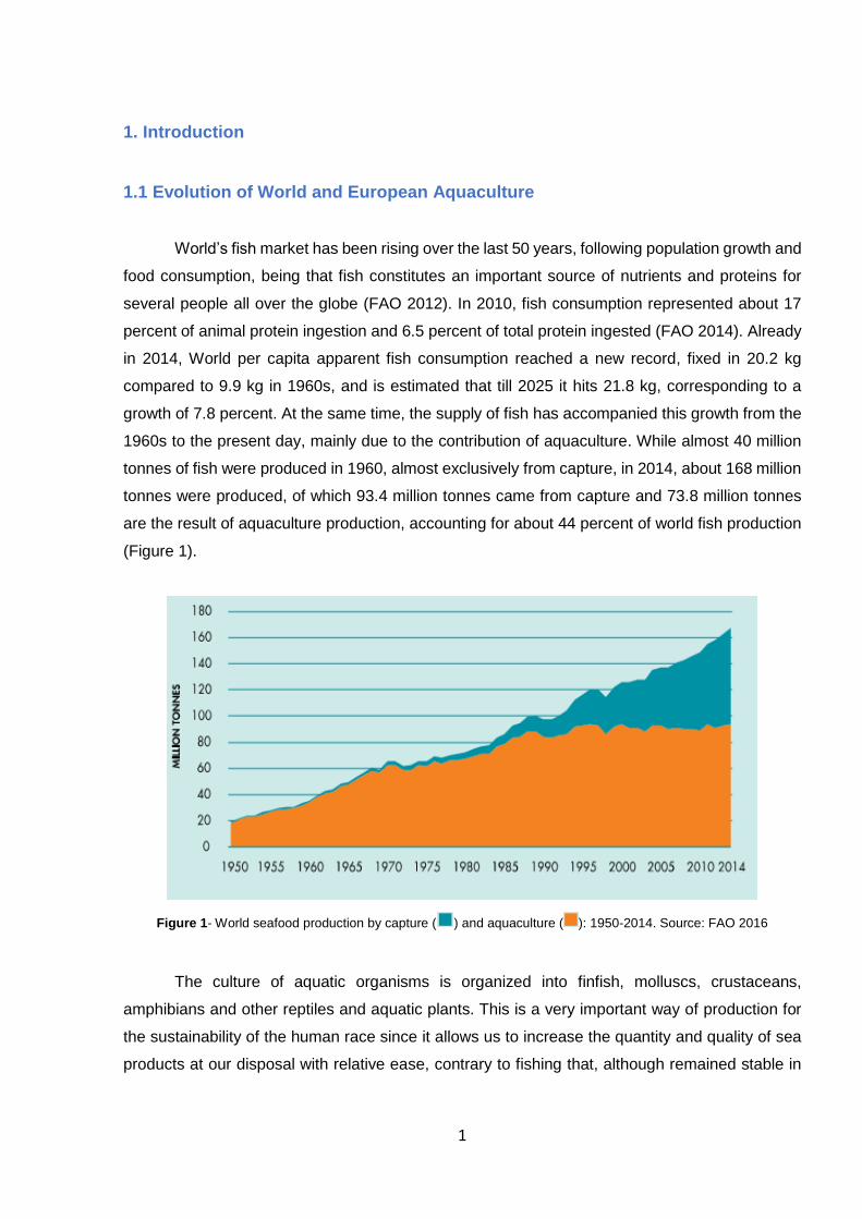
1960s to the present day, mainly due to the contribution of aquaculture. While almost 40 million
tonnes of fish were produced in 1960, almost exclusively from capture, in 2014, about 168 million
tonnes were produced, of which 93.4 million tonnes came from capture and 73.8 million tonnes
are the result of aquaculture production, accounting for about 44 percent of world fish production
(Figure 1).
Figure 1- World seafood production by capture ( ) and aquaculture ( ): 1950-2014. Source: FAO 2016
The culture of aquatic organisms is organized into finfish, molluscs, crustaceans,
amphibians and other reptiles and aquatic plants. This is a very important way of production for
the sustainability of the human race since it allows us to increase the quantity and quality of sea
products at our disposal with relative ease, contrary to fishing that, although remained stable in
2
the last years, is too much dependent on non-controllable factors such as food supply, predation,
water pollution and global warming (United Nations Development Programme 2012).
The main producers come from Asia, where about 55 percent of the aquatic animals
consumed are produced in aquaculture, being China particularly important because is one of the
pioneers in fish cultivation and has produced about 58 795 thousand tons in 2014. In Europe,
although it only represents 18 percent of the supply, aquaculture production has been increasing,
doubling the percentage of cultivated fish in 1995. Thus, according to the Food and Agriculture
Organization, 2014 Europe’s production reached 2,930 million tonnes, with the Southern Europe
countries, where Portugal is included, contributed with 595 thousand tonnes (FAO 2016).
More specifically, in Portugal and the same year, according to Instituto Nacional de
Estatistica, about 10,791 tonnes of marine animals were produced in aquaculture. In our territory
fish are the main group cultivated, of which we can emphasize Scophtalmus maximus (turbot),
Sparus aurata (gilthead seabream), Dicentrarchus labrax (European seabass) and Oncorhynchus
mykiss (rainbow trout). Regarding the exploitation regime, we can affirm that in fresh water
Portugal produces exclusively in an intensive way, while in brackish and marine waters 47.8
percent of the production was obtained extensively, 39.2 percent intensively and the remaining
percentage by the semi-intensive regime (INE 2016).

3
1.2 Characterization of the species
Oncorhynchus mykiss (Walbaum, 1792) is a fusiform fish of the Actinopterygii class,
Teleostei infra-class and Salmonidae family. It is a freshwater fish with a brownish or yellowish
body, with black spots over the length of the body and a pinkish stripe that extends from the gills
to the caudal fin, which is a specific characteristic of this species (Figure 2). Its origin is the Pacific
coast of the United States, although at present it is widely distributed, both because of its high
resistance and environmental adaptability and of its crescent demand. Its introduction in Europe
took place at the end of the 19th century and is currently being created all over Europe, being
mainly exported by countries such as Denmark, Poland and Sweden. In our country, the creation
of O. mykiss dates from the beginning of the 60s, with the first unit to be installed on the Coura
River (A Pesca e a Aquicultura na Europa 2012).
Figure 2- Oncorhynchus mykiss (Walbaum, 1792)
Its production in Portugal occurs exclusively under intensive mode in fresh water and, in
2014, 787 tons of O. mykiss were produced (INE 2016). O. mykiss growth and maturation are
influenced by water temperature and feed, which must be high in protein, and maturity is reached
between 3 and 4 years of age. Regarding to breeding, this process should ideally be performed
with water temperatures below 12º C. After hatching, the larval culture of these animals is made
in circular fiberglass or concrete tanks, under conditions of regular flow and balanced distribution
of animals. At the moment little fish reach a weight of 50 grams and a size of 8 to 10 centimetres,
they are transferred to growing tanks, in floating cages placed in ponds or rectangular concrete
fish farms, over a river. Although these cages do not require human intervention for water renewal,
in fish farms it represents a task that can be performed by two techniques: through an open
system, passing the river water through a channel, or with a closed (recirculation) system, which
circulates the water in the tanks and recycles it. After reaching this stage, commercial trout are
harvested with a net or pumped by a system that takes them ashore to then be packaged and
transported. Besides few studies have been performed to evaluate the presence of pathological

4
agents on portuguese trout farms, epidemic cases occurred mainly by bacterial Aeromonas
salmonicida (Saraiva et al. 1989), responsible for furunculosis, and Yersinia ruckeri (Sousa 1996).
1.3 Difficulties of aquaculture production
Although our coast has a supposed great potential for aquaculture practice, the truth is
that the coasts of the west and north of our territory suffer a great influence of the Atlantic Ocean
by its currents and waves, affecting the ambient and water temperature, especially in the winter
(Ramalho & Dinis 2010). A recent method that is being used in fish farms is the offshore, where
fish cages are moved and submerged in deeper water in order to increase fish welfare, decrease
pathologies and dilute wastes from fish production (Naylor & Burke 2005). Although this method
assumes an ecological and spacial concern which is of relevant importance for aquaculture
producers to obtain their farming licenses, the offshore technology represents a challenge to fish
farmers since these structures are very susceptible to storms and wind (Stickney et al. 2006),
with some reported cases of cage damages, resulting in considerable economical losses around
the world.
Investment in fish feed is also very important, representing a large share of aquaculture
expenditure. The food supplied must be adapted to the species, nutritional needs and stage of
life, whereby producers, by relying on nutritional management companies, benefit from better
information on handling, packaging and nutritional value, as well as tools that allow them to
evaluate production indexes such as feed conversion and growth rates to quickly detect food
deficiencies and corrected them (Ramalho & Dinis 2010; Hasan & New 2013).
Another obstacle that influences marine products is the poor modernization, either by the
protection policies of the coastal zones or by the costs inherent in it, which are incompatible with
our small and familiar farms (MADRP-DGPA 2007). Recirculating aquaculture systems (RAS) are
innovative and extremely efficient, using tanks that permit intensive fish production with limited
water exchange, due to biofiltration and other environmental correction resources that keep water
clean resulting in an healthier environment (Timmons & Ebeling 2013). Beyond the above profits,
RAS brings to producers higher flexibility to choose a place to install a fish farm, higher stock
density and production by area unit, drastic reduction of the effluent volume, water parameters
control such as pH, oxygenation and temperature, as well as fast access to feed and cultivated
animals that lead to enhanced performance. Besides all these advantages, RAS is still not being
widely explored in Portugal because of the high upfront investment in material and infrastructures,
as well as the need of experient and trained staff to monitor and operate the systems (Rawlinson
& Forster 2000).

5
1.4 Haematology
Blood studies in fish are relatively recent being the first reports of observation of erythrocytes in
1845 by Gulliver, and of leukocytes in 1905 by Drzewina (Oria 1932; Ellis 1976; Ellis 1977). The
execution of blood smears is of great importance since it allows us to assess the immunological
status of the fish in a very quick and representative way, in which only one drop of blood is enough
for a preliminary evaluation. However, its realization requires practice since the distinction
between the various lymphoid cells represents a complex task. To a better comprehension of this
subject, a brief summary of the most representative lymphoid cells on O. mykiss’s blood will be
presented (Figure 3).
Lymphocytes are the most representative lymphoid cells in O. mykiss blood. They are
predominantly rounded cells, of variated size, with basophilic cytoplasm and without granulations.
Its nucleus presents dense chromatin and is frequently observed an high nucleus/cytoplasm rate.
Although they may resemble thrombocytes, lymphocytes show cytoplasmic projections, which
facilitates their differentiation (Tavares-Dias & Moraes 2004).
The neutrophils are predominantly rounded, whose cytoplasm has very fine greyish
granulations and the nucleus has the form of a rod, peripheral, with the nuclear chromatin not
very compact and without a visible nucleolus. These cells have high migratory activity, like that
A
B
C D
Figure 3 - Blood O. mykiss smears stained with Wright’s stain. Neutrophils were labelled using the Antonow’s technique presenting abundant peroxidase granules. Letters represent: (A) monocyte; (B) thrombocyte; (C) lymphocyte; (D) neutrophil.

6
occurring in mammals, and a strong non-specific cytotoxic activity (Griffin 1984; Sasaki et al.,
2002). In addition to these characteristics, the presence of peroxidase, a lysosomal enzyme,
promotes the oxidation of compounds by hydrogen peroxide in the phagocytosis process (Oliveira
et al. 1997).
The monocytes are recognized as cells with few pseudopods, large numbers of
mitochondria and vacuoles, and some endoplasmic reticulum and Golgi complex. Its nucleus
occupies a third to a quarter of the cell and using optical microscope it is possible to observe them
quite dense, surrounded by a basophilic cytoplasm, distinctive from other cellular groups (Dogget
& Harris 1989). There is some confusion about the denomination of these cells due to their
phagocytic action, with some authors considering them macrophages. However, the
monocyte/macrophage designation is simple since monocytes act as circulating cells while
macrophages migrate to tissues and other places like the peritoneal cavity and natatory bladder
(Lorenzi 1999; Lamas et al. 1994).
The last lymphoid cells are the thrombocytes, which are observed under the optical
microscope as elliptic cells, with fusiform and colorated nucleus. These cells have the same
functions of platelets in mammals and can be found in birds, reptiles, amphibians and fish, with
the function of haemostasis and homeostasis (Roberts 1981). Thrombocytes are also in
discussion about their inclusion as lymphoid cells, being that past researchers have demonstrated
the phagocytic function of thrombocytes, suggesting these cells to make part of the defence cell
group (Hill & Rowley 1988). There is also evidence of presence of pattern recognition receptors
(PRR) on thrombocytes membrane that act by recognition of pathogen-associated molecular
pattern (PAMP) and damage-associated molecular pattern (DAMP) present on pathogens and
host cells respectively.

7
1.5 Immune system and inflammation
Besides fish being one of the primitive vertebrates, they possess phagocytic mechanisms
that work side by side with humoral and cell-mediated immunity (Roberts 2012). After infection,
immune system can act by two different ways, according to response speed and specifity. The
first response is from the innate system, that is fastest entering in action but low specific. Although
that, fish innate system is able to discriminate self and non-self organisms, and his role is to
protect the host by inhibiting pathogen’s multiplication (Jaqueline Parkin 2001). Later comes the
adaptive immunity, a pathogen specific response caused by antigen presentation from myeloid
cells to lymphocytes, with antibody and memory cell production for that kind of infection, on a
process that is influenced by temperature (Ellis 1999). Lymphocytes are cells of great relevance
for adaptive immunity, once different cell type lead to antigen recognition, specificity and memory.
These cells can be divided into B-cells responsible for antibody production (on fish the most
produced antibodies on systemic circulation are IgM, while IgT is widely present on mucosal
surface),and T-cells that mediate cell-mediated immunity. T-cells can be subdivided according to
their direct action on killing infected and abnormal cells (cytotoxic T-cells), or to them modulation
activity to other cells through cytokine production (helper T-cells) (Gudding et al. 2014).
Phagocytosis is the primordial cell defence mechanism activated after injury or infection
and is modulated by phagocytes, that can be divided into professional phagocytes (such as
neutrophils, monocytes and macrophages), with receptors on their surface able to detect harmful
objects, and non-professional phagocytes (Corbel 1975). In aquaculture, there is a particular
interest at developing disease’s resistance by increasing the phagocytic activity of defence cells.
The phagocytic process occurs due to recognition and attachment to a foreign particle, with
posterior engulfment and digestion. (Roberts 2012). Macrophages are cells that enter in action
after different types of activation, being the most common designation M1 or M2, according to
lymphocyte T helper 1 (Th1) or T helper 2 (Th2) cytokine induction (Mantovani 2002). However,
recent studies have enlarged the macrophages phenotype to four, according to different
environmental signals (Forlenza et al., 2011). Firstly, we have innate activation of macrophages,
that occurs after an isolated microbial stimulus which is recognized by macrophage’s Toll-like
Receptors (TLR), Cluster of differentiation (CD)14 or other PRR’s. Similarly to the above, we have
classical activation, that requires the same stimulus of innate activation plus the presence of the
cytokine Interferon ɣ (IFN-ɣ) (Dalton et al. 1993). The third and fourth phenotypes, alternative
activated macrophages and regulatory macrophages, are associated with bacterial interaction
with Interleukin-4/Interleukin-13 and Interleukin-10 respectively.
Other host defence mechanisms that try to contain the propagation or eliminate the cause
of host-tissue damage are called by some authors as nonspecific mediators of immunity. They

8
act as physical barriers to invading organisms before cell activation occurs of which we can
highlight skin and mucus. The mucosal surfaces of fishes (gill, skin, and gastrointestinal tract)
form a thin physical barrier between the external environment and the internal milieu, and they
are important sites of microbial exposure. Cutaneous mucus is considered the first line of defence
against infection through skin epidermis and it acts as a natural, physical, biochemical, dynamic,
and semipermeable barrier that enables the exchange of nutrients, water, gases, odorants,
hormones, and gametes (Esteban 2012). Furthermore, mucus in fish has the ability to trap and
immobilize pathogens before epitelial surface contact, due to its constant secretion and
substitution (Mayer 2003). In fish mucus, immune system components have been identified such
as enzymes, proteases, antimicrobial peptides, lectins, proteins and immunoglobulins, that can
provide a protective role for aquatic organisms.
After infection or tissue lesion, an inflammatory process begins in order to repair the
damage and stablish homeostasis (Kiron 2012). Inflammation mechanism is multi-factorial and
still few studied in fish (Ellis 2001), but is yet known that vascular phenomena occurs, initiating a
fast and local increased blood flow, resulting in hyperaemia. After this, local inflammatory
mediators are produced, promoting a process called chemotaxis, calling neutrophils and
monocytes to further phagocytosis (Kindt et al. 2007). Phagocytic cells are also capable of inhibit
the colonization, survival and proliferation of microorganisms with a set of antimicrobial agents
such as lysozyme, complement factors (Barton 2008), antiproteases (Secombs 1996), and
bactericidal reactive oxygen species (Ellis 2001).
Lysozyme is a phagocytic cell’s enzyme which hydrolyses N-acetylmuramic acid and N-
acetylglucosamine which are constituents of the peptidoglycan layer of bacterial cell walls. The
complement system can be activated by antigen-antibody reactions or by the so called alternative
route, via binding to microbial cell wall polysaccharides, which results in opsonization and/or lysis
of foreign cells (Bayne & Gerwick 2001). Antiproteases are substances that have yet been found
in O. mykiss serum (Ellis et al. 1981) and their role is to inhibit the proteases released from
bacteria that help these pathogens to use host proteins as substrate for their maintenance. The
main protease inhibitors are α1-anti-protease, α2-anti-plasmin and α2-Macroglobulin (α2M) (Ellis
2001). Neutrophils contain myeloperoxidase (MPO) in their cytoplasmic granules (Afonso et al.
1997). MPO in the presence of halide ions and H2O2 can kill bacteria by halogenation of the
bacterial cell walls as well as production of bactericidal hypohalite ions (Klebanoff & Clark 1978).

9
1.6 Water biotic and abiotic factors in fish haematology and immunity
1.6.1 Fish growth and maturation and genetical improvements
All physiological processes on animal organism occurs due to energy trades, coming from
their intermediary metabolism with further channelling of the amount required for each biological
task, according to each life phase (Myrick 2011). Fish growth and maturation are two of the most
exigent phases of young fish which demand a large quantity of energy and nutrients. Besides
there are not many studies that compare fish growth and haematological/leukocytary parameters,
DeWilde and Houston have demonstrated that erythrocyte number decreases with O. mykiss fish
development and weight gain. Likewise this fact, is also demonstrated that younger fish possess
higher erythropoietic activity than older and bigger fish (Zhiteneva & Gorislavskaya 1986), which
make believe that different size and age aquatic animals produce different haematological values.
Although relevant, this parameter shouldn’t be considered isolatedly, once fish growth is affected
by nutritional and ecophysiological conditions such as ambiental and water temperature,
photoperiod and water physical and chemical parameters (Chaudhuri et al. 1986).
Posterior studies have been performed on teleosts to verify the influence of fish sex and
gonadal maturation on haematological/leukogram values. One of these studies have compared
female and male O. mykiss, evaluating their leukogram, resulting in no significant differences
between different sex groups (Ranzani-Paiva et al. 1998a). On the other hand, the same author
observed significant differences between erythrogram/leukogram results and gonadal maturation,
whose results presented increased erythrocyte, neutrophil and monocyte number, with decrease
of lymphocyte number (Ranzani-Paiva et al. 1998b), suggesting that gonadal maturation might
be one of the physiological factors responsible for blood cell variations.
Increased aquaculture’s products demand has opened way for genetical improvements
on fish production, with O. mykiss triploid or 3N fish started being developed in middle 80´s, by
subjecting eggs to thermal or pressure shocks shortly after fertilization (Lincoln & Scott 1983).
This technology consists in selected genetic organisms that have high percentage of sterile
females. This is one of the genetical developments widely used in aquaculture, since it allows
transference of energy that would be used in reproduction to growth. Investigations into non-
specific, humoral immune parameters comparing diploid and triploid Salmo salar groups
produced interesting findings, with antiprotease activity results presenting lower concentrations
in triploids (Langston et al. 1997).

10
1.6.2 Stress and parasitism
Fish immune system might be affected by regular farming stressing procedures that are
identified at four levels: handling, confinement, stocking and transport (Wendelaar Bonga 1997;
Costas et al. 2012). These procedures develop physiological deregulation, followed by changes
on metabolism and cell processes, especially on leukocyte synthesis, with innate defence
mechanisms commitment and increasing the predisposal to pathological situations (Ellis 2001,
Tort 2011). One of the most used indicator to evaluate teleost fish on stressing processes is
cortisol (Mommsen et al. 1999), with increased plasma concentration in affected animals. High
cortisol plasma levels may lead to decreased lymphocyte number, supressed phagocytic activities
in head kidney and blood, and increased susceptibility to infection (Ortuño et al. 2001). Studies
made to evaluate cortisol influence on disease predisposal were performed, resulting in increased
susceptibility to furunculosis infection by A. salmonicida after exogen cortisol administration
(Pickering 1989; Pickering et al. 1992). Stress factors such as transport, anoxia, social conflict,
handling, injection and crowding have also been studied resulting in decreased number of
lymphocytes and thrombocytes and increased number of circulating neutrophils in several
species. (Pickering and Pottinger 1987; Pulsford et al. 1994; Espelid et al. 1996).
Fish exposition to pathogenic agents may generate physiological response similar to
stressant situations, resulting in host immune suppression (Ruane et al. 2000). Pathogens can
live on host without cause them any immunological change. This coexistence benefits both
organisms but when a change on the ambient-pathogen-host system occurs, fish defences are
at risk, which can result in a pathological process (Tavares-Dias et al. 1999), that in extreme
cases ends with death. Infectious agents have different incidence along the year, with bacteria
and parasite infections being associated with spring die-offs and viruses commonly affecting
fishes in autumn. A study developed on O. mykiss parasited with protozoan Ichthyophonus hoferi
resulted in a lower level of erythrocyte number and haemoglobin concentration (Rand & Cone
1990). The same study revealed an immunosuppression after parasite infection resulting in
leukopenia. Another study with Vibrio anguillarum, have produced low leukocyte levels too, but
was more specific about leukocitary changes, showing decreased lymphocyte levels and
increased neutrophil and monocyte levels (Lamas et al. 1994). O. mykiss was also infected with
Renibacterium salmoninarum revealing some differences comparing to the results above,
particularly at thrombocyte levels that were increased in infected animals (Bruno & Munro 1986).

11
1.6.3 Water dissolved oxygen and pH
Oxygen concentration is a water parameter of high relevance for fish population, with O2
playing an essential role on metabolic processes of nutrient assimilation (Castagnoli 2000).
Dissolved oxygen values vary on inverse sense of temperature, and oxygen solubility clearly
affects inorganic phosphate intra-erythrocitary such as adenosine triphosphate (ATP) and
guanosine triphosphate (GTP) (Val 2000). Although some fish can tolerate low oxygen
concentration, it clearly affects their health and performance (Noga & Francis-Floyd 1991), being
one of the most stressing ambiental factors for fish that requires adaptative adjustments at all
biological organization levels (Baldisseroto 2002). Researchers have submitted O. mykiss to low
concentrations of oxygen which resulted in increased gill ventilation to regulate oxygen
distribution and avoid blood pH decrease. These authors have also concluded that low oxygen
concentration affects hematological parameters, increasing haematocrit and haemoglobin
concentration without erythrocyte number alteration (Holeton & Randall 1967). On an opposite
situation, hyperoxia can lead to decreased ventilation frequency, resulting in CO2 accumulation in
blood, respiratory acidosis (Dejours 1977) and imbalance in gill ion concentration (Brauner et al.
1999). Salmon fish farms usually supplement oxygen to supersaturate the water with O2, since
they believe it can improve fish growth and increase pathogen resistance (Caldwell and Hinshaw
1994). Although immune response is still not correlated with water oxygen saturation, some
studies have demonstrated that moderate hyperoxia can in fact improve fish growth (Hosfeld et
al. 2008).
Water pollution may change biochemical quality of aquatic animals, causing several
problems on fish performance due to water acidification. pH variations can decrease fish activity
and swimming skills and alkaline pH levels are required for farming fish performance, with values
varying from 7.0 to 8.5 (Baldisseroto 2002). Acid pH is known to affect haemoglobin oxygenation
capacity, resulting in low oxygen arterial levels that demand circulating erythrocytes to
compensate oxygen transport (Houston & Gingra-Bedard 1994). In addition to other physiological
changes, hematologic and leukocyte alterations have also been reported with pH variations.
Diverse fish species studies have concluded that low pH levels presented increased haematocrit,
total leukocytes and thrombocyte, neutrophil and monocyte percentage (Giles et al. 1984; Dheer
et al. 1987; Rambhaskar & Srinivasa-Rao 1989).

12
1.6.4 Water Temperature
Fluctuations in water temperature are a seasonal phenomenon which influences humoral,
cell mediated and non-specific (physical barriers) defence mechanisms in fish. Seasonal variation
affects mainly the temperated zone, due to photoperiod and temperature changes that modulate
immunologic mechanisms of fish (Zapata et al. 1992). Haematological changes in O. mykiss
during the year resulted in reduction of erythrocyte number, haematocrit and haemoglobin
concentration on the transition autumn/winter (Lane 1979), while summer values showed
increased number of erythrocytes and mean corpuscular volume, with opposite decreased
haemoglobin concentration and mean corpuscular haemoglobin concentration (Rehulka 1997).
Leukogram changes are also observed resulting in higher leukocyte average number during
spring, while lymphocyte average number decreased and neutrophil average number increased
during summer months. (Houston et al. 1996). There are also evidences indicating that fish
possess low immunological activity on winter, similarly to what happen with amphibian and
reptiles (Zapata et al. 1992).
It is globally recognized that environmental factors and water temperature affect teleost
growth, reproduction, survival and metabolism. A study focused on the performance of female O.
mykiss, comparing diploid and triploid animals at chronic high temperatures, have showed that
triploid animals were more susceptible to water temperature variations in terms of growth and
survival (Ojolick et al. 1995). Increased water temperature leads to more biological and metabolic
activity of tropical fish, with consequent increase in the respiratory and cardiac frequencies due
to bigger oxygen demand (Baldisserotto 2002). Haematocrit, haemoglobin concentration and
erythrocyte number show tendency to increase with water temperature elevation (Martinez et al.
1994). Water temperature variations produce diverse leukogram discrepancies in different teleost
species. In 2002, studies have concluded that temperatures between 15 and 17 ºC produces
bigger monocyte, granulocyte and B lymphocyte activation on O. mykiss after inoculation with A.
salmonicida than with temperatures between 10 and 12 ºC. However, the development of a
specific antibody response against infection seemed to be more effective at lower temperatures
(Kollner & Kotterba 2002). Also on study specie, phagocytosis increases with temperature
elevation, compensating the lymphocytopenia (Houston et al. 1996). Between phagocytic cells,
neutrophils seemed to be the most resistant defence cells to immunosuppression with low
temperature (Ainsworth et al. 1991). Another study developed on Salmo trutta lacrustis (brown
trout) kept at 15 ºC for 77 days have produced lowest leukocyte and thrombocyte values, with no
significative alteration on number of lymphocytes or neutrophils (Rahkonem & Pasternack 1998).
13
1.7 Scope of study
Although aquaculture is a valid alternative for food sustainability, it represents a big
investment and is still somehow insecure due to problems of population density, fish immune
system and pathologies associated with some mortality and consequently monetary losses. In
view of these problems and the scarcity of studies in natural conditions of this species, the
objective of this work is to verify the influence of water temperature on the immunological status
of O. mykiss under culture conditions, through monthly samplings along all seasons of the year.
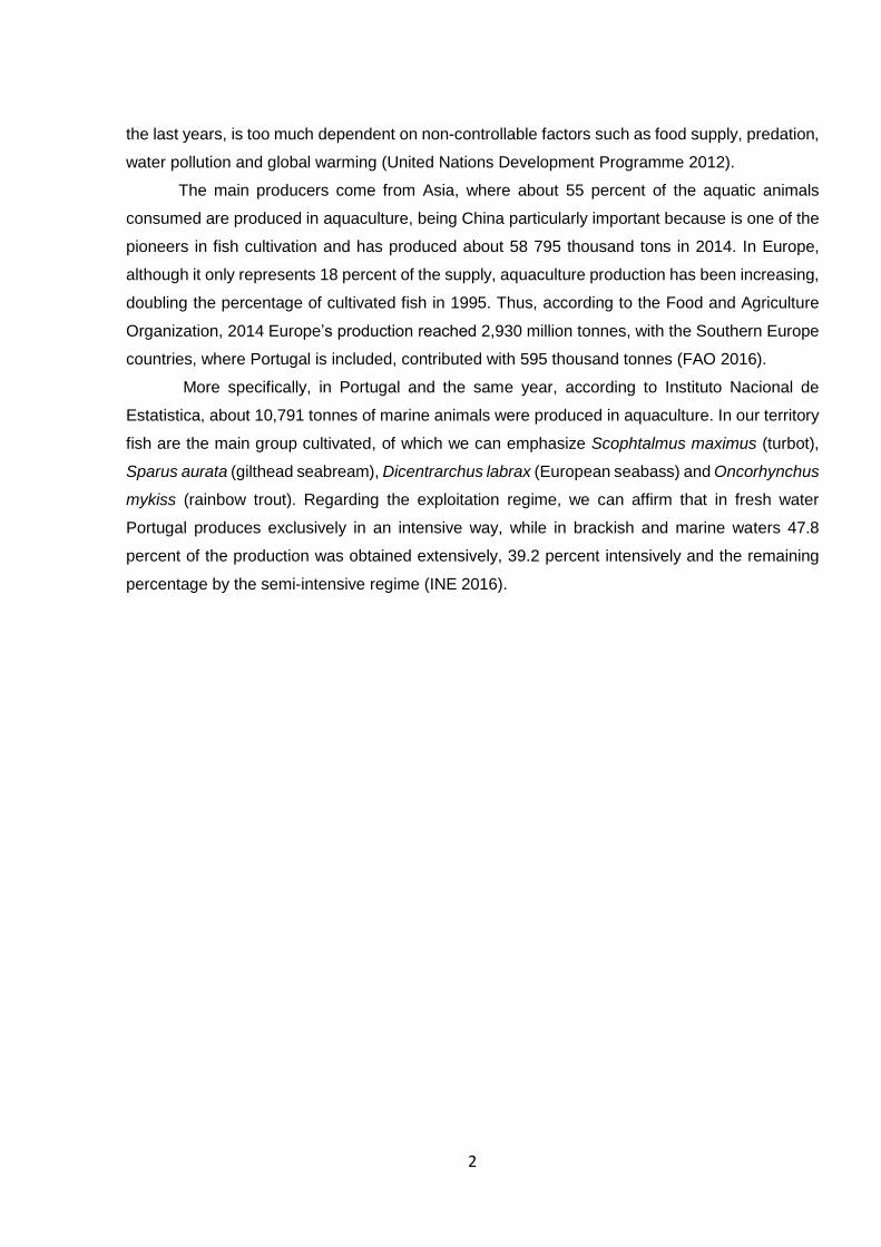
The chosen aquaculture was Quinta do Salmão, installed on the Portuguese district of Vila
Real, in the municipality of Montealegre, at Rio Rabagão’s dam, where O. mykiss are cultivated
on cages on an intensive way (Figure 4). These cages could be circular or rectangular and
rainbow trouts were separated in three different groups: one group of diploid small fishes (average
fish weight of 6.5 grams at the beginning of the samplings), constituted by male and female fish
that are descendants from reproductors of this aquaculture; one group of imported triploid female
small fishes (average fish weight of 7.5 grams at first sampling); and one group of medium sized
female animals (average fish weight of 300 grams at first time sampling) that were used on this
aquaculture as reproductors. As this fish farm is placed on a valley and cages are placed on the
river and not covered, fish are clearly susceptible by water temperature fluctuations which might
affect animal’s immune defences.
The immune status of two O. mykiss populations genetically different was evaluated,
throughout a year. Moreover, female diploid fish of bigger size were also evaluated at the same
time to assess eventual size/age effects. Fish cellularity and humoral parameters as lysozyme,
peroxidase and antiprotease activity were measured to identify periods of immunosuppression at
each group, in order to later implementation of prophylactic measures that may improve the
immune resistance of fish in critical periods.

14
Figures 4- Images of Quinta do Salmão aquaculture, at Rio Rabagão’s dam, Pisões.
2. Materials and Methods
2.1 Sampling
The sampling of animals was carried out monthly between June 2016 and March 2017 with water
temperature recording as shown (table I). Each time sampling, blood was collected from 10 small
diploid fish, 10 small triploid fish and 5 medium/large animals. The animals were anesthetized
with 2-phenolyethanol (Merck) and mucus was gently collected in a 15 ml Falcon tube and stored
at -20 ºC until assayed. Afterwards, fish were weighed and measured. The blood was then
collected through the caudal vein through a vacuum system with heparin, stored in heparinized
tubes and kept on ice until arrival at the CIIMAR facilities.
Table I. Time sampling and water temperature registration
Time Sampling Water Temperature
Time 1 – 21/06/16 18.0 ºC
Time 2 – 08/07/16 22.0 ºC
Time 3 – 07/09/16 22.0 ºC
Time 4 – 02/11/16 15.4 ºC
Time 5 – 25/11/16 11.0 ºC
Time 6 – 19/12/16 09.2 ºC
Time 7 – 30/01/17 06.6 ºC
Time 8 – 24/02/17 08.0 ºC
Time 9 – 24/03/17 08.0 ºC

15
2.2 Haematological analysis
Upon arrival in the laboratory, the smears from heparinized blood were run through a uniform
blood droplet and air dried, the rest of the blood being centrifuged at 10,000 × g for 10 min at 4
ºC and plasma was collected and frozen at -80 °C for humoral parameter quantification. Note that
for the first time sampling no blood smears were performed. After air drying, the slides were fixed
with a solution of formaldehyde-ethanol (90% absolute ethanol to 10% of 37% formaldehyde) for
one minute (Kaplow 1965). Neutrophils were identified by detection of peroxidase activity,
following a protocol described by Afonso et al (1998). Afterwards, slides were stained with the
Wright’s stain (Haemacolor, Merck) and observed under oil immersion (1000X). Immune cells
were identified and a differential count of neutrophils, monocytes, lymphocytes and thrombocytes
was made in a total of 200 cells/smear.
2.3 Humoral parameters analytical procedures
1) Lysozyme: Lysozyme activity was measured using a turbidimetric assay as described by
Costas et al. (2011). First, a solution of Micrococcus lysodeikticus (0.5 mg ml-1, 0.005 M
sodium phosphate buffer, pH 6.2) was prepared. After this 15 µl of plasma and 250 µl of the
above suspension were added to a microplate to achieve a final volume of 265 µl. The
reaction was carried out at 25º C and the absorbance (450 nm) was measured after 0.5 and
4.5 minutes in a Synergy HT microplate reader, Biotek (Figure 5). Lyophilized hen egg white
lysozyme (Sigma) was serially diluted in sodium phosphate buffer (0.05 M, pH 6.2) and used
to develop a standard curve. The amount of lysozyme in the sample was calculated using the
formula of the standard curve. All analysis was conducted in duplicates.

16
Figure 5- Synergy HT microplate reader, Biotek, before reading lysozyme activity (450 nm).
2) Peroxidase activity: Total peroxidase activity in plasma was measured following the
procedure described by Quade and Roth (1997). Firstly, we diluted samples in HBSS on a
1:10 dilution. After this a 15 µl of the dilution above were diluted in 250 µl of HBSS without
Ca2+ and Mg2+ in flat bottomed 96-well plates. Then, 50 µl of 20 mM 3,3’,5,5’-
tetramethybenzidine hydrochloride (TMB; Sigma) and 50 µl of 5 mM hydrogen peroxide were
added (Figure 6). The colour change reaction was stopped after 2 minutes by adding 50 µl
of 2M sulphuric acid (Figure 7) and the optical density was read at 450 nm in a Synergy HT
microplate reader, Biotek. The wells without plasma were used as blanks. The peroxidase
activity (units ml-1 plasma) was determined defining one unit of peroxidase as that which
produces an absorbance change of 1 Optical Density (OD).

17
Figure 6- Microplate after adding hydrogen peroxide.
Figure 7- Microplate after adding sulphuric acid.
3) Antiprotease Activity: The method described by Ellis (1990) was modified and adapted for
96-well microplates (Machado et al. 2015). At first, 10 µl of plasma were incubated with the
same volume of trypsin solution (5 mg ml-1 in NaHCO3 5 mg ml-1, pH 8.3) for 10 minutes at 22
ºC in polystyrene microtubes. To the incubation mixture, 100 µl of phosphate buffer
(NaH2PO4,13.9 mg ml-1, pH 7.0) and 125 µl of azocasein (20 mg ml-1 in NaHCO3, 5 mg ml-1,
pH 8.3) were added and incubated for 1 h at 22 ºC. After this, 250 µl of trichloroacetic acid
were added to the microtubes and incubated for 30 min at 22 ºC. Finally, the mixture was
centrifuged at 10,000 × g for 5 min at room temperature. The blank was made using
phosphate buffer saline instead of plasma and trypsin, and the reference sample was
obtained using phosphate buffered saline instead of plasma (Figure 8). To calculate the
percentage of trypsin activity we used the following formules:

18
% non-inhibited trypsin = (Sample absorbance × 100) / Reference sample
% inhibited trypsin = 100 - % non-inhibited trypsin
Figure 8- Antiprotease microplate before reading.
2.4 Statistical analysis
The groups of animals were divided by size/genetic group and, for each time sampling
and parameter, mean and standard deviation were calculated. Data were analysed for
normality and homogeneity of variance and Log or Arc Sen transformed before statistical
treatment when needed. Data were analysed by one-way ANOVA (Tukey post hoc test) when
normality and homogeneity was observed. Alternatively, Nonparametric Kruskal-Wallis test
was performed to find significative changes of sampling parameters between time sampling
groups. The performance of statistical analyses occurred under STATISTICA 13 program for
WINDOWS. The level of significance used was p ≤ 0.05 for all statistical tests.

19
3. Results
Haematological studies have shown that, on diploid fish, water temperature variation have not affected neutrophils, lymphocytes and
thrombocytes, just producing little changes on monocyte count, with a significant increase between 22.0 ºC 8.0 ºC (Table II).
Table II- Relative proportion of diploid O. mykiss peripheral blood leukocytes (thrombocytes, lymphocytes, monocytes and neutrophils) for each time sampling
22.0 ºC 22.0 ºC 15.4 ºC 11.0 ºC 9.2 ºC 6.6 ªC 8.0 ºC 8.0 ºC
Neutrophils (% WBC)
3.65 ± 2.07 6.10 ± 5.89 2.10 ± 1.62 3.40 ± 2.64 3.3 ± 2.58 4.65 ± 1.55 3.15 ± 2.04 4.45 ± 3.68
Monocytes (% WBC)
3.15 ± 2.46 0.90 ± 0.54 b 2.20 ± 1.12 2.10 ± 1.77 2.35 ± 2.66 2.50 ± 1.22 3.80 ± 2.87 a 2.20 ± 1.55
Lymphocytes (% WBC)
62.40 ± 8.30 61.70 ± 12.44 60.90 ± 7.17 68.85 ± 6.60 57.50 ± 7.45 59.60 ± 14.80 55.25 ± 5.71 57.55 ± 10.23
Thrombocytes (% WBC)
30.80 ± 7.69 31.30 ± 14.33 34.80 ± 6.33 25.65 ± 6.99 36.85 ± 7.30 33.25 ± 14.74 37.80 ± 7.18 35.80 ± 8.00
Values are expressed as means ± SD (n=10). Different letters mean significant differences among water temperature variations (One-way ANOVA, p ≤0.05)
On triploid fish, several significative cell count variations have been observed, with the most relevant changes being registed on lymphocytes and
thrombocytes count, that evolved on a wave form, with lymphocyte percentage hitting their maximum value (72.80 % ± 8.48 %) with a water
temperature value of 6.6 ºC, and thrombocytes obtaining highest cell values (47.45 % ± 9.47 %) at 8 ºC. (Table III).

20
Table III- Relative proportion of triploid O. mykiss peripheral blood leukocytes (thrombocytes, lymphocytes, monocytes and neutrophils) for each time sampling
22.0 ªC 22.0 ºC 15.4 ºC 11.0 ºC 9.2 ºC 6.6 ªC 8.0 ªC 8.0 ªC
Neutrophils (% WBC)
2.85 ± 2.34 b 3.35 ± 3.15 3.5 ± 3.49 6.35 ± 3.20 a 2.95 ± 1.42 4.85 ± 2.98 3.15 ± 1.69 2.35 ± 0.67
Monocytes (% WBC)
3.15 ± 2.25 1.35 ± 1.07 2.60 ± 1.37 1.85 ± 0.98 2.55 ± 1.82 3.30 ± 1.85 2.25 ± 1.57 2.15 ± 1.21
Lymphocytes (% WBC)
52.95 ± 11.08 bc 58.80 ± 6.73abc 49.00 ± 9.21c 65.75 ± 8.19 ab 49.00 ± 10.13 c 72.80 ± 8.48 a 48.90 ± 9.65c 48.05 ± 9.3c
Thrombocytes (% WBC)
41.05 ± 9.09 a 36.50 ± 7.57ab 44.90 ± 9.89a 26.05 ± 9.68 bc 45.50 ± 11.78 a 19.05 ± 7.68 c 45.70 ± 9.21a 47.45 ± 9.47a
Values are expressed as means ± SD (n=10). Different letters mean significant differences among water temperature variations (One-way ANOVA, p ≤0.05).
The medium fish group alterations were observed on neutrophils that have shown higher values at the first two smear group and lymphocyte and
thrombocyte population that have produced higher cell proportion on the last sampling times (Table IV).
Table IV- Relative proportion of medium O. mykiss peripheral blood leukocytes (thrombocytes, lymphocytes, monocytes and neutrophils) for each time sampling
22.0 ªC 22.0 ªC 15.4 ºC 11.0 ªC 9.2 ªC 6.6 ªC 8.0 ªC 8.0 ªC
Neutrophils (% WBC)
7.30 ± 4.35 ab 7.60 ± 2.85 a 1.20 ± 0.87 c 4.60 ± 4.64 3.80 ± 2.16 4.60 ± 2.99 1.90 ± 0.80 bc 5.20 ± 5.27
Monocytes (% WBC)
3.20 ± 1.50 1.80 ± 0.68 4.30 ± 1.29 1.70 ± 0.68 2.30 ± 1.33 3.20 ± 1.54 3.00 ± 1.76 2.30 ± 0.93
Lymphocytes (% WBC)
56.60 ± 6.30 58.30 ± 4.26 60.80 ± 7.49 53.30 ± 2.40 47.30 ± 6.50 b 69.40 ± 10.12 a 49.00 ± 6.20 b 58.60 ± 8.41
Thrombocytes (% WBC)
32.90 ± 7.50 32.30 ± 3.20 33.70 ± 6.80 40.40 ± 3.84 a 46.60 ± 5.34 a 22.80 ± 6.42 b 46.10 ± 5.40 a 33.90 ± 7.70
Values are expressed as means ± SD (n=5). Different letters mean significant differences among water temperature variations (One-way ANOVA, p ≤0.05).

21
The antiprotease activity has produced significative alterations along temperature variations
showing tendency to inhibit more trypsin with cooler water temperatures. Medium sized fish
produced no significative differences between time samplings on this parameter (figure 9).
Figure 9- Antiprotease activity of different O. mykiss groups along sampling time
Values are expressed as means ± SD (n=10 for diploid and triploid fish and n=5 on medium fish). Different letters mean
significant differences among water temperature variations to each group (One-way ANOVA and Kruskal Wallis p
≤0.05).
Lysozyme followed the tendency to increase its concentration with water temperature decrease
(figure 10). Highest activity results have been constant to all different groups but different
temperatures were observed on the lowest enzyme concentration among the three fish groups.
0
10
20
30
40
50
60
70
80
90
100
T1 T2 T3 T4 T5 T6 T7 T8 T9
% in
hib
ite
d try
psin
Water Temperature ( ªC)
Antiprotease activity
Diploid Triploid Medium
de
de cd
abc ab bcd abc a
c
c
aa a ab
a
a
ababcde
22.0 22.0 18.0 15.4 11.0 9.2 6.6 8.0 8.0

22
Figure 10- Lysozyme activity of different O. mykiss groups along temperature variation.
Values are expressed as means ± SD (n=10 for diploid and triploid fish and n=5 on medium fish). Different letters mean
significant differences among water temperature variations to each group (One-way ANOVA and Kruskal Wallis p
≤0.05).
Peroxidase activity has produced opposite results comparing to the other two humoral analysis
(figure 11), resulting in highest concentration on warmer temperatures.
Figure 11 - Peroxidase activity of different O. mykiss groups along sampling time.
Values are expressed as means ± SD (n=10 for diploid and triploid fish and n=5 on medium fish). Different letters mean
significant differences among water temperature variations to each group(One-way ANOVA and Kruskal Wallis p
≤0.05).
0
20
40
60
80
100
120
T1 T2 T3 T4 T5 T6 T7 T8 T9
µg
/mg
pro
tein
Water Temperature ( ºC)
Diploid Triploid Medium
a
bb
cb
abab
a
ab
cbc
a
abab ab ab
ab
ab
abc
abc
abc
abc
abc
abc
abc
abc abc
22.022.018.0 15.4 11.0 9.2 6.6 8.0 8.0
0
200
400
600
800
1000
1200
1400
1600
1800
T1 T2 T3 T4 T5 T6 T7 T8 T9
un
its m
l-1
Water Temperature ( ºC)
Peroxidase activity
Diploid Triploid Medium
a
aa
b
a
ab
ac
acd
abc
cded ee
ecd
abc
ab
a
bc
ab
abab
abab
abbc
abcde
22.022.018.0 15.4 11.0 9.2 6.6 8.0 8.0

23
4. Discussion
Haematological results have shown that, on diploid fish, temperature alteration had few
influence on blood cell count. However, on triploids, several significative cell count changes have
been observed, following the tendency for cell count fluctuation described from past studies on
O. mykiss (Ojolick et al. 1995). On triploid group, neutrophils have significally increased between
time 2 and 5, but the major cell type count differences were observed on lymphocytes, that varied
on a wave form during time, tending to show highest cell percentage at lower values of water
temperature. Thrombocyte counts has suffered variations too, with more relevance for their
decrease on September and January. Analogous results on lymphocytes have been observed on
other studies with study species (Houston et al. 1996), reinforcing the idea that cooler
temperatures influentiate positively the number of circulating lymphocyte cells. Already at the
medium sized fishes, neutrophils have been highly identified at first and second smear groups,
which makes believe that, on this fish group, these cells are more common on summer months.
On the opposite sense have evolved thrombocyte cells, whose results registered highest values
at lower temperatures, showing similarity to thrombocytary variations on S. trutta studies
(Rahkonem & Pasternack 1998) and appearing to be the leukocitary cell group with more
production rate in cooler waters. After this comparative study, we can consider that water
temperature variation affects fish haematological values, specially on triploid and medium fishes,
showing highest values of lymphocyte and thrombocyte cells at cooler temperatures, and
phagocytic cells being more prevalent on warmer waters.
Already on humoral parameters, antiproteases analysis have showed an increased activity
at water temperatures ranges close to optimum rearing temperatures (8-15 ºC), similar to
observed data in D. labrax that have produced higher antiprotease activity at temperatures
between 14 and 19 ºC. (Valero et al. 2014). Likewise results observed with environmental
temperature variations in Gadus morhua (cod) (Magnadóttir et al. 1999), antiprotease activity on
O. mykiss have also showed relevant decreased values on summer months. Is also relevant to
highlight intense activity of this humoral parameter was observed in October, November and
March, with more than 85.5 percent of inhibited trypsin being registed. Another curious fact is that
diploid fish have produced higher antiprotease mean values than triploid, going along with the
results of previous studies on S. salar that also produced diploids higher antiprotease
concentration than triploids (Langston et al. 1997), suggesting that genetic variations might
produce different antiprotease activity. It’s also remarkable that the group of medium fishes has
showed no significant differences during time and presented lower antiprotease concentration
than diploid fish, appearing to be the group less affected by our dependent factor, which might
suggest that fish age can regulate this enzyme production.

24
Although it is usually assumed that adaptive immune parameters tend to be suppressed
at low temperatures, some studies in S. aurata have been reported resulting in increased cellular
and humoral immune parameters at cooler temperatures (Machado et al. 2015; Guerreiro et al.
2016). In our study, fishes have verified their maximum lysozyme concentrations on March, at 8
ºC, with diploid and triploid groups obtaining respectively 35.38 and 38.98 µg per milligram of
protein. On a simplistic form, our results provide the idea that, at cooler temperatures (near 10
ºC), phagocytic cells produce more lysozyme, making fish immune system more able to block
and lyse bacterial cells, through a modulation still not well understood, but that could be caused
by interaction of environmental variations, stress and higher infection incidence. Since monocytes
are thought to be the main producers of lysozyme, slight correlation was observed between
enzyme concentration and monocyte cell count. Higher monocitary presence and lysozyme
reading peaks have been observed on same time samplings, as well as increased average
percentage of monocytes on medium group during time, matching with the markable increased
enzyme presence on the same sampling group. Considering medium fish group, the evolution of
lysozyme concentration has resulted in some contrasts with the diploid and triploid variations
along the year, once medium sized fish enzyme values developed on unequal way, with the
lowest concentration of lysozyme at 9.2 ºC, on December, and the highest enzyme concentration
at 8 ºC, on March, which suggests that this parameter might have been affected not only by water
temperature but also from fish growth and maturation. Another recent study on O. mykiss has
resulted in increased lysozyme concentration along the year occurred due not only by
temperature but also by photoperiod (Papežíková et al. 2016), which might be another plausible
explanation for our results.
Contrarily to the other humoral analysis performed on this thesis, peroxidase
concentration has produced the highest concentration values at warmer temperatures,
contrasting with lower enzyme scores on cooler water, results that had yet been registered in the
past on Labeo rohita (roho labeo) species (Swain et al. 2007). In our study, little changes were
noticed on peroxidase absorbance peaks among groups, with highest enzyme values being
obtained on summer months for diploid and triploid fish groups while medium fishes showed
strong enzyme activity on summer and autumn sampling months. Although this fact and once
lower peroxidase activity is registed on winter months with temperatures near 10 ºC, water
temperature seems to clearly modulate this enzyme. Concordance have also been found between
myeloperoxidase activity and neutrophils presence on blood smears, with high cell percentage
on the same sampling time of high plasmatic presence of the enzyme. Little discordances
between these two parameters might occur due to neutrophil temperature dependent activation
that leads to degranulation and consequent high presence of myeloperoxidase, without
proportional increased cell count. Another explanation could be the increased abundance of
25
opportunistic bacterial pathogens that might occur due to temperature augmentation, which in
turn could induce peroxidase release by activated neutrophils. In order to confirm this hypothesis,
a screening of opportunistic pathogens would be necessary.
5. Conclusions
Compilating all data is possible to hypothesize that water temperature has an important
role and affects the dynamics of fish cellularity and humoral parameters. As poikilothermic
animals, fish’s physiological processes are proven to be influenced and modulated by thermic
variations and many studies have showed that usually cooler temperatures depress non-specific,
humoral and cell-mediated defence mechanisms. The present study was important to show that,
on O. mykiss, water temperature differentially conditionate leukocitary population and plasmatic
immune defences, and that genetic and size differences on animals produce several variations
and, occasionally, opposite results. However, it’s not well clear how immune defences act isolated
and it is important to eliminate other possible factors that, on this study, might have interacted
with temperature like pH, water oxygenation, photoperiod, fish growth and weight, fish maturation,
nutrition and stress factors.
It was also of relevant interest, to discover if water temperature oscillations can
differentially influence healthy and infected animal’s immune system, by creation and evaluation
of groups in different immunitary condition, once pathogens by their own are proven to
immunostimulate fish and few studies have been performed with these characteristics. Further
studies can also be performed on the sequence of this project, adding data by analytical
procedures to other humoral parameters of plasma such as alternative complement pathway
activity, bactericidal activity, nitric oxide, proteases and Immunoglobulin M studies, as well as all
mucus analyses which might elucidate and provide stronger results.

26
6. Bibliography
Afonso A, Ellis AE & Silva MT (1997) “The leucocyte population of the
unstimulated peritoneal cavity of rainbow trout (Oncorhynchus mykiss)”. Fish &
Shellfish Immunology 7, 335–348.
Afonso A, Lousada S, Silva J, Ellis AE & Silva MT (1998) “Neutrophil and macrophage responses to
inflammation in the peritoneal cavity of rainbow trout Oncorhynchus mykiss: A light and electron
microscopic cytochemical study” Disease Aquatic Organisms 34, 27-37.
Ainsworth AJ, Dexiang C, Watestratt PR & Greenway T (1991) “Effect of temperature on the immune system of
channel catfish (Ictalurus punctatus). I. Leucocyte distribution and phagocyte function in the anterior
kydney at 10ºC”. Comparative Biochemistry and Physiology, 100A (4), 907-912.
Baldisseroto B (2002) Fisiologia de peixes aplicada à piscicultura. Editora UFSM: Santa Maria, 211.
Barton GM, (2008) “A calculated response: control of inflammation by the innate imune system”. Journal of
Clinical Investigation 118, 413-420.
Bayne CJ & Gerwick L (2001) “The acute phase response and innate immunity of fish” Developemental and
Comparative Immunology 25: 725-743.
Brauner CJ (1999) “The effect of diet and short duration hyperoxia exposure on seawater transfer in coho
salmon smolts (Oncorhynchus kisutch)” Aquaculture 177 (1–4) 257–265.
Bruno DW & Munro LS (1986) “Haematological assessment of rainbow trout, Salmo gairdnieri Richardson, and
Atlantic salmon, Salmo salar, infected with Renibacterium salmoniarum". Journal of Fish Diseases 9,
195-204.
Caldwell CA & Hinshaw J (1994) “Physiological and haematological responses in rainbow trout subjected to
supplemental dissolved oxygen in fish culture” Aquaculture 126 (1–2), 183-193.
Castagnolli N (2000) “Piscicultura intensiva e sustentável”. Aquicultura no Brasil: Bases para um
desenvolvimento sustentável, 399.
Chaudhuri SH, Pandit T & Banerjee S (1986) “Size and sex related of some blood parameters of Sarotherodon
mossambica”. Environmental and Ecological Statistics 4 (1), 61-63.
Comissão Europeia (2012) “Salão Seafood: o local dos primeiros passos“ A Pesca e a Aquicultura na Europa
3-7.
Corbel MJ, (1975) “Immune-Response in Fish – Review” Journal of Fish Biology 7, 539-563.
Costas B, Aragão C, Soengas JL, Míguez JM, Rema P, Dias J, Afonso A, Conceição LEC, (2012) “Effects of
dietary amino acids and repeated handling on stress response and brain monoaminergic
neurotransmitters in Senegalese sole (Solea senegalensis Kaup, 1858) juveniles” Comparative
Biochemistry and Physiology part A 161, 18-26.
Costas B, Conceicao LEC, Dias J, Novoa B, Figueiras A, Afonso A, (2011) “Dietary arginine and repeated
handling increase disease resistance and modulate innate imune mechanisms of Senegalese sole (Solea
Senegalensis Kaup, 1858)”. Fish and Shellfish Immunology 31, 838-847.
Dalton DK, Pitts-Meek S, Keshav S, Figari IS, Bradley A & Stewart TA (1993) “Multiple defects of immune cell
function in mice with disrupted interferongamma genes” Science 259 (5102), 1739–1742.
Dejours P, Toulmond A, Truchot JP (1977) “The effect of hyperoxia on the breathing of marine fishes”
Comparative Biochemistry and Physiology Part A: Physiology 58 (4), 409-411
Dheer JMS, Dheer TR & Mahajan CL (1987) “Haematological and haematopoietic responses to acid stress in
an air-breathing freshwater fish, Channa Punctatus Bloch” Journal of Fish Biology 30, 577-588.
Dogget TA & Harris JE (1989) “Ultrastructure of the peripheral blood leucocytes of Oreochromis mossambicus”
Journal of Fish Biology 33, 747-756.
Ellis AE (1976) “The leucocytes of fish. A review” Journal of Fish Biology 8, 143-156.
Ellis AE (1977) “Leucocytes and related cells in the plaice. Pleuronectes platessa” Journal of Fish Biology 11,
453-491.
Ellis AE (1981) “Non-specific defence mechanisms in fish and their role in disease processes” Development of
Specifications for Biotechnology Pharmaceutical Products 49, 337-352.
Ellis AE (1999) “Immunity to bacteria to fish” Fish Shellfish Immunol 9, 291-308.

27
Ellis AE (2001) “Innate host defence mechanisms of fish against viruses and bacteria” Developmental and
Comparative Immunology 25, 827-839.
Ellis AE, (1990) “Serum antiproteases in fish” Techniques in Fish Immunology ed.1 FairHaven: SOS.
Espelid S, Løkken GB, Steiro K, Bøgwald J, (1996) “Effects of cortisol and stress on the immune system in
Atlantic Salmon (Salmo salar L.)” Fish Shellfish Immunology 6, 95 – 110.
Esteban, MA (2012) “An Overview of the Immunological Defenses in Fish Skin” ISNR Immunology Article ID
853470, 29 pages
FAO (2012) “World review of fisheries and aquaculture” The State of World Fisheries and Aquaculture 3-5.
FAO (2014) “World review of fisheries and aquaculture” The State of World Fisheries and Aquaculture 3-4.
FAO (2016) “Box 1: Feed production and management practices in aquaculture” The State of World Fisheries
and Aquaculture 20-28.
Fevolden SE, Refstie T & Roed KH (1991) “Selection for high and low cortisol stress response in Atlantic salmon
(Salmo salar) and rainbow trout (Oncorhynchus mykiss)” Aquaculture 95, 53-65.
Fevolden SE, Refstie T & Roed KH (1992) “Disease resistance in rainbow trout (Oncorhynchus mykiss) selected
for stress response” Aquaculture 104, 19-29.
Forlenza M, Fink IR, Raes G & Wiegertjes GF (2011) “Heterogeneity of macrophage activation in fish”
Developmental and Comparative Immunology 35, 1246–1255.
Giles MA, Majewski HS & Hobden B (1984) “Osmoregulation and haematological responses of rainbow trout
(Salmo gairdneri) to extended environmental acidification” Canadian Journal of Fisheries and Aquatic
Sciences 41, 1696-1694.
Griffin BR (1984) “Random and directed migration of trout (Salmo gairdneri) leukocytes: activation by antibody,
complement and normal serum components” Developmental and Comparative Immunology 8, 589-
597.
Gudding R, Lillehaug A & Evensen O (2014) Fish Vaccination Wiley Blackwell, 104-115.
Guerreiro I, Serra CR, Enes P, Couto A, Salvador A, Costas B & Oliva-Teles A (2016) “Effect of short chain
fructooligosaccharides (scFOS) on immunological status and gut microbiota of gilthead sea bream
(Sparus aurata) reared at two temperatures” Fish & Shellfish Immunology 49, 122-131.
Hasan MR & New MB (2013) “On-farm feeding and feed management in aquaculture” FAO Fisheries and
Aquaculture Technical Paper No. 583.
Hill DJ & Rowley AF (1988) “The thromboxane mimetic, U-46619, induces the aggregation of fish thrombocytes”
British Journal of Haematology 33, 247-254.
Holeton GF & Randall DJ (1967) “The effect of hypoxia upon the partial pressure of gases in the blood and
water afferent and efferent to the gills of rainbow trout” Journal of Experimental Biology 46, 317-327.
Hosfeld CD, Engevik A, Mollan T, Lunde TM, Waagbø R, Olsen AB, Breck O, Stefansson S & Fivelstad S (2008)
“Long-term separate and combined effects of environmental hypercapnia and hyperoxia in Atlantic
salmon (Salmo salar L.) smolts” Aquaculture 280 (1–4), 146–153.
Houston AH & Gingra-Bedard JH (1994) “Variables versus constant temperature acclimation regimes: effects
on haemoglobin isomorph profile in goldfish, Crassus auratus” Fish Physiology and Biochemistry 13,
445-450.
Houston AH, Dobric N & Kahurananga R (1996) “The nature of haematological response in fish. Studies on
rainbow trout Oncorhynchus mykiss exposed to stimulated winter, Spring and summer conditions” Fish
Physiology and Biochemistry 15(4), 339-347.
Instituto Nacional de estatística (2016) “Aquicultura e salicultura” Estatísticas da Pesca 2015 75-80.
Jaqueline Parkin BC (2001) “An overview of the imune system” The Lancet 357, 1777-1789.
Kaplow LS (1965) “Simplified Myeloperoxidase Stain Using Benzidine Dihychloride. The Journal of
Haematology 26, 215.
Kiron V (2012) “Fish imune system and its nutritional modulation for preventive health care” Animal Feed
Science and Technology 173, 111-133.
Klebanoff SJ & Clark RD (1978) The Neutrophil: Function and Clinical Disorders
North Holland Publishing Company, Amsterdam 409-466.
28
Kollner B & Kotterba G (2002) “Temperature dependent activation of leucocyte populations of rainbow trout,
Oncorhynchus mykiss, after intraperitoneal immunisation with Aeromonas salmonicida” Fish & Shellfish
Immunology 12, 35-48.
Lamas J, Santos Y, Bruno DW Toranzo AE & Anadón R (1994) “Non-Specific cellular responses of rainbow
trout to Vibrio anguillarum and its extracellular products (EPCs)” Journal of Fish Biology 45(5), 839-
854.
Lane HC (1979) “Progressive changes in haematology and tissue water of sexually mature trout, Salmo
gairdneri Richardson during the autumn and winter” Journal of Fish Biology 15, 425-436.
Langston AE, McGruer S, Ellis AE & Johnstone R (1997) “The comparison of some non-specific immune
parameters in diploid and triploid Atlantic salmon” Developmental & Comparative Immunology 21, 216.
Lincoln RF & Scott AP (1983) “Production of all-female triploid rainbow trout“ Aquaculture 30, Issues 1–4, 375-
380.
Lorenzi TF (1999) Manual de hematologia propedêutica e clínica. São Paulo, MDSI, 641p.
Machado M, Azeredo R, Díaz-Rosales P, Afonso A, Peres H, Oliva-Teles A & Costas B (2015) “Dietary
tryptophan and methionine as modulators of European seabass (Dicentrarchus labrax) immune status
and inflammatory response” Fish & Shellfish Immunology 42 (2), 253-362.
MADRP-DGPA (2007) “Plano estratégico Nacional para a pesca 2007-2013” MADRP-DGPA p.27.
Magnadóttir B, , Jónsdóttir H, Helgason S, Björnsson B, Jørgensen TØ & Pilström L (1999) “Humoral immune
parameters in Atlantic cod (Gadus morhua L.): I. The effects of environmental temperature” Comparative
Biochemistry and Physiology Part B: Biochemistry and Molecular Biology, 122, Issue 2, 173–180.
Mantovani A, Sozzani S, Locati M, Allavena P & Sica A (2002) “Macrophage
polarization: tumor-associated macrophages as a paradigm for polarized M2
mononuclear phagocytes” Trends of Immunology 23 (11), 549-555.
Martínez FJ, García-Riera MP, Canteras M, Costa J, & Zamora S (1994) “Blood parameters in Rainbow Trout
(Oncorhynchus mykiss): Simultaneous influence of various factors” Comparative Biochemistry and
Physiology 107A, 95-100.
Mayer L (2003) “Mucosal immunit,” Pediatrics, vol. 111, no. 6, pp. 1595–1600.
Mommsen TP, Vijayan MM & Moon TW (1999) “Cortisol in teleosts: dynamics, mechanisms of action, and
metabolic regulation” Fish Biology and Fisheries 9, 211 – 268.
Myrick CA (2011) “Aquaculture | Physiology of Fish in Culture Environments” Encyclopedia of Fish
Physiology, 2084-2089.
Naylor R & Burke M (2005) "Aquaculture and ocean resources: raising tigers of the sea" Annual Review of
Environmental Resources 30, 185–218.
Noga EJ & Francis-Floyd R (1991) “Medical management of channel catfish: The environment” Compendium
on Continuing Education for the Practising Veterinarian 13(1), 160-166.
Ojolick EJ, Cusack R, Benfey TJ & Kerr SR (1995) “Survival and growth of all-female diploid and triploid rainbow
trout (Oncorhynchus mykiss) reared at chronic high temperature” Aquaculture 131, Issues 3–4, 177-187.
Oliveira LW, Moura WL, Matushima ER & Egami MI (1997) “Detecção citoquímica da mieloperoxidase em
eosinófilos e mononucleares azurófilos do sangue de Caiman crocodilus Ycare (Daudin, 1802) (Reptilia,
Crocodilia)” In Reunião Anual da Sociedade Brasileira para progresso da ciência, 49, 878.
Oria J (1932) “Elementos figurados do sangue de alguns teleósteos fluviais brasileiros (Nematognathas,
Characidaeos, Gymnotideos, Poeciliideos). I. Eritrócitos: formas normais, formas jovens e formas
involuídas” Ann. Faculdade de Medicina de São Paulo 8, 43-68.
Ortuño J Esteban MA & Meseguer J (2001) “Effects of short-term crowding stress on the gilthead seabream
(Sparus aurata L.) innate imune response” Fish Shellfish Immunology 11, 187 – 197.
Papežíková I, Mareš J, Vojtek L, Hyršl P, Marková Z, Šimková A, Bartoňková J, Navrátil S & Palíková M (2016)
“Seasonal changes in immune parameters of rainbow trout (Oncorhynchus mykiss), brook trout

29
(Salvelinus fontinalis) and brook trout × Arctic charr hybrids (Salvelinus fontinalis × Salvelinus alpinus
alpinus)” Fish and Shellfish Immunology 57, 400-405.
Pickering AD & Pottinger TG (1987) “Crowding causes prolonged leukopenia in salmonid fish, despite interrenal
acclimation” Journal of Fish Biology 30, 701-712.
Pickering AD (1989) “Environmental stress and survival of Brown trout, Salmo truta” Freshwater Biology 21,
47-55.
Pickering AD Pottinger TG & Hurley MA (1992) “Consistency in the stress response of individuals of two strains
of rainbow trout, Oncorhynchus mykiss” Aquaculture 103, 275-289.
Pulsford, AL, Lemaire-Gony S, Tomlinson M, Collingwood N, Glynn PJ (1994) “Effects of acute stress on the
immune system of the dab, Limanda limanda” Comparative Biochemistry and Physiology 109C, 129
– 139.
Quade MJ & Roth JA (1997) “A Rapid, direct assay to measure degranulation of bovine neutrophil primary
granules” Veterinary Immunology nad Immunopathology 58, 239-248.
Rahkonen R & Pasternack M (1998) “Effect of experimental Diphyllobothrium dendriticum infection on the blood
leucocyte pattern of brown trout at two temperature levels” Boreal Environmental Research 3, 381-386.
Ramalho A, Dinis MT, “Portuguese Aquaculture: current status and future perspectives” World Aquaculture,
26-31.
Rambhaskar B & Srinivasa-Rao K (1989) “Influence of environmental variables on haematological, and
compedium of normal haematological ranges in milkfish, Chanos chanos (Forskal), in brackishwater
culture” Aquaculture 83, 123-136.
Rand TG & Cone DK (1990) “Effects of Ichthyophonus hoferi on condition Indices and blood chemistry of
experimentally infected rainbow trout (Oncorhynchus mykiss)” Journal of Wildlife Diseases 26(3), 323-
328.
Ranzani-Paiva MJT, Eiras AC & Felizardo NN (1998a) “Análises hematológicas em tilápia-do-Nilo Oreochromis
niloticus, da represa de Guarapiranga, São Paulo” In: Reunião anual do instituto da pesca, 7.
Ranzani-Paiva MJT, Tabata YA & Eiras AC (1998b) “Hematologia comparada entre diploides e triploides de
truta arco-íris, Oncorhynchus mykiss Walbaum (Pisces, Salmonidae)” Revista brasileira de Zoologia
15, 1093-1102.
Rawlinson P & Forster A (2000) “The Economics of Recirculation Aquaculture” IIFET 1-13.
Rehulka J (1997) “Haematological parameters in rainbow trout, Oncorhynchus mykiss, in cage culture”
Zyvocisná Vyroba 42, 159-164.
Roberts RJ (1981) Patologia de los peces Madrid: Mundi-Prensa. 366-372.
Roberts RJ (2012) Fish Pathology Wiley-Blackwell 144-167.
Roberts RJ and Bullock AM (1980) “The skin surface ecosystem of teleost fishes” Royal Society of Edinburgh
79B, 87-91.
Ruane NM, Nolan DT, Rotllant J, Costelloe J & Wendelaar Bonga SE (2000) “Experimental exposure of rainbow
trout Oncorhynchus mykiss (Walbaum) to the infective stages of the sea louse Lepeophtheirus salmonis
(Kroyer) influences the physiological response to an acute stressor” Fish & Shellfish Immunol. 10, 451-
463.
Saraiva AJ, Eiras JC & Cruz C (1989) “Nota sobre a ocorrência de Aeromonas salmonicida em Portugal”
Publicação do Instituto de Zoologia "Dr. Augusto Nobre" (Faculdade de Ciências do Porto), N°
209.
Sasaki Y, Maita M, & Okamoto N (2002) “Rainbow trout are responsible for non-specific cytotoxicity” Fish &
Shellfish Immunology 12, 243-252.
Secombs CJ (1996) “The nonspecific immune system: cellular defence” The fish immune system. Academic
Press Inc. San Diego, USA. p. 378.
Sousa JA (1996) “Estudo Epidemiológico em Duas Truticulturas do Norte de Portugal e Caracterização dos
Agentes Bacterianos e Virais de Maior Impacto em Aquacultura” Dissertação de Doutoramento em
Biologia apresentada à Faculdade de Ciências da Universidade do Porto – FCUP p.395.
Stickney RR, Costa-Pierce B, Baltz DM, Drawbridge M, Grimes C, Phillips S & Swann DL (2006) “Toward
Sustainable Open Ocean Aquaculture” United States Fisheries 31, (12): 607–610.
Stoskopf MJ (1992) Fish Medicine W.B. Saunders Company 149-150.
30
Swain P, Dash S, Sahoo PK, Routray P, Sahoo SK, Gupta SD, Meher PK & Sarangi N (2007) “Non-specific
immune parameters of brood Indian major carp Labeo rohita and their seasonal variations” Fish &
Shellfish Immunology 22, 38-43.
Tavares-Dias M & Moraes FR (2004) “Características hematológicas dos peixes teleósteos: Linfócitos”
Hematologia dos Peixes Teleósteos 34-36.
Tavares-Dias M, Schalch SHC, Martins ML, Silva ED, Moraes FR & Perecin D (1999) “Hematologia de
teleósteos brasileiros com infecção parasitária. I. Variáveis do Leporinus macrocephalus Garavello &
Britski, 1988 (Anostomidae) e Piaractus mesopotamicus Holmberg, 1887 (Characidae)” Acta
Scientiarum 21, 337-342.
Timmons MB & Ebeling JB (2013). Recirculating Aquaculture 3rd ed. Ithaca Publishing Company Publishers,
3.
Tort L (2011) “Hormonal responses to stress-Impact of Stress in Health and Reproduction” Encyclopedia of
Fish Physiology 1541-1552.
United Nations Development Programe (2012) “Vol.I: Transforming Markets to Restore and Protect the Global
Ocean.” Catalysing Ocean Finance 6-12, 27-33.
Val AL (2000) “Organic phosphates in the red blood cells of fish” Comparative Biochemistry and Physiology
125A, 417-435.
Valero Y, García-Alcázar A, Esteban MÁ, Cuesta A & Chaves-Pozo E (2014) “Seasonal variations of the
humoral immune parameters of European sea bass (Dicentrarchus labrax L.)” Fish and Shellfish
Immunology 39, 185-187.
Wendelaar Bonga SE (1997) “The stress response in fish” Physiological Reviews 7, 591-625.
Zapata AG, Varas A & Torroba M (1992) “Seasonal variations in the immune system of lower vertebrates”
Immunology Today 13 (4), 142-147.
Zhiteneva LD & Gorislavskaya MM (1986) “Hematological parameters of herring, Clupea pallasi, in relation to
its physiological conditions” Journal of Ichthyology 26 (2), 106-116.

31
7. Appendix
Figure 12- Scientific poster presented at the 2nd Aquaimprove congress, that took place on 17th March, at CIIMAR/UP, Porto, Portugal