Embed Size (px)
Citation preview

Editorial
Relationship Between Social Factors and Cardiovascular Diseases
Original Articles
Impact of Risk Factors for Coronary Artery Disease on Hospital Costs of Patients Undergoing Myocardial Revascularization Surgery in the Brazilian Unified Health System (SUS)
Prevalence of Atherosclerotic Lesions in the Left Internal Thoracic Artery, Evidenced by Selective Angiographic Findings
Correlation between Clinical and Educational Factors and Delayed Hospital Arrival in Myocardial Infarction
Decrease in the Inflammatory Marker TNF-α after Consumption of Flaxseed by Hypercholesterolemic Rabbits
Evolution of Mortality from Diseases of the Circulatory System and of Gross Domestic Product per Capita in the Rio de Janeiro State Municipalities
Predictors of Coronary Artery Obstructive Disease in Acute Pulmonary Edema of Unclear Origin
Volu
me
31 -
Núm
ero
2 | M
arch
/ A
pril
| ISS
N 2
359-
4802
| IS
SN o
nlin
e 2
359-
5647
Study with a Portable Gas Analyzer of the 6-Minute Walk Test in Heart Failure with Normal Ejection Fraction
Drug-eluting stents Versus Coronary Artery Bypass Grafting in Multivessel Disease and Left Main Obstruction: Meta-analysis of Randomized Clinical Trials
Factors Associated with Post-Sternotomy Mediastinitis. Case-Control Study
Review Article
Chagas Disease Cardiomyopathy
Case Reports
Severe Mitral Regurgitation by Hyperthyroidism in the Absence of Left Ventricular Dilatation
Improvement of Pacing-Induced Dyssynchrony by Right Ventricular Septal Stimulation in a Child with Tetralogy of Fallot
Erratum
News
See in The Next Edition

SUMARY
87
90
97
107
114
123
133
143
152
163
• Editorial
Relationship between Social Factors and Cardiovascular Diseases ................................................................................ Claudio Tinoco Mesquita
• Original Articles
Impact of Risk Factors for Coronary Artery Disease on Hospital Costs of Patients Undergoing Myocardial Revascularization Surgery in the Brazilian Unified Health System (SUS) ....................................................................
João Luis Barbosa, Clarissa Antunes Thiers, Carlos Felipe dos Santos Cunha, Juliana Moutella, Bernardo Rangel Tura, Giulia Principe Orsi, Karen Feldman, Nathália Rodrigues da Silva, Luiz Felipe Faria
Prevalence of Atherosclerotic Lesions in the Left Internal Thoracic Artery, Evidenced by Selective Angiographic Findings .............................................................................................................................................................
Hadrien Felipe Meira Balzan, Rafael Vinicius Lube Battilani, Otávio Celeste Mangili, Marcos Franchetti, Leonardo Celeste Mangili, Julio de Paiva Maia, Dorane Dias de Moura, Bruna Felipe de Melo Lage
Correlation between Clinical and Educational Factors and Delayed Hospital Arrival in Myocardial Infarction .......... Andressa Sardá Maiochi Takagui, Daniel Medeiros Moreira, Ana Teresa Glaser Carvalho, Thays Fraga Duarte, Roberto
Léo da Silva, Tammuz Fattah
Decrease in the Inflammatory Marker TNF-α after Consumption of Flaxseed by Hypercholesterolemic Rabbits .................................................................................................................................................................................
Maynara Leonardi Schuh Martins, Aniely Bacelar Rocco de Lima, Ana Flavia Champoski, Pamela Cristiani Pereira, Fernando Martins, Carlos Tanizawa, Leonardo Précoma, Patrícia Campelo, Luiz César Guarita-Souza, Dalton Bertolim Précoma
Evolution of Mortality from Diseases of the Circulatory System and of Gross Domestic Product per Capita in the Rio de Janeiro State Municipalities ................................................................................................................................
Gabriel Porto Soares, Carlos Henrique Klein, Nelson Albuquerque de Souza e Silva
Predictors of Coronary Artery Obstructive Disease in Acute Pulmonary Edema of Unclear Origin ....................... Maria das Neves Dantas da Silveira Barros, Vander Weyden Batista de Sousa, Isabelle Adjanine Borges de Lima, Cecília
Raquel Bezerra Marinho Nóbrega, Isabelle Conceição Albuquerque Machado Moreira, Suzana Marine Martins Dourado, Bruna Maria Simões Andrade, Virgínia da Silva Batista, Maria Cleide Freire Clementino da Silva, Luís Cláudio Correia
Study with a Portable Gas Analyzer of the 6-Minute Walk Test in Heart Failure with Normal Ejection Fraction José Antônio Caldas Teixeira, Leandro Rocha Messias, Kátia Pedreira Dias, Washington Luiz Batista da Costa, Roberto
Macedo Cascon, Sandra Marina Ribeiro de Miranda, Pedro Soares Teixeira, Juliana Grael Jorge, Antonio Claudio Lucas da Nobrega, Denizar V. Araujo
Drug-eluting stents Versus Coronary Artery Bypass Grafting in Multivessel Disease and Left Main Obstruction: Meta-analysis of Randomized Clinical Trials .............................................................................................
Pedro José Negreiros de Andrade, Hermano Alexandre Lima Rocha, João Luiz de Alencar Araripe Falcão, Antonio Thomaz de Andrade, Breno de Alencar Araripe Falcão
Factors Associated with Post-Sternotomy Mediastinitis. Case-Control Study ............................................................. Débora Cristine Gomes Pinto, Antônio Fernandino de Castro Bahia Neto, Flávia Lage Gonçalves, Isabel Cristina Gomes,
Eduardo Back Sternick, Alessandra Maciel Almeida, Nulma Souto Jentzsch

• Review Article
Chagas Disease Cardiomyopathy ........................................................................................................................................... Marcus Vinicius Simões, Minna Moreira Dias Romano, André Schmidt, Káryta Suely Macedo Martins, José Antonio
Marin-Neto
• Case Reports
Severe Mitral Regurgitation by Hyperthyroidism in the Absence of Left Ventricular Dilatation ........................... Antonio José Lagoeiro Jorge, Wolney de Andrade Martins, Eliza de Almeida Gripp, Breno Macêdo de Almeida, Camila
Cezário Rocha Paz Figueroa, Cíntia Lobo Sabino
Improvement of Pacing-Induced Dyssynchrony by Right Ventricular Septal Stimulation in a Child with Tetralogy of Fallot .....................................................................................................................................................................
Alexander González Guillen, Michel Cabrera Ortega, Francisco Díaz Ramírez, Dunia Bárbara Benítez Ramos
• Erratum ......................................................................................................................................................................................
• News ...........................................................................................................................................................................................
• See in The Next Edition ......................................................................................................................................................
173
190
193
196
197
198

ISSN 2359-4802 / IJCS ONLINE: ISSN 2359-5647
EditorCláudio Tinoco Mesquita – Hospital Universitário Antônio Pedro (HUAP), Universidade Federal Fluminense (UFF), Niterói, Rio de Janeiro, RJ – Brazil
Associated EditorsClério Francisco Azevedo Filho (Cardiovascular Imaging Area) – Universidade do Estado do Rio de Janeiro (UERJ), Rio de Janeiro, RJ - Brazil
Gláucia Maria Moraes de Oliveira (Clinical Cardiology Area) – Departamento de Clínica Médica, Faculdade de Medicina (FM), Universidade Federal do Rio de Janeiro (UFRJ), Rio de Janeiro, RJ - Brazil
BrazilAndréia Biolo – Faculdade de Medicina, Universidade Federal do Rio Grande do Sul (UFRGS), Porto Alegre, RS – BrazilAngelo Amato Vincenzo de Paola – Escola Paulista de Medicina (EPM), Universidade Federal de São Paulo (UNIFESP), São Paulo, SP – BrazilAntonio Cláudio Lucas da Nóbrega – Centro de Ciências Médicas, Universidade Federal Fluminense (UFF), Niterói, Rio de Janeiro, RJ – BrazilAri Timerman – Unidades de Internação, Instituto Dante Pazzanese de Cardiologia (IDPC), São Paulo, SP - BrazilArmando da Rocha Nogueira – Departamento de Clínica Médica, Universidade Federal do Rio de Janeiro (UFRJ), Rio de Janeiro, RJ - BrazilCarísi Anne Polanczyk – Hospital de Clínicas de Porto Alegre, Universidade Federal do Rio Grande do Sul (UFRGS), Porto Alegre, RS – BrazilCarlos Eduardo Rochitte – Departamento de Cardiopneumologia, Hospital das Clínicas da Faculdade de Medicina da Universidade de São Paulo (HCFMUSP), São Paulo, SP – BrazilCarlos Vicente Serrano Júnior – Faculdade de Medicina da Universidade de São Paulo, Instituto do Coração (InCor), São Paulo, SP – BrazilCláudio Gil Soares de Araújo – Instituto do Coração Edson Saad, Universidade Federal do Rio de Janeiro (UFRJ), Rio de Janeiro, RJ - BrazilCláudio Pereira da Cunha – Departamento de Clínica Médica, Universidade Federal do Paraná (UFPR), Paraná, PR – BrazilCláudio Tinoco Mesquita – Hospital Universitário Antônio Pedro (HUAP), Universidade Federal Fluminense (UFF), Niterói, Rio de Janeiro, RJ – BrazilDenílson Campos de Albuquerque – Faculdade de Ciências Médicas, Universidade do Estado do Rio de Janeiro (UERJ), Rio de Janeiro, RJ – BrazilDenizar Vianna Araujo – Departamento de Clínica Médica, Universidade do Estado do Rio de Janeiro (UERJ), Rio de Janeiro, RJ – BrazilEsmeralci Ferreira – Hospital Universitário Pedro Ernesto (HUPE), Universidade do Estado do Rio de Janeiro (UERJ), Rio de Janeiro, RJ - BrazilEvandro Tinoco Mesquita – Hospital Universitário Antônio Pedro (HUAP), Universidade Federal Fluminense (UFF), Niterói, Rio de Janeiro, RJ – BrazilFernando Nobre – Faculdade de Medicina de Ribeirão Preto (FMRP), Universidade de São Paulo, São Paulo, SP – BrazilGabriel Blacher Grossman – Serviço de Medicina Nuclear, Hospital Moinhos de Vento, Porto Alegre, RS – BrazilHenrique César de Almeida Maia – Governo do Distrito Federal (GDF), Brasília, DF - BrazilHumberto Villacorta Júnior – Hospital Universitário Antônio Pedro (HUAP), Universidade Federal Fluminense (UFF), Niterói, Rio de Janeiro, RJ – BrazilIran Castro – Fundação Universitária de Cardiologia (FUC), Instituto de Cardiologia do Rio Grande do Sul (IC), Porto Alegre, RS – BrazilJoão Vicente Vitola – Quanta Diagnóstico e Terapia (QDT), Curitiba, PR – BrazilJosé Geraldo de Castro Amino – Sessão Clínica, Instituto Nacional de Cardiologia (INC), Rio de Janeiro, RJ – BrazilJosé Márcio Ribeiro – Clínica Médica (Ambulatório), União Educacional Vale do Aço (UNIVAÇO), Ipatinga, MG - Brazil Leonardo Silva Roever Borges – Departamento de Pesquisa Clínica, Universidade Federal de Uberlândia (UFU), MG – Brazil
Leopoldo Soares Piegas – Fundação Adib Jatene, Instituto Dante Pazzanese de Cardiologia (IDPC/FAJ), São Paulo, SP - BrazilLuís Alberto Oliveira Dallan – Serviço Coronariopatias, Instituto do Coração (INCOR), São Paulo, SP - BrazilMarcelo Iorio Garcia – Clínica de Insuficiência Cardíaca, Universidade Federal do Rio de Janeiro (UFRJ), Rio de Janeiro, RJ – BrazilMarcelo Westerlund Montera – Centro de Insuficiência Cardíaca, Hospital Pró Cardíaco (PROCARDIACO), Rio de Janeiro, RJ – BrazilMarcio Luiz Alves Fagundes – Divisão de Arritmia e Eletrofisiologia, Instituto Nacional de Cardiologia Laranjeiras (INCL), Rio de Janeiro, RJ – BrazilMarco Antonio Mota Gomes - Fundação Universitária de Ciências da Saúde Governador Lamenha Filho (UNCISAL), Maceió, AL - BrazilMarco Antonio Rodrigues Torres – Departamento de Medicina Interna, Hospital de Clínicas de Porto Alegre, Porto Alegre, RS – BrazilMarcus Vinicius Bolivar Malachias – Instituto de Pesquisas e Pós-graduação (IPG), Faculdade de Ciências Médicas de Minas Gerais (FCMMG), Belo Horizonte, MG – BrazilMaria Eliane Campos Magalhães – Departamento de Especialidades Médicas, Universidade do Estado do Rio de Janeiro (UERJ), Rio de Janeiro, RJ – BrazilMário de Seixas Rocha – Unidade Coronariana, Hospital Português, Salvador, BA – BrazilMaurício Ibrahim Scanavacca – Unidade Clínica de Arritmia, Instituto do Coração do Hospital das Clínicas da FMUSP, São Paulo, SP – BrazilNadine Oliveira Clausell – Faculdade de Medicina, Universidade Federal do Rio Grande do Sul (UFRGS), Porto Alegre, RS – BrazilNazareth de Novaes Rocha – Centro de Ciências Médicas, Universidade Federal Fluminense, UFF - Rio de Janeiro, RJ – BrazilNelson Albuquerque de Souza e Silva – Departamento de Clínica Médica, Universidade Federal do Rio de Janeiro (UFRJ), Rio de Janeiro, RJ – BrazilPaola Emanuela Poggio Smanio – Seção Médica de Medicina Nuclear, Instituto Dante Pazzanese de Cardiologia (IDPC) São Paulo, SP - BrazilPaulo Cesar Brandão Veiga Jardim – Liga de Hipertensão Arterial, Universidade Federal de Goiás (UFGO), Goiânia, GO – BrazilRonaldo de Souza Leão Lima – Pós-Graduação em Cardiologia, Universidade Federal do Rio de Janeiro (UFRJ), Rio de Janeiro, RJ – BrazilSalvador Manoel Serra – Setor de Pesquisa Clínica, Instituto Estadual de Cardiologia Aloysio de Castro (IECAC), Rio de Janeiro, RJ – BrazilSandra Cristina Pereira Costa Fuchs – Departamento de Medicina Social, Universidade Federal do Rio Grande do Sul (UFRGS), Porto Alegre, RS – BrazilTiago Augusto Magalhães – Ressonância Magnética e Tomografia Cardíaca, Hospital do Coração (HCor), São Paulo, SP – BrazilWalter José Gomes – Departamento de Cirurgia, Universidade Federal de São Paulo (UFESP), São Paulo, SP – BrazilWashington Andrade Maciel – Serviço de Arritmias Cardíacas, Instituto Estadual de Cardiologia Aloysio de Castro (IECAC), Rio de Janeiro, RJ – BrazilWolney de Andrade Martins – Centro de Ciências Médicas, Universidade Federal Fluminense (UFF), Niterói, Rio de Janeiro, RJ – Brazil
Guilherme Vianna e Silva (Interventionist Cardiology Area) – Texas Heart Institute, USAJoão Augusto Costa Lima (Integrative Imaging Area) – Johns Hopkins Hospital – Baltimore, USALauro Casqueiro Vianna (Multiprofessional Area) – Faculdade de Educação Física, Universidade de Brasília (UnB), Brasília, DF – BrazilMiguel Mendes (Ergometric and Cardiac Rehabilitation Area) – Sociedade Portuguesa de Cardiologia, PortugalRicardo Mourilhe-Rocha (Heart Failure and Myocardiopathy Area) – Hospital Universitário Pedro Ernesto, Universidade do Estado do Rio de Janeiro (UERJ), Rio de Janeiro, RJ - Brazil
EDITORIAL BOARD

ExteriorAmalia Peix - Instituto de Cardiología y Cirugía Cardiovascular, Havana – Cuba Amelia Jiménez-Heffernan - Hospital Juan Ramón Jiménez, Huelva – SpainAna Isabel Venâncio Oliveira Galrinho - Hospital Santa Marta, Lisboa – PortugalAna Maria Ferreira Neves Abreu - Hospital Santa Marta, Lisboa – PortugalAna Teresa Timóteo - Hospital Santa Marta, Lisboa – PortugalCharalampos Tsoumpas - University of Leeds, Leeds – EnglandChetal Patel - All India Institute of Medical Sciences, Delhi – IndianEdgardo Escobar - Universidad de Chile, Santiago – Chile Enrique Estrada-Lobato - International Atomic Energy Agency, Vienna – Austria Erick Alexanderson - Instituto Nacional de Cardiología - Ignacio Chávez, Ciudad de México – México Fausto Pinto - Universidade de Lisboa, Lisboa - Portugal Ganesan Karthikeyan - All India Institute of Medical Sciences, Delhi – IndianGuilherme Vianna e Silva - Texas Heart Institute, Texas – USA
Horacio José Faella - Hospital de Pediatría S.A.M.I.C. “Prof. Dr. Juan P. Garrahan”, Caba – ArgentinaJames A. Lang - Des Moines University, Des Moines – USA James P. Fisher - University of Birmingham, Birmingham – England João Augusto Costa Lima - Johns Hopkins Medicine, Baltimore – USA Jorge Ferreira - Hospital de Santa Cruz, Carnaxide, PortugalManuel de Jesus Antunes - Centro Hospitalar de Coimbra, Coimbra – Portugal Marco Alves da Costa - Centro Hospitalar de Coimbra, Coimbra – Portugal Maria João Soares Vidigal Teixeira Ferreira - Universidade de Coimbra, Coimbra – PortugalMassimo Francesco Piepoli - Ospedale “Guglielmo da Saliceto”, Piacenza – ItalyNuno Bettencourt - Universidade do Porto, Porto – PortugalRaffaele Giubbini - Università degli Studi di Brescia, Brescia – ItalyRavi Kashyap - International Atomic Energy Agency, Vienna – Austria Roberto José Palma dos Reis - Hospital Polido Valente, Lisboa – PortugalShekhar H. Deo - University of Missouri, Columbia – USA
BIENNIUM BOARD 2018/2019
SOCIEDADE BRASILEIRA DE CARDIOLOGIA/ BRAZILIAN SOCIETY OF CARDIOLOGY
President
Oscar Pereira Dutra
Vice-President
José Wanderley Neto
Scientific Director
Dalton Bertolim Précoma
Financial Director
Denilson Campos de Albuquerque
Administrative Director
Wolney de Andrade Martins
Government Liaison Director
José Carlos Quinaglia e Silva
Information Technology Director
Miguel Antônio Moretti
Communication Director
Romeu Sergio Meneghelo
Research Director
Fernando Bacal
Assistance Quality Director
Evandro Tinoco Mesquita
Specialized Departments Director
Audes Diógenes de Magalhães Feitosa
State and Regional Relations Director
Weimar Kunz Sebba Barroso de Souza
Cardiovascular Health Promotion Director - SBC/Funcor
Fernando Augusto Alves da Costa
Chief Editor of the Arquivos Brasileiros de Cardiologia
Carlos Eduardo Rochitte
Chief Editor of the International Journal of Cardiovascular Sciences
Claudio Tinoco Mesquita
PRESIDENTS OF STATE AND REGIONAL SOCIETIES
SBC/AL – Edvaldo Ferreira Xavier Júnior
SBC/AM – João Marcos Bemfica Barbosa Ferreira
SBC/BA – Emerson Costa Porto
SBC/CE – Maria Tereza Sá Leitão Ramos Borges
SBC/DF – Ederaldo Brandão Leite
SBC/ES – Fatima Cristina Monteiro Pedroti
SBC/GO – Gilson Cassem Ramos
SBC/MA – Aldryn Nunes Castro
SBC/MG – Carlos Eduardo de Souza Miranda
SBC/MS – Christiano Henrique Souza Pereira
SBC/MT – Roberto Candia
SBC/NNE – Maria Alayde Mendonca da Silva
SBC/PA – Moacyr Magno Palmeira
SBC/PB – Fátima Elizabeth Fonseca de Oliveira Negri
SBC/PE – Audes Diógenes de Magalhães Feitosa
SBC/PI – Luiza Magna de Sá Cardoso Jung Batista
SBC/PR – João Vicente Vitola
SBC/RN – Sebastião Vieira de Freitas Filho
SBC/SC – Wálmore Pereira de Siqueira Junior
SBC/SE – Sheyla Cristina Tonheiro Ferro da Silva
SBC/TO – Wallace André Pedro da Silva
SOCERGS – Daniel Souto Silveira
SOCERJ – Andréa Araujo Brandão
SOCERON – Fernanda Dettmann
SOCESP – José Francisco Kerr Saraiva
PRESIDENTS OF DEPARTAMENTS AND STUDY GROUPS
SBC/DA – Maria Cristina de Oliveira Izar
SBC/DCC – João Luiz Fernandes Petriz
SBC/DCC/CP – Andressa Mussi Soares
SBC/DCM – Marildes Luiza de Castro
SBC/DECAGE – Elizabeth da Rosa Duarte
SBC/DEIC – Salvador Rassi
SBC/DERC – Tales de Carvalho
SBC/DFCVR – Antoinette Oliveira Blackman
SBC/DHA – Rui Manuel dos Santos Povoa
SBC/DIC – Marcelo Luiz Campos Vieira
SBCCV – Rui Manuel de Sousa S. Antunes de Almeida
SOBRAC – Jose Carlos Moura Jorge
SBHCI – Viviana de Mello Guzzo Lemke
DCC/GAPO – Pedro Silvio Farsky
DERC/GECESP – Antonio Carlos Avanza Jr
DERC/GECN – Rafael Willain Lopes
DERC/GERCPM – Mauricio Milani
DCC/GECETI – Luiz Bezerra Neto
DCC/GECO – Roberto Kalil Filho
DEIC/GEICPED – Estela Azeka
DCC/GEMCA – Roberto Esporcatte
DEIC/GEMIC – Fabio Fernandes
DCC/GERTC – Juliano de Lara Fernandes
DEIC/GETAC – Silvia Moreira Ayub Ferreira

INTERNATIONAL JOURNAL OF CARDIOVASCULAR SCIENCES
The International Journal of Cardiovascular Sciences (ISSN 2359-4802) bi-monthly edited by SBC:
Av. Marechal Câmara, 160 - 3º andar - Sala 33020020-907 • Centro • Rio de Janeiro, RJ • Brazil
Telephone number: (21) 3478-2700 e-mail: [email protected]
<www.onlineijcs.org>
Volume 31, Nº 2, March/April 2018Indexing: Index Medicus Latino-Americano – LILACS and Scientific Electronic Library Online - SciELO
Commercial DepartmentTelephone Number: (11) 3411-5500 e-mail: [email protected]
Editorial Production SBC - Gerência Científica - Núcleo de Publicações
Desktop Publishing and Graphic DesignAlodê Produções Artísticas & Eventos
Former SOCERJ Magazine (ISSN 0104-0758) up to December 2009; Revista Brazileira de Cardiologia
(print ISSN 2177-6024 and online ISSN 2177-7772) from January 2010 up to December 2014.
International Journal of Cardiovascular Sciences (print ISSN 2359-4802 and online ISSN 2359-5647)
from January 2015.
ÓRGÃO OFICIAL DA SOCIEDADE BrazilEIRA DE CARDIOLOGIA - SBC
PUBLICAÇÃO BIMESTRAL / PUBLISHED BIMONTHLY INTERNATIONAL JOURNAL OF CARDIOVASCULAR SCIENCES
(INT J CARDIOVASC SCI)
This work is available per guidelines from the Creative Commons License. Attribution 4.0 International. Partial or total reproduction of this work is permitted upon citation.

DOI: 10.5935/2359-4802.20180007
87
EDITORIAL
International Journal of Cardiovascular Sciences. 2018;31(2)87-89
Mailing Address: Claudio Tinoco MesquitaProfessor da Universidade Federal Fluminense. Programa de Pós-Graduação em Ciências Cardiovasculares. Hospital Universitário Antonio Pedro.Av. Marquês do Paraná 303, Centro, Niterói, RJ – BrazilE-mail: [email protected]
Relationship between Social Factors and Cardiovascular DiseasesClaudio Tinoco MesquitaUniversidade Federal Fluminense, Niterói, RJ – Brazil
Cardiovascular Diseases / mortality; Cardiovascular Diseases / epidemiology; Cardiovascular Diseases / prevention and control; Myocardial Ischemia; Risk Factors; Socioeconomic Factors.
Keywords
“It’s a recession when your neighbor loses his job; it’s a depression when you lose your own.”
– Harry Truman
For decades, western countries have witnessed cardiovascular diseases leading the statistics for cause of death. In Brazil, diseases of the circulatory system (DCS) represent a major cause of death, accounting, in 2011, for 28.6% of all mortality causes.1
In a multinational endeavor, the reduction in the risk of premature death due to cardiovascular diseases has been defined as a United Nations Organization sustainable development goal for 2030.2 Although DCS are the major cause of death worldwide, industrialized countries have shown a decline in death due to DCS. This reduction in cardiovascular mortality has also occurred in Brazil. Mansur & Favarato3 have shown that significant and constant reduction from 1980 to 2012, probably secondary to the easier diagnosis and treatment of systemic arterial hypertension, the main cardiovascular risk factor. Of the several actions that contributed to decrease cardiovascular mortality, the following stand out: cardiovascular prevention with better control of risk factors; access to new drugs to manage dyslipidemia and prevent myocardial infarction, such as aspirin; fighting smoking and sedentary lifestyle; and the most effective treatment of cardiovascular diseases already established, cardiovascular surgeries and percutaneous procedures.2
In this issue of the International Journal o f Cardiovascular Sciences, Soares et al.4 report on relevant
and innovative data in cardiology: the relationship of cardiovascular mortality with macroeconomics indicators. Correlating gross domestic product per capita (GDPpc) data from several municipalities of the Rio de Janeiro state in recent decades with the reduction in mortality due to DCS, those authors have reported that the decline in mortality has been preceded by a GDPpc elevation, with a strong correlation between that indicator and the mortality rates. Those authors have concluded that the GDPpc variation associated strongly with the decline in mortality due to DCS.4
Although relevant, the association between social factors and cardiovascular diseases has been little studied. In 2015, the American Heart Association published a document aimed at raising awareness about the influence of social factors on the incidence, treatment and outcomes of cardiovascular diseases.5 The World Health Organization defines the social components of health as “the circumstances under which individuals are born, grow, live, work and age, in addition to the systems used to cope with diseases”. Of the several social factors related to cardiovascular diseases, education stands out, and studies have shown that individuals with lower educational levels have greater prevalence of cardiovascular risk factors, higher incidence of cardiovascular events and higher cardiovascular mortality rate regardless of other demographic factors.5 A lower educational level is associated with several risk factors, such as higher sedentary lifestyle rates.6 Other studies have pointed to the combination of emotional stress and low socioeconomic status in patients experiencing an episode of acute coronary syndrome as a determinant of greater vulnerability to subsequent anxiety and depression, factors associated with worse prognosis.7 Andrade et al.8 have reported that certain social factors, such as the number of elderly people, the illiteracy rate and the human development index,

88
10.0
8.0
4.0
6.0
2.0
0.0
–2.0
–4.0
–6.01977 1982 1987 1992 2002 20121997 2007 2017
Annual percentage variation of the GDP in Brazil between 1979 and 2016
Figure 1 – Annual percentage variation of the gross domestic product (GDP) in Brazil between 1979 and 2016. Source: Fundação Getúlio Vargas - National Account Center - several publications from 1947 to 1989; IBGE. Research Executive Board. National Account Coordination. https://agenciadenoticias.ibge.gov.br (accessed on February 19, 2018).
Claudio Tinoco
Relationship Between Social Factors and Cardiovascular Diseases
Int J Cardiovasc Sci. 2018;31(2)87-89
Editorial
contribute to mortality due to ischemic heart disease in Brazil. Those authors have found an inverse relationship between GDP and cardiovascular mortality, as well as a lower cardiovascular mortality rate in the most populous cities, which might have more resources to cope with acute complications of ischemic heart disease. It is worth noting that those authors have found a relationship of cardiovascular mortality with the distance between the patients’ household and the healthcare centers, indicating that patients living on the periphery of larger cities have higher cardiovascular complication rates.8
Figure 1 shows worrisome data by illustrating the behavior of GDP in Brazil in recent decades. After a variable period of GDP growth, Brazil faced two consecutive years of GDP reduction, and the Rio de Janeiro state was particularly affected from the social
viewpoint, with regression in several social development indicators, consequent to the significant crisis in the oil sector. Such data added to the increase in obesity and in the prevalence of diabetes in Brazil might have accounted for the interruption in the decline in cardiovascular mortality reported by Mansur & Favarato3 since 2010 in Brazil, contributing to the unprecedented increase in cardiovascular mortality after years of progressive drops. In the United States, a similar phenomenon has been recently observed and has raised adverse expectations regarding the cardiovascular mortality decline trajectory.9 We congratulate the authors on their study that evidences the importance of improving the population’s life conditions to reduce cardiovascular mortality. Public health managers should strive not to miss any opportunity in that area.

89
1. Soares GP, Brum JD, Oliveira GM, Klein CH, Souza e Silva NA. Evolution of socioeconomic indicators and cardiovascular mortality in three Brazilian states. Arq Bras Cardiol. 2013;100(2):147-56.
2. Mesquita CT, Leão M. Cardiology and sustainable development. Int J Cardiovasc Sci. 2018;31(1):1-3.
3. Mansur Ade P, Favarato D. Trends in mortality rate from cardiovascular disease in Brazil, 1980-2012. Arq Bras Cardiol. 2016;107(1):20-5.
4. Soares GP, Klein CH, Souza e Silva NA. Evolution of mortality from diseases of the circulatory system and of gross domestic product per capita in the Rio de Janeiro State Municipalities. Int J Cardiovasc Sci. 2018. [in press].
5. Havranek EP, Mujahid MS, Barr DA, Blair IV, Cohen MS, Cruz-Flores S, et al; American Heart Association Council on Quality of Care and Outcomes Research, Council on Epidemiology and Prevention, Council on Cardiovascular and Stroke Nursing, Council on Lifestyle and Cardiometabolic Health, and Stroke Council. Social determinants of risk
and outcomes for cardiovascular disease: a scientific statement from the American Heart Association. Circulation. 2015;132(9):873-98.
6. Teresa A, Carvalho G, Duarte TF, Maiochi AS, Leo R, Moreira DM. Correlation between physical activity and clinical variables in patients with acute myocardial infarction. Int J Cardiovasc Sci. 2018;31(1):22-5.
7. Steptoe A, Molloy GJ, Messerly-Bürgy N, Wikman A, Randall G, Perkins-Porras L, et al. Emotional triggering and low socio-economic status as determinants of depression following acute coronary syndrome. Psychol Med. 2011;41(9):1857-66.
8. de Andrade L, Zanini V, Batilana AP, de Carvalho EC, Pietrobon R, Nihei OK, et al. Regional Disparities in Mortality after Ischemic Heart Disease in a Brazilian State from 2006 to 2010. PLoS One. 2013;8(3):e59363.
9. Sidney S, Quesenberry CP Jr, Jaffe MG, Sorel M, Nguyen-Huynh MN, Kushi LH, et al. Recent trends in cardiovascular mortality in the United States and public health goals. JAMA Cardiol. 2016;1(5):594-9.
References
Claudio Tinoco
Relationship Between Social Factors and Cardiovascular Diseases
Int J Cardiovasc Sci. 2018;31(2)87-89
Editorial
This is an open-access article distributed under the terms of the Creative Commons Attribution License

DOI: 10.5935/2359-4802.20170098
90International Journal of Cardiovascular Sciences. 2018;31(2)90-96
ORIGINAL ARTICLE
Mailing Address: João Luis BarbosaAv: Embaixador Abelardo Bueno, 3250/BL 2/603. Postal Code: 22775040, Barra da Tijuca, Rio de Janeiro, RJ – Brazil.E-mail: [email protected], [email protected]
Impact of Risk Factors for Coronary Artery Disease on Hospital Costs of Patients Undergoing Myocardial Revascularization Surgery in the Brazilian Unified Health System (SUS)João Luis Barbosa,1 Clarissa Antunes Thiers,1 Carlos Felipe dos Santos Cunha,2 Juliana Moutella,2 Bernardo Rangel Tura,1 Giulia Principe Orsi,2 Karen Feldman,2 Nathália Rodrigues da Silva,2 Luiz Felipe Faria3
Instituto Nacional de Cardiologia (INC);1 Universidade Estácio de Sá;2 Universidade Federal Fluminense (UFF),3 Rio de Janeiro, RJ – Brazil
Manuscript received May 31, 2016, revised manuscript July 22, 2017, accepted July 31, 2017
Abstract
Background: Cardiovascular diseases are a major cause of mortality and morbidity. Myocardial revascularization surgery may be indicated for the relief of symptoms and to reduce mortality. However, surgery is a costly procedure and the impact of the number of cardiovascular risk factors on the cost of the procedure has not been established.
Objectives: To identify the impact of risk factors for coronary artery disease on myocardial revascularization surgery cost.
Methods: We selected 239 patients undergoing myocardial revascularization surgery at the National Institute of Cardiology in the period from 01 January to 31 December 2013. We included patients aged over 30 years, with indication for the procedure. Patients undergoing combined procedures were excluded.
Results: Seven patients had only one risk factor, 32 patients had two risk factors, 75 patients had 3 risk factors, 78 patients had four risk factors, 36 patients had 5 risk factors and 11 patients presented 6 risk factors. The total costs, on average, was R$ 14,143.22 in the group with 1 risk factor, R$ 18,380.40 in the group with 2 risk factors, R$ 21,229.51 in the group with 3 risk factors, R$ 24,620.86 in the group with 4 risk factors, R$ 21,337.92 in the group with 5 risk factors and R$ 36,098,35 in the group with 6 risk factors (p = 0.441).
Conclusion: This study demonstrates that, in a public referral center for highly complex cardiology procedures, there was no significant correlation between the number of cardiovascular risk factors and hospitalization costs. (Int J Cardiovasc Sci. 2018;31(2)90-96)
Keywords: Coronary Artery Disease; Myocardial Revascularization / economics; Risk Factors; Hospital Costs; Unified Health System.
Introduction
Cardiovascular diseases are a major cause of mortality and morbidity.1 In a national context, the prevalence of ischemic heart disease has increased in the past years, leading to an increase in hospitalizations and health costs.2 Myocardial revascularization surgery (MRS) is an expensive therapy, indicated to selected patients. Clinical conditions of patients prior to MRS can have an important influence on the procedure costs. However, there is little information regarding the impact of cardiovascular risk factors related to the development of coronary artery disease (CAD) on
MRS costs at a national level. The aim of this study was to investigate the impact of risk factors on MRS costs in the Brazilian Unified Health System (SUS).
Methods
This was an observational, prospective, unicenter study. A total of 239 consecutive patients who had undergone MRS at the National Institute of Cardiology were selected. This is a public tertiary hospital that serves SUS users referred for high complexity cardiology procedures from 01 January 2013 to 31 December 2013.
91
Barbosa et al.
Impact of risk factors on MRS costs
Int J Cardiovasc Sci. 2018;31(2)90-96
Original Article
We included patients aged over 30 years, of both sexes, with indication for MRS and CAD confirmed by coronary angiography. Patients who had undergone MRS combined with other surgeries including valve surgeries, carotid endarterectomy, vascular surgeries were excluded. Systemic hypertension, diabetes mellitus, dyslipidemia, current or past smoking, sedentary lifestyle, chronic renal failure and obesity were considered risk factors for CAD. Hospitalization costs related to medications, laboratory tests, imaging tests, materials, and healthcare professionals, provided by the cost center, were collected from patients’ medical records. We used the micro-costing method, in which each intervention performed was individually counted for the total hospitalization costs. The values used as basis of cost estimation were obtained from the Table of Procedures and Medications of SUS Managing System (SIGTAP).
Exploratory analysis of the frequencies of categorical variables was performed. Continuous variables were presented as mean, median and other measures of central tendency, dispersion and data ordering, as appropriate. Categorical variables were analyzed by the chi-square test. P-values < 0.05 were considered statistically significant. The SPSS 20.0 (IBM) was used for the analysis. The present study was approved by the Ethics Committee (approval number 648089), and the study was performed according to the Helsinki declaration.
Results
A total of 239 patients presenting from 1 to 6 cardiovascular risk factors were evaluated. Seven patients had only one risk factor, 32 patients had two risk factors, 75 patients had three risk factors, 78 four risk factors, 35 had five risk factors and 11 patients had six risk factors.
Patients’ characteristics and definitions of cardiovascular risk factors are described in Table 1 and Table 2, respectively.
Patients with a higher number of comorbidities showed higher BMI as compared with patients with less risk factors (p < 0.001). Mean age was not significantly different between the groups.
The prevalence of cardiovascular risk factors was variable among the subjects, and the most frequent ones were systemic arterial hypertension and dyslipidemia, found in 95.8% and 76.6% of patients, respectively. The prevalence of the risk factors analyzed in the study is shown in Figure 1.
Table 3 displays hospitalization costs analyzed by the micro-costing approach, stratified as medications, laboratory tests, imaging tests, materials, professionals and common costs.
The occurrence of complications during hospitalization was not significantly different between the groups (Table 4). Deaths were proportional to the number of subjects in each group, with no significant differences between the groups. The numbers of hospital days and ICU days were not different between the groups.
Discussion
Results of this study represent the costs of MRS alone, encompassing the whole hospitalization period, in a referral hospital for cardiology diseases in the SUS.
A number of studies have suggested that demographic characteristics of patients, including older age, female sex, left ventricular ejection fraction, number of coronaries involved, previous surgeries and high number of comorbidities, may significantly affect MRS hospital costs.3 However, an analysis under this perspective has not been performed in Brazil yet.
Patients of the present study showed a higher prevalence of hypertension, diabetes mellitus, and left coronary artery lesion as compared with patients of similar reports.4
In all categories, there was a direct relationship between costs and the number of risk factors, with no statistical significance though. Other studies have shown a positive correlation between cardiovascular risk factors and hospital costs.5,6 Nevertheless, there is evidence suggesting that local factors, such as the country and even the level of hospital complexity may influence the effects of cardiovascular risk factors on hospital costs.7
In the present study, no significant differences in demographic variables, cause of hospitalization, ventricular function or angiographic data were found between the groups. There were differences in the clinical history and comorbidities between the groups; these differences, though, were expected, since the characterization of the groups was based on the presence and the number of comorbidities.
In addition, no differences were found with respect to patients’ complications, which account for a considerable percentage of hospitalization costs, not only for the increase in the hospital or ICU stay, but also for the increased use of resources.8 Nevertheless, other studies have reported a correlation between risk factors and complications during hospitalization,9 which may lead to higher hospital-related costs.

92Barbosa et al.
Impact of risk factors on MRS costs
Int J Cardiovasc Sci. 2018;31(2)90-96
Original Article
Table 1 – Patients’ characteristics
Number of cardiovascular risk factors
1 2 3 4 5 6 p
Demographic profile
Number of patients /group 7 32 75 78 36 11
Age, mean (± SD) 62.6 (8.7) 63.6 (8.0) 62.0 (10.0) 62.0 (8.1) 59.3 (8.2) 59.3 (8.8) 0.387
Male, n 6 24 51 52 28 7 0.699
Anthropometric data, mean (± SD)
Weight (Kg) 67.1 (10.0) 73.8 (16.4) 73.1 (11.9) 77.7 (13.1) 79.9 (15.4) 87.6 (15.4) 0.002
Height (m) 1.62 (0.09) 1.66 (0.10) 1.65 (0.10) 1.64 (0.08) 1.62 (0.06) 1.67 (0.12) 0.421
BMI (kg/m2) 25.7 (3.3) 26.5 (4.0) 26.9 (3.8) 28.9 (4.2) 30.3 (5.4) 31.4 (4.3) < 0.001
Cause of hospitalization, n
Stable CAD without angina 0 3 4 2 2 1
Stable angina 5 18 40 45 18 3
Unstable angina 0 4 14 14 6 2 0.743
NSTEMI 1 4 11 7 5 3
STEMI 1 2 5 8 5 2
Others 0 1 1 2 0 0
Clinical history, n
Systemic arterial hypertension 2 29 73 78 36 11 < 0.001
Diabetes mellitus 0 5 25 44 26 10 < 0.001
Dyslipidemia 2 11 60 69 31 10 < 0.001
Current smoking 2 3 12 26 19 5 < 0.001
Past smoking 1 10 25 28 13 6 0.624
Sedentary lifestyle 0 1 5 14 21 10 < 0.001
Previous myocardial infarction 2 16 43 40 19 7 0.710
Previous coronary angioplasty 0 1 6 11 3 1 0.479
Arrhythmia 0 0 2 2 1 0 0.930
Family history of CAD 0 2 10 11 12 4 0.009
Peripheral artery disease 0 1 6 7 4 2 0.615
Carotid artery disease 0 0 2 4 0 0 0.495
Chronic kidney disease 0 0 3 9 5 2 0.087
Chronic obstructive pulmonary disease 0 1 6 1 3 0 0.299
Alcoholism 0 2 5 4 1 0 0.869
Illicit drug use 0 0 1 3 0 0 0.588
Previous stroke 0 1 3 2 3 0 0.681
Hypothyroidism 0 1 1 5 1 0 0.571
Obesity 0 3 12 33 17 8 < 0.001

93
Barbosa et al.
Impact of risk factors on MRS costs
Int J Cardiovasc Sci. 2018;31(2)90-96
Original Article
Table 2 – Definitions of cardiovascular risk factors
Risk factor Definition
Systemic arterial hypertension Arterial pressure ≥ 140x90 mmHg (measured by the physician)
Diabetes mellitus Fasting glucose ≥ 126 mg/dL on more than one occasion or non-fasting glucose ≥ 200 mmHg
Dyslipidemia LDL-cholesterol ≥ 130 mg/dL or total cholesterol total ≥ 200 mg/dL or Triglycerides ≥ 150 mg/dL
Current smoking Self-reported use of any tobacco product within the last 30 days
Past smoking Self-reported use of any tobacco product in the past, and cessation at least 30 days before the study
Sedentary lifestyle Practice of physical activities for less than 150 minutes a week
Family history of CAD CAD in first-degree relatives younger than 55 years (men) or 65 years (women)
Chronic kidney disease Glomerular filtration rate lower than 90 mL/min
Obesity Body mass index ≥ 30 kg/m2
LDL: Low density lipoprotein; CAD: coronary artery disease
Continuation
Left ventricular function, n
Normal 3 17 44 49 21 5
Mild dysfunction 2 7 11 4 5 4 0.998
Moderate dysfunction 2 7 9 10 2 1
Severe dysfunction 0 1 11 15 7 1
Mean left ventricular ejection fraction (%) 56 56 56 56 55 56 0.999
Angiographic data, n
LCA lesions 1 11 26 22 7 7 0.112
Three vessel disease 5 23 46 52 24 5 0.453
Hospitalization data
Days of hospital stay 22.8 29.8 31.4 34.1 31.1 41.1 0.527
Days of ICU stay 5 5 6 8 8 17 0.080
Duration of ECC (minutes) 115 96 101 99 85 93 0.102
Surgeries without ECC 1 4 3 6 3 1 0.695
BMI: body mass index; CAD: Coronary artery disease; NSTEMI: non-ST segment elevation myocardial infarction; STEMI: ST-elevation acute myocardial infarction; ICU: intensive care unit; LCA: left coronary artery; ECC: extracorporeal circulation
Also, we found no significant differences in hospitalization data between the groups. This finding is relevant, since duration of hospital stay and ICU stay are strong determinants of total hospitalization costs.10
In this study, the micro-costing method enabled a more accurate estimation of the hospitalization costs at patient level, including a more refined
analysis of the costs related to medication, laboratory tests, complementary imaging tests, materials and professionals.
Clinical scores used to assess complication and mortality risk in MRS, such as EuroSCORE11 and the STS score,12 estimate the occurrence of events based on the presence of comorbidities and cardiovascular risk

94
16000
14000
12000
10000
8000
6000
4000
2000
01 2 3 4 5 6
Costs of medications
Costs of laboratory tests
Costs of complementary imaging tests
Common costs
Total costs
Cost
s (Br
azilia
n re
als)
Number of risk factors
Figure 1 – Average costs stratified by patients categorized by number of cardiovascular risk factors (in Brazilian reals)
Barbosa et al.
Impact of risk factors on MRS costs
Int J Cardiovasc Sci. 2018;31(2)90-96
Original Article
Table 3 – Hospitalization costs (in Brazilian reals) by number of cardiovascular risk factors
Number of cardiovascular risk factors
1 2 3 4 5 6 p
Medications 1,809.58 3,358.82 4,372.45 5,461.54 4,090.17 7,661.63 0.946
Laboratory tests 451.24 530.30 613.99 563.60 601.21 824.16 0.685
Complementary imaging tests 284.30 547.22 609.90 534.02 598.29 872.86 0.448
Materials 2,170.45 2,181.64 2,535.56 2,616.95 2,213.19 3,279.91 0.600
Professionals 5,835.67 7,346.73 7,996.76 9,137.31 8,276.91 12,874.42 0.393
Common costs 3,591.97 4,415.69 5,100.86 6,307.44 5,558.16 10,585.36 0.186
Total costs 14,143.22 18,380.40 21,229.51 24,620.86 21,337.92 36,098.35 0.441
Table 4 – Complications during hospitalization by number of cardiovascular risk factors
Number of cardiovascular risk factors
1 2 3 4 5 6 p
Infectious complications 1 5 11 9 7 3 0.763
Cardiovascular complications 1 6 11 12 6 1 0.984
Arrhythmias 2 3 8 5 6 2 0.329
Bleeding 0 3 7 6 1 0 0.664
General complications 3 13 35 23 17 5 0.317
Death 1 4 11 6 3 2 0.998

95
1. World Health Organization (WHO). The global burden of disease: 2004 update. Geneva (Switzerland); 2008.
2. Brasil. Ministério da Saúde. Secretaria Executiva. Datasus. Informações de saúde. [Acesso em 2013 Junho 20]. Disponível em: http://www.datasus.gov.br.
3. Saleh SS, Racz M, Hannan E. The effect of preoperative and hospital characteristics on costs for coronary artery bypass graft. Ann Surg. 2009;249(2):335-41. doi: 10.1097/SLA.0b013e318195e475.
4. Toor I, Bakhai A, Keogh B, Curtis M, Yap J. Age ≥75 years is associated with greater resource utilization following coronary artery bypass grafting. Interact CardioVasc Thorac Surg. 2009;9(5):827-31. doi: 10.1510/icvts.2009.210872.
5. Mauldin PD, Weintraub WS, Becker ER. Predicting hospital costs for first-time coronary artery bypass grafting from preoperative and postoperative variables. Am J Cardiol. 1994;74(8):772-5. PMID: 7942547.
6. Smith LR, Milano CA, Molter BS, Elbeery JR, Sabiston DC Jr, Smith PK. Preoperative determinants of postoperative costs associated with coronary artery bypass graft surgery. Circulation. 1994;90(5 Pt 2):II124-8. PMID: 7955238.
7. Gaughan J, Kobel C, Linhart C, Mason A, Street A, Ward P; EuroDRG group. Why do patients having coronary artery bypass grafts have different costs or length of stay? An analysis across 10 European countries. Health Econ. 2012;21 Suppl 2:77-88. doi: 10.1002/hec.2842.
8. Girardi PB, Hueb W, Nogueira CR, Takiuti ME, Nakano T, Garzillo CL, et al. Comparative costs between myocardial revascularization with or without extracorporeal circulation. Arq Bras Cardiol. 2008; 91 (6): 369-76. doi: http://dx.doi.org/10.1590/S0066-782X2008001800003.
9. Wrobel K, Stevens SR, Jones RH, Selzman CH, Lamy A, Beaver TM, et al. Influence of baseline characteristics, operative conduct, and postoperative course on 30-day outcomes of coronary artery bypass grafting among patients with left ventricular dysfunction: results from the Surgical Treatment for Ischemic Heart Failure (STICH) trial. Circulation. 2015;132(8):720-30. doi: 10.1161/CIRCULATIONAHA.114.014932.
10. Badreldin AMA, Doerr F, Kroener A, Wahlers T, Hekmat K. Preoperative risk stratification models fail to predict hospital cost of cardiac surgery patients. J Cardiothorac Surg. 2013 May 9;8:126. doi: 10.1186/1749-8090-8-126.
11. Roques F, Nashef SA, Michel P, Gauducheau E, de Vincentiis C, Baudet E, et al. Risk factors and outcome in European cardiac surgery: analysis of the EuroSCORE multinational database of 19030 patients. Eur J Cardiothorac Surg. 1999;15(6):816-22. PMID: 10431864.
12. Shahian DM, O'Brien SM, Filardo G, Ferraris VA, Haan CK, Rich JB, et al; Society of Thoracic Surgeons Quality Measurement Task Force. The Society of Thoracic Surgeons 2008 cardiac surgery risk models: part 1-coronary artery bypass grafting surgery. Ann Thorac Surg. 2009;88(1 Suppl):S43-62. doi: 10.1016/j.athoracsur.2009.05.055.
References
Barbosa et al.
Impact of risk factors on MRS costs
Int J Cardiovasc Sci. 2018;31(2)90-96
Original Article
factors. However, in this study involving patients with one to six risk factors, complications rates and costs were not different between the groups.
One limitation of this study was the fact that the groups with the highest and the lowest numbers of risk factors were also the groups with the lowest number of patients, which may make the detection of significant differences between the groups difficult. In addition, the lack of significant differences may be due to the small number of patients in some groups.
Our results may contribute to a better control of costs and optimization of resource allocation by public health managers. The use of the micro-costing approach places the costs of each patient as a priority, taking into account the costs of each intervention the patient receives during hospital stay.
Further studies may use the micro-costing method to get a more detailed understanding of the costs of the MRS procedure in the public and in the private health systems.
Author contributions
Conception and design of the research: Barbosa JL. Acquisition of data: Barbosa JL, Cunha CFS, Moutella J, Orsi GP, Feldman K, Silva NR, Faria LF. Analysis and interpretation of the data: Barbosa JL, Thiers CA, Cunha CFS, Moutella J, Tura BR, Orsi GP, Feldman K,
Silva NR, Faria LF. Statistical analysis: Barbosa JL. Obtaining financing: Barbosa JL. Writing of the manuscript: Barbosa JL, Thiers CA. Critical revision of the manuscript for intellectual content: Barbosa JL, Thiers CA, Tura BR. Supervision / as the major investigador: Barbosa JL.
Potential Conflict of Interest
No potential conflict of interest relevant to this article was reported.
Sources of Funding
There were no external funding sources for this study.
Study Association
This article is part of the thesis of Doctoral submitted by João Luís Barbosa , from Universidade Federal do Rio de Janeiro (UFRJ).
Ethics approval and consent to participate
This study was approved by the Ethics Committee of the Instituto Nacional de Cardiologia under the protocol number 648089. All the procedures in this study were in accordance with the 1975 Helsinki Declaration, updated in 2013. Informed consent was obtained from all participants included in the study.

96Barbosa et al.
Impact of risk factors on MRS costs
Int J Cardiovasc Sci. 2018;31(2)90-96
Original Article
This is an open-access article distributed under the terms of the Creative Commons Attribution License
DOI: 10.5935/2359-4802.20170099
Abstract
Background: By observing the high prevalence of failures in the surgical treatment of myocardial revascularization (MR), with the use of the Left Internal Thoracic Artery (LITA) as a graft, evidenced by the international literature, it was sought to demonstrate the prevalence of lesions that would not allow the use of LITA as a graft in myocardial revascularization surgery, with possible alteration in the surgical management performed by the cardiac surgeon, and reduction of the morbimortality of these patients.
Objectives: To evaluate the prevalence of atherosclerotic lesions of the LITA, through selective preoperative angiography, in patients submitted to coronary angiography and indicated for myocardial revascularization. We also analyzed other lesions that made the use of LITA unfeasible as a main graft in cases of myocardial revascularization surgery (MRS).
Methods: This was a cross-sectional, prevalence study that evaluated, through selective angiography, the LITA of 39 patients with a median age of 63 years, submitted to coronary angiography, with indication of Coronary Artery Bypass Graft (CABG). Categorical variables were compared by chi-square test and Fisher's exact test. The single continuous variable, age, was tested for normality by the Kolmogorov-Smirnov test, described in median (P25; P75) and the groups compared with the Mann-Whitney test. The level of statistical significance adopted was p < 0.05. The analyzes were performed in SPSS® software version 20.
Results: It was identified the presence of 7.7% of disorders in the LITA that made it unfeasible to be used. In all of the patients there was no specific symptomatology evidencing the lesion. No variable was shown as a predictor for the occurrence of the outcomes.
Conclusion: The prevalence of the lesions found in the study was significant, indicating that a preoperative evaluation of LITA could bring future benefits to the patients submitted to CABG. (Int J Cardiovasc Sci. 2018;31(2)97-106)
Keywords: Atherosclerosis; Myocardial Revascularization; Mammary Arteries; Coronary Angiography.
97International Journal of Cardiovascular Sciences. 2018;31(2)97-106
ORIGINAL ARTICLE
Mailing Address: Hadrien Felipe Meira BalzanAvenida Guedner, s/n. Postal Code: 87050-390, Jardim Aclimação, Maringá, PR – Brazil.E-mail: [email protected]
Prevalence of Atherosclerotic Lesions in the Left Internal Thoracic Artery, Evidenced by Selective Angiographic FindingsHadrien Felipe Meira Balzan,1 Rafael Vinicius Lube Battilani,1 Otávio Celeste Mangili,1 Marcos Franchetti,2 Leonardo Celeste Mangili,3 Julio de Paiva Maia,2 Dorane Dias de Moura,1 Bruna Felipe de Melo Lage4
Centro Universitário de Maringá (CESUMAR),1 Centro de Diagnósticos Paraná – Cedipar,2 Cardioclínica Maringá,3 PR, Centro Universitário de Várzea Grande (UNIVAG),4 Várzea Grande, MT – Brazil
Manuscript received November 23, 2016; revised manuscript June 09, 2017; accepted July 07, 2017.
Introduction
Among the principal means available for the diagnosis
of atherosclerotic coronary disease, coronary angiography,
an invasive imaging method, is the choice to examinate the
evaluation of patients with high cardiovascular risk, calculated
through non-invasive scores. Coronary angiography is also
indicated in patients who present angina and symptoms of
heart failure, and these recommendations are based on a high
degree of evidence.1
After the diagnosis of coronary artery disease is
established, and according to the characteristics perceived
by analyzing the images, three main therapeutic approaches
are recommended: clinical treatment, percutaneous coronary
intervention, and myocardial revascularization surgery.

98Balzan et al.
Atherosclerosis in the internal thoracic artery
Int J Cardiovasc Sci. 2018;31(2)97-106
Original Article
Following the establishment of the criteria for surgical
indication, one should proceed to choose the type of graft
to be used by the surgeon. Among the arterial options, the
best choice is the internal thoracic artery.2
In 1986, a study performed at the Cleveland Clinic
demonstrated superiority in the use of the LITA, or
left internal thoracic artery, compared to the use of the
saphenous vein when anastomosed to the left anterior
descending branch of the left coronary artery, with patency
indices (90%) in 10 years.3 This study was confirmed by
Boylan MJ et al,4 with a 20-year follow-up and maintenance
of patency rates of around 90% of patients undergoing LITA
graft surgery. Studies indicate that only 4% of LITA present
atherosclerosis, and only 1% are considered major stenoses.5
The possible disadvantages are the presence of spasms,
possibility of atrophy when used to revascularize an artery
without significant stenosis, and in case of bilateral use (LITA
and right internal thoracic artery), a possible increase in the
incidence of sternal infections in obese and diabetic patients.2
On the other hand, the PREVENT IV study observed 1539
patients undergoing myocardial revascularization, with
LITA grafting for 12-18 months after surgery, evidencing
a considerable rate of graft failure in LITA of about 8.6%.6
Recently, Shavadia et al.7 (2015), followed 5276 patients
who underwent coronary artery bypass grafting, where
281 patients had graft failure after 12 months of follow-up,
demonstrating the presence of lesions that made the use of
the LITA unfeasible.
This is an analytical, cross-sectional, prevalence study
performed through the analysis of images by interventional
cardiologists obtained through angiography, which
quantified the prevalence of left internal thoracic artery
stenosis in patients submitted to coronary artery bypass
grafting (CABG). The images analyzed were from patients
who were admitted to the hemodynamic service between
January 2012 and August 2016.
Methods
The study was carried out with patients who underwent
coronary angiography at the hemodynamics department of
the Centro de Diagnósticos Paraná - CEDIPAR - Hospital
Paraná, in the city of Maringá, PR.
Patients were selected independently of age, sex and
comorbidities, and were indicated by interventional
cardiologists of the CEDIPAR hemodynamic service to
surgical correction of the lesions, CABG, based on the
severity of the coronary artery lesions found.
After completion of the cardiac catheterization examination by radial route, and the need for coronary artery bypass grafting was confirmed, a Simmons 1 or 2 catheter was inserted, depending on the conformation of the aortic arch of the patient, and a selective catheterization of the artery left subclavian and internal thoracic with manual injection or through injection pump, of approximately 10 mL of contrast, iodixanol.
Data were collected from all patients in the study, including comorbidities and life habits, such as smoking, sedentarism, diabetes mellitus type 1 and 2, systemic arterial hypertension, hypercholesterolemia, previous history of AMI with supra-ST-segment elevation, without ST-segment elevation, and previous ischemic and hemorrhagic stroke. The degree of stenosis of the LITA was not evaluated, considering only the presence or absence of lesion. This data was organized and tabulated using the Microsoft Excel 2010® program.
The primary objective of this study was to identify the presence of atherosclerotic lesions in the LITA, analyzed by means of angiography, in patients with indication for coronary artery bypass grafting, and the quantification of lesions that would not allow LITA to be used as a graft for the anterior descending branch of the left coronary artery.
The following study respected ethical standards, since it was submitted to the ethics and research committee, through the Plataforma Brasil® applying the free and informed consent form for all patients, and its approval was registered by opinion 1,651,761 (CAAE: 57529416.0.0000.5539).
Statistical analysis
Patients were divided into groups with and without changes in the LITA. Categorical variables were described in percentages and groups compared with chi-square test and Fisher's exact test. The only continuous variable, age, was tested for normality by the Kolmogorov-Smirnov test and, because it had no normal distribution, was described in the median (P25; P75) and the groups compared with the Mann-Whitney test. The level of statistical significance was p < 0.05. The analyzes were performed in SPSS® software version 20.
Results
This study analyzed the prevalence of atherosclerotic lesions and other lesions that made the use of the LITA unfeasible in 39 patients who were candidates for CABG. The median age (25th percentile, 75th percentile) of the

99
40
35
3025
2015
105
0Present Absent
Injuries
Injuries
Graph 1 – Prevalence of injuries
Table 1 – Values of p
Values for p
Gender 0.508
Sedentarism 0.480
Family history for CHD 0.711
Smoker 0.101
Ex-smoker 0.674
High blood pressure 0.457
Type 2 diabetes mellitus 0.637
AMI without supra ST 0.597
Unstable angina 0.457
Dyslipidemia 0.444
Balzan et al.
Atherosclerosis in the internal thoracic artery
Int J Cardiovasc Sci. 2018;31(2)97-106
Original Article
patients was 63 years, being 79.5% male and 20.5% female.
The prevalence of 7.7% of lesions in the LITA was identified
(Graph 1). A case of LITA stenosis was observed, with
> 70% of obstruction, and two lesions that made the LITA
unfeasible as a graft, being collateral circulation to lower
limbs through the LITA and epigastric, and a total occlusion
of the subclavian artery in a portion proximal. The analysis
of categorical variables using the chi-square test, and
Fisher's exact test of predictors for outcomes, took into
account patient's age, smoking, sedentary lifestyle, type 1
and type 2 diabetes mellitus, systemic arterial hypertension,
hypercholesterolemia, previous history of AMI with
supra-ST segment elevation, without ST-segment elevation,
and previous ischemic and hemorrhagic encephalic vascular
accident, where no variable was shown as a predictor factor
for the occurrence of outcomes (Table 1). All patients were
asymptomatic with regard to LITA alterations.
Discussion
After the determination of the degree of coronary lesions,
evidenced by the cinecoronariography examination, and
based on its severity, the conduct to be taken is determined
by choosing among: clinical treatment, percutaneous coronary
intervention or myocardial revascularization surgery.
The criteria for indication of myocardial revascularization
surgery are based on two main objectives, being them the
improvement of survival and improvement of symptoms.
When we think of improved survival, the main indication
(Class IB) is for patients with significant stenosis (> 50%
of the diameter) of the trunk of the left coronary artery.8-9
When analyzing other anatomical regions out of the trunk
of the left coronary artery, there is a surgical indication for
improvement of survival (Class IB) in cases of significant
stenosis (> 70% of diameter) in three main coronary arteries,
without involvement of the proximal region of the anterior
descending artery, or if there is involvement of the proximal
region of the anterior descending artery, the association with
a major coronary artery.10-11 Still related to the improvement
of survival, the surgical procedure is indicated for patients
post cardiac arrest with presumed ischemia, mediated by
ventricular tachycardias due to significant stenosis (> 70% of
the diameter) in coronary artery (Class IB).12 Related to the
improvement of symptoms, there is indication for surgery

100Balzan et al.
Atherosclerosis in the internal thoracic artery
Int J Cardiovasc Sci. 2018;31(2)97-106
Original Article
when one or more significant stenosis of coronary arteries may be revascularized, or cases of unacceptable angina in patients undergoing drug treatment.2
After the establishment of the criteria for surgical indication, one should proceed to choose the type of graft to be used by the surgeon. The arterial options include the internal thoracic, radial, gastroepiploic, and inferior epigastric, and among the veins, the saphenous vein is chosen. The efficacy of the procedure is directly related to graft viability. According to the American Heart Association's latest Guideline for coronary artery bypass grafting (CABG), the use of the left internal thoracic artery (LITA) is preferred to revascularize the left anterior descending artery (AD) when indicated (Class IB). In cases of non-viability of the LITA, it is recommended to use the right internal thoracic artery (Class IC).2
The internal thoracic artery, described by the Jena Nomina Anatomica in 1936, originates in the subclavian artery, appearing antero-inferiorly in the first part of the subclavian, about 2 cm above the clavicle, medially to the first rib.13 In 4-30% of patients may arise from a common trunk along with other arteries that also originate in the subclavian, such as the thyrocervical trunk, suprascapular and lower thyroid arteries.14 After its origin, it continues its course posterior to the brachiocephalic vein and medially to the scalene muscle anteriorly, descending vertically near the sternal border, and later crossing the six upper costal cartilages, ending at a bifurcation at the level of the sixth rib, giving rise to the superior epigastric and musculophrenic arteries.15
The PREVENT IV study analyzed 1539 patients through selective angiography, in order to describe the number of LITA grafts for the anterior descending (AD) graft, in a period of 12 to 18 months after being submitted to coronary artery bypass grafting. We found 132 patients with significant stenosis of LITA, being considered as significant, a stenosis greater than or equal to 75% of the vessel diameter. Among the patients under study, 61 had total occlusion of the LITA, three had a subtotal stenosis, between 95 and 99%, and a stenosis between 75-95%. The same study carried out a four-year follow-up of these patients, in order to evaluate the rate of major outcomes, such as death, AMI, and revascularization, and to compare it with the group without significant stenosis.16 In cases of stenosis (32% vs. 16.5%), clearly demonstrating the negative impact of graft patency on the prognosis of patients submitted to surgical treatment. The study considers as one of the main predictors of failure, non-significant left-sided stenosis, less than 75%; however, the study did not evaluate the presence of previous lesions
through a control angiography performed prior to surgery, and it was not possible to relate the non-viability after previous atherosclerotic disease, or even its contribution to the long-term stenosis process.16
The results of our study show that in the selected population of patients who are candidates for CABG, the prevalence of atherosclerotic lesions and lesions that impair LITA is significant, and this artery is not routinely evaluated by interventional cardiology, and it is difficult to make a clinical diagnostic, since patients are usually asymptomatic, and the changes are only evidenced by selective angiography. In one patient in our study, who underwent selective angiography during catheterization, the presence of atherosclerotic lesion in LITA was evidenced, with stenosis >70% (Figure 1). In order to differentiate from a possible vasospasm, infusion of 200mcg intra-arterial nitroglycerin was performed, where the subocclusive lesion remained (Figure 2). Taking into account the small number of the sample, 39 patients underwent cardiac catheterization with indication of CABG, the result is relevant, since in this patient the graft alteration used for myocardial revascularization was chosen, excluding the use of the LITA , due to the great risk of treatment failure and increased mortality.
In 1993, Sons et al.17 demonstrated the high prevalence of atherosclerotic lesions of LITA in patients with functional heart disease. The study analyzed 117 patients, all of whom had coronary artery disease (CAD), associated with valve abnormalities, or some other cardiac pathology. Atherosclerosis of the LITA was found in 11.1% of all patients investigated, indicating that risk factors such as the presence of peripheral arterial disease and hyperlipidemia deserve special attention in a patient with an indication for CABG, due to the high prevalence of the association of these factors with atherosclerosis of the main graft used for this surgery at the present time.
Chen et al.18 carried out a prospective study of LITA in eighty-six patients with indication for performing CABG. The investigation was performed through selective angiography of the LITA, during cardiac catheterization, seeking to evidence the presence of significant stenosis that could render the graft unfeasible. A significant lesion in the internal thoracic artery (1.2%) was found at the right subclavian artery, along with five other lesions (5.8%) that made its use unfeasible. The author considers as the only and important risk factor, the female sex. The study concludes that selective angiography of LITA during catheterization, especially in patients with indication for CABG, is a safe and necessary procedure, and should be

101
Figure 1 – LITA with stenosis.
Figure 2 – LITA with stenosis after nitroglycerin infusion.
Balzan et al.
Atherosclerosis in the internal thoracic artery
Int J Cardiovasc Sci. 2018;31(2)97-106
Original Article
performed due to the high prevalence of lesions that may make it unfeasible, with possible future complications to the patient submitted to the surgical treatment.
In contrast, Perić et al.19 also analyzed by means of selective angiography the characteristics of LITA and its anatomical variations, in 80 randomly selected patients, and different from that found in the studies cited previously, no patient presented atherosclerosis in LITA. However, the degree of anatomical variations was greater, about 13.25% of
the LITAs evaluated, proving that the indication of selective angiography in all patients who would use the LITA graft for myocardial revascularization may be necessary.
The disagreement regarding the presence of atherosclerosis between the studies, and even in our study, can be attributed to the population used as the basis for each author, since Sons et al.17 and Chen et al.18 evaluated patients candidates for surgical correction of myocardium, as the present study, unlike Perić et al.19, who randomly

102
Figure 3 – LITA with presence of collateral circulation.
Balzan et al.
Atherosclerosis in the internal thoracic artery
Int J Cardiovasc Sci. 2018;31(2)97-106
Original Article
selected patients. All the studies present a reduced number of patients evaluated, which hinders both the homogeneity of the data and the agreement between the prevalence of the lesions. What is clear is the need to evaluate preoperative LITA in patients candidates for CABG, since in addition to diagnosing lesions that will impair the effectiveness of CABG, it is also possible to identify possible changes that cause consequences for patients , such as lower limb ischemia in cases of IAC acting as collateral circulation, and subclavian steal syndrome in patients with significant subclavian occlusion or stenosis.
The presence of chronic aortoiliac occlusive disease is considered an important predictor for the development of anatomical alterations involving LITA. Usually these patients develop collateral perfusions in order to reconstruct the arterial system of the pelvis and lower limbs, avoiding the ischemia of the same. The LITA, together with the superior and inferior epigastric arteries, work as the main parietal collateral pathway in the reconstruction of the external iliac artery, and if this graft is used for myocardial revascularization, the patient may have an acute ischemia of the lower limbs in the post-surgery.20-25 The presence of this collateral pathway is of such importance that the LITA becomes one of the main arteries responsible for irrigation of the lower limbs, accounting for 38% of the blood flow in the region, and doubling the volume of blood (LITA) every minute.26 Studies show that the presence of LITA and epigastric as a collateral route to the lower extremities is not an uncommon finding in patients with AOD, and its
prevalence is estimated in up to 12% of cases in that AOD was greater than or equal to 75% of the vessel diameter.25 In our study, one patient presented LITA and epigastric as a collateral route for irrigation to the legs (Figure 3), alteration identified by preoperative selective angiography in patients with coronary artery bypass indication, without the presence of clinical changes which give evidence of the existence of AOD.26 The identification of LITA as a collateral route occurred through the progression of contrast to lower vessels, upper and lower epigastric arteries (Figure 3), and continuity to the level of the pelvic vessels (Figure 4). We did not investigate alterations that might indicate the presence of AOD in the present study. However, authors such as Kim et al.25 point out that the presence of weakened femoral pulses in the affected extremity, with decreased amplitude and volume, and alterations in the ankle index doppler probe, less than 0.7, are important indicators of the presence of AOD. After identification of the presence of collateral circulation to the lower limbs by LITA and epigastric lesions, the presence of aortoiliac-atherosclerotic lesions at the site was demonstrated by selective aorto-iliac angiography (Figure 5).
Another alteration found in our study was a patient with total occlusion of the subclavian artery, characterized by complete interruption in contrast progression, at the proximal level of the subclavian, evidenced by selective angiography (Figure 6). Such alteration may compromise the results of CABG, where the presence of occlusion is one of the main causes of recurrent angina in the postoperative

103
Figure 4 – Collateral circulation of the LITA to the pelvic region.
Figure 5 – Aorta with suboclusive atherosclerotic lesions.
Balzan et al.
Atherosclerosis in the internal thoracic artery
Int J Cardiovasc Sci. 2018;31(2)97-106
Original Article
period, leading the patient to perform a myocardial
revascularization surgery without effectiveness, since the
blood flow of the LITA has its inverted direction, due to
subclavian occlusion.27 In addition, patients may have a
characteristic clinical presentation, due to vertebral artery
flow also being directed to the subclavian, with symptoms
such as dizziness, syncope and vertigo due to the ischemia
generated by the deviation of the blood supply. The entire
clinical picture generated by this pathophysiology is called
the subclavian steal syndrome, and these patients can be
screened for both upper limb blood pressure difference, as
well as for murmur survey at subclavian level, and pulse
difference. Patients with total occlusion of the subclavian
artery are at high risk of developing this syndrome
before and after CABG.28-30 The patient in this study was
asymptomatic when submitted to angiographic evaluation.
The limitation of this study was the small population
analyzed, resulting in an absence of relationship between

104
Figure 6 – Total occlusion of the left subclavian artery.
Balzan et al.
Atherosclerosis in the internal thoracic artery
Int J Cardiovasc Sci. 2018;31(2)97-106
Original Article
the predictors and the outcome. However, we consider this prevalence value to be significant, since a CABG with arterial graft failure can lead to serious complications, increasing mortality, and decreasing the effectiveness of the procedure.
Conclusion
In this study, the prevalence of atherosclerotic lesions and lesions that made the use of the LITA as an arterial graft unfeasible in patients candidates for CABG, evidenced by coronary angiography, were 7.7%. Thus, it is prudent to consider the preoperative assessment of the LITA in patients with indication for CABG, especially in the presence of clinical evidence of subclavian occlusion and AOD.
Author contributions
Conception and design of the research: Balzan HFM, Battilani RVL, Mangili OC, Franchetti M, Mangili LC, Maia JP. Acquisition of data: Balzan HFM, Battilani RVL, Moura DD, Lage BFM. Analysis and interpretation of the data: Balzan HFM, Battilani RVL, Mangili OC, Franchetti M, Mangili LC, Maia JP, Moura DD, Lage BFM. Statistical analysis: Balzan HFM, Battilani RVL, Mangili OC, Mangili LC, Maia JP. Obtaining financing:
Balzan HFM, Battilani RVL, Mangili OC, Franchetti M. Writing of the manuscript: Balzan HFM, Battilani RVL, Mangili OC, Franchetti M, Moura DD, Lage BFM. Critical revision of the manuscript for intellectual content: Balzan HFM, Battilani RVL, Mangili OC, Franchetti M, Mangili LC, Maia JP, Moura DD, Lage BFM. Supervision / as the major investigador: Balzan HFM, Battilani RVL, Mangili OC, Franchetti M.
Potential Conflict of Interest
No potential conflict of interest relevant to this article was reported.
Sources of Funding
There were no external funding sources for this study.
Study Association
This study is not associated with any thesis or dissertation work.
Ethics approval and consent to participate
This article does not contain any studies with human participants or animals performed by any of the authors.

105
1. Cesar LA, Ferreira JF, Armaganijan D, Gowdak LH, Mansur AP, Bodanese LC et al; Sociedade Brasileira de Cardiologia. Guideline for stable coronary artery disease. Arq Bras Cardiol. 2014;103(2 Suppl 2):1-56. doi: http://dx.doi.org/10.5935/abc.2014S004.
2. Hillis LD, Smith PK, Anderson JL, Bittl JA, Bridges CR, Byrne JG, et al; American College of Cardiology Foundation/American Heart Association Task Force on Practice Guidelines. 2011 ACCF/AHA guideline for coronary artery bypass graft surgery: executive summary. A report of the American College of Cardiology Foundation/American Heart Association Task Force on Practice Guidelines. J Thorac Cardiovasc Surg. 2012;143(1):4-34. doi: 10.1016/j.jtcvs.2011.10.015. Erratum in: J Thorac Cardiovasc Surg. 2012;143(5):1235.
3. The BARI Investigators. The final 10-year follow-up results from the BARI randomized trial. J Am Coll Cardiol. 2007;49(15):1600-6. doi: 10.1016/j.jacc.2006.11.048.
4. Boylan MJ, Lytle BW, Loop FD, Taylor PC, Borsh JA, Goormastic M, et al. Surgical treatment of isolated left anterior descending coronary stenosis: comparison of left internal mammary artery and venous autograft at 18 to 20 years of follow-up. J Thorac Cardiovasc Surg. 1994;107(3):657-62. PMID: 8127094.
5. Stephan WJ, O'Keefe JH Jr, Piehler JM, McCallister BD, Dahiya RS, Shimshak TM, et al. Coronary angioplasty versus repeat coronary artery bypass grafting for patients with previous bypass surgery. J Am Coll Cardiol. 1996;28(5):1140-6. doi: 10.1016/S0735-1097(96)00286-0.
6. Hess CN, Lopes RD, Gibson CM, Hager R, Wojdyla DM, Englum BR, et al. Saphenous vein graft failure after coronary artery bypass surgery: insights from PREVENT IV. Circulation. 2014;130(17):1445-51. doi: 10.1161/CIRCULATIONAHA.113.008193.
7. Shavadia J, Norris CM, Graham MM, Verma S, Ali I, Bainey KR. Symptomatic graft failure and impact on clinical outcome after coronary artery bypass grafting surgery: results from the Alberta Provincial Project for Outcome Assessment in Coronary Heart Disease registry. Am Heart J. 2015;169(6):833-40. doi: 10.1016/j.ahj.2015.02.022.
8. Caracciolo EA, Davis KB, Sopko G, Kaiser GC, Corley SD, Schaff H, et al. Comparison of surgical and medical group survival in patients with left main equivalent coronary artery disease. Long-term CASS experience. Circulation. 1995;91(9):2335-44. PMID: 7729019.
9. Dzavik V, Ghali WA, Norris C, Mitchell LB, Koshal A, Saunders LD, et al; Alberta Provincial Project for Outcome Assessment in Coronary Heart Disease (APPROACH) Investigators. Long-term survival in 11,661 patients with multivessel coronary artery disease in the era of stenting: a report from the Alberta Provincial Project for Outcome Assessment in Coronary Heart Disease (APPROACH) Investigators. Am Heart J. 2001;142(1):119-26. doi: 10.1067/mhj.2001.116072.
10. Jones RH, Kesler K, Phillips HR 3rd, Mark DB, Smith PK, Nelson CL, et al. Long-term survival benefits of coronary artery bypass grafting and percutaneous transluminal angioplasty in patients with coronary artery disease. J Thorac Cardiovasc Surg. 1996;111(5):1013-25. PMID: 8622299.
11. Smith PK, Califf RM, Tuttle RH, Shaw LK, Lee KL, Delong ER, et al. Selection of surgical or percutaneous coronary intervention provides differential longevity benefit. Ann Thorac Surg. 2006;82(4):1420-8. doi: 10.1016/j.athoracsur.2006.04.044.
12. Every NR, Fahrenbruch CE, Hallstrom AP, Weaver WD, Cobb LA. Influence of coronary bypass surgery on subsequent outcome of patients resuscitated from out of hospital cardiac arrest. J Am Coll Cardiol. 1992;19(7):1435-9. PMID: 1593036.
13. Sajja LR, Mannam G. Internal thoracic artery: anatomical and biological characteristics revisited. Asian Cardiovasc Thorac Ann. 2015;23(1):88-99. doi: 10.1177/0218492314523629.
14. Henriquez-Pino JA, Gomes WJ, Prates JC, Buffolo E. Surgical anatomy of the internal thoracic artery. Ann Thorac Surg. 1997;64(4):1041-5. PMID: 9354524.
15. Bruneton JN, Dalfin FY, Caramella E, Roux P, Héry M. Value of ultrasound in localizing the internal mammary vessels. Eur J Radiol. 1986;6(2):142-4. PMID: 3522233.
16. Harskamp, RE, Alexander JH, Ferguson TB Jr, Hager R, Mack MJ, Englum B, et al. Frequency and predictors of internal mammary artery graft failure and subsequent clinical outcomes: Insights From the Project of Ex-vivo Vein Graft Engineering via Transfection (PREVENT) IV Trial. Circulation. 2016;133(2):131-8. doi: 10.1161/CIRCULATIONAHA.115.015549.
17. Sons HJ, Godehardt E, Kunert J, Lösse B, Bircks W. Internal thoracic artery: prevalence of atherosclerotic changes. J Thorac Cardiovasc Surg. 1994;106(6):1192-5. PMID: 8246559.
18. Chen CW, Lin TK, Chen BC, Lin CT, Liu CJ, Lin CL. Preoperative semi-selective left internal mammary artery angiography: easy, safe, necessary and worthy. J Cardiovasc Surg (Torino). 2004;45(2):107-10. PMID: 15179343.
19. Perić M, Sagić D, Mirić M, Bojić M. [Angiographic characteristics of the internal thoracic artery–anatomic variations and their surgical importance]. Med Pregl. 2000;53(5-6):245-9. PMID: 11089364.
20. Melissano G, Di Credico G, Chiesa R, Grossi A. The use of internal thoracic arteries for myocardial revascularization may produce acute leg ischemia in patients with concomitant Leriche’s syndrome. J Vasc Surg 1996;24(4):698. PMID: 8911420.
21. Dietzek AM, Goldsmith J, Veith FJ, Sanchez LA, Gupta SK, Wengerter KR. Interruption of critical aortoiliac collateral circulation during nonvascular operations: a cause of acute limb-threatening ischemia. J Vasc Surg. 1990;12(6):645-51. doi: 10.1067/mva.1990.25254.
22. Kitamura S, Inoue K, Kawachi K, Morita R, Seki T, Taniguchi S, et al. Lower extremity ischemia secondary to internal thoracic-coronary artery bypass grafting. Ann Thorac Surg. 1993;56(1):157-9. PMID: 8328849.
23. Tsui SS, Parry AJ, Large SR. Leg ischaemia following bilateral internal thoracic artery and inferior epigastric artery harvesting. Eur J Cardiothorac Surg. 1995;9(4):218-20. PMID: 7605648.
24. Yapici F, Tuygun AG, Tarhan IA, Yilmaz M, Tuygun AK, Yapici N, et al. Limb ischemia due to use of internal thoracic artery in coronary bypass. Asian Cardiovasc Thorac Ann. 2002;10(3):254-5. doi: 10.1177/021849230201000315.
25. Kim J, Won JY, Park SI, Lee DY. Internal thoracic artery collateral to the external iliac artery in chronic aortoiliac occlusive disease. Korean J Radiol. 2003;4(3):179-83. doi: 10.3348/kjr.2003.4.3.179.
26. Yurdakul M, Tola M, Özdemir E, Bayazit M, Cumhur T. Internal thoracic artery-inferior epigastric artery as a collateral pathway in aortoiliac occlusive disease. J Vasc Surg. 2006;43(4):707-13. doi: 10.1016/j.jvs.2005.12.042.
27. Gomes V, Roman M, Barcellos C, Lasevitch R, Hickmann P, Alcalde R, et al. Prevalence of stenosis of the left subclavian artery in CABG candidates: a multicenter registry. Rev Bras Cardiol Invas. 2008;16(3):307-11. doi: http://dx.doi.org/10.1590/S2179-83972008000300011.
28. Schatzl S, Karnik R, Gattermeier M. Coronary subclavian steal syndrome: an extracoronary cause of acute coronary syndrome. Wien Klin Wochenschr. 2013;125(15-16):437-8. doi: 10.1007/s00508-013-0386-3.
29. Rogers JH, Calhoun RF 2nd. Diagnosis and management of subclavian artery stenosis prior to coronary artery bypass grafting in the current era. J Card Surg. 2007;22(1):20-5. doi: 10.1111/j.1540-8191.2007.00332.x.
30. Kargiotis O, Siahos S, Safouris A, Feleskouras A, Magoufis G, Tsivgoulis G. Subclavian steal syndrome with or without arterial stenosis: a review. J Neuroimaging. 2016;26(5):473-80. doi: doi: 10.1111/jon.12371.
References
Balzan et al.
Atherosclerosis in the internal thoracic artery
Int J Cardiovasc Sci. 2018;31(2)97-106
Original Article

106Balzan et al.
Atherosclerosis in the internal thoracic artery
Int J Cardiovasc Sci. 2018;31(2)97-106
Original Article
This is an open-access article distributed under the terms of the Creative Commons Attribution License

DOI: 10.5935/2359-4802.20170093
Introduction
Myocardial ischemia and, consequently, acute myocardial infarction (AMI) are mentioned as one of the main causes of worldwide morbimortality. Estimated at around 30% in the 1950s, hospital mortality due to AMI showed a significant decline in the last decades, both in Europe and the United States, as well as in Brazil.1-3 Currently, with the use of thrombolytics or primary angioplasty, its occurrence is estimated at between 8 and 10%, mainly due to the benefits of early recanalization of the coronary artery related to the event.
The Delta Time (∆-T) between the onset of the first symptoms and the arrival at the emergency service is directly related to the disease morbimortality, and rapid specialized care is essential. However, it is estimated that only 20% of individuals with chest pain reports reach the emergency unit within 2 hours of symptom onset.4
Among the prehospital factors that hinder the early care of AMI are: the patient's lack of awareness of chest pain symptoms as being a sign of infarction; attributing the symptoms to other conditions or to a
107International Journal of Cardiovascular Sciences. 2018;31(2)107-113
ORIGINAL ARTICLE
Mailing Address: Andressa Sardá Maiochi TakaguiRua Olívio Domingos Brugnago, 195 − Vila Nova. Postal Code: 89259-260, Jaraguá do Sul, SC – BrazilE-mail: [email protected]; [email protected]
Correlation between Clinical and Educational Factors and Delayed Hospital Arrival in Myocardial InfarctionAndressa Sardá Maiochi Takagui,1 Daniel Medeiros Moreira,1,2 Ana Teresa Glaser Carvalho,1 Thays Fraga Duarte,1 Roberto Léo da Silva,1 Tammuz Fattah1
Instituto de Cardiologia de Santa Catarina (ICSC),1 Universidade do Sul de Santa Catarina (UNISUL),2 Florianópolis, SC – Brazil
Manuscript received February 08, 2017, revised manuscript June 01, 2017, accepted July 11, 2017.
Abstract
Background: Acute myocardial infarction is one of the main causes of morbidity and mortality in the world, and one of the factors with the greatest prognostic impact is early specialist care, but there are still many factors that delay patient's arrival at the hospital.
Objective: To correlate social, educational, cognitive and clinical factors with time to hospital arrival after the onset of acute myocardial infarction's first symptoms.
Methods: Time interval to search for medical care was measured by patient's report of the onset of infarction's first symptoms and hospital admission verified through electronic medical data of the emergency service. The correlation between delta-T and other variables was performed through Kendall's correlation. Values of p < 0.05 were considered statistically significant.
Results: There was no correlation between delta-T and scholarity, or between delta - T and Mini Mental State Examination performance, as well as no association between the presence of hypertension, diabetes mellitus, dyslipidemia, family history, sedentary lifestyle or smoking with arrival time at the hospital. Comparisons between delta-T and marital status were also not statistically significant. Transfer from another health service and city of origin were the most determinant delay factors in our population's arrival at the hospital.
Conclusion: The present study suggests that, in our population, educational, social and cognitive factors are not directly related to the delay in arriving at the hospital. (Int J Cardiovasc Sci. 2018;31(2)107-113)
Keywords: Myocardial Infarction; Indicators of Morbidity and Mortality; Myocardial Ischemia; Chest Pain; Emergency Medical Services; Risk Factors.

108
common illness (influenza or muscle pain, for instance); lack of knowledge on the benefits of early diagnosis and treatment; and non-availability of standardized extra-hospital emergency care to all.5
Previous studies have already shown a correlation between low level of schooling and delay in seeking medical care after chest pain onset.6 Supposedly, individuals with greater intellectual capacity would be more capable to recognize their symptoms as potentially severe and would seek health care earlier.
This study aims to correlate social, educational, cognitive and clinical factors with the time of arrival at the hospital after the onset of AMI first symptoms.
Methods
This research used the Catherine database Heart Study, a prospective cohort study with registration on ClinicalTrials.gov NCT03015064, which exclusively used its database.
Briefly, this is a prospective cohort in which patients from Instituto de Cardiologia de Santa Catarina (ICSC) with a diagnosis of the first AMI are being evaluated. Data have been collected since July 2016, and the study is expected to be completed by December 2020, also intending to include other public hospitals in the State of Santa Catarina. All patients included in the database until December 2016 participated in the current analysis.
The inclusion criteria were age older than 18 years; AMI diagnosis established by the presence of suggestive precordial pain, associated with electrocardiogram with a new ST-segment elevation at the J point in two contiguous leads, with limits of ≥ 0.1 mV in all leads, except for leads V2 and V3, to which the limits of ≥ 0.2 mV in men ≥ 40 years, ≥ 0.25 mV in men < 40 years and ≥ 0.15 mV in women are applied; or the presence of precordial pain suggestive of AMI associated with elevation in troponin I or creatine kinase MB Isoenzyme (CKMB) levels above the 99th percentile of the upper reference limit. The exclusion criteria considered for the study were the absence of the established criteria for AMI, presence of previous AMI, and disagreement with the Free and Informed Consent Terms.
Data collection was performed through an individual interview and complemented with data obtained from the Micromed® electronic medical record. The questionnaire included different clinical and social variables, as well as
a specific test for cognitive assessment, the Mini-Mental State Examination (MMSE), which was applied to all study patients during the length of stay at the institution, usually between the second and the fifth days. Among the social variables, gender, age, marital status, origin and level of schooling were assessed. The clinical variables included the presence of classic risk factors, physical activity, drug and alcohol consumption, and time of symptom onset, among others. Additionally, all study participants are being followed up at 30 days and 1 year, through medical records or by telephone contact, when the records are not available, to assess relevant clinical events such as acute intrastent thrombosis, restenosis, AMI, unstable angina, cerebrovascular accident, bleeding, rehospitalization and death. Such assessments, however, will be addressed in a future study of the Catarina Heart Study.
The primary outcome of this study was the correlation between years of schooling with the ∆-T, characterized by the interval between the onset of the first ischemic symptoms and the time of admission at the referral hospital emergency unit, as documented in the electronic medical record. The secondary outcomes were the correlation between ∆-T and MMSE performance, as well as the association between ∆-T and marital status and ∆-T and the presence of classic risk factors for coronary artery disease (systemic arterial hypertension, diabetes mellitus, dyslipidemia, smoking, sedentary lifestyle and family history).
Statistical analysis
For the analytical evaluations, a sample of 92 patients was calculated to find a correlation of 0.3, with 90% power and alpha of 0.05. The obtained data were tabulated and analyzed through the Statistical Package for Social Science (SPSS), version 13.0 for Windows. Continuous variables were expressed as mean and standard deviation, or median and interquartile range, and evaluated by the two-tailed Mann-Whitney U test. Normality was assessed by the Kolmogorov-Smirnov test. Age was the only variable that showed a normal distribution and, thus, it was expressed as mean and standard deviation. Variables such as level of schooling, ∆-T and MMSE performance did not show a normal distribution, being expressed as median and interquartile range. Associations between quantitative variables were evaluated by the Kendall correlation, since the correlated variables did not have a parametric distribution. Categorical variables were expressed
Takagui et al.
Infarction and delay in hospital care
Int J Cardiovasc Sci. 2018;31(2)107-113
Original Article

109
Table 1 – Characteristics of the studied population
Variables Values
Age, mean ± SD 59.32 ± 11.57
Male, n (%) 75 (69.4)
Ethnicity n (%)
White 96 (88.9)
Black 3 (2.8)
Mixed-race 9 (8.3)
Marital status, n (%)
Single 7 (6.5)
Married/in a stable relationship 75 (69.4)
Divorced 14 (13.0)
Widowed 12 (11.1)
STEMI, n (%) 51 (47.2)
Origin, n (%)
São José 29 (26.9)
Other municipalities 79 (73.1)
Referred, n (%) 57 (52.8)
Risk factors, n (%)
Systemic arterial hypertension
62 (57.4)
Diabetes Mellitus 24 (22.2)
Dyslipidemia 35 (32.4)
Family history, n (%) 44 (40.7)
Smoker 34 (31.5)
Ex-smoker 31 (28.7)
Sedentary lifestyle 59 (54.6)
SD: standard deviation; STEMI: ST-elevation myocardial infarction
as numeric values with the respective percentages, and analyzed by the chi-square test or Fisher’s test. Values of p < 0.05 were considered significant, and confidence intervals were set at 95%.
The study was carried out in accordance with Resolution 466/2012 of the National Health Council and was approved by the institution's ethics committee. All participants signed the Free and Informed Consent form.
Results
We evaluated 107 patients who were admitted to the ICSC from July to December 2016, in addition to a single patient admitted to another public hospital in the city of Florianópolis, Santa Catarina, Brazil. The sample consisted of 75 men (69.4%), and the mean age was 59.32 ± 11.57 years. Most of them were married/in a stable relationship (69.4%) and self-reported their ethnicity as White (88.9%). Regarding the origin, 29 patients (26.9%) came from São José (SC), where the institution is located, while 79 (73.1%) came from other municipalities. These and other characteristics of the studied population are shown in table 1.
The median duration of schooling was 5 years, with an Interquartile Range (IQR) of 3 to 8 years. The median score in the MMSE was 25 (IQR: 22 to 27).
The median ∆-T was 4 hours and 51 minutes (IQR: 2 hours and 11 minutes - 13 hours and 45 minutes), with a minimum ∆-T of 26 minutes, and a maximum of 90 hours and 39 minutes. These values were significantly lower when only ST-elevation myocardial infarctions were considered, with a median of 3 hours and 49 minutes (IQR: 1 hour and 56 minutes - 11 hours and 46 minutes), when compared to 9 hours and 40 minutes (IQR: 2 hours and 40 minutes - 18 hours and 01 minute) for non-ST elevation MI (NSTEMI) (p = 0.04). Additionally, patients from São José took less time to arrive at the hospital, with a median of 2 hours and 17 minutes (IQR: 1 hour and 21 minutes - 12 hours and 19 minutes), when compared with the median of 6 hours and 17 minutes (IQR: 2 hours and 42 minutes - 14 hours and 03 minutes) from other municipalities (p = 0.02); patients who were transferred from other health services had a greater median time (9 hours and 04 minutes; IQR: 3 hours and 19 minutes - 16 hours and 33 minutes) compared to those who spontaneously came to the hospital (2 hours and 57 minutes ; IQR: 1 hour and 45 minutes - 6 hours and 07 minutes), with p = 0.007.
There was no correlation between years of schooling and ∆-T (r = -0.032, p = 0.645), or between MMSE performance and ∆-T (r = -0.073; p = 0.283), even when infarctions with and without ST-elevation were evaluated separately (Table 2). When the risk factors were analyzed, there was no association between the presence of systemic arterial hypertension, diabetes mellitus, dyslipidemia, family history, smoking or sedentary lifestyle with time of arrival at the hospital (Table 3). Comparisons between ∆-T and marital status
Takagui et al.
Infarction and delay in hospital care
Int J Cardiovasc Sci. 2018;31(2)107-113
Original Article

110
Table 2 – Correlations between arrival time, schooling and Mini-Mental State Examination (MMSE) *
Type of AMI
∆-T
STEMI NSTEMI All
Value of r Value of p Value of r Value of p Value of r Value of p
Schooling level - 0.009 0.933 - 0.067 0.495 -0.032 0.645
MMSE - 0.061 0.545 - 0.140 0.146 - 0.073 0.283
* Evaluated by Kendall's correlation. STEMI: ST-elevation myocardial infarction; NSTEMI: non-ST-elevation myocardial infarction
were also not statistically significant, with married individuals showing a median time of 4 hours and 49 minutes (IQR: 2 hours and 07 minutes - 12 hours and 9 minutes), whereas the single, divorced and widowed individuals showed a median time of 5 hours and 20 minutes (IQR: 2 hours and 47 minutes - 17 hours and 59 minutes), with p = 0.335.
There was, however, a statistically significant association between the presence of systemic arterial hypertension and a worse performance in the MMSE, with the patients with systemic arterial hypertension obtaining a median score of 23 (IQR: 20.75 - 26.00), whereas those without systemic arterial hypertension had a median score of 27 (IQR: 25-28), with p < 0.001. The hypertensive group also had lower educational levels, with patients with systemic arterial hypertension
showing a median of 4 years of schooling (IQR: 2 - 8), whereas those without systemic arterial hypertension had 8 years (IQR: 4 - 11), p = 0.002. Moreover, the women in our sample had fewer years of schooling, with a median of 4 years (IQR: 2.5 - 7.5), compared to men, who had a median of 7 years (IQR: 4-8), with p = 0.04. The MMSE score was 23 (IQR: 21-26) for women and 26 (IQR: 23-28) for men (p = 0.04).
Discussion
This study showed no correlation between the time of arrival at the hospital and the level of schooling or performance in the MMSE, as well as no association between ∆-T and marital status, or between ∆-T and the presence of risk factors for coronary disease.
Table 3 – Comparisons between the presence of risk factors and time of arrival
Risk Factor
∆-T*
Yes No p value†
Systemic arterial hypertension 5h10min (2h13min - 12h44min ) 4h20min (2h10min - 16h41min) 0.805
Diabetes Mellitus 3h53min (1h54min - 16h30min) 5h03min (2h15min - 13h19min) 0.979
Dyslipidemia 7h02min (2h08min - 17h12min) 4h20min (2h17min - 12h19min) 0.413
Family history 4h00min (1h46min - 9h58min) 6h17min (2h24min - 18h22min) 0.093
Smoking 5h20min (2h44min - 13h39min) 4h47min (2h07min - 14h05min) 0.843
Sedentary lifestyle 4h49min (2h07min - 12h44min) 6h43min (2h37min - 16h32min) 0.359
* Values are shown as median (interquartile range); †assessed by the two-tailed Mann-Whitney test. h: hours; min: minutes
Takagui et al.
Infarction and delay in hospital care
Int J Cardiovasc Sci. 2018;31(2)107-113
Original Article

111
These data are different from those reported by Franco et al.,7 who assessed 112 patients with STEMI, with baseline characteristics similar to ours and showed a weak to moderate correlation (r = 0.24) between ∆-T and level of schooling. The comparison of marital status by the same authors showed that married, divorced and widowed patients had a higher ∆-T than the single individuals, but also without statistical significance (p = 0.06).
The MMSE median score found in our population was of 25, noting that it is considered a normal score when ≥ 27, and cognitive dysfunction (dementia) when ≤ 24 or ≤ 17 for illiterate or time of schooling less than 4 years.8 This result reflected the low educational levels in our sample, which were even lower in the group of women.
The ∆-T medians found in our study were 4 hours 51 minutes for the general population and 3 hours and 49 minutes, considering only patients with STEMI, similar to that reported by other national studies, such as the one by Bastos et al.4 in São José do Rio Preto (4 hours and 08 minutes) and by Franco et al.7 in Porto Alegre (3 hours and 11 minutes). Our result is also similar to that found in an international study, which found a median ∆-T of 3 hours and 30 minutes in the United States, 4 hours and 24 minutes in South Korea, 4 hours and 30 minutes in Japan and 30 minutes in England.9 However, our ∆-T is still much higher than that recommended by the American Heart Association guidelines.10 When available in a timely fashion (< 2 hours), primary angioplasty results in a benefit regarding mortality of 25 to 30%. The benefit of thrombolysis is maximal when administered within 2 hours of symptom onset, especially within the first 70 minutes, since resistance to thrombolysis is time-dependent.11
The GRACE study found that individuals with a previous history of AMI had arrived earlier at the hospital when compared to those without a history of angina, diabetes, heart failure, or hypertension.12 Our study did not include patients with prior AMI, but most individuals were aware of at least one risk factor for coronary heart disease and it was expected that they would seek care faster because they associated their symptoms with the heart; however, this was not confirmed in this study. Nevertheless, it was observed that the patients with arterial hypertension had a worse performance in the MMSE. This result may be simply related to the lower level of schooling observed in this group, or even suggest that hypertension is a factor associated with the identified cognitive decline.
The role of systemic arterial hypertension in determining loss of cognitive function, in the absence of a previous cerebral vascular accident, is still not a consensus in the literature. Some clinical studies have shown that hypertensive individuals show a poor performance in neuropsychological tests.13 Reports from the Framingham study observed an inverse association between blood pressure and cognition, concluding that elevated blood pressure might be related to the presence of cognitive decline.14 Other investigations found the inverse, that is, systemic arterial hypertension was associated with better cognitive function in the elderly.15 Finally, other studies did not demonstrate this association.16
The results of our study suggest that factors such as level of schooling, cognitive performance and marital status were not determinant for the delay in hospital arrival, similar to the data reported by Dracup and Moser.17
It is known that other prehospital factors influence the patient’s time of arrival, such as the region of origin, the availability of their own means of transportation or ambulance availability for transportation to the hospital, and the search or transfer to the closest health service, which, in most cases is not a specialized one and does not have the recommended resources for the care of infarction victims.18,19 Our study reflected the logistical and operational obstacles faced in the care of infarction in the public health system, since the factors that most often determined the delay in the specialized treatment of our population were the transfer from another health service and the region of origin.
Among this study limitations, the fact that we exclusively evaluated patients treated by the Brazilian Unified Health System and mostly in a single institution (only one patient was treated in another public hospital in Florianópolis), may have disregarded a portion of the population that, supposedly, could have better socioeconomic conditions and higher levels of schooling. Additionally, the small sample size may have influenced the lack of statistically significant associations, although there was enough power to evaluate the suggested correlations. Also, according to Bonnet and Wright,20 perhaps the data would be more accurate if the correlation had been calculated with a confidence interval, which was not used in the present study. However, these biases do not invalidate our study, because even if the sample were larger, it is possible that any found correlation would be weak and without clinical significance.
Takagui et al.
Infarction and delay in hospital care
Int J Cardiovasc Sci. 2018;31(2)107-113
Original Article

112
1. Nichols M, Townsend N, Scarborough P, Rayner M. Cardiovascular disease in Europe 2014: epidemiological update. Eur Heart J. 2014;35(42):2950-9. doi: 10.1093/eurheartj/ehu299.
2. Mozaffarian D, Benjamin EJ, Go AS, Arnett DK, Blaha MJ, Cushman M, et al; American Heart Association Statistics Committee and Stroke Statistics Subcommittee. Heart disease and stroke statistics - 2015 update: a report from the American Heart Association. Circulation. 2015;131(4):e29-322. doi: 10.1161/CIR.0000000000000152. Erratum in: Circulation. 2016;133(8):e417. Circulation. 2015;131(24):e535.
3. Rodriguez T, Malvezzi M, Chatenoud L, Bosetti C, Levi F, Negri E, et al. Trends in mortality from coronary heart and cerebrovascular diseases in the Americas: 1970-2000. Heart. 2006;92(4):453-60. doi: 10.1136/hrt.2004.059295.
4. Bastos AS, Beccaria LM, Contrin LM, Cesarino CB. Tempo de chegada do paciente com infarto agudo do miocárdio em unidade de emergência. Rev Bras Cir Cardiovasc. 2012;27(3):411-8.
5. Sociedade Brasileira de Cardiologia. [IV Guidelines of Sociedade Brasileira de Cardiologia for treatment of acute myocardial infarction with ST-segment elevation]. Arq Bras Cardiol. 2009;93(6 Suppl 2):e179-264. PMID: 20676461. Erratum in: Arq Bras Cardiol. 2010 Oct;95(4):553.
6. McGinn AP, Rosamond WD, Goff DC Jr, Taylor HA, Miles JS, Chambless L. Trends in prehospital delay time and use of emergency medical services for acute myocardial infarction: experience in 4 US communities from 1987-2000. Am Heart J. 2005;150(3):392-400. doi: 10.1016/j.ahj.2005.03.064.
7. Franco B, Rabelo ER, Goldemeyer S, Souza EM. Patients with acute myocardial infarction and interfering factors when seeking emergency care: implications for health education. Rev Latino Am Enfermagem. 2008;16(3):414-8. PMID: 18695815.
8. Mitchell AJ. A meta-analysis of the accuracy of the mini-mental state examination in the detection of dementia and mild cognitive impairment. J Psychiatr Res. 2009;43(4):411-31. doi: 10.1016/j.jpsychires.2008.04.014.
9. McKinley S, Dracup K, Moser DK, Ball C, Yamasaki K, Kim CJ, et al. International comparison of factors associated with delay in presentation for AMI treatment. Eur J Cardiovasc Nurs. 2004;3(3):225-30. doi: 10.1016/j.ejcnurse.2004.06.004.
10. O’Gara PT, Kushner FG, Ascheim DD, Casey DE Jr, Chung MK, de Lemos JA, et al; American College of Cardiology Foundation/American Heart Association Task Force on Practice Guidelines. 2013 ACCF/AHA guideline for the management of ST-elevation myocardial infarction: a report of the American College of Cardiology Foundation/American Heart Association Task Force on Practice Guidelines. Circulation. 2013;127(4):e362-425. doi: 10.1161/CIR.0b013e3182742cf6. Erratum in: Circulation. 2013;128(25):e481.
11. De Luca G, Suryapranata H, Ottervanger JP, Antman EM. Time delay to treatment and mortality in primary angioplasty for acute myocardial infarction every minute of delay counts. Circulation. 2004;109(10):1223-5. doi: 10.1161/01.CIR.0000121424.76486.20.
12. Goldberg RJ, Steg PG, Granger CB, Jackson EA, Budaj A, Brieger D, et al. Extent of, and factors associated with, delay to hospital presentation in patients with acute coronary disease. (The GRACE registry). Am J Cardiol. 2002;89(7):791-6. PMID: 11909560.
13. Kilander L, Nyman H, Boberg M, Hansson L, Lithell H. Hypertension is related to cognitive impairment: a 20- year follow-up of 999 men. Hypertension. 1998;31(3):780-6. PMID: 9495261.
14. Elias MF, Wolf PA, D'agostino RB, Cobb J, White LR. Untreated blood pressure level is inversely related to cognitive functioning: the Framingham Study. Am J Epidemiol. 1993;138(6):353-64. PMID: 8213741.
15. Guo Z, Viitanen M, Fratiglioni L, Winblad B. Low blood pressure and dementia in elderly people: the Kungsholmen Project. BMJ. 1996;312(7034):805-8. PMID: 8608286.
16. Haan MN, Weldon M. The influence of diabetes, hypertension, and stroke on ethnic differences in physical and cognitive functioning in an ethnically diverse older population. Ann Epidemiol. 1996;6(5):392-8. PMID: 8915470.
References
Conclusion
The present study suggests that, in our population, educational, social and cognitive factors were not directly related to the delay at hospital arrival. The factors that most often determined the delay were the transfer from another health service and the region of origin. A prolonged ∆-T, mainly due to prehospital delay, remains a challenging issue so that we can attain the maximum benefits from modern reperfusion therapies.
Author contributions
Conception and design of the research: Moreira DM. Acquisition of data: Takagui ASM, Carvalho ATG, Duarte TF. Analysis and interpretation of the data: Takagui ASM, Silva RL, Fattah T. Statistical analysis: Moreira DM. Writing of the manuscript: Takagui ASM. Critical revision of the manuscript for intellectual content: Moreira DM.
Potential Conflict of Interest
No potential conflict of interest relevant to this article was reported.
Sources of Funding
There were no external funding sources for this study.
Study Association
This article is part of completion work submitted by Andressa Sardá Maiochi Takagui, from Instituto de Cardiologia de Santa Catarina (ICSC).
Ethics approval and consent to participate
This study was approved by the Ethics Committee of the Instituto de Cardiologia de Santa Catarina under the protocol number 55450816.0.1001.0113. All the procedures in this study were in accordance with the 1975 Helsinki Declaration, updated in 2013. Informed consent was obtained from all participants included in the study.
Takagui et al.
Infarction and delay in hospital care
Int J Cardiovasc Sci. 2018;31(2)107-113
Original Article

113
17. Dracup K, Moser DK. Beyond sociodemographics: factors influencing the decision to seek treatment for symptoms of acute myocardial infarction. Heart Lung. 1997;26(4):253-62. PMID: 9257135.
18. Mussi FC, Passos LC, Menezes AA, Caramelli B. Impediments in the access to medical care: experiences of people with acute myocardial infarction. Rev Assoc Med Bras. 2007;53(3):234-9. doi: 10.1590/S0104-42302007000300021.
19. Luepker RV. Delay in acute myocardial infarction: Why don´t they come to the hospital more quick and what can we do to reduce delay? Am Heart J. 2005;150(3):368-70. doi: 10.1016/j.ahj.2005.05.012.
20. Bonett DG, Wright TA. Sample size requirements for estimating pearson, kendall and spearman correlations. Psychometrika. 2000;65(1):23-8.
This is an open-access article distributed under the terms of the Creative Commons Attribution License
Takagui et al.
Infarction and delay in hospital care
Int J Cardiovasc Sci. 2018;31(2)107-113
Original Article

DOI: 10.5935/2359-4802.20180002
114International Journal of Cardiovascular Sciences. 2018;31(2)114-122
ORIGINAL ARTICLE
Mailing Address: Maynara Leonardi Schuh Martins Rua Prof. Sebastião Paraná 762. Vila Izabel. Curitiba. Postal Code 80320-070, Novo Mundo, Curitiba, Paraná, PR – Brazil.E-mail: [email protected]; [email protected]
Decrease in the Inflammatory Marker TNF-α after Consumption of Flaxseed by Hypercholesterolemic RabbitsMaynara Leonardi Schuh Martins,1 Aniely Bacelar Rocco de Lima,1 Ana Flavia Champoski,1 Pamela Cristiani Pereira,1 Fernando Martins,2 Carlos Tanizawa,1 Leonardo Précoma,1 Patrícia Campelo,1 Luiz César Guarita-Souza,1 Dalton Bertolim Précoma1
Pontifícia Universidade Católica do Paraná (PUC-PR),1 Paraná, PR; Universidade Federal da Grande Dourados (UFGD),2 Mato Grosso do Sul, MS – Brazil
Manuscript received February 27, 2017; revised manuscript June 22, 2017; accepted July 07, 2017.
Abstract
Background: Functional foods such as flaxseed have been commonly consumed to prevent atherosclerosis.
Objectives: To assess the effects of flaxseed in atherogenesis in rabbits consuming a high-cholesterol diet.
Methods: Thirty male albino rabbits were randomized to three groups based on a 12-week dietary treatment: control group (G1), standard diet; high-cholesterol diet (G2), standard diet plus 0.25% cholesterol from lyophilized eggs; and high-cholesterol plus flaxseed (G3), similar diet as G2 plus flaxseed. Biochemical (total cholesterol [TC], high-density lipoprotein [HDL-C], low-density lipoprotein cholesterol [LDL-C], and triglycerides) and immunohistochemical (intercellular adhesion molecule 1 [ICAM-1] and tumor necrosis factor alpha [TNF-α]) analyses were performed in all groups. P values < 0.05 indicated statistical significance.
Results: At 12 weeks, serum TC levels increased significantly in G2 and G3 compared with G1. Serum LDL-C levels were higher in group G2, and the increase in group G3 was approximately six times lower than that in G2. HDL-C levels increased in all groups, with the highest increase observed in G2. Triglycerides levels in G3 decreased by ~70% and differed significantly in G1 and G3 (p = 0.034) and G2 and G3 (p = 0.015). ICAM-1 levels increased only in aortic segment 4 in G3. TNF-α levels in G3 were similar to those in the control group, while the levels in G2 were greater than twice as those in the control group (p < 0.05).
Conclusions: The group fed with a functional diet (flaxseed) showed decreased development of atherosclerosis, reduced serum triglycerides levels, and lower TNF-α levels on immunohistochemistry. (Int J Cardiovasc Sci. 2018;31(2)114-122)
Keywords: Rabbits; Flax / seeds; Atherosclerosis / prevention & control; Diet, Atherogenic; Cholesterol, Diet; Obesity, Sedentary Lifestyle.
Introduction
Atherosclerosis is a chronic inflammatory disease marked by endothelial dysfunction affecting the intimal layer of medium- and large-caliber arteries.1 This process occurs slowly over the course of decades and has the potential of evolving into more advanced lesions, which may rupture and trigger thrombus formation, potentially leading to acute myocardial infarction and death.2
Several factors are implicated in the etiology of atherosclerosis, including persistent elevation of
atherogenic serum lipoproteins (such as low-density
lipoprotein cholesterol [LDL-C]), which are associated
with lifestyle factors such as poor eating habits, sedentary
lifestyle, and obesity, as well as deposition of these
lipoproteins in the inner tunica.2,3
Many food products with functional/nutraceutical
properties have been studied with the aim of preventing
the development of atherosclerotic plaques and promote
positive cardiovascular effects. These products contain
dietary fibers, phytosterols, omega-3 fatty acids,

115
Martins et al.
Flaxseed Reduces TNF-α
Int J Cardiovasc Sci. 2018;31(2)114-122
Original Article
antioxidants, and polyphenols,4 which can significantly decrease the effect of atherosclerotic factors. One such product is flaxseed (Linum usitatissimum L.).
The nutritional composition of flaxseed comprises fats (41%), dietary fibers (28%), proteins (21%), moisture (7.7%), ashes (3.5%), and soluble sugars (1 – 2%),5,6 and their lipid content includes 51 – 55% of alpha-linolenic acid.7 Additionally, flaxseed is the richest source of lignan secoisolariciresinol-diglucoside (SDG), which has potent antioxidant and antiatherogenic properties.
Based on these considerations, the study of the functional properties of flaxseed becomes essential for the scientific community and the general population. The aim of the present study was to assess the potential effects of flaxseed on atherogenesis in rabbits subjected to a high-cholesterol diet (containing 0.25% cholesterol) of lyophilized eggs. For this purpose, we analyzed the lipid levels and performed immunohistochemical analysis of inflammatory markers in the aortic segments of the rabbits.
Methods
The present study was approved by the Animal Research Ethics Committee of PUC-PR under the protocol number 720/2012 and was conducted in the Laboratory of Experimental Surgery at Hospital Angelina Caron (HAC), the Experimental Pathology Laboratory at PUC-PR, and the Central Animal Facility at PUC-PR.
Animals
Thirty male New Zealand white rabbits (Oryctolagus cuniculus) weighing approximately 3 kg and with a mean age of 4 months were selected for the experiment. Calculation of the sample size was based on the study by Prim et al. (2008).8 The rabbits were kept in the animal facility at PUC-PR in a macroenvironment with light/dark cycles of 12/12 hours, fresh air changes, and temperature controlled between 19°C and 23°C. The animals were identified on an individual basis and maintained in individual cages cleaned daily.
Experimental period and group distribution
The study was conducted over 12 weeks (84 days). The animals were maintained in individual cages and randomized to three groups according to dietary treatment. The control group (G1, n = 10) received
a standard diet for rabbits (Nuvital® Nutrientes S.A., Colombo, PR, Brazil). The high-cholesterol diet group (G2, n = 10) received a standard diet for rabbits (Nuvital®) plus 0.25% cholesterol from lyophilized eggs. The high-cholesterol plus flaxseed diet group (G3, n = 9) received a standard diet for rabbits (Nuvital®), 0.25% cholesterol from lyophilized eggs, and 8 g of ground flaxseed per kg of body weight.
Preparation of supplementary feed
The high-cholesterol diet offered to groups G2 and G3 throughout the experimental period contained 0.25% cholesterol, which was added to induce atherosclerotic lesions in the aorta. In order to achieve that, 5 kg of standard feed (Nuvital®), weighed and ground for each animal group, was added to 1,800 g of egg powder diluted in 2,000 mL of water.
The flaxseed concentration was 8 g per kg of body weight. The seeds were ground in a food processor and stored under refrigeration before added to the feed.
The feed given to groups G2 and G3 was supplemented with lyophilized eggs. This mixture was processed using an industrial meat grinder (Poli®, model PCP-22LR-N, feeder #10, Siemsen, Brusque, SC, Brazil). For group G3, the mixture was supplemented with ground flaxseed. After grinding the mixture, the feed was pelleted, and the pellets were heated in an electric oven for 10 min at a temperature of 180°C.
The prepared feed was stored under refrigeration until use.
Animal preparation and sample collection
The animals were anesthetized, and blood was collected according to the method described by Précoma et al.9 and Alessi et al.10 Xylazine (Coopazine; Schering-Plough-Coopers, Cotia, SP, Brazil) at 5 mg/kg mixed with ketamine (Vetanarcol , König, Mairinque, SP, Brazil) at 30 mg/kg was administered intramuscularly into the rabbits’ thigh area. Biochemical analyses performed on days 1 and 84 included total cholesterol (TC), LDL-C, high-density lipoprotein (HDL-C), and triglycerides (TG), which were assessed using an automated equipment (ADVIA 1200, Siemens Healthcare Diagnostics, Inc., Newark, DE, USA) and commercial kits.
116Martins et al.
Flaxseed Reduces TNF-α
Int J Cardiovasc Sci. 2018;31(2)114-122
Original Article
Histological analysis
The thoracic aorta was removed at the level of the aortoiliac bifurcation following a midline thoracotomy and laparotomy, and cross sections with a thickness of 2 to 3 mm were cut from different aortic segments. Each segment was labeled, placed in a microtome (Leica® RM2145, Leica Microsystems Nussloch GmbH, Germany) for overnight processing, and dehydrated with a graded ethanol series (70%, 80%, and 90%). The tissue samples were subsequently diaphanized for 12 hours in xylol containing serial concentrations of paraffin. Paraffin blocks were obtained by embedding the samples in hot paraffin using a tissue embedding console system (Leica® EG1160, Leica Microsystems Nussloch GmbH, Germany), according to the standard procedure.
The paraffin blocks were cut into 5 μm sections using the microtome (Leica® RM 2145). Each section was stained with hematoxylin and eosin (HE) and orcein (elastic tissue staining) according to conventional techniques and mounted on permanent slides.
Blocks with four aortic segments were prepared by selecting one proximal segment near the aortic arch (point 1), one segment located in the thoracic aorta (point 2), one proximal segment in the abdominal aorta (point 3), and one distal segment in the abdominal aorta (point 4). These segments were selected according to the histological findings in HE staining.
Immunohistochemical analysis
The tissue microarray (TMA) technique was used for immunohistochemical analysis. The samples were incubated with intercellular adhesion molecule 1 (ICAM-1) monoclonal antibodies at a dilution of 1:50, and mixed with tumor necrosis factor alpha (TNF-α) monoclonal antibody for 1 hour. The antibodies were detected by incubating the slides with the substrate 3,3'-diaminobenzidine-tetrahydrochloride-dihydrate (DAB; DakoCytomation A/S, Glostrup, Denmark). Counterstaining was performed with Mayer’s hematoxylin, followed by sample dehydration with different concentrations of 100% ethanol and deparaffinization with xylol. The slides were mounted with Canada balsam. The protocol described above was standardized at our department. Positive and negative controls were used for each reaction.
The histological sections were digitized with a scanner (Axio Scan Z1, Zeiss, Jena, Germany) and the images were analyzed using Image-Pro Plus® software, version 4.5. The color morphometry method was applied, staining
the areas with positive antibody imaging in pink and those with negative imaging in brown. The data were transferred to an Excel spreadsheet and subjected to statistical analysis.
Statistical analysis
The results are expressed as mean, median, minimum and maximum, and standard deviation values. The assumption of normality of the variables was assessed using the Kolmogorov-Smirnov test. The variables not meeting the normality assumption were analyzed using the Kruskal-Wallis nonparametric test. P values below 0.05 were considered statistically significant. The data were analyzed using the IBM SPSS Statistics software, version 20.0.
Results
One death occurred in group G3 at experimental week 12, resulting in a total of 29 rabbits in the final study sample.
Lipid profile
Total cholesterol
Serum TC levels increased significantly in groups G2 and G3 compared with G1. This result demonstrates that the diet was effective in increasing cholesterol levels in the rabbits (Table 1).
LDL-C and HDL-Cl
There were no significant differences in the levels of LDL-C and HDL-C among the groups (Tables 2 and 3). However, serum LDL-C levels in the biochemical analysis were higher in group G2 at the end of the experiment. The increase in LDL-C in group G3 was approximately six times lower than that in G2. HDL-C levels increased in groups G1 (12.93 mg/dL), G2 (82.26 mg/dL), and G3 (12.03 mg/dL), with the highest increase observed in G2.
Triglycerides
The diet did not increase the TG levels in the study groups. In addition, TG levels in G3 decreased by approximately 70% (Table 4). At the end of the experiment, the results indicated significant differences in TG levels between G1 and G3 (p = 0.034), and between G2 and G3 (p = 0.015).

117
Martins et al.
Flaxseed Reduces TNF-α
Int J Cardiovasc Sci. 2018;31(2)114-122
Original Article
Table 1 – Mean, median, minimum and maximum, standard deviation, and p values for total cholesterol in all groups
Variable Group n Mean Median Minimum Maximum Standard deviation p value*
Total cholesterol (baseline)
Control (G1) 10 35.60 34.00 14.00 69.00 15.13
Cholesterol (G2) 10 24.60 24.50 12.00 43.00 9.51
Flaxseed (G3) 9 28.33 24.00 15.00 54.00 13.51 0.174
Total cholesterol (end of the study)
Control (G1) 10 23.64 21.50 8.90 57.00 15.72
Cholesterol (G2) 10 62.09 24.54 12.90 336.00 100.23
Flaxseed (G3) 8 36.21 34.20 16.40 72.00 17.32 0.204
Difference (end of the study - baseline)
Control (G1) 10 -11.96 -13.50 -31.00 6.00 12.50
Cholesterol (G2) 10 37.49 2.55 -28.00 305.00 97.79
Flaxseed (G3) 9 3.86 3.00 -19.00 18.00 10.83 0.029
*Nonparametric Kruskal-Wallis test, p < 0.05.
Immunohistochemical analysis
Immunohistochemistry was used to investigate the presence of ICAM-1 and TNF-α in all four aortic segments.
The results indicated that ICAM-1 levels increased only in aortic segment 4 in group G3; however, there was no significant difference in relation to median levels (Figure 1).
A similar result was found for TNF-α, that is, the level of this marker was significantly increased in the aortic segment 4 (Figure 2). TNF-α levels in group G3 were similar to those in the control group, while the levels in group G2 were greater than twice as those in the control group (p < 0.05).
Discussion
Atherosclerosis is a major cardiovascular disease, and the understanding of its development is required for its prevention and treatment. This knowledge can be acquired using experimental models. In this respect, several animal studies have shown that a high-fat diet leads to obesity, hypercholesterolemia, and other complications such as endothelial lesions.11 The experimental protocol in the present study was an early model of atherosclerosis and involved the addition of 0.25% cholesterol from lyophilized eggs to the diet of rabbits aiming at causing atherosclerotic lesions.12 The use of rabbits as an experimental model suits the objectives of this study because these animals develop aortic lesions fast.
This s tudy assessed the b iochemical and immunochemical changes caused by a high-cholesterol diet with and without flaxseed, which is considered a functional food.
The increase in TC found in the biochemical analysis in groups G2 and G3 was not expected in the group treated with flaxseed. A similar result was obtained by Dupasquier et al.,13 who observed no changes in TC in animals receiving a diet with 10% of flaxseed. This unexpected result may be explained by several factors, including the concentration of flaxseed used, the form of flaxseed administration (ground seeds), feed heating for the incorporation of components, and the duration of seed intake. Previous results have indicated that alpha-linolenic and linoleic acids are sensitive to light, heating, and oxygen, and undergo oxidation when exposed to temperatures between 120°C and 270°C.14 Therefore, the process of production of feed containing flaxseed in the present study may have caused a decrease in the level of these acids, thereby compromising their nutraceutical function.
LDL-C levels increased in the groups treated with high cholesterol; however, the levels remained similar between these groups. Similar results were obtained in a study in humans involving the consumption of flaxseed, in which the levels of TC and LDL-C remained unchanged,15 even though the experimental period of 4 weeks may not have been sufficient to cause this reduction. Zheng et al.16 conducted a randomized clinical trial in individuals diagnosed with

118Martins et al.
Flaxseed Reduces TNF-α
Int J Cardiovasc Sci. 2018;31(2)114-122
Original Article
Table 2 – Mean, median, minimum and maximum, standard deviation, and p values for LDL-C in all groups
Variable Group n Mean Median Minimum Maximum Standard deviation p value*
LDL-C (baseline)
Control (G1) 10 8.64 8.45 2.90 18.10 5.61
Cholesterol (G2) 10 7.83 6.30 1.20 27.10 7.60
Flaxseed (G3) 9 11.92 7.40 4.50 27.30 7.94 0.305
LDL- C (end of the study)
Control (G1) 10 21.57 16.45 3.90 81.00 21.49
Cholesterol (G2) 10 90.09 32.00 7.50 348.00 123.47
Flaxseed (G3) 8 26.95 22.00 2.40 93.00 29.82 0.145
Difference (end of the study - baseline)
Control (G1) 10 12.93 11.65 -5.70 63.50 19.71
Cholesterol (G2) 10 82.26 21.95 4.90 341.80 124.45
Flaxseed (G3) 9 12.03 0.50 -27.30 79.40 30.61 0.085
* Nonparametric Kruskal-Wallis test, p < 0.05.
Table 3 – Mean, median, minimum and maximum, standard deviation, and p values for HDL-C in all groups
Variable Group n Mean Median Minimum Maximum Standard deviation p value*
HDL-C (baseline)
Control (G1) 10 19.29 16.45 10.30 37.30 8.60
Cholesterol (G2) 10 17.15 17.70 9.80 25.40 5.78
Flaxseed (G3) 9 15.79 13.40 7.70 33.50 8.35 0.475
HDL-C (end of the study)
Control (G1) 10 21.55 22.50 1.00 41.90 12.80
Cholesterol (G2) 10 48.76 25.65 4.30 290.70 85.99
Flaxseed (G3) 8 28.16 26.25 4.20 60.00 17.71 0.780
Difference (end of the study - baseline)
Control (G1) 10 2.26 -2.15 -13.30 14.90 10.39
Cholesterol (G2) 10 31.61 9.00 -15.30 278.70 87.50
Flaxseed (G3) 9 9.24 8.00 -16.50 46.60 17.48 0.570
* Nonparametric Kruskal-Wallis test, p < 0.05.
hypercholesterolemia and used a lignan extract from flaxseed during an 8-week period. The LDL-C levels in the group that received 600 mg of the extract decreased between weeks 6 and 8. These authors concluded that the lignan extract decreased the concentration of cholesterol in a dose-dependent manner. One study investigated the effect of phytosterol intake and indicated that blood cholesterol levels decreased after 4 weeks.17 In addition, cholesterol levels remained unchanged between weeks 8 and 12,18,19 which suggested that seasonal factors may have affected the results.
In the present study, TC increased in all tested groups. Studies have shown that lignans present in flaxseed reduce serum cholesterol. This effect is probably related to their antioxidant properties, provided by secoisolariciresinol lignan (SDG), enterolactone, and enterodiol, which inhibit the peroxidation of polyunsaturated fatty acids in vitro and favor the decrease in cholesterol levels.20 One hypothesis to explain the discrepancy between the results of this study from others in the literature is the high concentration of ingested cholesterol.

119
16
14
12
10
8
6
4
2
0Control Cholesterol Flaxseed
MeanMean ± SEMean ± SD
ICAM
-1 –
segm
ent 4
Group
Figure 1 – Mean intercellular adhesion molecule 1 (ICAM-1) values at the animals’ aortic segment 4 in each group.
Martins et al.
Flaxseed Reduces TNF-α
Int J Cardiovasc Sci. 2018;31(2)114-122
Original Article
Table 4 – Mean, median, minimum and maximum, standard deviation, and p values for triglycerides in all groups
Variable Group n Mean Median Minimum Maximum Standard deviation p value*
Triglycerides (baseline)
Control (G1) 10 65.30 50.00 36.00 206.00 50.87
Cholesterol (G2) 10 59.50 56.50 25.00 146.00 32.74
Flaxseed (G3) 9 79.44 78.00 32.00 149.00 38.20 0.308
Triglycerides - (end of the study)
Control (G1) 10 67.50 65.00 7.00 228.00 63.27
Cholesterol (G2) 10 64.60 44.50 20.00 204.00 53.31
Flaxseed (G3) 8 22.50 14.00 11.00 53.00 15.26 0.042
Difference (end of the study - baseline)
Control (G1) 10 2.20 13.50 -56.00 47.00 33.98
Cholesterol (G2) 10 5.10 -17.50 -98.00 148.00 66.92
Flaxseed (G3) 9 -59.44 -63.00 -149.00 16.00 48.12 0.022
* Nonparametric Kruskal-Wallis test, p < 0.05.
Another hypothesis is the duration of the functional
diet (8 weeks), which may have been too short to
induce a significant reduction in LDL-C and TC.
In the present study, no differences in HDL-C levels
were observed among the three groups. Higher levels of
HDL-C increase the protection against the development
of atherosclerotic plaques in the heart. Studies have
shown that flaxseed intake at a dose of 15% was strongly
associated with protection from inflammation, estrogens,
and genotoxicity.21
The results that we found most fascinating were
those of TG levels. There was a significant decrease (by
approximately 70%) in this biochemical parameter in
group G3, which demonstrates that the administration of
feed containing flaxseed (a functional food that contains
primarily omega-3 fatty acids, fibers, and lignans) was

120
3.5
3.0
2.5
2.0
1.5
1.0
0.5
0.0
TNF-
α – s
egm
ent 4
Control Cholesterol
Group
FlaxseedMedian25%-75%Min-Max
Figure 2 – Mean tumor necrosis factor alpha (TNF-α) values at the animals’ aortic segment 4 in each group.
Martins et al.
Flaxseed Reduces TNF-α
Int J Cardiovasc Sci. 2018;31(2)114-122
Original Article
effective in decreasing TG levels. This result is in line with those from previous studies and suggests that dietary supplementation with omega-3 via the consumption of flaxseed helps decrease TG levels, possibly via the decrease of remaining chylomicron particles and the inhibition of the synthesis and secretion of very low-density lipoprotein (VLDL-C) by the liver.22
Cintra et al.23 tested the effect of the consumption of a high-fat flaxseed-based diet in Wistar rats and observed a reduction in TC and TG levels, higher fecal excretion of lipids, and lower deposition of cholesterol in the liver. This result was similar to that obtained by Bhathena (2003).23
Immunohistochemistry was used to investigate the presence of two markers involved in inflammation, a cellular adhesion molecule (ICAM-1) and a proinflammatory cytokine (TNF-α). Several studies have used ICAM-1 as a marker of inflammation. In a study with 60 rabbits (10 in the control group and 50 in the atherosclerotic group), animals fed a high-fat diet exhibited higher levels of ICAM-1 in the aorta compared with those in the control group (p < 0.01), which indicates a role for ICAM-1 as a predictor of cardiovascular disease.25
The importance of the present study lies in the analysis of ICAM-1 in four aortic segments. The results indicated an increase in ICAM-1 levels in all animal groups and all tissue sections; however, the results were not statistically
significant. A similar result was obtained in an animal study conducted by Prim et al.,8 and in human studies, which also showed nonsignificant reductions in the levels of ICAM-1 and VCAM-1 markers.26
We should emphasize that no antiinflammatory activity was observed in the group that received flaxseed (G3), suggesting that the findings of the antiinflammatory effect of omega-3 fatty acids are contradictory. In addition, the results of ICAM-1 suggest that this activity is dependent on the duration of consumption of omega-3 fatty acids.
The results of the TNF-α analysis indicated a significant difference among the groups. TNF-α levels were higher in G1 than G3. This result demonstrated an effect of flaxseed, but was not statistically significant. Moreover, the levels of TNF-α increased in G2, which indicates that this marker is associated with stress and inflammation. A similar result was observed in a previous study,27 in which the elevated levels of TNF-α were associated with recurrent coronary events after myocardial infarction.
Levels of the inflammatory marker TNF-α decreased significantly in group G3 (flaxseed group), which favors the hypothesis that flaxseed components can reduce antiinflammatory and antiatherogenic activity. A previous study has observed a decrease in TNF-α levels after the consumption of omega-3 fatty acids by most patients.28

121
1. Xavier HT, Izar MC, Faria Neto JR, Assad MH, Rocha VZ, Sposito AC, et al; Sociedade Brasileira de Cardiologia. [V Brazilian Guidelines on Dyslipidemias and Prevention of Atherosclerosis]. Arq Bras Cardiol. 2013;101(4 Suppl 1):1-20. doi: 105935/abc20135010.
2. Castro PS, Oliveira FL. Prevention of atherosclerosis and drug treatment of high-risk lipid abnormalities in children and adolescents. J Pediatr (Rio J). 2009;85(1):6-14. doi:10.2223/JPED.1852.
3. Schoenhagen P, Tuzcu EM. Atherosclerosis imaging in progression/regression trials: surrogate marker or direct window into the atherosclerotic disease process? Arq Bras Cardiol. 2008;91(6):385-98. PMID: 19142366.
4. Bloedon LT, Szapary PO. Flaxseed and cardiovascular risk. Nutr Rev. 2004;62(1):18-27. PMID: 14995053.
5. Kasote DM. Flaxseed phenolics as natural antioxidants. Int Food Res J. 2013;20(1):27-34.
6. Prasad K. Flaxseed and cardiovascular health. J Cardiovasc Pharmacol. 2009;54(5):369-77. doi: 10.1097/FJC.0b013e3181af04e5.
7. Padilha PC, Pinheiro RL. O papel dos alimentos funcionais na prevenção e controle do câncer de mama. Rev Bras Cancerol. 2004;50(3):251-60.
8. Prim CR, Baroncini LA, Précoma LB, Caron PH, Winter G, Poletti MO, et al. Effects of linseed consumption for a short period of time on lipid profile and atherosclerotic lesions in rabbits fed a hypercholesterolaemic diet. Br J Nutr. 2012;107(5):660-4. doi: 10.1017/S0007114511003539.
9. Précoma DB, Noronha L, Moura AV, Yamada AS, Knopfholz J, Dusilek CL, et al. Vascular radiolesion as a deleterious effect of high-dose-rate intraarterial brachytherapy with samarium-153 in hypercholesterolemic rabbits. Arq Bras Cardiol. 2006;87(4):512-9. PMID: 17128322.
10. Alessi A, França Neto OR, Brofman PR, Camila Prim C, Noronha L, Kuenzer RF, et al. Use of rosiglitazone before and after vascular injury in hypercholesterolemic rabbits: assessment of neointimal formation. Thromb J. 2008;6:12. doi: 10.1186/1477-9560-6-12.
11. Martins F, Campos SH, Pagan UL, Martinez PF, Okoshi K, Okoshi MP, et al. High-fat diet promotes cardiac remodeling in an experimental model of obesity. Arq Bras Cardiol. 2015;105(5):479-86. doi: 10.5935/abc.20150095.
References
Martins et al.
Flaxseed Reduces TNF-α
Int J Cardiovasc Sci. 2018;31(2)114-122
Original Article
Conclusion
The results of this study are aligned with others in the literature and showed a decreased development of atherosclerosis in the group fed a functional diet.
The results obtained in the flaxseed group showed significant changes in serum TG and decreased TNF-α levels on immunohistochemical analysis. These findings corroborate observations from previous studies that have shown flaxseed as having a strong tendency to act effectively in reducing TNF-α levels, thus preventing injury and development of atherosclerotic plaques in the intima of coronary arteries.
However, several differences may arise when changes are made to the experiment or study protocol, so it we suggest that a larger number of studies should be conducted directly comparing these functional seeds.
Acknowledgments
We are thankful to Jasmine Alimentos Ltda. (Curitiba, PR, Brazil) for donating the flaxseed used in the study. We are also grateful for the medical students (Pontifical Catholic University of Paraná) Carlos Humberto Guilman Tanizawa, Bruno Carnevalli, Alexandre da Silva Phaco Jr., and Eduardo Hubbe Buss for their assistance in feeding the animals.
Author contributions
Conception and design of the research: Martins MLS, Lima ABR, Champoski AF, Précoma L, Precoma DB.
Acquisition of data: Martins MLS, Lima ABR, Tanizawa C. Analysis and interpretation of the data: Martins MLS, Champoski AF, Pereira PC, Martins F, Precoma DB. Statistical analysis: Martins MLS, Campelo P. Obtaining financing: Martins MLS, Lima ABR, Pereira PC. Writing of the manuscript: Martins MLS, Martins F. Critical revision of the manuscript for intellectual content: Martins MLS, Lima ABR, Martins F, Campelo P, Souza LCG. Supervision / as the major investigador: Precoma DB.
Potential Conflict of Interest
No potential conflict of interest relevant to this article was reported.
Sources of Funding
There were no external funding sources for this study.
Study Association
This article is part of the thesis of master submitted by Maynara Leonardi Schuh Martins, from Pontifícia Universidade Católica do Paraná.
Ethics approval and consent to participate
This study was approved by the Ethics Committee of the Pontifícia Universidade Católica do Paraná under the protocol number 720-2012. All the procedures in this study were in accordance with the 1975 Helsinki Declaration, updated in 2013. Informed consent was obtained from all participants included in the study.

122
12. Bocan TM, Mueller SB, Mazur MJ, Uhlendorf PD, Brown EQ, Kieft KA. The relationship between the degree of dietary-induced hypercholesterolemia in the rabbit and atherosclerotic lesion formation. Atherosclerosis. 1993;102(1):9-22. PMID: 8257456.
13. Dupasquier CH, Weber AM, Ander BP, Rampersad PP, Steigerwald S, Wigle JT, et al. Effects of dietary flaxseed on vascular contractile function and atherosclerosis during prolonged hypercholesterolemia in rabbits. Am J Physiol Heart Circ Physiol. 2006;291(6):H2987-96, doi: 10.1152/ajpheart.01179.2005.
14. Marques AC, Hautrive TP, Moura GB, Callegaro MG, Hecktheuer LH. Effect of flaxseed (Linum usitatissimum L.) prepared by different methods on the biological response of rats. Rev Nutr. 2011;24(1):131-41.
15. Stuglin C, Prasad K. Effect of flaxseed consumption on blood pressure, serum lipids, hemopoietic system and liver and kidney enzymes in healthy humans. J Cardiovasc Pharmacol Ther. 2005;10(1):23-7. doi: 10.1177/107424840501000103.
16. Zheng Y, Weisenborn DP, Tostenson K, Kangas N. Energy analysis in the screw pressing of whole and dehulled flaxseed. Journal of Food Engineering. 2005;66(2):193-202. doi: 10.1016/j.jfoodeng.2004.03.005.
17. Kris-Etherton PM, Dietschy J. Design criteria for studies examining individual fatty acid effects on cardiovascular disease risk factors: human and animal studies. Am J Clin Nutr. 1997;65(5 Suppl):1590S-6S. PMID: 9129499.
18. Nguyen TT, Dale LC, Von Bergmann K, Croghan IT. Cholesterollowering effect of stanol ester in a US population of mildly hypercholesterolemic men and women: a randomized controlled trial. Mayo Clin Proc. 1999:74(12):1198-206. doi: 10.4065/74.12.1198.
19. Blair SN, Capuzzi DM, Gottlieb SO, Nguyen T, Morgan JM, Cater NB. Incremental reduction of serum total cholesterol and low-density lipoprotein cholesterol with the addition of plant stanol estercontaining spread to statin therapy. Am J Cardiol. 2000;86(1):46-52. PMID: 10867091.
20. Prasad K. Hypocholesterolemic and antiatherosclerotic effect of flax lignan complex isolated from flaxseed. Atherosclerosis. 2005;179(1):269-75. doi: 10.1016/j.atherosclerosis.2004.11.012.
21. Dikshit MA, Gomes Filho E, Eilati S, McGee C, Small C, Gao T. Flaxseed reduces the pro-carcinogenic micro-environment in the ovaries of normal hens by altering the PG and oestrogen pathways in a dose dependent manner. Br J Nutr. 2015;113(9):1384-95. doi: 10.1017/S000711451500029X.
22. AdkinsY, Kelley DS. Mechanisms underlying the cardioprotective effects of omega-3 polyunsaturated fatty acids. J Nutr Biochem. 2010:21(9):781-92. doi: 10.1016/j.jnutbio.2009.12.004.
23. Cintra DE, Costa AV, Penuzio Mdo C, Matta SL, Silva MT, Costa, NM. Lipid profile of rats fed high-fat diets based on flaxseed, peanut, trout or chicken skin. Nutrition. 2006:22(2):197-205. doi: 10.1016/j.nut.2005.09.003.
24. Bhathena SJ, Ali AA, Haudenschild C, Latham P, Ranich T, Mohamed AI, et al. Dietary flaxseed mealis more protective than soy protein concentrate against hypertriglyceridemia and steatosis of the liver in an animal model of obesity. J Am Coll Nutr. 2003;22(2):157-64. PMID: 12672712.
25. Li M, Wang X, Yang L, Gao C, Ma Y. Psychological stress increases expression of aortic plaque intercellular adhesion molecule-1 and seruminflammatory cytokines in atherosclerotic rabbit model. J Geriatr Cardiol. 2008;5(4):235-42.
26. Abe Y, El-Masri B, Kimball KT, Pownall H, Reilly CF, Osmundsen K, et al. Soluble cell adhesion molecules in hypertrigliceridemia and potential significance on monocyte adhesion. Aterioscler Thoromb Vasc Biol. 1998;18(5):723-31. PMID: 9598830.
27. Ridker PM, Rifai N, Pfeffer M, Sacks F, Lepage S, Braunwald E. Elevation of tumor necrosis factor-alpha and increased risk of recurrent coronary events after myocardial infarction. Circulation.2000;101(18):2149-53. PMID: 10801754.
28. Grunfeld C, Kotler DP, Shigenaga JK, Doerrler W, Tierney A, Wang J, et al. Circulating interferon-alpha levels and hypertriglyceridaemia in the acquired immunodeficiency syndrome. Am J Med. 1991;90(2):154-62. PMID: 1996584.
Martins et al.
Flaxseed Reduces TNF-α
Int J Cardiovasc Sci. 2018;31(2)114-122
Original Article
This is an open-access article distributed under the terms of the Creative Commons Attribution License

DOI: 10.5935/2359-4802.20180003
123International Journal of Cardiovascular Sciences. 2018;31(2)123-132
ORIGINAL ARTICLE
Mailing Address: Gabriel Porto Soares •Rua Abdo José Ferreira, 32. Postal Code: 27700-000, Tambasco, Vassouras, RJ – Brazil.E-mail: [email protected], [email protected]
Evolution of Mortality from Diseases of the Circulatory System and of Gross Domestic Product per Capita in the Rio de Janeiro State MunicipalitiesGabriel Porto Soares,1,2 Carlos Henrique Klein,1,3 Nelson Albuquerque de Souza e Silva,1 Glaucia Maria Moraes de Oliveira1
Universidade Federal do Rio de Janeiro (UFRJ),1 Universidade Severino Sombra,2 Escola Nacional de Saúde Pública – Fiocruz,3 Rio de Janeiro, RJ – Brazil
Manuscript received March 05, 2017, revised manuscript July 10, 2017, accepted August 21, 2017
Abstract
Background: Diseases of the circulatory system are the leading cause of death in Brazil and the world, falling progressively during the twentieth century, preceded by an increase in Gross Domestic Product.
Objective: To correlate balanced and adjusted mortality rates from circulatory system diseases in the municipalities of Rio de Janeiro state between 1979 and 2010 with the gross domestic product per capita (GDPpc) beginning in 1950.
Methods: Population and death data were obtained from the Department of Information and Computer Services at the National Health System/Brazilian Ministry of Health (Departamento de Informática do Sistema Único de Saúde - Ministério da Saúde - DATASUS-MS). Mortality rates were calculated for Ischemic Heart Disease (IHD), Cerebrovascular Disease (CBVD), and Circulatory System Disease (CSD); adjusted by the direct method; and balanced for ill-defined causes. The GDPpc data were obtained from the Institute of Applied Economic Research (Instituto de Pesquisas Econômicas Aplicadas - IPEA). Mortality rates were correlated with socioeconomic indicators using Pearson's linear correlation coefficient to determine the annual optimized lag time. Regression slope coefficients between the dependent disease and independent socioeconomic indicator were estimated.
Results: In recent decades, there has been a reduction in mortality from CSD in all Rio de Janeiro state municipalities, mainly due to a decline in mortality from CBVD. The decline in mortality from CSD was preceded by an increase in the GDPpc, and a strong correlation was observed between this index and mortality rates.
Conclusion: The evolution of the variation in GDPpc demonstrated a strong correlation with the reduction in CSD mortality. This relationship demonstrates the importance of improving the living conditions of the population to reduce cardiovascular mortality. (Int J Cardiovasc Sci. 2018;31(2)123-132)
Keywords: Stroke / complications; Mortality; Risk Factors; Gross Domestic Product.
Introduction
The health conditions of the populations are influenced in a complex way by social determinants, such as income and wealth distribution and education, as if these indicators were interdependent risk factors for the occurrence of diseases.1 During the 20th century, almost the entire world has experienced an improvement in socioeconomic indicators, in addition to a drop in the general mortality rates, with a consequent increase in the life expectancy of
the populations. Furthermore, there was a change in the epidemiological profile, in which communicable diseases were no longer the major causes of death, being replaced by non-communicable diseases, mainly diseases of the circulatory system (DCS), which are the leading cause of mortality worldwide, corresponding to approximately one third of all deaths. Nevertheless, the deaths from DCS have shown a progressive reduction from the mid-20th century in developed countries, and, in Brazil, that reduction has been observed since the 1970s.2-5

124Soares et al.
Cardiovascular Mortality X Gross Domestic Product
Int J Cardiovasc Sci. 2018;31(2)123-132
Original Article
In 2010 and according to data from the Brazilian Institute of Geography and Statistics (IBGE), the Rio de Janeiro State, then divided into 92 municipalities, had 15,989,929 inhabitants, with a population density of 365.23 inhabitants/km2. The Gross Domestic Product (GDP) of the Rio de Janeiro State corresponds to 11.3% of the Brazilian GDP.6 The Rio de Janeiro State municipalities have a very heterogeneous socioeconomic structure. Some municipalities, such as Porto Real, have a GDP per capita (GDPpc) that exceeds R$ 200,000.00, and others, such as Japeri, have a GDPpc of R$ 5,000.00, similar to that of some countries, such as Congo, Samoa and Swaziland, and much lower than that of the Brazilian mean of R$ 19,000.00.6 Some Rio de Janeiro State municipalities, such as São Francisco de Itabapoana, have a poverty index greater than 36%, while others, such as Niterói and Volta Redonda, have a poverty index lower than 10%. The poverty index considers three variables: the short duration of life (the population percentage that does not reach the age of 40 years), the lack of elementary education (the illiterate percentage of the population), the lack of access to public and private resources (the population percentage that has access to neither health care service nor potable water, and of malnourished children).7
Some studies have assessed the evolution of mortality from DCS and its major two subgroups in Brazil, ischemic heart diseases (IHD) and cerebrovascular diseases (CBVD),5, 8-11 however, studies correlating that mortality with socioeconomic indicators per municipality are rare.
Therefore, a study with the Rio de Janeiro State municipalities, which have a varied and heterogeneous socioeconomic structure, will allow us to build models about the evolution of the mortality rates from DCS and of GDPpc, estimating correlations between those variables aiming at suggesting factors involved in reducing the mortality rates from DCS, IHD and CBVD.
Methods
This study collected data on GDPpc and mortality in Rio de Janeiro State municipalities, which were analyzed according to the geopolitical structure of the year 1950, gathering the emancipated municipalities with their original headquarters from that date on. Those aggregates of municipalities caused a reduction in the total number of Rio de Janeiro State municipalities from 92 in 2010 to 56 aggregates for this study analysis.
In addition, those aggregates of municipalities were analyzed by region. This study used the regional division
proposed by the Rio de Janeiro State Secretariat of Health with a change, subdividing the Metropolitan region into the Metropolitan Belt, which comprises all municipalities in the region except for the municipalities of Rio de Janeiro and Niterói, which constituted two autonomous regions. The other regions, Mid-Paraíba, Mountain, Northern, Coastal Lowlands, Northwestern, Southern-Central, and Ilha Grande Bay, are those defined by the Rio de Janeiro State Secretariat of Health.12
The GDP data were obtained from the Applied Economic Research Institute (Instituto de Pesquisa Econômica Aplicada)13 for the years 1949, 1959, 1970, 1975, 1980 and 1985 to 2010. The population data were obtained from the IBGE6 for the general census years (1950, 1960, 1970, 1980, 1991, 2000 and 2010) and population counting (1996). Intercensal population estimates were calculated with the arithmetic method by use of the census years or population counting immediately before or after. Those estimates were performed for the fractions corresponding to the age groups, at 10-year intervals, for each sex. The GDPpc was calculated by dividing the absolute and the municipality GDP by the population in the corresponding year. Then the GDPpc was converted into dollars (1 dollar = 3.2 reais, currency exchange rate of April 2015).
To calculate the mortality rates, the mortality data restricted to adults aged 20 years and older from the database DATASUS-MS were analyzed.14 Such data were divided into the major fractions of interest in this study: DCS, corresponding to the codes listed in chapter VII of ICD-915 or chapter IX of ICD-10;16 IHD, corresponding to the codes 410-414 of ICD-9 or codes I20-I25 of ICD-10; CBVD, corresponding to the codes 430-438 of ICD-9 or codes I60-I69 of ICD-10. In addition, the deaths from ill-defined causes (IDC), listed in chapter XVI of ICD-9 and chapter XVIII of ICD-10, as well as the total of all-cause (AC) deaths were used in the analysis. The ICD-9 was in force until 1995, while ICD-10 has been since 1996. The crude and sex- and age-adjusted mortality rates were calculated by use of the direct method17 per 100,000 inhabitants. The mortality rates from IDC in Rio de Janeiro State have increased significantly since 1990,8 thus, compensation was performed, consisting in assigning to deaths from DCS, IHD and CBVD their part of deaths from IDC, corresponding to the fractions observed in the defined deaths, that is, excluded those from IDC. After compensation of the deaths from DCS, IHD and CBVD for those from IDC, sex- and age-adjusted mortality rates were estimated. The standard population for the adjustments was that of Rio de Janeiro State registered

125
Soares et al.
Cardiovascular Mortality X Gross Domestic Product
Int J Cardiovasc Sci. 2018;31(2)123-132
Original Article
in 2000 by the census, stratified into seven age groups (20-29 years; 30-39 years; 40-49 years; 50-59 years; 60-69 years; 70-79 years; and 80 years or older) for each sex. Those rates were denominated compensated and adjusted.
The mortality rates and GDPpc were correlated by estimating the Pearson coefficients of correlation18 in all combinations of time series allowed to determine the optimal annual lag, according to the availability of socioeconomic data, which could be 29 years maximally. The optimal annual lag was that with the highest Pearson linear coefficient in all series combined. In addition, the regression slope coefficients were estimated between the dependent variable mortality (DCS, IHD, CBVD) and the independent variable (GDPpc), multiplied by 100 dollars, in series with optimal lag, according to the coefficient of linear correlation.
The quantitative analyses were performed with the Excel-Microsoft19 and STATA programs.20
Results
The optimal GDPpc time lags (Table 1) with the mortality from DCS group and with the mortality from CBVD subgroup were very close, with respective means of 20.4 and 20.3 years in the Rio de Janeiro State; however, that with the mortality from IHD subgroup was lower, with a mean of 18.1 years. Regarding the regions, the highest time lags were of GDPpc with DCS in the Southern-Central region (mean of 24.3 years), and the lowest, of GDPpc with IHD in the Northern region (mean of 11.5 years). The highest time lag of GDPpc in the municipalities, which was 29 years, the maximum limit allowed by the data available, occurred with DCS in the municipalities of São Pedro da Aldeia, Paraíba do Sul, Vassouras, Nilópolis, São João de Meriti and Niterói; with CBVD, in Cabo Frio, Nilópolis, São João de Meriti and Niterói; and with IHD, in Vassouras, Nilópolis and Niterói. Some municipalities showed no time lag between the variable ‘mortality rate’ and GDPpc, which occurred with DCS in Porciúncula, with CBVD in Silva Jardim, Miracema and Porciúncula, and with IHD in Saquarema and Sapucaia.
The coefficients of correlation (Table 1) of GDPpc with DCS and CBVD were closer to the extreme value (-1.0), with means of -0.84 and -0.83, respectively; however, the coefficients of correlation of GDPpc with IHD were closer to absence of correlation (0), with mean of -0.62. The most extreme of those coefficients was that with DCS in Niterói (-0.99). Only the municipalities of São Pedro da Aldeia
and Cambuci showed positive coefficients of correlation of GDPpc with IHD, +0.49 and +0.20, respectively, but closer to the level of absence of correlation.
The evolution of the GDPpc in the Rio de Janeiro State municipalities over the past six decades showed a GDPpc elevation with heterogeneous distribution of the mean GDPpc values between the regions and the municipalities (Figure 1). The highest GDPpc values over the years were found in the capital of the Rio de Janeiro State, in Niterói, and in some more industrialized municipalities of inner state, such as Resende and Barra Mansa; and, in the past decade, in the coastal municipalities of the Northern and Coastal Lowlands regions, which concentrate the oil industry.
The death variations at every 100-dollar increment in GDPpc (Figure 2) were higher in the group of deaths from DCS, because that group includes the two subgroups, CBVD and IHD, showing an important mortality reduction related to GDP elevation. Such mortality reduction related to GDPpc elevation was very heterogeneous: there are municipalities where a 100-dollar increment in GDPpc correlated with a reduction by more than 60 deaths from DCS, such as in Cordeiro, a municipality of the Mountain region. However, in only two small municipalities, with less than 40,000 inhabitants aged 20 years or older in 2010, São Pedro da Aldeia and Cambuci, the GDPpc elevation correlated with a mild increase in the number of deaths from IHD. In addition, in four municipalities (Valença, Niterói, Rio de Janeiro and Nova Friburgo), the 100-dollar increment in GDPpc correlated with a higher reduction in deaths from IHD than from CBVD, a pattern that is opposite to those of the other municipalities, where the GDPpc increment correlated with a higher reduction in deaths from CBVD.
Discussion
Reductions in the mortality rates from DCS have been shown in the Rio de Janeiro State municipalities for the past three decades.21 In addition, GDPpc elevations have been observed in all municipalities studied (Figure 1) since 1950. They reflect the improvement in the socioeconomic indicators occurring all over Brazil, where the following aspects have been observed: income increase; mortality rate decrease; life expectancy increase; fertility decrease; child mortality reduction; and educational level increase resulting from illiteracy reduction. In addition, the improvement in the indicators in Brazil is also associated with the great income concentration.22-24

126Soares et al.
Cardiovascular Mortality X Gross Domestic Product
Int J Cardiovasc Sci. 2018;31(2)123-132
Original Article
Table 1 – Optimal time lag and Pearson coefficients of correlation between mortality from DCS, CBVD and IHD per 100,000 inhabitants and GDP per capita in the aggregates of the Rio de Janeiro State municipalities from 1979 to 2010
Regions/municipalities DCS OAL DCS Correl CBVD OAL CBVD Correl IHD OAL IHD Correl
Ilha Grande Bay
Angra 21 -0.90 21 -0.86 23 -0.78
Mangaratiba 21 -0.73 17 -0.68 23 -0.40
Parati 22 -0.77 22 -0.72 21 -0.47
Means 21.3 -0.80 20.0 -0.75 22.3 -0.55
Coastal Lowlands
Araruama 22 -0.90 22 -0.89 19 -0.31
Cabo Frio 21 -0.85 29 -0.82 20 -0.75
Casimiro 14 -0.83 17 -0.80 14 -0.65
São Pedro da Aldeia 29 -0.76 21 -0.81 12 0.49
Saquarema 24 -0.79 20 -0.68 0 -0.38
Means 22.0 -0.83 21.8 -0.80 13.0 -0.32
Southern-Central Region
Paraíba do Sul 29 -0.91 25 -0.86 3 -0.69
Sapucaia 16 -0.79 17 -0.81 0 -0.40
Três Rios 23 -0.90 24 -0.85 20 -0.80
Vassouras 29 -0.99 28 -0.97 29 -0.89
Means 24.3 -0.89 23.5 -0.87 13.0 -0.69
Metropolitan Belt
Duque de Caxias 26 -0.96 26 -0.93 20 -0.89
Itaboraí 6 -0.90 6 -0.85 15 -0.88
Itaguaí 21 -0.93 21 -0.84 20 -0.85
Magé 26 -0.76 26 -0.80 26 -0.58
Maricá 18 -0.93 20 -0.78 18 -0.78
Nilópolis 29 -0.95 29 -0.90 29 -0.90
Nova Iguaçu 26 -0.97 26 -0.96 27 -0.86
Rio Bonito 24 -0.92 25 -0.87 23 -0.69
São Gonçalo 23 -0.98 26 -0.98 20 -0.97
São João de Meriti 29 -0.95 29 -0.91 28 -0.84
Silva Jardim 1 -0.64 0 -0.60 5 -0.32
Means 20.8 -0.90 21.3 -0.86 21.0 -0.78
Mid-Paraíba Region
Barra do Piraí 23 -0.92 23 -0.93 26 -0.90
Barra Mansa 17 -0.84 17 -0.85 16 -0.83
Pirai 25 -0.72 26 -0.67 26 -0.64
Resende 23 -0.95 23 -0.95 25 -0.87
Rio Claro 26 -0.74 24 -0.73 6 -0.40

127
Soares et al.
Cardiovascular Mortality X Gross Domestic Product
Int J Cardiovasc Sci. 2018;31(2)123-132
Original Article
Continuation
Rio das Flores 17 -0.70 19 -0.65 16 -0.36
Valença 22 -0.93 21 -0.88 28 -0.87
Means 21.9 -0.83 21.9 -0.81 20.4 -0.69
Niterói
Niterói 29 -0.99 29 -0.97 29 -0.98
Northwestern Region
Cambuci 13 -0.82 19 -0.84 18 0.20
Itaocara 21 -0.88 22 -0.91 22 -0.67
Itaperuna 24 -0.89 25 -0.89 24 -0.69
Miracema 3 -0.81 0 -0.79 14 -0.50
Natividade 20 -0.79 22 -0.88 17 -0.72
Porciúncula 0 -0.78 0 -0.66 6 -0.54
Santo Ant de Padua 24 -0.90 25 -0.90 20 -0.92
Means 15.0 -0.84 16.1 -0.84 17.3 -0.55
Northern Region
Campos 21 -0.89 22 -0.92 28 -0.78
Macaé 5 -0.93 9 -0.93 3 -0.88
São João da Barra 19 -0.76 21 -0.80 12 -0.33
São Fidelis 19 -0.83 20 -0.80 3 -0.40
Means 16.0 -0.85 18.0 -0.86 11.5 -0.60
Rio de Janeiro
Rio de Janeiro 25 -0.98 25 -0.97 25 -0.95
Mountain Region
Bom Jardim 17 -0.85 22 -0.90 13 -0.80
Cachoeiras 29 -0.79 29 -0.71 7 -0.25
Cantagalo 21 -0.65 18 -0.67 22 -0.33
Carmo 18 -0.93 19 -0.93 19 -0.54
Cordeiro 28 -0.72 29 -0.79 27 -0.52
Duas Barras 15 -0.67 20 -0.60 13 -0.49
Nova Friburgo 18 -0.92 22 -0.91 17 -0.90
Petrópolis 24 -0.90 22 -0.88 29 -0.91
Santa Maria Madalena 19 -0.72 12 -0.77 25 -0.20
São Sebastião do Alto 17 -0.74 23 -0.88 19 -0.54
Sumidouro 23 -0.70 3 -0.67 17 -0.53
Teresópolis 22 -0.95 23 -0.97 19 -0.86
Trajano de Morais 13 -0.74 6 -0.70 9 -0.38
Means 20.3 -0.79 19.1 -0.80 18.2 -0.56
Means of the Rio de Janeiro State 20.4 -0.84 20.3 -0.83 18.1 -0.62
DCS: diseases of the circulatory system; CBVD: cerebrovascular diseases; IHD: ischemic heart diseases; OAL: optimal annual lag; Correl: coefficient of correlation.

128Soares et al.
Cardiovascular Mortality X Gross Domestic Product
Int J Cardiovasc Sci. 2018;31(2)123-132
Original Article
Figure 1 – Evolution of decennial GDP per capita in the Rio de Janeiro State municipalities from 1950 to 2009.
This study was aimed at demonstrating a correlation between the reduction in the mortality rates from DCS and their major subgroups, IHD and CBVD, occurring since 1980, and the improvement in socioeconomic indicators from the second half of the 20th century. Although GDPpc is a good socioeconomic indicator portraying an overview of the socioeconomic conditions of a certain place, it is not the best; however, because the GDP data of the Rio de Janeiro State municipalities are available and organized by municipality since 1920, we chose to correlate them with those mortality rates, considering the several time lags between those indices.13
In the period analyzed, from 1950 to 2010, the GDPpc increased in all municipalities, but heterogeneously. The highest GDPpc values were found in the municipalities of Rio de Janeiro and Niterói, the former is the current state capital, former capital of Brazil from 1763 to 196025 and of the extinct Guanabara State until 1975, while the latter was the former capital of the Rio de Janeiro State until 1975.26 Other municipalities had high GDPpc, being directly related to certain industrial activities as follows: Barra Mansa, related to the steelworks industry (Volta Redonda, which houses the headquarters of the Brazilian Steelworks Company, was aggregated to Barra Mansa

129
–70 –60 –50 –40 –30 –20 –10 0 10
Ilha
Gran
de B
ayCo
astal
Lowl
ands
South
ern-
Centr
alMe
tropo
litan B
eltMi
d-Pa
raíba
Niter
óiNo
rther
nthwe
stern
North
ern
Rio d
eJa
neiro
Moun
tain
Regress_DCS Regress_CBVD Regress_IHD
Angra dos Reis (Ilha Grande Bay)Mangaratiba (Ilha Grande Bay)
Parati (Ilha Grande Bay)
São Pedro da Aldeia (Coastal Lowlands)Araruama (Coastal Lowlands)
Saquarema (Coastal Lowlands)Casimiro de Abreu (Coastal Lowlands)
Cabo Frio (Coastal Lowlands)
Três Rios (Southern-Central)Sapucaia (Southern-Central)
Paraiba do Sul (Southern-Central)Vassouras (Southern-Central)
Duque de Caxias (Metropolitan Belt)Maricá (Metropolitan Belt)Itaguaí (Metropolitan Belt)
Itaboraí (Metropolitan Belt)Rio BoNiteróio (Metropolitan Belt)
Magé (Metropolitan Belt)Nova Iguaçu (Metropolitan Belt)São Gonçalo (Metropolitan Belt)
Nilópolis (Metropolitan Belt)São João de Meriti (Metropolitan Belt)
Resende (Mid-Paraíba)Barra Mansa (Mid-Paraíba)
Pirai (Mid-Paraíba)Valença (Mid-Paraíba)
Rio Claro (Mid-Paraíba)Barra do Piraí (Mid-Paraíba)Rio das Flores (Mid-Paraíba)
Niteróieroi (Niterói)
Natividade (Northernthwestern)Porciuncula (Northernthwestern)
Itaperuna (Northernthwestern)Santo Ant de Padua (Northernthwestern)
Miracema (Northernthwestern)Itaocara (Northernthwestern)
Cambuci (Northernthwestern)
Macaé (Northern)São João da Barra (Northern)
São Fidelis (Northern)Campos (Northern)
Rio de Janeiro (Rio de Janeiro)
Cantagalo (Mountain)Santa Maria Madalena (Mountain)
Sumidouro (Mountain)Nova Friburgo (Mountain)
Teresopolis (Mountain)Cachoeiras (Mountain)
São Sebastião do Alto (Mountain)Petrópolis (Mountain)
Duas Barras (Mountain)Bom Jardim (Mountain)
Trajano de Morais (Mountain)Carmo (Mountain)
Cordeiro (Mountain)
Figure 2 – Variations (linear regression model) in the deaths from diseases of the circulatory system (DCS), cerebrovascular diseases (CBVD) and ischemic heart diseases (IHD) per 100,000 inhabitants at each 100-dollar increment in the GDP per capita in the aggregates of the Rio de Janeiro State municipalities from 1979 to 2010.
Soares et al.
Cardiovascular Mortality X Gross Domestic Product
Int J Cardiovasc Sci. 2018;31(2)123-132
Original Article
because it was emancipated only in 195527); Angra dos
Reis, related to the naval industry; Resende, related to the
automotive industry; and municipalities related to the oil
industry, such as Duque de Caxias, Macaé, Campos dos
Goytacazes, Casimiro de Abreu, Cabo Frio and São João
da Barra.28 However, the big GDPpc increase of those
municipalities occurred only in the last years of the study,
probably not correlating with the reduction in deaths
from DCS, whose influence might be felt in future years.
We demonstrated that the mean coefficient of
correlation between GDPpc elevation since 1950 and
mortality from DCS in adults since 1979, with a time
lag of a little more than 20 years, of all Rio de Janeiro
State municipalities was negative and high (-0.84).

130Soares et al.
Cardiovascular Mortality X Gross Domestic Product
Int J Cardiovasc Sci. 2018;31(2)123-132
Original Article
Being negative indicates an inverse relationship, that is, the higher the GDPpc, the lower the mortality from DCS. This evidences that the improvement in the socioeconomic indicators preceded the reduction in cardiovascular deaths. The behavior of the CBVD subgroup was similar to that of the DCS, regarding both the correlation index of GDPpc and the time lag. Regarding the IHD subgroup, the correlation indices, although significant, were not that close to the negative maximum value, and the optimal time lag was also a little shorter, around 18 years. These differences in IHD as compared to DCS and CBVD might be due to the lowest mortality rates from IHD in almost all municipalities throughout the study period.21 This might have caused greater fluctuations in the IHD rates than in the others, which is even more evident when we observe that the municipalities with smaller populations have the lowest correlation indices and the greatest variations in optimal time lag.
The increase in GDPpc might have influenced on the reduction of the deaths from DCS. This impact varied in the different Rio de Janeiro State municipalities, in the Rio de Janeiro State regions, and even in the municipalities within the same region. In some municipalities, such as Carmo and Cordeiro in the Mountain region, and Nilópolis in the Metropolitan Belt, the 100-dollar increment in GDPpc was related to a reduction of more than 50 deaths per year from DCS. In other municipalities, however, such as Angra dos Reis in the Ilha Grande Bay region, Macaé in the Northern region, and Cantagalo in the Mountain region, that same increment in GDPpc related to a reduction of less than 10 deaths per year from DCS. In two of those municipalities, that phenomenon can be explained by the great elevation in the GDPpc of Macaé and Angra dos Reis in the study period, because, despite having a reduction in death from DCS similar to that of other municipalities, their great elevation in GDPpc made the variation in deaths as compared to the GDPpc increase smaller. The CBVD as compared to the IHD stand out as the group with the highest reduction in the number of deaths per year, although the higher mortality rates from CBVD in the initial years of the study should be considered. In addition, one can infer that the costs to prevent and reduce mortality from CBVD are lower than those estimated for IHD, because the reduction in the incidence of stroke, the major cause of death from CBVD, is closely related to the improvement in primary health care and arterial hypertension control, conditions affected by the global economic improvement reflected in
GDP increase.29-31 By providing important details when analyzing the Rio de Janeiro State municipalities, this study corroborates the clear inverse relationship between cardiovascular mortality rates and GDPpc. The inverse relationship of those variables has been suggested in the study32 relating the Brazilian GDPpc between 1947 and 2004 to the mortality from IHD in the Rio de Janeiro State between 1980 and 2002, also showing the time lag between those variables. In addition, the use of the Human Development Index (HDI) showed an inverse relationship with the mortality rates from CBVD in the administrative regions of the Rio de Janeiro municipality, and to every 0.05 reduction in the HDI, there was a 65% increase in the number of deaths from CBVD.33
One limitation of this study is the quality variation in the completion of death certificate over time and in the municipalities studied. However, death certificates are the best mortality data source available. A more serious limitation was the difficulty to obtain the economic data of the years before 1980, because they sometimes had a decennial periodicity and only those of the years of the IBGE census could be found, which determined the use of interpolation for the unavailable years. The compensation of the number of deaths from DCS, CBVD and IHD considering the deaths from IDC might have caused inaccuracy in the estimated mortality rates. Another limitation is the analysis with possible maximal time lag of 29 years, because, in some municipalities, the optimal time lag coincided with that value, and, thus, the actual value might have been greater; however, that happened in only 5 of 56 municipalities.
Conclusion
From 1979 to 2010, there was an important reduction in mortality from DCS in the Rio de Janeiro State municipalities, especially in the CBVD subgroup. The decrease in mortality from DCS was preceded by periods of GDPpc elevation, and the evolutionary variation of that indicator showed an important correlation with the reduction in mortality. A regional pattern for that correlation that indicated the importance of improving the population life conditions to reduce cardiovascular mortality could not be identified.
Author contributions
Conception and design of the research: Soares GP, Klein CR, Souza e Silva NA, Oliveira GMM. Acquisition of

131
1. Moonesinghe R, Bouye K, Penman-Aguilar A. Difference in health inequity between two population groups due to a social determinant in health. Int J Envorin Res Public Health. 2014;11(12):13074-83. doi: 10.3390/ijerph111213074.
2. World Health Organization. (WHO). Media Centre. The top 10 causes of death. [Access in 2015 May 15]. Available from: http://www.who.int/mediacentre.
3. Prata PR. The epidemiologic transition in Brazil. Cad Saúde Publica. 1992;8(2):168-75. doi: http://dx.doi.org/10.1590/S0102-311X1992000200008.
4. Yunes J, Ronchezel VS. Trends in general, infant and proportional mortality in Brazil. Rev Saúde Pública. 1974;8:3-48. doi: http://dx.doi.org/10.1590/S0034-89101974000500002.
5. Lolio CA, Lotufo PA. Mortality trends due to myocardial ischemia in capital cities of the metropolitan areas of Brazil, 1979-89. Arq Bras Cardiol. 1995;64(3):213-6. PMID: 7487506.
6. Brasil. Ministério do Planejamento, Orçamento e Gestão. Instituto Brasileiro de Geografia e Estatística. (IBGE). [Acesso em 2015 fev 10]. Disponível em: http://www.ibge.gov.br.
7. Santos VC, Lemos JJ. Mapeamento da pobreza no Estado do Rio de Janeiro: um estudo através de análise multivariada. In: 52 Congresso Brasileiro de Economia e Sociologia Rural (SOBER). Cuiabá; 2004. Anais.
8. Soares GP, Brum JD, Oliveira GM, Klein CH, Souza e Silva NA. Mortalidade por doenças isquêmicas do coração, cerebrovasculares e causas mal definidas nas regiões do Estado do Rio de Janeiro, 1980-2007. Rev SOCERJ. 2009;22(3):142-50.
9. Mansur AP, Favarato D. Mortality due to cardiovascular diseases in Brazil and in the metropolitan region of São Paulo: a 2011 update. Arq Bras Cardiol. 2012;99(2):755-61. doi: http://dx.doi.org/10.1590/S0066-782X2012005000061.
10. Soares GP, Brum JD, Oliveira GM, Klein CH, Souza e Silva NA. [All-cause and cardiovascular diseases mortality in three Brazilian states, 1980 to 2006]. Rev Panam Salud Publica. 2010;28(4):258-66.
11. Godoy MF, Lucena JM, Miquelin AR, Paiva FF, Oliveira DL, Augustin Jr JL, et al. Cardiovascular mortality and its relation to socioeconomic levels among inhabitants of São José do Rio Preto, São Paulo State, Brazil. Arq Bras Cardiol. 2007;88(2):176-82. doi: http://dx.doi.org/10.1590/S0066-782X2007000200011.
12. Rio de Janeiro (Estado). Secretaria de Estado de Saúde do Rio de Janeiro. Deliberação CIB no 1452 de 09 de novembro de 2011. Aprova a configuração das regiões de saúde do Estado do Rio de Janeiro. Diário Oficial; 22 de novembro; 2011.
13. Instituto de Pesquisa Econômica Aplicada (IPEA). IPEADATA. [Acesso em 2014 jan 30]. Disponível em: http://www.ipeadata.gov.br.
14. Brasil. Ministério da Saúde. DATASUS. Informações de Saúde. Estatísticas Vitais. [Acesso em 2014 fev 15]. Disponível em: http://www.datasus.gov.br.
15. Organização Mundial de Saúde. (OMS). Manual da classificação internacional de doenças, lesões e causas de óbitos. 9ª. rev. São Paulo; 1978.
16. Organização Mundial de Saúde. (OMS). Classificação estatística internacional de doenças e problemas relacionados à saúde: classificação internacional de doenças. 10ª. rev. São Paulo: EDUSP; 1995.
17. Vermelho LL, Costa AJL, Kale PL. Indicadores de saúde. In: Medronho RA. Epidemiologia. São Paulo: Editora Atheneu; 2008.
18. Pagano M, Gauvreau K. Princípios de bioestatística. São Paulo: Pioneira Thompson Learning; 2004.
19. Microsoft Excel. Microsoft Corporation. Versão 2007. Redmond (Washington); 2007.
20. Statistics/Data Analysis. STATA Corporation: STATA, Version 12.1. University of Texas (USA); 2011.
21. Soares GP, Klein CH, Silva NA, Oliveira GM. Evolution of cardiovascular diseases mortality in the counties of the state of Rio de Janeiro from 1979 to 2010. Arq Bras Cardiol. 2015;104(5):356-65. doi: http://dx.doi.org/10.5935/abc.20150019.
22. Luna FV, Klein HS. Desigualdade e indicadores sociais no Brasil. In: Schwartzman LF, Schwartzman IF, Schwartzman FF, Schwartzman ML (orgs.). O sociólogo e as políticas públicas. Rio de Janeiro: Editora FGV; 2009. p. 97-116. ISBN 978-85-225-0736-8.
23. Becker FR. Demografia e educação no brasil: as desigualdades regionais. In: 15 Encontro Nacional de Estudos populacionais, ABEP. Caxambu (MG); 18-22 set; 2006.
24. Brito F. Transição demográfica e desigualdades sociais no Brasil. R bras Est Pop, São Paulo; 2008;25(1):5-26.
25. Brasil. Lei número 2.874, de 19 de setembro de 1956. Dispõe sobre a mudança da Capital Federal e dá outras providências. Presidência da República. Casa Civil. Subchefia para Assuntos Jurídicos. Diário Oficial da União de 20 de setembro de 1956.
26. Brasil. Lei Complementar número 20, de 1º de julho de 1974. Dispõe sobre a criação de Estados e Territórios. Presidência da República. Diário Oficial da União, de 1 de julho de 1974.
References
Soares et al.
Cardiovascular Mortality X Gross Domestic Product
Int J Cardiovasc Sci. 2018;31(2)123-132
Original Article
data: Soares GP, Klein CR, Souza e Silva NA, Oliveira GMM. Analysis and interpretation of the data: Soares GP, Klein CR, Souza e Silva NA, Oliveira GMM. Statistical analysis: Soares GP, Klein CR, Souza e Silva NA, Oliveira GMM. Writing of the manuscript: Soares GP, Klein CR, Souza e Silva NA, Oliveira GMM. Critical revision of the manuscript for intellectual content: Soares GP, Klein CR, Souza e Silva NA, Oliveira GMM.
Potential Conflict of Interest
No potential conflict of interest relevant to this article was reported.
Sources of Funding
There were no external funding sources for this study.
Study Association
This article is part of the thesis of Doctoral submitted by Gabriel Porto Soares, from Universidade Federal do Rio de Janeiro.
Ethics approval and consent to participate
This article does not contain any studies with human participants or animals performed by any of the authors.

132
27. Rio de Janeiro (Estado). Lei Estadual n.º 2185. Dispõe sobre o desmembramento de Barra Mansa. Diário Oficial do Estado. Rio de Janeiro, 17 de julho de 1954.
28. Federação das Indústrias do Estado do Rio de Janeiro (FIRJAN). Retratos regionais: perfil econômico regional – 6ª ed. Rio de Janeiro; 2015.
29. Rashid P, Leonard-Bee J, Bath P. Blood pressure reduction and secondary prevention of stroke and other vascular events: a systematic review. Stroke. 2003;34(11):2741-8. doi: 10.1161/01.STR.0000092488.40085.15.
30. Wilkinson RG, Marmot MG. Social determinants of health: the solid facts. 2nd ed. Copenhagen (Denmark): WHO: 2003. ISBN: 9289013710.
31. Wilkinson RG, Pickett K. O nível: por que uma sociedade mais igualitária é melhor para todos. Rio de Janeiro; Editora Civilização Brasileira; 2015. ISBN: 978.85.200-0922-2.
32. Tura, BR, Souza e Silva, NA, Pereira, BB: Associação entre renda per capita e mortalidade por doença cardiovascular. Rev SOCERJ. 2006;19(3):215-8.
33. Fonseca RH. Análise espacial da mortalidade por doença cerebrovascular no municípío do Rio de Janeiro, 2002 a 2007: correlação com dados demográficos e socioeconômicos. [Tese]. Rio de Janeiro: Universidade Federal do Rio de Janeiro; 2013.
Soares et al.
Cardiovascular Mortality X Gross Domestic Product
Int J Cardiovasc Sci. 2018;31(2)123-132
Original Article
This is an open-access article distributed under the terms of the Creative Commons Attribution License

DOI: 10.5935/2359-4802.20180013
133International Journal of Cardiovascular Sciences. 2018;31(2)133-142
ORIGINAL ARTICLE
Mailing Address: Maria das Neves Dantas da Silveira Barros•Rua Cardeal Arcoverde, 85/1501 Bloco A. Postal Code: 52011-240, Graças, Recife, PE – Brazil.E-mail: [email protected]; [email protected]
Predictors of Coronary Artery Obstructive Disease in Acute Pulmonary Edema of Unclear OriginMaria das Neves Dantas da Silveira Barros,1,2,3 Vander Weyden Batista de Sousa,2 Isabelle Adjanine Borges de Lima,2 Cecília Raquel Bezerra Marinho Nóbrega,2 Isabelle Conceição Albuquerque Machado Moreira,2 Suzana Marine Martins Dourado,2 Bruna Maria Simões Andrade,2 Virgínia da Silva Batista,2 Maria Cleide Freire Clementino da Silva,2 Luís Cláudio Correia1
Escola Bahiana de Medicina e Saúde Pública - EBMSP,1 Salvador, BA; Pronto-Socorro Cardiológico de Pernambuco/Universidade de Pernambuco - Procape/UPE;2 Coordenação de Aperfeiçoamento de Pessoal de Nível Superior/ Programa de Doutorado Sanduíche no Exterior CAPES/PDSE,3 – Brazil
Manuscript received November 27, 2016, revised manuscript August 08, 2017, accepted August 21, 2017
Abstract
Background: Cardiogenic Acute Pulmonary Edema (APE) is considered one of the main medical emergencies, and it is the extreme manifestation of acute heart failure. The main etiology of heart failure is ischemic heart disease. To date, the definition of ischemic etiology in acute pulmonary edema was based on criteria such as: clinical history of ischemic heart disease, noninvasive examinations and, in other patients, coronary angiography. Classified as such, ischemic heart disease has been shown to be its main etiology. The high prevalence between these two diseases was evaluated, but not by the exclusive angiographic criterion, the gold standard of this pathology and the reason of this study.
Objective: To evaluate the predictors of obstructive coronary artery disease in patients with acute pulmonary edema of unclear origin.
Method: Patients admitted to a cardiovascular disease referral emergency unit were recruited to undergo coronary angiography if the acute pulmonary edema etiology was not adequately elucidated. Obstructive coronary disease was considered if at least one epicardial vessel had 70% of occlusion.
Results: Obstructive coronary disease was classified by coronary angiography in 149 consecutively evaluated patients, and coronary artery obstruction was the outcome variable of the predictor model. Among the variables related to coronary disease, the predictor variables were the history of coronary artery disease (p < 0.001) and myocardium segmental deficit at the echocardiogram (p < 0.02).
Conclusion: The antecedent of coronary disease and the myocardium segmental deficit at the echocardiogram were able to discriminate patients with acute pulmonary edema associated with obstructive coronary disease. Troponin values classified by two cardiologists as secondary to an acute non-ST-segment elevation myocardial infarction, and chest pain preceding the clinical picture were not able to discriminate patients with or without coronary obstruction and thus, the diagnosis of obstructive coronary disease should not be pursued based on the troponin value and/or chest pain preceding the clinical picture. (Int J Cardiovasc Sci. 2018;31(2)133-142)
Keywords: Heart Failure; Pulmonary Edema; Coronary Artery Disease; Risk Factors; Myocardial Ischemia.
Introduction
Acute Pulmonary Edema (APE) is the extreme expression of acute heart failure (HF) in the emergency room, being classified as cardiogenic (APE) or non-cardiogenic (Acute Respiratory Distress Syndrome – ARDS). The etiology and pathophysiological mechanisms differ between them. The first occurs due to the increase
in hydrostatic pressure within the pulmonary capillary, and the second type is a consequence of the increased permeability of the alveolar-capillary membrane. It is important to consider the clinical history to identify each of these types, as the diseases related to each type are well recognized; however, misdiagnosis can occur due to the coexistence of diseases in the same clinical scenario. APE corresponds to the second highest hospital

134Barros et al.
Acute pulmonary edema. Coronary artery disease.
Int J Cardiovasc Sci. 2018;31(2)133-142
Original Article
cost among the modalities of acute HF, with the first one being cardiogenic shock.1 APE 30-day mortality is still considered high. In the study by Figueras et al.,2 it was observed that the mortality rate in 216 consecutive patients admitted with APE in a single center was 14.1%.
Severe Coronary Artery Disease (CAD) is often the anatomical substrate in patients with APE, as shown by Graham et al.3 in a study of 119 patients with APE, of which 71 (60%) had a significant coronary lesion; 35 (49%), lesion in three epicardial vessels; and 7 (10%), left coronary trunk lesion.
Ischemic heart disease is the most frequent etiology of chronic HF worldwide4 and in Brazil.5 APE is a severe clinical expression of acute HF and obstructive CAD (arterial lumen mostly occluded) is often related to this clinical syndrome. The present study aimed to determine the predictors of obstructive CAD in this extreme HF clinical condition. We believe that we can contribute to the decision-making concerning patients who have this syndrome and with a suspected ischemic substrate for its occurrence.
Methods
An observational, cross-sectional, diagnostic study with prospective data collection was performed, analyzing 149 patients between 40 and 80 years of age, consecutively admitted to a public emergency unit, specialized in cardiology, after registration in the hospital admission authorization form and patient history was reviewed by the research team. If the APE etiology was not clearly defined, such as uremic syndrome, previously known valvulopathy, well-defined cardiomyopathy at the echocardiogram and ST-segment elevation myocardial infarction, the patient was invited to participate in the study. Kidney dysfunction with creatinine >2 mg% was an exclusion criterion.
Creatinine values above 2mg% were included in the study if a coronary angiography (CA) had been requested by the attending physician prior to the study protocol evaluation. After signing the Free and Informed Consent form (CAAE 05503912.6.0000.5544), the CA was requested, aiming at defining the degree of coronary obstruction. Patients who had undergone the examination 1 year prior to the study and had normal results or had CAD definition without new therapeutic possibilities were not submitted to a new examination. The CA was analyzed by a single physician who was only aware of the reason for the hospitalization, APE. Obstructive CAD was considered severe if more than two-thirds (70%) of
the arterial lumen, including total vessel occlusion, were occupied and if an epicardial vessel was affected: right coronary artery and its branches, circumflex artery and its branches, anterior descending artery, diagonal artery and 50% if the left main coronary artery was affected. The CA was part of the research protocol because, as part of the primary objective, it was necessary to determine the predictors of severe obstructive coronary disease, and this is the gold standard examination for the investigation of this pathology.
The electrocardiogram was performed on the first and second days, and was analyzed by an arrhythmologist, a specialist on the subject. All patients underwent Doppler echocardiography. Ultrasensitive troponin (Roche laboratory equipment) was performed on the first and second days and two cardiologists were asked to interpret the results if the obtained values were compatible with APE precipitated by or associated with an acute coronary event. The diseases considered to be present, such as systemic arterial hypertension, diabetes mellitus, prior stroke, valvulopathy, asthma and chronic obstructive pulmonary disease, were taken into account based on the information provided by the patient. Smoking was considered when the patient was a current smoker, or when the patient had been an ex-smoker for less than 2 years.
Statistical analysis
To create the explanatory model for the occurrence of obstructive coronary disease, the following variables were collected: age, gender, presence of chest pain on admission, suggestive of myocardial ischemia prior to dyspnea, presence of previous dyspnea, history of coronary disease (classic angina pectoris, history of acute myocardial infarction, surgical or percutaneous myocardial revascularization); electrocardiographic alteration suggestive of myocardial ischemia (ST depression ≥ 0.05 mV − or negative T wave ≥ 2 mm in leads correlated to a coronary artery occlusion on admission and/or electrocardiographic evolution of an acute ischemic event, such as a new q wave and/or dynamic change in ST or T); high-sensitivity troponin T value and its evolution in the first 48 hours, with measurements made on the first and second days; ejection fraction at the echocardiogram and myocardium segmental deficit at the echocardiogram; blood pressure at admission, and risk factors for atherosclerotic disease and atherosclerosis-related diseases, such as peripheral obstructive arterial disease and stroke, systemic arterial hypertension, diabetes mellitus and smoking.

135
Barros et al.
Acute pulmonary edema. Coronary artery disease.
Int J Cardiovasc Sci. 2018;31(2)133-142
Original Article
To characterize the study population and perform the bivariate analysis, the continuous variables were shown as means and standard deviation, when they had a normal distribution, or by medians and interquartile ranges. The normality hypothesis was verified by the Kolmogorov-Smirnov test. When searching for an association between the obstructive coronary artery disease predictive variables, Pearson's chi square test was used for the comparison of proportions, whereas the Mann-Whitney test was used for the comparison of medians and the Student's t test for the comparison of the means. The variables that showed statistical significance of up to 10% (p < 0.10) were eligible for the multivariate model. The logistic regression model was applied. The Hosmer-Lemeshow test was used for the model assessment, and model performance was assessed through the area under the curve (AUC) of the Receiver Operating Characteristic (ROC) curve. The statistical software Stata for Windows, version 12, was used for the statistical analyses.
Results
Of the 149 patients, 89 (59%) had severe obstructive CAD. Most patients were elderly and female (Table 1).
A moderate lesion alone was observed in 9 patients (6%) of the 149 coronary angiograms evaluated. In most cases, the affected coronary artery was restricted to an epicardial vessel, with the right coronary artery being the one most frequently involved. An affected left main coronary artery and the involvement of three vessels were observed in 11 (12%) and 15 (17%) of the 89 patients, respectively, as described in Table 2.
Table 3 shows the detailed involvement of the epicardial vessels. It was observed that severe coronary artery disease more often affected the population with diabetes, peripheral obstructive arterial disease, those with a history of coronary artery disease and stroke.
As for the ejection fraction at the echocardiogram, the mean value was above 45% and in most cases, it was evaluated using the Teichholz method. The median troponin I on the first day was 0.086 ng/mL (normal value 0.014 ng/mL), and the interquartile range was 0.036 and 0.201 ng/mL in patients with APE. Troponin was considered positive when two independent cardiologists classified it as suggestive of an APE-related acute coronary event, a fact observed in 77 patients (51.3%) (Table 1).
Mean systolic and diastolic blood pressures were high. The most often found alteration in the electrocardiogram was ST depression, verified in most assessed patients (53.5%).
An intracoronary thrombus was visualized in 4 (3%) of the 122 patients in which CA was performed during hospitalization. One had elevation of CK-MB mass and troponin, with very high values – respectively, 86 mg/mL (reference value of 4.54 mg/mL) and 1.29 ng/mL (reference value of 0.014 ng/mL) –, suggesting values compatible with coronary artery occlusion. The electrocardiogram showed ST depression of 1 mV in the inferior wall, V4 to V6 and ST elevation of 1 mV in aVR and V1. The CA showed a thrombus in the anterior descending artery and severe lesions in the diagonal and right coronary arteries.
The univariate analysis of the possible predictors of obstructive CAD is described in Table 4. Ischemic chest pain, diabetes mellitus, systemic arterial hypertension, history of CAD, history of obstructive peripheral arterial disease, stroke, ejection fraction, myocardium segmental deficit at the echocardiography, presence of Q wave and evaluation of the absolute measurement of troponin on the first day were associated with obstructive CAD (p < 0.1).
The variables that were included in the multivariate model and the independent variables of obstructive CAD were history of CAD (p < 0.000) and myocardium segmental deficit (p < 0.02), as described in Table 5.
The model performance was evaluated by the area under the curve (AUC) of receiver operating characteristic (ROC) curve, as shown in Figure 1. The AUC was shown to have a good discriminatory power with a C-statistics of 0.905 (95% confidence interval - 95%CI: 0.862-0.954).
Discussion
APE is a severe expression of acute heart failure (AHF) in the emergency room. It has been commonly interpreted in the presence of a positive necrosis marker as an expression of unstable coronary disease, as defined by Figueras et al.2 and Pena-Gil et al.6 However, it is known that AHF, alone, alters the marker – either CKMB or troponin – and therefore it is not a reliable marker for the diagnosis of acute coronary syndrome.7 Thus, this diagnosis is difficult in the absence of clear and evident electrocardiographic alterations of an arterial occlusion, such as ST-elevation with its respective classical evolution at the electrocardiogram.

136Barros et al.
Acute pulmonary edema. Coronary artery disease.
Int J Cardiovasc Sci. 2018;31(2)133-142
Original Article
Table 1 – Baseline characteristics of patients with acute pulmonary edema of unclear origin
Characteristic Total (149)
Age 65.4 ± 10.2
Male gender 54 (36.2)
Previous dyspnea 118 (79.2)
Ischemic pain 30 (20.1)
Diabetes 71 (47.6)
Hypertension 121 (81.2)
Smoking 86 (57.7)
History of CAD 44 (29.5)
History of POAD 16 (10.7)
Previous Stroke 18 (12.1)
SBP on admission 171.8 ± 42.7
DBP at admission 99.6 ± 23.3
Ejection fraction 46.4 ± 14.6
Ejection fraction <40% 56 (37.6)
Segmental myocardium deficit 53 (35.6)
Creatinine> 1.2 mg% 44 (29.5)
Creatinine mg% (131 patients) 1.0 (0.8; 1.31)
BNP pg / mL (123 patients) 3.856 (1.441; 6.634)
ST depression *, in mm 54 (53.5)
ST elevation *, in mm 32 (31.7)
Negative T wave * ≥ 2 mm 44 (43.6)
Q Wave ≥ 0.03" 53 (52.5)
Positive troponin ng/mL 76 (51.0)
Value of Troponin ng/mL (1st measurement) 0.087 (0.036; 0.201)
ECG
Atrial fibrillation 9 (6.0)
RBBB 3 (2.0)
LBBB 28 (18.8)
LVH 72 (48.3)
Results shown as n (%), mean ± standard deviation or median (P25, P75). * Among patients without left bundle branch block (101 patients). This elevation was not considered as acute myocardial infarction with ST-elevation, but elevation of other etiologies, such as left ventricular hypertrophy and early repolarization. CAD: coronary artery disease; POAD: peripheral obstructive artery disease; SBP: systemic arterial pressure; DBP: diastolic blood pressure; BNP: B-type natriuretic peptide; ECG: electrocardiogram; RBBB: right bundle branch block; LBBB: left bundle branch block; LVH: left ventricular hypertrophy.
The mean systolic and diastolic blood pressures were
high in the APE population, and the majority had an
ejection fraction above 45% (Teichholz method). As for
the troponin values, which are indicative of a probable
acute myocardial infarction associated with APE, were
assessed by two independent cardiologists and these

137
Barros et al.
Acute pulmonary edema. Coronary artery disease.
Int J Cardiovasc Sci. 2018;31(2)133-142
Original Article
Table 2 – Coronary angiography in patients with acute pulmonary edema of unclear origin
Coronary disease %
Without coronary disease 61 (41%)
Exclusive moderate CAD 9 (6%)
Moderate and severe CAD* 30 (20%)
Severe CAD 89 (59%)
ADA Lesion 46 (52%)
LMCA 11 (12%)
CX Lesion 47 (53%)
RCA Lesion 55 (62%)
* Severe CAD ≥ 70% or sub-occluded artery. CAD: coronary artery disease; ADA: anterior descending artery; LMCA: left main coronary artery; CX: circumflex artery; RC: right coronary artery.
Table 3 – Detailed topography of the 89 patients with severe obstructive lesions in patients with acute pulmonary edema of unclear origin
Coronary artery vessels n (%)
With LMCA 11 (12)
1 vessel with obstructive CAD 37 (42)
2 vessels with obstructive CAD 26 (29)
3 vessels with obstructive CAD 15 (17)
Topography
RCA (55 patients with severe lesion) 55 (62)
Proximal 20 (36)
Medium 27 (49)
Distal 13 (24)
ADA (46 patients with severe lesion) 46 (52)
Proximal 22 (48)
Ostial 9 (20)
Distal 11 (24)
Cx Artery (47 patients with severe lesion) 47 (53)
Proximal 13 (28)
Distal 23 (48)
Marginal of Cx 34 (72)
LMCA: left main coronary artery; CAD: Coronary artery disease; RCA: right coronary artery; ADA: anterior descending artery; Cx: circumflex artery.

138Barros et al.
Acute pulmonary edema. Coronary artery disease.
Int J Cardiovasc Sci. 2018;31(2)133-142
Original Article
Table 4 – Predictors of obstructive coronary artery disease (CAD) in acute pulmonary edema of unclear origin
Predictors
Obstructive CAD
OR (IC95%) p value
Yes (n = 89) No (n = 60)
Agea 65.9 ± 9.1 64.7 ± 11.7 1.01 (0.98-1.05) 0.459
Male gender 31 (34.8) 23 (37.7) 0.88 (0.45-1.74) 0.719
Previous dyspnea 68 (76.4) 51 (83.6) 0.63 (0.28-1.46) 0.287
Ischemic pain 25 (28.1) 5 (8.2) 3.18 (0.83-12.3) 0.093
Diabetes 53 (59.5) 19 (31.1) 3.25 (1.64-6.47) 0.001b
Hypertension 77 (86.5) 45 (73.8) 2.28 (1.00-5.25) 0.053
Smoking 54 (60.7) 33 (54.1) 1.38 (0.71-2.70) 0.346
History of CAD 40 (44.9) 4 (6.6) 11.4 (3.81-34.2) 0.000†
History of POAD 15 (16.8) 1 (1.6) 12.2 (1.56-94.7) 0.017†
Previous Stroke 16 (18.0) 2 (3.3) 6.56 (1.44-29.7) 0.015†
SBP on admissiona 170.6 ± 37.7 175.6 ± 51.8 1.00 (0.99-1.01) 0.496
DBP on admissiona 100.9 ± 24.0 99.4 ± 25.8 1.00 (0.99-1.02) 0.714
Ejection fractiona 44.1 ± 13.6 50.1 ± 15.6 0.97 (0.95-0.99) 0.018†
Myocardium segmental deficit 46 (51.7) 7 (11.5) 8.33 (3.41-20.4) 0.000†
Creatinine ≥ 1.2 mg% 32 (36.0) 12 (19.7) 2.12 (0.97-4.66) 0.060
ST depression, mmd 34 (38.2) 20 (32.8) 1.13 (0.50-2.55) 0.763
Dynamic ST depression mmd 27 (30.3) 16 (26.2) 1.17 (0.53-2.57) 0.699
ST elevation mmd 22 (24.7) 10 (31.2) 1.52 (0.40-1.79) 0.356
Q wave > 0.03"d 38 (42.7) 15 (24.6) 2.53 (1.11-5.77) 0.027†
Negative T wave mmd 30 (33.7) 14 (23.0) 1.73 (0.75-3.94) 0.194
Dynamic T waved 18 (20.2) 10 (16.4) 1.24 (0.50-3.07) 0.638
LBBB 17 (19.1) 12 (19.7) 0.89 (0.38-2.07) 0.788
LVH 34 (38.2) 24 (39.3) 0.84 (0.37-1.89) 0.672
Positive troponin ng/mL 48 (53.9) 29 (47.5) 1.29 (0.67-2.48) 0.442
Troponin (1st day) ng/m 0.11 (0.056; 0.35) 0.054 (0.025; 0.13) 2.69 (1.30-5.58) ‡ 0.008†
BNP 4.586 (1.862; 9.064) 2.223 (965; 4.960) 1.07 (1.01-1.15) // 0.047
a Mean ± standard deviation; b statistically significant association (p < 0.05); c median as reference; d in patients without left bundle branch block (101 patients); this elevation was not considered as acute myocardial infarction with ST elevation, but elevation of other etiologies, such as left ventricular hypertrophy and early repolarization; f increased chance of obstructive coronary disease at each increase of 1,000 BNP units. Results expressed as n (%), mean ± standard deviation and median - P25; P75. OR: odds ratio; 95% CI: 95% confidence interval; POAD: peripheral obstructive arterial disease; SBP: systemic blood pressure; DBP: diastolic blood pressure; LBBB: left bundle branch block; LVH: left ventricular hypertrophy; BNP: B-type natriuretic peptide.
values were not able to differentiate between obstructive
and non-obstructive CAD. The troponin elevation
occurred, respectively, in 63% and 48% of the cases.
This points to the following question: were more
than half of the patients with APE going to have
an acute myocardial infarction? Probably not, since
the association between APE and acute myocardial
infarction was more expected to be found during a
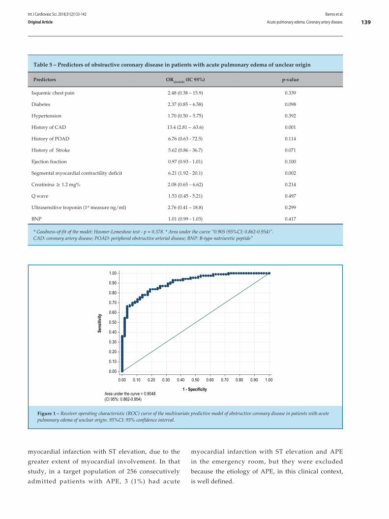
139
0.00
0.10
0.20
0.30
0.40
0.50
0.60
0.70
0.80
0.90
1.00
Sens
itivit
y
0.00 0.10 0.20 0.30 0.40 0.50 0.60 0.70 0.80 0.90 1.00
1 - SpecificityArea under the curve = 0.9048(CI 95%: 0.862-0.954)
Figure 1 – Receiver operating characteristic (ROC) curve of the multivariate predictive model of obstructive coronary disease in patients with acute pulmonary edema of unclear origin. 95%CI: 95% confidence interval.
Table 5 – Predictors of obstructive coronary disease in patients with acute pulmonary edema of unclear origin
Predictors ORajustada (IC 95%) p-value
Isquemic chest pain 2.48 (0.38 – 15.9) 0.339
Diabetes 2.37 (0.85 – 6.58) 0.098
Hypertension 1.70 (0.50 – 5.75) 0.392
History of CAD 13.4 (2.81 – .63.6) 0.001
History of POAD 6.76 (0.63 - 72.5) 0.114
History of Stroke 5.62 (0.86 - 36.7) 0.071
Ejection fraction 0.97 (0.93 - 1.01) 0.100
Segmental myocardial contractility deficit 6.21 (1.92 - 20.1) 0.002
Creatinina ≥ 1.2 mg% 2.08 (0.65 – 6.62) 0.214
Q wave 1.53 (0.45 - 5.21) 0.497
Ultrasensitive troponin (1st measure ng/ml) 2.76 (0.41 – 18.8) 0.299
BNP 1.01 (0.99 - 1.03) 0.417
* Goodness-of-fit of the model: Hosmer-Lemeshow test - p = 0.378. * Area under the curve “0.905 (95%CI: 0.862-0.954)”.CAD: coronary artery disease; POAD: peripheral obstructive arterial disease; BNP: B-type natriuretic peptide"
Barros et al.
Acute pulmonary edema. Coronary artery disease.
Int J Cardiovasc Sci. 2018;31(2)133-142
Original Article
myocardial infarction with ST elevation, due to the
greater extent of myocardial involvement. In that
study, in a target population of 256 consecutively
admitted patients with APE, 3 (1%) had acute
myocardial infarction with ST elevation and APE
in the emergency room, but they were excluded
because the etiology of APE, in this clinical context,
is well defined.

140Barros et al.
Acute pulmonary edema. Coronary artery disease.
Int J Cardiovasc Sci. 2018;31(2)133-142
Original Article
In the study by Figueras et al.,2 acute myocardial infarction with ST elevation was present in 30% of patients admitted with this pathology.
Classifying serial troponin measurements, increased in APE, as secondary to an acute coronary event is not reasonable in this model and brings costly consequences for the health system,8 since the presence of CAD in acute HF increases the time and costs of hospitalization, according Purek et al.9
It is noteworthy the fact that alterations in the electrocardiogram, which may suggest ischemia, such as changes in ST (depression) and T wave, were not part of the predictor model, suggesting that such changes can be found with or without obstructive CAD.10 This was verified in the series of nine cases by Littmann,11 in which negative and deep T waves and QT increase were not associated with coronary disease at the electrocardiogram after 24 hours of APE. One must always consider that other causes of ST depression may occur, such as left ventricular hypertrophy and drug use (such as digoxin) and, perhaps, the relative ischemia itself, which may occur during an episode of APE, where hypoxia is invariably present and, when associated with an increase in troponin, can be considered a type-2 acute myocardial infarction.10
The exact mechanism of the found alterations is poorly defined, attributing the electrophysiological responses of myocardial cells to adrenergic stimulation and myocardial hypoxia, which usually accompany the clinical condition of APE. In this series, only four patients underwent invasive investigation.
That study was not intended to evaluate mortality, but it was 15% over a one-year period – emphasizing that contact was established with only 48% of the study population. In the study by Figueras et al.,2 mortality at 30 days was 14%. That demonstrates disease severity, even in the present day.
Severe valvular disease occurred in 15 cases (10%). It is worth remembering that no prior valvulopathy was known and that 11 cases (73%) had severe aortic stenosis, which is characteristic of the study population’s profile, whose mean age was 65.4 ± 10.2.
This study shows that severe obstructive coronary disease was present in most patients with acute pulmonary edema, but variables commonly used in the emergency room to identify its presence failed, such as the interpretation of troponin levels and chest pain
preceding the clinical picture. Of course, we expect to find the predictors related to this condition – such as history of coronary disease and the segmental deficit at the echocardiogram – in this pathology.
Study limitations and prospects
The present study showed that obstructive coronary disease is prevalent in patients with APE: 59% of patients had some artery with severe obstructive lesion as demonstrated by the invasive hemodynamic study. Percutaneous intervention was not performed in most individuals with this condition, that is, increased troponin and obstructive CAD; as previously mentioned, the research was not designed to answer this question.
Moreover, considering the high frequency of troponin positivity in all the acute conditions affecting the heart, and the intersection of symptoms in the different etiologies, one expects a misdiagnosis when there is no specific marker for a given disease, such as the electrocardiogram in acute myocardial infarction with ST elevation and total atrioventricular block. This has occurred in clinical practice, in which symptoms associated with myocardial ischemia and positive troponin have elicited the request for a CA, interpreting the whole set as an acute ischemic syndrome.
Another limitation would be the CAD progression in patients who were submitted to CA in less than 1 year and the disease occurred during this period. It is assumed, however, that any research process restricted to a single research center results in limitations, considering the differences of conduct and clinical interpretations found in health units.
Conclusion
In hospitalized individuals with acute pulmonary edema of unclear origin, prior history of coronary artery disease and segmental myocardial contractility deficit were the independent predictors of obstructive coronary disease. Of these two predictors, the previous history of coronary artery disease is the main probabilistic determinant, practically guaranteeing the presence of obstructive coronary disease. However, in the absence of a previous history of coronary artery disease or stroke, the probability of obstructive coronary disease is an intermediate one, being modulated by two independent predictors: ejection fraction and myocardial contractility

141
1. Harjola VP, Costa S, Sund R, Ylikangas S, Siirilä-Waris K, Melin J, et al; FINN-AKVA Study Group. The type of acute heart failure and the costs of hospitalization. Int J Cardiol. 2010;145(1):103-5. doi: 10.1016/j.ijcard.2009.05.058.
2. Figueras J, Peña C, Soler-Soler J. Thirty day prognosis of patients with acute pulmonary oedema complicating acute coronary syndromes. Heart. 2005;91(7):889-93. doi: 10.1136/hrt.2004.043703.
3. Graham SP, Vetrovec GW. Comparison of angiographic findings and demographic variables in patients with coronary artery disease presenting with acute pulmonary edema versus those presenting with chest pain. Am J Cardiol. 1991;68(17):1614-8. PMID: 1746462.
4. Ponikowski P, Voors AA, Anker SD, Bueno H, Cleland, JG, Coats AJ, et al. 2016 ESC Guidelines for the diagnosis and treatment of acute and chronic heart failure: the Task Force for the diagnosis and treatment of acute and chronic heart failure of the European Society of Cardiology (ESC). Eur Heart J. 2016;37(27):2129-200. doi: 10.1093/eurheartj/ehw128.
5. Albuquerque DC, Souza Neto JD, Bacal F, Rohde LE, Bernardez-Pereira S, Berwanger O, et al; Investigadores Estudo BREATHE. I Brazilian registry of heart failure – clinical aspects, care quality and hospitalization outcomes. Arq Bras Cardiol. 2015;104(6):433-42. doi: 10.5935/abc.20150031. Erratum in: Arq Bras Cardiol. 2015;105(2):208.
6. Pena-Gil C, Figueras J, Soler-Soler J. Acute cardiogenic pulmonary edema: relevance of multivessel disease, conduction abnormalities and silent ischemia. Int J Cardiol. 2005;103(1):59-66. doi: 10.1016/j.ijcard.2004.08.029.
7. Haaf P, Drexler B, Reichlin T, Twerenbold R, Reiter M, Meissner J, et al. High-sensitivity cardiac troponin in the distinction of acute myocardial infarction from acute cardiac noncoronary artery disease. Circulation. 2012;126(1):31-40. doi: 10.1161/CIRCULATIONAHA.112.100867.
8. Felker GM, Teerlink JR. In Mann DL; Douglas PZ; Libby P; Bonow RO. Acute Heart Failure. Braunwald’s Heart Disease. 10th ed. Elsevier. 2015. p. 484-511.
9. Purek L, Christ A, Klima T, Pfisterer ME, Perruchoud AP, Mueller C. Coronary artery disease and outcome in acute congestive heart failure. Heart. 2006;92(5):598-602. doi: 10.1136/hrt.2005.066464.
10. Thygesen K, Alpert JS, Jaffe AS, Simoons ML, Alpert JS, White HD, et al; Writing Group on the Joint ESC/ACCF/AHA/WHF Task Force for the Universal Definition of Myocardial Infarction; ESC Committee for Practice Guidelines (CPG) Third universal definition of myocardial infarction. Eur Heart J. 2012;33(20):2551-67. doi: 10.1093/eurheartj/ehs184.
11. Littmann L. Large T wave inversion and QT prolongation associated with pulmonary edema: a report of nine cases. J Am Coll Cardiol. 1999;34(4):1106-10. PMID: 10520798.
References
Barros et al.
Acute pulmonary edema. Coronary artery disease.
Int J Cardiovasc Sci. 2018;31(2)133-142
Original Article
deficit. The value of troponin did not independently predict the presence of obstructive coronary disease in a scenario of acute heart failure, and the identification of the presence of obstructive coronary disease involved few revascularization procedures. This raises questions about the usefulness of pursuing the diagnosis of coronary disease in this clinical scenario.
Author contributions
Conception and design of the research: Barros MNDS, Correia LC. Acquisition of data: Barros MNDS, Sousa VWB, Lima IAB, Nóbrega CRBM, Moreira ICAM, Dourado SMM, Andrade BMS, Batista VS, Silva MCFC, Correia LC. Analysis and interpretation of the data: Barros MNDS, Correia LC. Statistical analysis: Barros MNDS, Correia LC. Obtaining financing: Barros MNDS, Correia LC. Writing of the manuscript: Barros MNDS, Correia LC. Critical revision of the manuscript for intellectual content: Barros MNDS, Correia LC.
Potential Conflict of Interest
No potential conflict of interest relevant to this article was reported.
Sources of Funding
There were no external funding sources for this study.
Study Association
This article is part of the thesis of Doctoral submitted by Maria das Neves Dantas da Silveira Barros from Escola Bahiana de Medicina e Saúde Pública – EBMSP.
Ethics approval and consent to participate
This study was approved by the Ethics Committee of the Faculdade Bahiana de Medicina e Saúde Publica under the protocol number 05503912.6.0000.5544. All the procedures in this study were in accordance with the 1975 Helsinki Declaration, updated in 2013. Informed consent was obtained from all participants included in the study.

142
This is an open-access article distributed under the terms of the Creative Commons Attribution License
Barros et al.
Acute pulmonary edema. Coronary artery disease.
Int J Cardiovasc Sci. 2018;31(2)133-142
Original Article

DOI: 10.5935/2359-4802.20180001
143International Journal of Cardiovascular Sciences. 2018;31(2)143-151
ORIGINAL ARTICLE
Mailing Address: José Antônio Caldas Teixeira •Rua Presidente João Pessoa, 248. Postal Code: 24220-331, Icaraí, Niterói, RJ – Brazil.E-mail: [email protected], [email protected]
Study with a Portable Gas Analyzer of the 6-Minute Walk Test in Heart Failure with Normal Ejection FractionJosé Antônio Caldas Teixeira,1 Leandro Rocha Messias,1 Kátia Pedreira Dias,1 Washington Luiz Batista da Costa,1 Roberto Macedo Cascon,2 Sandra Marina Ribeiro de Miranda,1 Pedro Soares Teixeira,3 Juliana Grael Jorge,4 Antonio Claudio Lucas da Nobrega,1 Denizar V. Araujo5
Universidade Federal Fluminense (UFF),1 Clínica Fit Center,2 Complexo Hospitalar de Niterói,3 Serviço de Arritmia da Rede Dor,4 Universidade do Estado do Rio de Janeiro,5 Rio de Janeiro, RJ – Brazil
Manuscript received February 06, 2017, revised manuscript June 01, 2017, accepted July 31, 2017
Abstract
Background: Few studies have used portable gas analyzers during the 6-minute walk test (6MWT) in patients with heart failure and normal ejection fraction (HFNEF).
Objectives: To analyze the kinetics of hemodynamic, ventilatory, and metabolic variables in patients with HFNEF during the T6m using a portable gas analyzer.
Methods: Prospective, analytical study with an intentional, non-probabilistic, convenience sample. In total, 24 patients with HFNEF and past hospital admissions due to a clinical diagnosis of heart failure (HF) were included using the 2007 criteria established by the European Society of Cardiology. Three assessments were performed: 6MWT familiarization, 6MWT with the portable gas analyzer, and cardiopulmonary exercise test (CPET).
Results: The heart rates (HRs) and the peak VO2 at the end of the 6MWT corresponded to 85.7% and 86.45% of the values obtained during the CPET. The final HRs after the T6m were equivalent to those obtained at the CPET anaerobic threshold (AT), with relative VO2 values at the end of the 6MWT above the VO2 of the CPET AT. There was no difference between the maximum respiratory quotient (RQ) values in these two tests, which were both above 1.0. The VE/VO2 slope descended initially and then ascended significantly after the fifth minute of the test, estimating the identification of the AT.
Conclusions: In patients with HFNEF, the 6MWT represents an almost maximum effort, and is performed above the CPET AT and 85% above the maximum HR and the CPET peak VO2, with a maximum RQ similar to that in the CPET. (Int J Cardiovasc Sci. 2018;31(2)143-151)
Keywords: Heart Failure; Blood Gas Analysis; Stroke Volume; Exercise; Walk Test.
Introduction
Heart failure (HF) evokes the image of a dilated
heart with reduced systolic function and ejection
fraction (HF with reduced ejection fraction, HFREF).
However, the study by Burkhoff et al.1 has shown that a
large proportion of patients with HF symptoms has an
ejection fraction within the normal range, and are then
classified as having HF with normal ejection fraction
(HFNEF).2,3 The limited tolerance to physical effort in
HF is often the first and main clinical characteristic of the disease.4 In the identification of this exercise intolerance, the 6-minute walk test (6MWT) has been used as an instrument to evaluate the progression after different interventions.5
Only a few studies have been conducted using portable gas analyzers to describe and analyze the behavior of variables in the 6MWT and possible characteristics or markers that may help guide the therapy and improve the prognosis in HF,6-10 particularly in patients with HFNEF.

144Teixeira et al.
Gas Analysis in the 6-Minute Walk Test in HFNEF
Int J Cardiovasc Sci. 2018;31(2)143-151
Original Article
The aim of this study was to describe and analyze the kinetic behavior at each minute of hemodynamic, ventilatory, and metabolic variables in patients with HFNEF during the 6MWT using a portable gas analyzer, and to compare the findings with those obtained during a cardiopulmonary exercise test (CPET).
Methods
Prospective, analytical study with a non-probabilistic, intentional, and convenience sample, as set by the adopted criteria, carried out at the Hospital Universitário Antônio Pedro (HUAP) of the Universidade Federal Fluminense (UFF) between March 2010 and July 2013. The study project was approved by the Ethics Research Committee at HUAP under the number 152A/2010, and all participants signed an informed consent form.
The inclusion criteria adopted to characterize HFNEF were those described by Paulus et al.,11 in addition to the following factors: (A) complaints suggestive of HF (dyspnea, fatigue, and/or edema); (B) report of prior hospitalization due to decompensated heart disease, but at the moment of the test presenting with a functional class II to III according to the New York Heart Association (NYHA); (c) age > 18 years; (d) disease duration greater than 6 months; (e) use of medication; and (F) stable disease.
The exclusion criteria adopted were: (A) chronic obstructive pulmonary disease (COPD) based on clinical criteria, (B) functional class IV or other criteria contraindicating the CPET,12 and (C) participation in supervised cardiac rehabilitation programs.
The patients were instructed to maintain the current medications. The tests comprised three moments: conventional 6MWT (6MWT1), 6MWT coupled to a portable gas analyzer (6MWT2), and CPET on a treadmill. Due to a matter related to the hospital's flow, some patients were first assigned randomly to the 6MWT and others to the CPET.
The 6MWT was performed in a hallway with an extension of 30 meters. The first 6MWT (6MWT1) had a learning effect as objective, as recommended in the literature.6 The second 6MWT (6MWT2) was performed at least 3 days and no more than 3 weeks after the first, when the patients repeated the 6MWT, but this time they were connected to a portable gas analyzer. The maximum interval between the 6MWT and the CPET was also 3 weeks. Both the 6MWT and the CPET were scheduled by the same evaluator and applied by the same team to avoid application variability.
We used for the assessments the metabolic analyzer MedGraphics (MGC) VO2000 (Imbrasport, Porto Alegre, RS, Brazil), the system Ergo PC Elite 13, and the treadmill Centurion 300 (MicroMed, Brasília, DF, Brazil). The gas analyzer was calibrated before each test by the autocalibration system in a ventilated environment. The biological control of the calibration was performed monthly and the control by the equipment's representative (CAEL, Rio de Janeiro, RJ, Brazil) was conducted every 3 months.
In the CPET, each patient underwent a 2-minute baseline collection followed by a 1-minute warm-up at 1 km/h and 0º slope before starting the ramp protocol. In order to analyze and assess the CPET variables, we used the software ErgoPCElite for Windows 13W (MicroMed, Brasília, DF, Brazil). The perceived exertion (PE), assessed by the Borg scale (variation 0-10), and the hemodynamic and electrocardiographic variables were recorded at every minute. During the recovery phase, the patient remained seated. Two referees analyzed the report of the test to obtain the following information: VE/VCO2 slope value, presence of oscillatory breathing (OB), and establishment of the ventilatory threshold I, referred from now on as the anaerobic threshold (AT). In order to determine the AT, we used the curves of the ventilatory equivalents of the VO2 and VCO2, in addition to the curves of VO2 and VCO2 expired fractions, as recommended by the CPET guideline of the American Heart Association (AHA).13
The evaluations were performed while the patients maintained the use of their usual medications, during the same time of the day, and at least 2 hours after the last meal. Peak VO2 was defined as the highest VO2 value obtained up to the final 30 seconds or 10 seconds into the immediate recovery. In order to determine the occurrence of OB and the value of the VE/VCO2 slope, we followed the AHA guideline13 and the recommendations by Guazzi et al.14 Since spirometry was not performed, the ventilatory reserve was not considered in the analysis. Only one CPET was performed, as recommended by Scott et al.15
The maximum estimated heart rate (HR) was obtained using the formula by Tanaka et al.16 and was used to calculate the chronotropic index.
During the 6MWT, we recorded the HR (Polar monitor, model T31, Oulu, Finland) at each minute, along with the PE according to the Borg scale and the capillary O2 saturation using a pulse oximeter (Onyx, Minneapolis, MN, USA).

145
Teixeira et al.
Gas Analysis in the 6-Minute Walk Test in HFNEF
Int J Cardiovasc Sci. 2018;31(2)143-151
Original Article
Table 1 – Clinical characteristics of the study population
Variables n = 24 %
Sexmale 7 29.2
female 17 70.8
Mean SD
EF (%) 64.7 8.2
Weight (kg) 84.3 16.6
Height (cm) 160.2 7.4
BMI 32.9 6.7
Waist (cm) 104.4 13.3
Hip (cm) 110.8 11.6
W/H 0.944 0.086
Age (years) 59.6 10.2
Abbreviations: EF: ejection fraction; BMI: body mass index; W/H: waist-to-hip ratio; SD: standard deviation.
In order to analyze the gases expired during the 6MWT and obtain the values of the variables, we used the same VO2000 in a portable mode connected wirelessly to a computer. We used the software Aerograph, version 4.3 (Imbrasport, Porto Alegre, RS, Brazil), which organized the data for later analysis.
During the 6MWT, the patient connected to the gas analyzer remained seated for 4 minutes and 30 seconds. After the values of the variables were collected during this resting period, the patient stood up for 30 seconds before initiating the 6MWT, which was performed according to the guidelines of the American Thoracic Society (ATS).5
All patients completed the 6 minutes of walk. The gas analysis ended after the fifth minute of recovery. The maximum blood pressure (BP) considered in the analysis was the one obtained immediately after the effort, i.e., after the sixth minute of the walk when the patient sat down and the BP was then measured between 30-40 seconds during recovery.
Statistical analysis
The numerical data are expressed as mean ± standard deviation (SD) since all the variables included had an approximately normal distribution (p value of the Kolmogorov-Smirnov test > 0.05). Categorical data are expressed as frequency (n) and percentage (%).
The variation of the hemodynamic, metabolic, and ventilatory measurements between the tests (assessments) CPET and 6MWT were analyzed by Student's t test for paired samples.
A descriptive graphic analysis was performed, presenting the mean and SD values of the measurements obtained at each minute of the variables HR and VO2, indicating, for each one of these variables, the maximum CPET values and values equivalent to the AT.
In order to evaluate the correlation levels between the 6MWT distance and the peak VO2 and CPET peak VO2, we used Pearson's correlation coefficient (r).
We adopted a significance level of 5%. The statistical analysis was performed using the software SAS 6.11 (SAS Institute, Inc., Cary, NC, USA).
Results
A total of 24 patients were evaluated. Of these, 22 underwent both tests (CPET and 6MWT) while two underwent only the CPET; thus, the comparison between the CPET and 6MWT included only data of 22 patients. Table 1 shows the baseline characteristics of the study population. There was a predominance of the female gender (70.8%) and obesity (58.3%). Hypertension was present in 100.0% of the patients.

146Teixeira et al.
Gas Analysis in the 6-Minute Walk Test in HFNEF
Int J Cardiovasc Sci. 2018;31(2)143-151
Original Article
Most patients (70.8%) used beta-blockers. The profile of the medications used by the patients showed the usual therapeutic approach for hypertension: beta-blockers, angiotensin-converting enzyme inhibitors (25.0%) or angiotensin receptor blockers (54.1%), calcium channel blockers (37.5%), thiazides (29.1%), and vasodilators (48.5%).
A stratification of the patients according to the results of the CEPT and following the classification by Weber17 showed a higher percentage of patients categorized as class B (30.4%).
The patients had a mean E/E' of 15.9 ± 4.3, which fulfilled one of the criteria for diagnosis of HFNEF.11
The mean walked distance was 419.2 ± 76.5 meters during the 6MWT1 and 446.2 ± 67.7 meters during the 6MWT2, with a significant difference between both groups (p = 0.002).
Table 2 shows the maximum values of the variables obtained in the 6MWT2 and in the CPET. There was no difference between the maximum respiratory quotient (RQ) values of the two tests, and their maximum values were greater than 1.0. The results showed increased values at the end of the 6MWT2 (1.04), and of 22 patients who underwent the 6MWT2, 11 had an RQ ≥ 1.0 (50% of the group). Of these 11 patients, 7 obtained an RQ ≥ 1.10 (31.8% of the sample).
Since BP measurement during the 6MWT was not feasible, BP was only measured in the baseline condition and immediately after the 6MWT. Considering that prior spirometry was not performed, other ventilatory parameters were not assessed.
The longitudinal analysis of the HR during the 6MWT2 showed that the group reached stable HR values after the second minute of the 6MWT2 (T3). The maximum HR
Table 2 – Maximum variables obtained in the 6-minute walk test (6MWT) and the cardiopulmonary exercise test (CPET) (n = 22)
Mean values - 6MWTversus CPET
6MWT SD CPET SD p < 0.05
Max SBP 187.04 26.60 215.3 27.4 0.002
Max DBP 97.80 13.7 104.1 16.0 0.09
Max DP 20951 5497 27524 6130 0.0004
CircPot 2533 957 3462 1266 0.002
Max HR 110.9 18.1 125.4 19.6 0.004
HR1Rec 23.1 10.1 17.2 10.1 0.035
ChronI 0.42 0.2 0.57 0.1 0.002
O2 pulse 10.5 3.6 10.5 3.5 0.95
Max RQ 1.06 0.24 1.08 0.09 0.47
VE/VCO2 slope 22.3 4.8 23.2 4.4 0.10
VP 8.31 1.92 9.59 2.11 0.0002
Peak VO2 (mL.kg-1. min-1) 13.8 4.1 15.9 5.4 0.025
T1/2 61.4 16.5 111.2 25.7 0.0001
OUES 1.26 0.74 1.34 0.59 0.10
AT VO2 11.76 3.79
Max SBP: maximum systolic blood pressure; Max DBP: maximum diastolic blood pressure; Max DP: maximum double product; CircPot: circulatory power; Max HR: maximum heart rate; HR1Rec: heart rate in the first minute of recovery; ChronoI: chronotropic index; O2 pulse: oxygen pulse (mL.kg-1.min-1/bpm); Max RQ: maximum respiratory quotient; VE/VCO2 slope: ventilation minute to carbon dioxide production ratio; VP: ventilatory power; peak VO2 (mL.kg-1. min-1): oxygen consumption peak; T1/2: time (in seconds) of the recovery kinetics of oxygen consumption up to half of the value of peak effort; OUES: oxygen uptake efficiency slope; AT: anaerobic threshold; 6MWT2: second 6-minute walk test; CPET: cardiopulmonary exercise test; SD: standard deviation. All heart rate values are represented in beats per minute.

147
160
140
120
100
80
60
40
20T1 T2 T3 T4 T5 T6 T7
108 – HR at the AT HFNEF
126 – maxHR in the CPET
108.9
HR (b
pm)
Figure 1 – Longitudinal progression of the heart rate (HR) during the second 6-minute walk test (6MWT2) in the study participants. HFNEF: heart failure with normal ejection fraction; CPET: cardiopulmonary exercise test; maxHR: maximum heart rate; AT: CPET anaerobic threshold; T1: time zero of the 6MWT (baseline conditions); T2, T3, T4, T5, T6, T7: first, second, third, fourth, fifth, and sixth minutes of the 6MWT, respectively.
Teixeira et al.
Gas Analysis in the 6-Minute Walk Test in HFNEF
Int J Cardiovasc Sci. 2018;31(2)143-151
Original Article
during the 6MWT2 (108.9 bpm) was similar to the HR at the CPET AT (108 bpm), shown by the dashed line (Figure 1), and 85.7% compared with the CPET maximum HR (126 bpm), highlighted in the Figure.
The longitudinal progression of the relative VO2 (mL.kg-1.min-1) during the 6MWT2 in the study participants is shown in Figure 2. Note in the dashed line the VO2 value of the CPET AT (11.76 mL.kg-1. min-1) and in the highlighted area, the value of 15.9 mL.kg-1. min-1 of the CPET peak VO2 (Figure 2).
The longitudinal progression of the production of carbon dioxide (VCO2) during the 6MWT2 occurred in a similar way to that of the VO2, reaching maximum values in the 6MWT2 of 11.5 mL.kg-1.min-1 (not displayed).
Pearson's correlation (r) levels found between the maximum walked distance in the 6MWT2 with the peak VO2 obtained in the same test and between the walked distance with the CPET peak VO2 were r = 0.528 (p = 0.014) and r = 0.532 (p = 0.013), respectively.
Discussion
A review of the literature on the subject of this study – 6MWT and HFNEF – has shown that most studies in this area are focused on HFREF. Therefore, it became difficult to compare the results found in the present study, which included patients with HFNEF, with data from the literature. Due to that, the discussion below will be based on similar results found in studies with HFREF.
Riley et al.,6 Foray et al.,7 Faggiano et al.,8 and Kervio et al.10 analyzed the kinetics of variables using a
portable gas analyzer and demonstrated that the kinetics and other cardiovascular parameters are decreased in HFREF in response to the effort imposed by the 6MWT. However, no studies have described how the kinetics of patients with HFREF respond during this test.
Among the study patients, there was a predominance of patients of the female gender, with hypertension, and with an increased body mass index (BMI),3,11 characteristics that confirm the typical clinical profile of patients with HFNEF.
The average distance walked is within the values that indicate a good prognosis, i.e., above 300 meters.5,18 A significant percentage (81.81%) equaled or increased in the 6MWT2 the distance walked during the 6MWT1 but did not obtain values of clinical significance, i.e., above 50 meters.5 This reinforces the importance of conducting at least one learning and familiarization test due to a potential influence on the results of the 6MWT.6,19
The distances obtained are aligned with the average values found in studies with patients with HFREF, such as those by Kervio et al.10 (452.6 ± 18.7 meters), Faggiano et al.8 (419 ± 120 meters), and Guimarães et al.9 (470 ± 48 meters).
The correlation levels found between the 6MWT2 and the peak VO2 in the 6MWT1 and with the CPET peak VO2 are aligned with findings in the literature, despite some discrepancies. Some authors report a good correlation between the distance of the 6MWT and the peak VO2, with an average of 0.73 in patients with HF6,19 (all with HFREF). Riley et al.6 found a high correlation (r = 0.63), whereas Lucas et al.20 and Roul et al.21 found a

148
18
16
14
12
10
8
6
4
2
0T1 T2 T3 T4 T5 T6 T7
11.76 – VO2 at the CPET AT
13.7
15.9 CPET peak VO2
VO2 (m
L.kg
–1.m
in–1
)
Figure 2 – Longitudinal progression of the relative VO2 (mL/kg/min-1) during the 6MWT2. HFNEF: heart failure with normal ejection fraction; CPET: cardiopulmonary exercise test; VO2: oxygen consumption (mL.kg-1.min-1); T1: time zero of the 6MWT (baseline conditions); T2, T3, T4, T5, T6, T7: first, second, third, fourth, fifth, and sixth minutes of the 6MWT, respectively, AT: anaerobic threshold.
Teixeira et al.
Gas Analysis in the 6-Minute Walk Test in HFNEF
Int J Cardiovasc Sci. 2018;31(2)143-151
Original Article
low correlation with the walked distance in the 6MWT (r = 0.28 and r = 0.24, respectively). Such discrepancies could be attributed to different methodologies of the 6MWT and the type of ergometers used.
The HFREF studies found in the literature assessing the 6MWT with portable gas analyzers and performed in a hallway were: Riley et al.,6 Foray et al.,7 Faggiano et al.,8 and Kervio et al.10 Guimarães et al.9, in turn, used the gas analyzer during two 6MWT tests performed on a treadmill, also in patients with HFREF.
In regards to HR parameters, the study group showed more stable values after the second minute of the 6MWT (T3). Kervio et al.10 highlight that earlier achievement of a stable state reflect better clinical conditions and, therefore, less severe ones.
The HR at the end of the 6MWT was 85.7% of the CPET maximum HR, which is similar to the percentage values of the peak VO2 in the 6MWT in relation to the CPET peak VO2.
It is noteworthy that the final HR in the 6MWT was similar to the HR in the CPET AT, reinforcing that the 6MWT represents an intense effort, carried out at the level of or above the CPET AT in patients with HF.6-8,10
Despite the authors’ claim that patients with HFNEF present HR alterations in the first minute of recovery (HR1Rec)22 and the fact that there are no data in the literature related to the 6MWT, if we consider the recommended value of 12 bpm, this value was within the normal limits both in the T6M as well as in the CPET.23
The chronotropic index analysis demonstrated a chronotropic incompetence, even if we consider the use of beta-blockers (normal > 0.60), both in relation to the 6MWT as well as to the CPET, as found in the literature.24
The maximum O2 pulse in the 6MWT and the CPET showed no difference between the two tests, yielding reduced pulse O2 values in relation to the predicted one (< 85%), around 70%, and with absolute values below 12 mL.kg-1.min-1/bpm, which is considered of poor prognosis.25
In relation to the two ventilatory variables analyzed, no difference in the VE/VCO2 slope was observed. The VE/VCO2 slope values are within the values cited as indicative of good prognosis (< 30),13 and this fact associated with the lack of difference between the two tests can reinforce the prognostic ability of the 6MWT. The ventilatory power (VP) analysis also showed a difference between the two tests, with higher values for the CPET. The VP, which combines the response of the SBP with the VE/VCO2 slope (VP = SBP x VE/VCO2
slope)26 showed a difference probably due to the higher SBP in the CPET. However, good prognosis values (> 3.5) were observed for both tests.26
The RQ is a criterion to obtain intense (> 1.0) or maximum effort in exercises of increasing intensity (> 1.15).13 There was no difference between the maximum RQ values between the two tests. The data found here are in agreement with those found by Kervio et al.,10 who consider the RQ in the 6MWT as reflecting intense effort. Both in the population with HFREF10 and in patients with HFNEF in the study, 50% of each sample obtained an

149
Teixeira et al.
Gas Analysis in the 6-Minute Walk Test in HFNEF
Int J Cardiovasc Sci. 2018;31(2)143-151
Original Article
RQ > 1.0. Other authors agree and claim that the greater the functional deficiency of the studied cohort, the more the 6MWT would be executed close to the maximum.6-8
In the analysis of the VE/VO2, the curve first descended and then ascended, which became significant after the fifth minute of the test (T6 and T7). Such progression is one of the criteria used to identify the CPET AT.13,24 When this concept is transferred to the 6MWT, it confirms that it represents an effort similar to or slightly above the AT.
In regards to the VE/VCO2, the values stabilized after the second minute of effort (T3), with peak values slightly above those of the CPET AT (24.5 ± 3.1 in the 6MWT versus 23.6 of the CPET AT).The VE/VCO2 stabilized in values that do not reflect ventilatory inefficiency.
The relative VO2 in the 6MWT showed stable values and its progression in patients with HFNEF did not differ from that found in similar studies in patients with HFREF, both in terms of progression during the 6MWT as well as in regards to the percentages obtained in relation to the CPET:6-8,10 it reached values above the VO2 corresponding to the CPET AT and of significant percentages of the CPET peak VO2 (86.45%).
The variable VCO2 tended to stabilize after T5 (the fourth minute of the 6MWT), which contrasts with the data presented by Kervio et al.,10 in which this variable did not reach a stable state until the end of the 6MWT in patients with HFREF.
The patients with HFREF in the study by Kervio et al.10 performed their 6MWT above the relative VO2 of the CPET AT, which is aligned with findings in patients with HFNEF in this study. The VO2 at the CPET AT obtained by Kervio et al.10 (11.7 ± 0.6 mL.kg-1.min-1) is also very similar to that found in this study (11.76 mL.kg-1.min-1).
It is interesting to note that 7 out of 22 patients in the present study had peak VO2 in the 6MWT equal to or greater than the CPET peak VO2, representing 31.8% of the total sample. This percentage value is aligned and supersedes those by Faggiano et al.8 (27.0%) in a HFREF population.
The high intensity of the 6MWT for patients with HF is reinforced by findings from the study by Kervio et al.,10 Faggiano et al.,8 and Foray et al.,7 which demonstrated that the 6MWT leads to a demand above 85% of the values of the CPET relative peak VO2.
Faggiano et al.8 found a peak VO2 at 86% of the CPET, which represented 73.0% of the VO2 of the CPET AT. The present study showed similar percentages in
relation to the CPET, but the VO2 at the CPET AT presented higher percentages, corresponding to 85.03% of the peak VO2 in the 6MWT in patients with HFNEF. Guimarães et al.9 analyzed the results of a single CPET and 6MWT on a treadmill, with the participants connected to a gas analyzer and, using an incentive, found a peak VO2 at 90% of the CPET.
The values of the CPET relative peak VO2 are aligned with the findings by Guazzi et al.27 These authors found CPET peak VO2 values of 15 mL.kg-1.min-1 in HFNEF patients.
The oxygen uptake efficiency slope (OUES) showed lower values in the 6MWT, but no difference in values was observed between the 6MWT and the CPET. Although the literature indicates that reduced values in both HFREF and HFNEF,13 the HFNEF patients evaluated obtained values above 1.2, which are considered to be of poor prognosis.28
The metabolic variable with the most difference was the kinetics of oxygen consumption during recovery (T1/2), which showed a significantly greater recovery time in the CPET. The T1/2 values in the 6MWT did not fulfill the criteria of poor prognosis.13,24,29 Considering that the value of 90 seconds29 in the CPET, the study participants exceeded this value.
Conclusion
There is an actual possibility of patients with HFNEF to be are able to perform a 6MWT at maximum or almost maximum intensity. This estimate is based on the following observations: high percentages obtained in peak values in the 6MWT in relation to the maximum value of the variables HR (85.7%) and relative VO2 (86.4%) in the CPET, similar RQ values, similar peak VO2 values in the 6MWT and at the CPET AT, and the progression of the VE/VO2, which after reaching a nadir, showed a trend to curve upward.
It should be noted that in relation to the reviewed studies, all conducted in patients with HFREF, the assessed variables in HFNEF showed, on average, an equal profile during the 6MWT.
Study limitations
Some limitations in this study are identified:
• Reduced size of the study sample. Further studies with an increased number of patients are suggested to validate some of the conclusions.

150
1. Burkhoff D, Maurer MS, Packer M. Heart failure with a normal ejection fraction. is it really a disorder of diastolic function? Circulation. 2003;107(5):656-8. PMID: 12578861.
2. Vasan RS, Benjamin EJ, Levy D. Prevalence, clinical features and prognosis of diastolic heart failure: an epidemiologic perspective. J Am Coll Cardiol. 1995;26(7):1565-74. doi: 10.1016/0735-1097(95)00381-9.
3. Zile MR, Kjellstrom B, Bennett T, Cho Y, Baicu CF, Aaron MF, et al. Effects of exercise on left ventricular systolic and diastolic properties in patients with heart failure and a preserved ejection fraction versus heart failure and a reduced ejection fraction. Circ Heart Fail. 2013;6(3):508-16. doi: 10.1161/CIRCHEARTFAILURE.112.000216.
4. Downing J, Balady GJ. The role of exercise training in heart failure. J Am Coll Cardiol. 2011;58(6):561-9. doi: 10.1016/j.jacc.2011.04.020.
5. ATS Committee on Proficiency Standards for Clinical Pulmonary Function Laboratories. ATS statement: guidelines for the six-minute walk test. Am J Respir Crit Care Med. 2002;166(1):111-7. doi: 10.1164/ajrccm.166.1.at1102.
6. Riley M, McParland J, Stanford CF, Nicholls DP. Oxygen consumption during corridor walk testing in chronic cardiac failure. Eur Heart J.1992;13(6):789-93. PMID: 1623869.
7. Foray A, Williams D, Reemtsma K, Oz M, Mancini D. Assessment of submaximal exercise capacity in patients with left ventricular assist devices. Circulation. 1996;94(9 Suppl):II-222-6. PMID: 8901750.
8. Faggiano P, D’Aloia A, Gualeni A, Lavatelli A, Giordano A. Assessment of oxygen uptake during the 6-minute walking test in patients with heart failure: preliminary experience with a portable device. Am Heart J. 1997;134(2 Pt 1):203-6. PMID: 9313598.
9. Guimarães GV, Bellotti G, Bacal F, Mocelin A, Bocchi EA. Can the cardiopulmonary 6-minute walk test reproduce the usual activities of
patients with heart failure? Arq Bras Cardiol. 2002;78(6):553-60. http://dx.doi.org/10.1590/S0066-782X2002000600003.
10. Kervio G, Ville NS, Leclercq C, Daubert JC, Carre F. Cardiorespiratory adaptations during the six-minute walk test in chronic heart failure patients. Eur J Cardiovasc Prev Rehabil. 2004;11(2):171-7. PMID: 15187823.
11. Paulus WJ, Tschöpe C, Sanderson JE, Rusconi C, Flachskampf FA, Rademakers FE, et al. How to diagnose diastolic heart failure: a consensus statement on the diagnosis of heart failure with normal left ventricular ejection fraction by the Heart Failure and Echocardiography Associations of the European Society of Cardiology. Eur Heart J.2007;28(20):2539-50. doi: 10.1093/eurheartj/ehm037.
12. Fletcher GF, Ades PA, KligfieldP, Arena R, Balady GF, Bittner VA, et al; American Heart Association Exercise, Cardiac Rehabilitation, and Prevention Committee of the Council on Clinical Cardiology, Council on Nutrition, Physical Activity and Metabolism, Council on Cardiovascular and Stroke Nursing, and Council on Epidemiology and Prevention. Exercise standards for testing and training: a scientific statement from the American Heart Association. Circulation. 2013;128(8):873-934. doi: 10.1161/CIR.0b013e31829b5b44.
13. Balady GJ, Arena R,Sietsema K, Myers J, Coke L, Fletcher, GF, et al; American Heart Association Exercise, Cardiac Rehabilitation, and Prevention Committee of the Council on Clinical Cardiology; Council on Epidemiology and Prevention; Council on Peripheral Vascular Disease; Interdisciplinary Council on Quality of Care and Outcomes Research. Clinician’s Guide to cardiopulmonary exercise testing in adults a scientific statement from the American Heart Association. Circulation. 2010;122(2):191-225. doi: 10.1161/CIR.0b013e3181e52e69.
14. Guazzi M, Raimondo R, Vicenzi M, Arena R, Proserpio C, Sarzi Braga S, et al. Exercise oscillatory ventilation may predict sudden cardiac death in heart failure patients. J Am Coll Cardiol. 2007;50(4):299-308. doi: 10.1016/j.jacc.2007.03.042.
References
Teixeira et al.
Gas Analysis in the 6-Minute Walk Test in HFNEF
Int J Cardiovasc Sci. 2018;31(2)143-151
Original Article
• The predominance of the female gender and overweight/obesity, characteristic of HFNEF, may have influenced the variables related to functional capacity in both 6MWT and CPET.
• The peripheral determinants of peak VO2, such as the oxygen transportation system or changes in peripheral muscles, were not considered in the functional assessment.
• The use of beta-blockers and other medications may have influenced the interpretation of variables that integrate HR or BP in their calculation.
Author contributions
Conception and design of the research: Teixeira JAC, Nobrega ACL, Araujo DV. Acquisition of data: Teixeira JAC, Messias LR, Dias KP, Costa WLB, Cascon RM, Miranda SMR, Teixeira PS, Jorge JG. Analysis and interpretation of the data: Teixeira JAC, Messias LR, Dias KP, Costa WLB, Teixeira PS, Nobrega ACL, Araujo DV. Writing of the manuscript: Teixeira JAC, Teixeira PS, Jorge JG. Critical revision of the manuscript
for intellectual content: Teixeira JAC, Nobrega ACL, Araujo DV. Bibliographic Survey: Teixeira JAC, Messias LR, Miranda SMR. Bibliographic Review: Costa WLB, Cascon RM.
Potential Conflict of Interest
No potential conflict of interest relevant to this article was reported.
Sources of Funding
There were no external funding sources for this study.
Study Association
This article is part of the thesis of associate professor submitted by José Antônio Caldas Teixeira, from Universidade Federal Fluminense.
Ethics approval and consent to participate
This article does not contain any studies with human participants or animals performed by any of the authors.

151
15. Scott JM, Haykowsky MJ, Eggebeen J, Morgan TM, Brubaker PH, Kitzman DW. Reliability of peak exercise testing in patients with heart failure with preserved ejection fraction. Am J Cardiol. 2012;110(12):1809-13. doi: 10.1016/j.amjcard.2012.08.015.
16. Tanaka H, Monahan KD, Seals DR. Age-predicted maximal heart rate revisited. J Am Coll Cardiol. 2001;37(1):153-6. PMID: 11153730.
17. Task Force of the Italian Working Group on Cardiac Rehabilitation and Prevention (Gruppo Italiano di Cardiologia Riabilitativa e Prevenzione, GICR), Working Group on Cardiac Rehabilitation and Exercise Physiology of the European Society of Cardiology. Statement on cardiopulmonary exercise testing in chronic heart failure due to left ventricular dysfunction: recommendations for performance and interpretation. Part III: Interpretation of cardiopulmonary exercise testing in chronic heart failure and future applications. Eur J Cardiovasc Prev Rehabil. 2006;13(4):485-94. doi: 10.1097/01.hjr.0000201518.43837.bc.
18. Arslan S, Erol MK, GundogduF, Sevimli S, Aksakal E, Senocak H, et al. Prognostic value of 6-minute walk test in stable outpatients with heart failure. Tex Heart Inst J. 2007;34(2):166-9. PMID: 17622362.
19. Faggiano P, D’Aloia A, Gualeni A, Brentana L, Dei Cas L. The 6 minute walking test in chronic heart failure: indications, interpretation and limitations from a review of the literature. Eur J Heart Fail. 2004;6(6):687-91. doi: 10.1016/j.ejheart.2003.11.024.
20. Lucas C, Stevenson LW, Johnson W, Hartley H, Hamilton MA, Walden J, et al. The 6-min walk and peak oxygen consumption in advanced heart failure: aerobic capacity and survival. Am Heart J. 1999;138(4 Pt 1):618-24. PMID: 10502205.
21. Roul G, Germain P, Bareiss P. Does the 6-min walk test predict the prognosis in patients with NYHA class II or III chronic heart failure? Am Heart J. 1998;136(3):449-57. PMID: 9736136.
22. Guazzi M, Myers J, Peberdy MA, Bensimhon D, Chase P, Arena R. Cardiopulmonary exercise testing variables reflect the degree of diastolic dysfunction in patients with heart failure–normal ejection
fraction. J Cardiopulm Rehabil Prev. 2010;30(3):165-72. doi: 10.1097/HCR.0b013e3181d0c1ad.
23. Cole CR, Foody JM, Blackstone EH, Lauer MS. Heart rate recovery after submaximal exercise testing as a predictor of mortality in a cardiovascularly healthy cohort. Ann Intern Med. 2000;132(7):552-5. PMID: 10744592.
24. Meneghelo RS, Araújo CG, Stein R, Mastrocolla LE, Albuquerque PF, Serra SM, et al; Sociedade Brasileira de Cardiologia. [III Guidelines of Sociedade Brasileira de Cardiologia on the exercise test]. Arq Bras Cardiol. 2010;95(5 supl. 1):1-26. doi: http://dx.doi.org/10.1590/S0066-782X2010000800001.
25. Oliveira RB, Myers J, Araújo CG, Abella J, Mandic S, Froelicher V. Maximal exercise oxygen pulse as a predictor of mortality among male veterans referred for exercise testing. Eur J Cardiovasc Prev Rehab. 2009;16(3):358-64. doi: 10.1097/HJR.0b013e3283292fe8.
26. Forman DE, Guazzi M, Myers J, Chase P, Bensimhon D, Cahalin LP, et al. Ventilatory power: a novel index that enhances prognostic assessment of patients with heart failure. Circ Heart Fail. 2012;5(5):621-6. doi: 10.1161/CIRCHEARTFAILURE.112.968529.
27. Guazzi M, Dickstein K, Vicenzi M, Arena R. Six-minute walk test and cardiopulmonary exercise testing in patients with chronic heart failure: a comparative analysis on clinical and prognostic insights. Circ Heart Fail. 2009;2(6):549-55. doi: 10.1161/CIRCHEARTFAILURE.109.881326.
28. Hollenberg M, Tager IB. Oxygen uptake efficiency slope: an index of exercise performance and cardiopulmonary reserve requiring only submaximal exercise. J Am Coll Cardiol. 2000;36(1):194-201. PMID: 10898434.
29. Cohen-Solal A, Tabot JY, Logeart D, Bourgoin P, Tokmakova M, Dahan M. A non-invasively determined surrogate of cardiac power (‘circulatory power’) at peak exercise is a powerful prognostic factor in chronic heart failure. Eur Heart J. 2002;23(10):806-14. doi: 10.1053/euhj.2001.2966.
This is an open-access article distributed under the terms of the Creative Commons Attribution License
Teixeira et al.
Gas Analysis in the 6-Minute Walk Test in HFNEF
Int J Cardiovasc Sci. 2018;31(2)143-151
Original Article

DOI: 10.5935/2359-4802.20180005
Abstract
Background: The choice between percutaneous coronary intervention (PCI) and coronary artery bypass grafting (CABG) remains controversial.
Objective: To conduct a meta-analysis of randomized studies comparing drug-eluting stents (DES) and CABG in multivessel disease or obstruction of the left main coronary artery.
Method: Electronic databases were searched systematically to evaluate results of randomized trials comparing PCI with DES versus CABG in multivessel disease and obstruction of the left main coronary artery. Ten studies were identified.
Results: In the aggregated results (n = 9268), mortality at 30 days and incidence of stroke favored PCI (0.8% versus 1.5%, p = 0.005; 0.4% versus 1.5%, p < 0.0001, respectively). There was no difference in mortality at 1 year (3.4% versus 3.5%, p = 0.50). The late mortality favored CABG (10.1% versus 8.5%, p = 0.01). In patients with diabetes derived from four studies (n = 3830), late mortality favored CABG (12.5% versus 9.7%, p < 0.0001). In six studies of left main coronary artery obstruction (n = 4700), the incidence of stroke favored PCI (0.3% versus 1.5%, p < 0.001) and there was no difference in mortality at 30 days (0.8% versus 1.3%, p = 0.15), mortality at 1 year, or late mortality (8.1% versus 8.1%). The subgroups with high SYNTAX score and diabetes were those influencing most strongly and adversely the PCI results.
Conclusion: When compared with PCI, CABG was superior in regards to late mortality and inferior in regards to 30-day mortality and incidence of stroke. Diabetes and SYNTAX score strongly impacted the results. (Int J Cardiovasc Sci. 2018;31(2)152-162)
Keywords: Myocardial Revascularization; Drug Eluting Stents; Randomized Controlled Trials as Topic; Meta-Analysis.
152International Journal of Cardiovascular Sciences. 2018;31(2)152-162
ORIGINAL ARTICLE
Mailing address: Pedro José Negreiros de AndradeFrancisco Holanda, 992, Ap.: 1101. Postal Code 60130-040, Dionisio Torres, Fortaleza, CE – Brazil.E-mail: [email protected]; [email protected]
Drug-eluting stents Versus Coronary Artery Bypass Grafting in Multivessel Disease and Left Main Obstruction: Meta-analysis of Randomized Clinical TrialsPedro José Negreiros de Andrade,1,2 Hermano Alexandre Lima Rocha,1,3 João Luiz de Alencar Araripe Falcão,1,2 Antonio Thomaz de Andrade,1 Breno de Alencar Araripe Falcão1,4
Hospital Dr. Carlos Alberto Studart Gomes, Messejana,1 Universidade Federal do Ceará (UFC),2 Centro Universitário Christus,3 Universidade Estadual do Ceará (UECE),4 Fortaleza, CE – Brazil
Manuscript received February 13, 2017; revised manuscript May 25, 2017; accepted July 11, 2017
Introduction
Percutaneous coronary intervention (PCI or angioplasty)
and coronary artery bypass grafting (surgery or CABG)
are well-accepted, safe, and effective alternatives in the
treatment of coronary insufficiency. A large number of
randomized clinical trials has been published comparing
both procedures.1-12 In light of these studies, there seems to
be a slight superiority of surgery over PCI in the ability to
reduce anginal symptoms and a significant difference in its
ability to prevent new revascularization procedures. Such
studies are generally undersized to evaluate outcomes like
death, stroke, and acute myocardial infarction (AMI).
The objective of this study was to perform a meta-analysis
of randomized clinical trials comparing PCI and CABG in
multivessel disease and obstruction of the left main coronary
artery in the era of drug-eluting stents, with emphasis on
mortality and stroke.
Methods
Randomized studies comparing PCI with drug-eluting
stents versus CABG in multivessel lesions and/or obstruction
of the left main coronary artery published between January
2002 and November 2016 were searched in the databases
MEDLINE and Cochrane Library, and in bibliographical

153
Andrade et al.
Stent versus surgery: randomized trials
Int J Cardiovasc Sci. 2018;31(2)152-162
Original Article
references of reviews published on the subject. The date of January 2002 was chosen as the initial period since drug-eluting stents began to be established as a therapeutic method after that. Clinical trials were included in the review if they were randomized, had compared surgery and coronary angioplasty, used drug-eluting stents, involved exclusively multivessel disease or left main coronary artery obstruction, had a minimum follow-up of 1 year, and were published in international journals with an impact factor > 2.0. We used the following terms in the search: coronary artery bypass surgery, coronary stents, and randomized controlled trials. Studies exclusively using balloon or bare-metal stents, or which assessed predominantly one-vessel disease were not included. Studies using drug-eluting and bare-metal stents1,4 were included as studies of the drug-eluting stent era. Works resulting from observational studies (registries) or only published as meetings proceedings were not considered.
We identified 10 randomized studies that satisfied the requirements: LE MANS,1 SYNTAX,2-3 CARDia,4 Boudriot et al.,5 PRECOMBAT,6 VA CARDS,7 FREEDOM,8 BEST,9 NOBLE,10 and EXCEL.11 Three authors (PJNA, BAAF, and JLAAF) evaluated the studies, which were all considered to be of high quality.
The main outcomes of interest were mortality and stroke. The incidence of AMI was not evaluated because the definition of this event varied widely in the studies. We also did not evaluate the incidence of new revascularization, because the superiority of surgery on this outcome is well established. Mortality was divided into early mortality, mortality at 1 year, and late mortality. Early mortality was defined as death occurring up to 30 days after the procedure, including deaths occurring after randomization but before the procedure. This mortality was obtained from seven studies, whereas three studies did not provide this information.2,4,7 Mortality at 1 year was defined as death occurring up to 1 year after the procedure, including early mortality. This mortality was obtained from nine studies, while one study did not provide such information.9 Late mortality was defined as death recorded at the end of follow-up, after at least 3 years. This mortality was obtained from eight studies, six of which performed a follow-up for 5 years, one for 3 years2 and one for 10 years.1 We were unable to obtain this information from two studies.5,7 For the incidence of stroke, we considered the events occurring up to 1 year after the procedure. In eight studies, we obtained the results up to 30 days and in one of them,2 up to 1 year, while in one of the studies, this information was unavailable.9 We evaluated separately the results of studies in the left main coronary artery and
late mortality in the subgroup of patients with diabetes.
We also performed analysis of combined major adverse
cardiac and cerebrovascular events (MACCE) and
assessed the variables age, gender, presence of diabetes,
SYNTAX score, and compromised ejection fraction in
subgroups based on data published in five trials.2,4,6,8,9
Combined MACCE comprised death, AMI, and new
revascularization in two of these trials,6,9 and death, AMI,
and stroke in the remaining ones.
In order to aggregate the outcomes of mortality and stroke,
as well as those of MACCE (in subgroups), we considered
whenever possible the absolute number of events and the
number of patients followed up. Otherwise, percentages were
transformed into absolute numbers.
Statistical analysis
We measured the relative risk and the risk difference
after grouping the results of each outcome. In order to assess
the statistical significance of the differences between the
drug-eluting stent and the surgery groups, we performed
a meta-analysis using the Mantel-Haenszel method, with
a fixed-effect model. We calculated the heterogeneity of
the studies using Cochran's Q test and the significance of
the measure of the meta-analytic effect using the Z test.
The differences between the results in the stent and CABG
groups were considered significant if p < 0.05.
The statistical analyses were performed using the
program Review Manager (RevMan), version 5.3
(Copenhagen: The Nordic Cochrane Centre, The Cochrane
Collaboration, 2014).
In order to represent the heterogeneity of the studies, we
constructed Forest plots. We used the risk difference to plot
these graphs since this is a more stable index. We refrained
from using odds ratio or relative risk due to the inclusion of
some clinical trials with zero or near zero events.
Results
Characteristics of the trials are shown in table 1
The studies included a total of 9268 patients (4642 in
the stent group and 4626 in the CABG group). The mean
age of the patients was 64 years, 75% were male, 51%
were diabetic, 24% were smokers, 64% were hypertensive,
and 31% had unstable angina. The mean ejection fraction
(reported in seven studies) was 59%, the mean EuroSCORE
(reported in five studies) was 2.9, and the mean SYNTAX
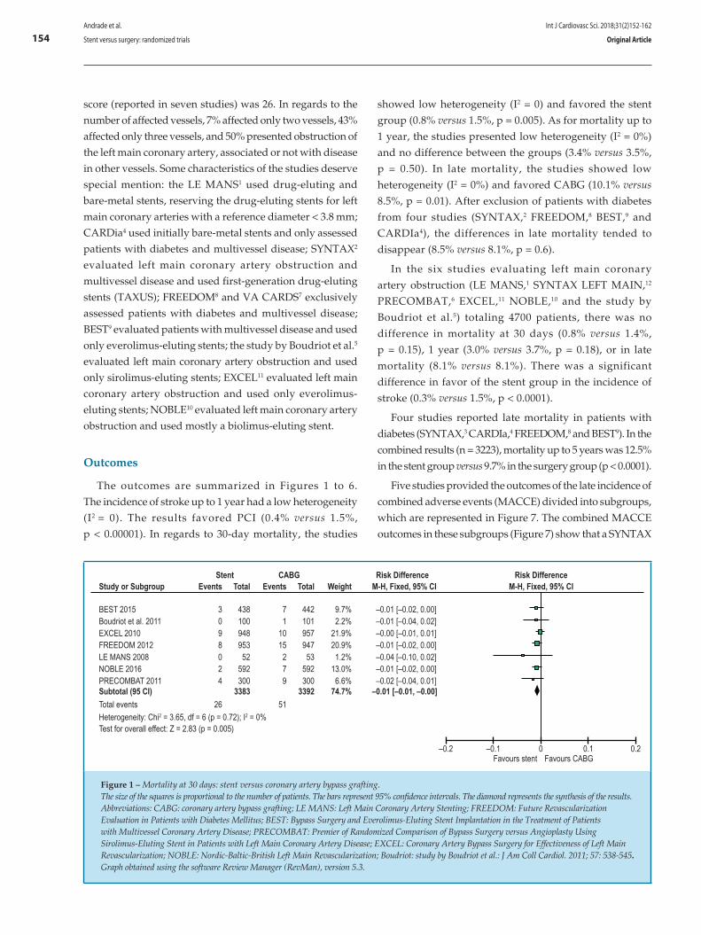
154
Study or SubgroupStent
Events Total Events TotalCABG
WeightRisk Difference Risk Difference
M-H, Fixed, 95% CI M-H, Fixed, 95% CI
–0.2 –0.1 0.20 0.1Favours stent Favours CABG
Heterogeneity: Chi2 = 3.65, df = 6 (p = 0.72); I2 = 0%Test for overall effect: Z = 2.83 (p = 0.005)
Total events 26 51
BEST 2015 3 438 7 442 9.7% –0.01 [–0.02, 0.00]Boudriot et al. 2011 0 100 1 101 2.2% –0.01 [–0.04, 0.02]EXCEL 2010 9 948 10 957 21.9% –0.00 [–0.01, 0.01]FREEDOM 2012 8 953 15 947 20.9% –0.01 [–0.02, 0.00]
NOBLE 2016 2 592 7 592 13.0% –0.01 [–0.02, 0.00]
Subtotal (95 CI) 3383 74.7%3392 –0.01 [–0.01, –0.00]PRECOMBAT 2011 4 300 9 300 6.6% –0.02 [–0.04, 0.01]
LE MANS 2008 0 52 2 53 1.2% –0.04 [–0.10, 0.02]
Figure 1 – Mortality at 30 days: stent versus coronary artery bypass grafting.The size of the squares is proportional to the number of patients. The bars represent 95% confidence intervals. The diamond represents the synthesis of the results.Abbreviations: CABG: coronary artery bypass grafting; LE MANS: Left Main Coronary Artery Stenting; FREEDOM: Future Revascularization Evaluation in Patients with Diabetes Mellitus; BEST: Bypass Surgery and Everolimus-Eluting Stent Implantation in the Treatment of Patients with Multivessel Coronary Artery Disease; PRECOMBAT: Premier of Randomized Comparison of Bypass Surgery versus Angioplasty Using Sirolimus-Eluting Stent in Patients with Left Main Coronary Artery Disease; EXCEL: Coronary Artery Bypass Surgery for Effectiveness of Left Main Revascularization; NOBLE: Nordic-Baltic-British Left Main Revascularization; Boudriot: study by Boudriot et al.: J Am Coll Cardiol. 2011; 57: 538-545. Graph obtained using the software Review Manager (RevMan), version 5.3.
Andrade et al.
Stent versus surgery: randomized trials
Int J Cardiovasc Sci. 2018;31(2)152-162
Original Article
score (reported in seven studies) was 26. In regards to the number of affected vessels, 7% affected only two vessels, 43% affected only three vessels, and 50% presented obstruction of the left main coronary artery, associated or not with disease in other vessels. Some characteristics of the studies deserve special mention: the LE MANS1 used drug-eluting and bare-metal stents, reserving the drug-eluting stents for left main coronary arteries with a reference diameter < 3.8 mm; CARDia4 used initially bare-metal stents and only assessed patients with diabetes and multivessel disease; SYNTAX2 evaluated left main coronary artery obstruction and multivessel disease and used first-generation drug-eluting stents (TAXUS); FREEDOM8 and VA CARDS7 exclusively assessed patients with diabetes and multivessel disease; BEST9 evaluated patients with multivessel disease and used only everolimus-eluting stents; the study by Boudriot et al.5 evaluated left main coronary artery obstruction and used only sirolimus-eluting stents; EXCEL11 evaluated left main coronary artery obstruction and used only everolimus-eluting stents; NOBLE10 evaluated left main coronary artery obstruction and used mostly a biolimus-eluting stent.
Outcomes
The outcomes are summarized in Figures 1 to 6. The incidence of stroke up to 1 year had a low heterogeneity (I2 = 0). The results favored PCI (0.4% versus 1.5%, p < 0.00001). In regards to 30-day mortality, the studies
showed low heterogeneity (I2 = 0) and favored the stent group (0.8% versus 1.5%, p = 0.005). As for mortality up to 1 year, the studies presented low heterogeneity (I2 = 0%) and no difference between the groups (3.4% versus 3.5%, p = 0.50). In late mortality, the studies showed low heterogeneity (I2 = 0%) and favored CABG (10.1% versus 8.5%, p = 0.01). After exclusion of patients with diabetes from four studies (SYNTAX,2 FREEDOM,8 BEST,9 and CARDIa4), the differences in late mortality tended to disappear (8.5% versus 8.1%, p = 0.6).
In the six studies evaluating left main coronary artery obstruction (LE MANS,1 SYNTAX LEFT MAIN,12 PRECOMBAT,6 EXCEL,11 NOBLE,10 and the study by Boudriot et al.5) totaling 4700 patients, there was no difference in mortality at 30 days (0.8% versus 1.4%, p = 0.15), 1 year (3.0% versus 3.7%, p = 0.18), or in late mortality (8.1% versus 8.1%). There was a significant difference in favor of the stent group in the incidence of stroke (0.3% versus 1.5%, p < 0.0001).
Four studies reported late mortality in patients with diabetes (SYNTAX,3 CARDIa,4 FREEDOM,8 and BEST9). In the combined results (n = 3223), mortality up to 5 years was 12.5% in the stent group versus 9.7% in the surgery group (p < 0.0001).
Five studies provided the outcomes of the late incidence of combined adverse events (MACCE) divided into subgroups, which are represented in Figure 7. The combined MACCE outcomes in these subgroups (Figure 7) show that a SYNTAX

155
–0.2 –0.1 0.20 0.1Favours stent Favours CABG
Heterogeneity: Chi2 = 7.31, df = 8 (p = 0.50); I2 = 0%Test for overall effect: Z = 0.25 (p = 0.080)
Total events 143 145Subtotal (95% CI) 4191 70.3%4138 –0.00 [–0.01, 0.01]
Boudriot et al. 2011 2 100 5 101 1.7% –0.03 [–0.08, 0.02]CARDIa 2010 8 254 8 246 4.2% –0.00 [–0.03, 0.03]EXCEL 2010 38 948 38 948 16.0% –0.00 [–0.02, 0.02]FREEDOM 2012 32 953 30 953 16.1% –0.00 [–0.01, 0.02]
NOBLE 2016 9 592 17 592 10.0% –0.01 [–0.03, 0.00]PRECOMBAT 2011 6 300 8 300 5.1% –0.01 [–0.03, 0.02]SYNTAX LM 2009 39 891 30 849 14.7% 0.01 [–0.01, 0.03]VA-CARDIS 2013 8 101 5 97 1.7% 0.03 [–0.04, 0.10]
LE MANS 2008 1 52 4 52 0.9% –0.06 [–0.14, 0.02]
Study or SubgroupStent
Events Total WeightRisk Difference Risk Difference
M-H, Fixed, 95% CICABG
Events Total M-H, Fixed, 95% CI
Figure 2 – Mortality at 1 year: stent versus coronary artery bypass grafting.The size of the squares is proportional to the number of patients. The bars represent 95% confidence intervals. The diamond represents the synthesis of the results.Abbreviations: CABG: coronary artery bypass grafting; SYNTAX: Synergy between PCI with Taxus and Cardiac Surgery; CARDia: Coronary Artery Revascularization in Diabetes; LE MANS: Left Main Coronary Artery Stenting; FREEDOM: Future Revascularization Evaluation in Patients with Diabetes Mellitus; VA CARDS: Coronary Artery Revascularization in Diabetes; BEST: Bypass Surgery and Everolimus-Eluting Stent Implantation in the Treatment of Patients with Multivessel Coronary Artery Disease; NOBLE: Nordic-Baltic-British Left Main Revascularization; Boudriot: study by Boudriot et al.: J Am Coll Cardiol. 2011; 57: 538-545. Graph obtained using the software Review Manager (RevMan), version 5.3.
–0.2 –0.1 0.20 0.1Favours stent Favours CABG
Heterogeneity: Chi2 = 6.85, df = 7 (p = 0.44); I2 = 0%Test for overall effect: Z = 2.58 (p = 0.010)
Total events 435 353Subtotal (95% CI) 4292 70.9%4175 0.02 [0.00, 0.03]
BEST 2015 28 437 22 440 7.3% 0.01 [–0.02, 0.04]CARDIa 2010 35 254 31 248 4.2% 0.01 [–0.05, 0.07]EXCEL 2010 71 913 53 903 15.2% 0.02 [–0.00, 0.04]FREEDOM 2012 114 894 83 859 14.7% 0.03 [0.00, 0.06]
NOBLE 2016 36 592 33 592 9.9% 0.01 [–0.02, 0.03]PRECOMBAT 2011 17 279 23 275 4.6% –0.02 [–0.07, 0.02]SYNTAX LM 2009 123 871 92 805 14.0% 0.03 [–0.00, 0.06]
LE MANS 2008 11 52 16 53 0.9% –0.09 [–0.26, 0.08]
Study or SubgroupStent
Events Total WeightRisk Difference Risk Difference
M-H, Fixed, 95% CICABG
Events Total M-H, Fixed, 95% CI
Figure 3 – Late mortality: stent versus coronary artery bypass grafting.The size of the boxes is proportional to the number of patients. The bars represent 95% confidence intervals. The diamond represents the synthesis of the results.Abbreviations: CABG: coronary artery bypass grafting; SYNTAX: Synergy between PCI with Taxus and Cardiac Surgery; CARDia: Coronary Artery Revascularization in Diabetes; LE MANS: Left Main Coronary Artery Stenting; FREEDOM: Future Revascularization Evaluation in Patients with Diabetes Mellitus; VA CARDS: Coronary Artery Revascularization in Diabetes; BEST: Bypass Surgery and Everolimus-Eluting Stent Implantation in the Treatment of Patients with Multivessel Coronary Artery Disease; PRECOMBAT: Premier of Randomized Comparison of Bypass Surgery versus Angioplasty Using Sirolimus-Eluting Stent in Patients with Left Main Coronary Artery Disease; EXCEL: Coronary Artery Bypass Surgery for Effectiveness of Left Main Revascularization; NOBLE: Nordic-Baltic-British Left Main Revascularization Study; Boudriot: study by Boudriot et al.: J Am Coll Cardiol. 2011; 57: 538-545. Graph obtained using the software Review Manager (RevMan), version 5.3.
Andrade et al.
Stent versus surgery: randomized trials
Int J Cardiovasc Sci. 2018;31(2)152-162
Original Article
score in the upper tertile and the occurrence of diabetes had
a strong negative influence on the PCI outcome. In patients
in the lower SYNTAX tertile and in those without diabetes,
there was no significant difference in terms of MACCE
between the CABG and PCI groups. The elderly condition
and the female gender contributed to the difference in results
but to a lesser degree. An ejection fraction < 50% did not
contribute significantly to the difference in results.

156
–0.2 –0.1 0.20 0.1Favours stent Favours CABG
Heterogeneity: Chi2 = 7.53, df = 8 (p = 0.48); I2 = 0%Test for overall effect: Z = 5.37 (p = 0.00001)
Total events 16 65Subtotal (95% CI) 4204 70.5%4184 –0.01 [–0.02, –0.01]
Boudriot et al. 2011 0 100 2 101 1.7% –0.02 [–0.05, 0.01]CARDIa 2010 1 254 7 248 4.2% –0.02 [–0.05, –0.05]EXCEL 2010 5 948 12 948 15.9% –0.01 [–0.02, 0.00]FREEDOM 2012 3 953 16 947 16.0% –0.01 [–0.02, –0.00]
NOBLE 2016 1 593 4 592 10.0% –0.01 [–0.01, 0.00]PRECOMBAT 2011 0 300 2 300 5.0% –0.01 [–0.02, 0.00]SYNTAX LM 2009 6 903 19 897 15.1% –0.01 [–0.03, –0.00]VA-CARDIS 2013 0 101 1 97 1.7% –0.01 [–0.04, 0.02]
LE MANS 2008 0 52 2 54 0.9% –0.04 [–0.10, 0.02]
Study or SubgroupStent
Events Total WeightRisk Difference Risk Difference
M-H, Fixed, 95% CICABG
Events Total M-H, Fixed, 95% CI
Figure 4 – Stroke: stent versus coronary artery bypass grafting.The size of the boxes is proportional to the number of patients. The bars represent 95% confidence intervals. The diamond represents the synthesis of the results.Abbreviations: CABG: coronary artery bypass grafting; SYNTAX: Synergy between PCI with Taxus and Cardiac Surgery; CARDia: Coronary Artery Revascularization in Diabetes; LE MANS: Left Main Coronary Artery Stenting; FREEDOM: Future Revascularization Evaluation in Patients with Diabetes Mellitus; VA CARDS: Coronary Artery Revascularization in Diabetes; BEST: Bypass Surgery and Everolimus-Eluting Stent Implantation in the Treatment of Patients with Multivessel Coronary Artery Disease; PRECOMBAT: Premier of Randomized Comparison of Bypass Surgery versus Angioplasty Using Sirolimus-Eluting Stent in Patients with Left Main Coronary Artery Disease; EXCEL: Coronary Artery Bypass Surgery for Effectiveness of Left Main Revascularization; NOBLE: Nordic-Baltic-British Left Main Revascularization Study; Boudriot: study by Boudriot et al.: J Am Coll Cardiol. 2011; 57: 538-545. Graph obtained using the software Review Manager (RevMan), version 5.3.
–0.2 –0.1 0.20 0.1Favours stent Favours CABG
Heterogeneity: Chi2 = 3.60, df = 5 (p = 0.61); I2 = 0%Test for overall effect: Z = 1.33 (p = 0.18)
Total events 71 87Total (95% CI) 2349 100.0%2341 –0.01 [–0.02, 0.00]
Boudriot et al. 2011 2 100 5 101 4.3% –0.03 [–0.08, 0.02]EXCEL 2010 38 948 38 948 40.4% 0.00 [–0.02, 0.02]LE MANS 2008 1 52 4 52 2.2% –0.06 [–0.14, 0.02]NOBLE 2016 9 592 17 592 25.2% –0.01 [–0.03, 0.00]
SYNTAX LM 2013 15 357 15 348 15.0% –0.00 [–0.03, 0.03]PRECOMBAT 2011 6 300 8 300 12.8% –0.01 [–0.03, 0.02]
Study or SubgroupStent
Events Total WeightRisk Difference Risk Difference
M-H, Fixed, 95% CICABG
Events Total M-H, Fixed, 95% CI
Figure 5 – Studies including the left main coronary artery. Mortality at 1 year: Stent versus coronary artery bypass grafting.The size of the boxes is proportional to the number of patients. The bars represent 95% confidence intervals. The diamond represents the synthesis of the results.Abbreviations: CABG: coronary artery bypass grafting; LE MANS: Left Main Coronary Artery Stenting; SYNTAX LM: left main coronary artery subgroup of SYNTAX (Synergy between PCI with Taxus and Cardiac Surgery); PRECOMBAT: Premier of Randomized Comparison of Bypass Surgery versus Angioplasty Using Sirolimus-Eluting Stent in Patients with Left Main Coronary Artery Disease; EXCEL: Coronary Artery Bypass Surgery for Effectiveness of Left Main Revascularization; NOBLE: Nordic-Baltic-British Left Main Revascularization; Boudriot: study by Boudriot et al.: J Am Coll Cardiol. 2011; 57: 538-545. Graph obtained using the software Review Manager (RevMan), version 5.3.
Andrade et al.
Stent versus surgery: randomized trials
Int J Cardiovasc Sci. 2018;31(2)152-162
Original Article
Discussion
Several systematic reviews, collaborative studies, and
meta-analyses13-18 have been published comparing PCI and
CABG. The most important ones included studies of the
balloon and bare-metal stent era or left out important recent
studies.14-16 The main differential of the present meta-analysis
is the large number of included studies and patients and
the fact that it is up-to-date and included only trials of the
drug-eluting stent era.

157
–1 –0.5 10 0.5Favours stent Favours CABG
Heterogeneity: Chi2 = 5.99, df = 4 (p = 0.20); I2 = 33%Test for overall effect: Z = 0.01 (p = 1.00)
Total events 177 87Total (95% CI) 2193 100.0%2171 0.00 [–0.02, 0.02]
EXCEL 2010 71 913 53 903 41.6% 0.02 [–0.00, 0.04]LE MANS 2008 11 52 16 53 2.4% –0.09 [–0.26, 0.08]NOBLE 2016 36 592 33 592 27.1% 0.01 [–0.02, 0.03]PRECOMBAT 2011 17 279 23 275 12.7% –0.02 [–0.07, 0.02]SYNTAX LM 2013 42 357 50 348 16.2% –0.03 [–0.08, 0.02]
Study or SubgroupStent
Events Total WeightRisk Difference Risk Difference
M-H, Fixed, 95% CICABG
Events Total M-H, Fixed, 95% CI
Figure 6 – Studies including the left main coronary artery. Late mortality: stent versus coronary artery bypass grafting.The size of the boxes is proportional to the number of patients in the trial. The bars represent 95% confidence intervals. The diamond represents the synthesis of the results.Abbreviations: CABG: coronary artery bypass grafting; LE MANS: Left Main Coronary Artery Stenting; SYNTAX LM: left main coronary artery subgroup of SYNTAX (Synergy between PCI with Taxus and Cardiac Surgery); PRECOMBAT: Premier of Randomized Comparison of Bypass Surgery versus Angioplasty Using Sirolimus-Eluting Stent in Patients with Left Main Coronary Artery Disease; EXCEL - Coronary Artery Bypass Surgery for Effectiveness of Left Main Revascularization; NOBLE: Nordic-Baltic-British Left Main Revascularization; Boudriot: study by Boudriot et al.: J Am Coll Cardiol. 2011; 57: 538-545. Graph obtained using the software Review Manager (RevMan), version 5.3.
Andrade et al.
Stent versus surgery: randomized trials
Int J Cardiovasc Sci. 2018;31(2)152-162
Original Article
In the evaluation of the results, it is important to highlight
the superiority of PCI in the mortality at 30 days. This is
in line with a prior systematic review17 and with the New
York registry.19 The difference is obviously not applicable
to patients with lesions of high angiographic complexity,
as seen in the analysis of the survival curves of aggregated
results from SYNTAX LM and PRECOMBAT.20 A greater
incidence of stroke in the surgical group had already been
suggested in prior systematic reviews,14,17 and in the light of
the data presented here, this fact becomes indisputable. It
is worth mentioning a reduced incidence of stroke in more
recent studies, reflecting a greater care taken by surgeons
while manipulating the aorta. The similarity of the mortality
results at 1 year is aligned with a prior systematic review,
which included studies of the era prior to drug-eluting
stents.17 The difference favoring surgery in regards to late
mortality is consistent with another meta-analysis16 and also
with a recently published collaborative study.21 It should be
emphasized that the difference found was due to the large
number of patients with diabetes in the studies of the drug-
eluting stent era, which disappeared in the aggregated results
when these studies were excluded. These data confirm those of
the collaborative study by Hlatki et al.15, which demonstrated
a lower overall mortality at 5 years with surgery, but no
difference among nondiabetic patients. We should emphasize
that the study by Hlatki et al.15 included trials of the balloon
era in which two-vessel disease predominated, while in the
present review there was a predominance of three-vessel
disease and obstruction of the left main coronary artery.
In regards to the results of obstruction of the left main
coronary artery, it is important to remember that the group
of patients with this type of obstruction comprised for a long
time a forbidden territory for angioplasty. LE MANS was the
first randomized study that attempted to compare stent and
surgery in left main coronary artery obstruction, with results
similar or even superior to those with PCI. However, this
was a small study (105 patients), which has been criticized
for not having used grafting of internal thoracic artery in
approximately 25% of the cases. After that, emerged the
results of the SYNTAX12 subgroup with left main coronary
artery obstruction and of the PRECOMBAT trial and the
study by Boudriot et al.5, which led to the improvement of
the recommendations of PCI in left main artery obstruction.
Despite that, the American guidelines only changed the
recommendation to IIA in patients with a low SYNTAX
score and IIB in patients with intermediate SYNTAX scores.18
We should emphasize that such recommendations are
restricted to patients with a high surgical risk. In the present
study, which combined the results of six studies with 4700
patients, the outcomes of PCI with drug-eluting stents were
equal or even greater than those with CABG. In light of these
evidence and recent results of NOBLE and EXCEL, we believe
that the American and Brazilian guidelines22,23 may be soon
modified to improve the classification of PCI with drug-eluting
stents, mainly in left main coronary artery obstruction.
In relation to the results in patients with diabetes, it is
important to remember that the evidence contrary to PCI
in diabetes has its origin in the balloon era, from occasional

158
–0.2 –0.1 0.20 0.1Favours stent Favours CABG
Heterogeneity: Chi2 = 3.63, df = 4 (p = 0.46); I2 = 0%Test for overall effect: Z = 4.90 (p = 0.00001)
Total events 419 301Subtotal (95% CI) 1714 14.1%1708 0.00 [–0.02, 0.02]
BEST 2015 34 177 17 186 1.5% 0.10 [0.03, 0.17]EXCEL 2016 51 256 47 259 2.1% 0.02 [–0.05, 0.09]FREEDOM 2012 257 953 179 947 7.8% 0.08 [0.04, 0.12]PRECOMBAT 2011 23 102 15 90 0.8% 0.06 [–0.05, 0.17]SYNTAX LM 2009 54 226 43 226 1.9% 0.05 [–0.03, 0.12]
Study or Subgroup1.7.1 Patients with diabetes
Heterogeneity: Chi2 = 5.24, df = 4 (p = 0.26); I2 = 24%Test for overall effect: Z = 0.54 (p = 0.59)
Total events 207 204Subtotal (95% CI) 1338 11.2%1395 0.00 [–0.02, 0.03]
BEST 2015 24 185 23 186 1.5% 0.01 [–0.06, 0.07]EXCEL 2016 28 294 46 364 2.7% –0.03 [–0.08, 0.02]FREEDOM 2012 75 329 58 340 2.8% 0.06 [–0.00, 0.12]PRECOMBAT 2011 16 128 13 104 0.9% 0.00 [–0.09, 0.09]SYNTAX LM 2009 64 402 64 401 3.3% –0.00 [–0.03, 0.12]
1.7.9 SYNTAX lower tertile
Heterogeneity: Chi2 = 2.11, df = 4 (p = 0.72); I2 = 0%Test for overall effect: Z = 3.63 (p = 0.0003)
Total events 196 148Subtotal (95% CI) 816 7.0%871 0.07 [0.03, 0.11]
BEST 2015 13 66 10 79 0.6% 0.07 [–0.05, 0.19]EXCEL 2016 37 220 30 217 1.8% 0.03 [–0.04, 0.10]FREEDOM 2012 56 182 41 192 1.5% 0.09 [0.01, 0.18]PRECOMBAT 2011 14 58 13 68 0.5% 0.05 [–0.09, 0.19]SYNTAX LM 2009 76 290 54 315 2.5% 0.09 [0.03, 0.16]
1.7.10 SYNTAX upper tertile
Heterogeneity: Chi2 = 0.73, df = 3 (p = 0.87); I2 = 0%Test for overall effect: Z = 2.56 (p = 0.01)
Total events 145 114Subtotal (95% CI) 688 5.8%710 0.05 [0.01, 0.09]
BEST 2015 22 134 13 137 1.1% 0.07 [–0.01, 0.15]EXCEL 2016 43 226 28 215 1.8% 0.06 [–0.01, 0.13]FREEDOM 2012 67 256 61 289 2.2% 0.05 [–0.02, 0.12]PRECOMBAT 2011 13 72 12 69 0.6% 0.01 [–0.12, 0.13]
1.7.6 Female gender
Heterogeneity: Chi2 = 4.47, df = 2 (p = 0.11); I2 = 55%Test for overall effect: Z = 1.28 (p = 0.20)
Total events 40 27Subtotal (95% CI) 149 1.2%143 0.06 [–0.03, 0.05]
BEST 2015 7 17 4 17 0.1% 0.18 [–0.13, 0.49]EXCEL 2016 20 111 20 115 0.9% 0.01 [–0.09, 0.11]FREEDOM 2012 13 21 3 11 0.1% 0.35 [0.01, 0.68]
1.7.7 Reduced ejection fraction
Heterogeneity: Chi2 = 3.87, df = 2 (p = 0.14); I2 = 48%Test for overall effect: Z = 2.09 (p = 0.04)
Total events 312 269Subtotal (95% CI) 1823 15.1%1840 0.03 [0.00, 0.05]
BEST 2015 60 421 43 425 3.5% 0.04 [–0.00, 0.09]EXCEL 2016 109 782 111 786 6.5% –0.00 [–0.04, 0.03]FREEDOM 2012 143 620 115 629 5.2% 0.05 [0.00, 0.09]
1.7.8 Non-reduced ejection fraction
Heterogeneity: Chi2 = 1.83, df = 3 (p = 0.61); I2 = 0%Test for overall effect: Z = 1.42 (p = 0.16)
Total events 281 252Subtotal (95% CI) 1830 14.1%1842 0.02 [–0.01, 0.04]
BEST 2015 33 261 30 256 2.1% 0.01 [–0.05, 0.07]EXCEL 2016 86 692 88 707 5.8% –0.00 [–0.03, 0.03]PRECOMBAT 2011 29 198 27 210 1.7% 0.02 [–0.05, 0.08]SYNTAX LM 2009 133 679 107 669 5.6% 0.04 [–0.00, 0.08]
1.7.2 Patients without diabetes
Heterogeneity: Chi2 = 13.75, df = 3 (p = 0.003); I2 = 78%Test for overall effect: Z = 3.20 (p = 0.001)
Total events 366 289Subtotal (95% CI) 1952 16.1%1956 0.04 [0.01, 0.06]
BEST 2015 45 304 34 325 2.6% 0.04 [–0.01, 0.10]EXCEL 2016 94 722 107 742 6.0% –0.01 [–0.05, 0.02]FREEDOM 2012 188 698 118 658 5.6% 0.09 [0.05, 0.13]PRECOMBAT 2011 39 228 30 231 1.9% 0.04 [–0.02, 0.11]
1.7.5 Male gender
Heterogeneity: Chi2 = 6.69, df = 2 (p = 0.04); I2 = 70%Test for overall effect: Z = 0.41 (p = 0.68)
Total events 102 95Subtotal (95% CI) 862 7.1%846 0.01 [–0.02, 0.04]
BEST 2015 26 209 17 190 1.6% 0.03 [–0.03, 0.10]EXCEL 2016 55 482 68 485 4.0% –0.03 [–0.07, 0.02]PRECOMBAT 2011 21 171 10 171 1.4% 0.06 [0.00, 0.12]
1.7.4 Nonelderly
Heterogeneity: Chi2 = 1.74, df = 2 (p = 0.42); I2 = 0%Test for overall effect: Z = 1.91 (p = 0.06)
Total events 138 127Subtotal (95% CI) 824 7.2%967 0.03 [–0.00, 0.07]
BEST 2015 41 229 30 252 2.0% 0.06 [–0.00, 0.12]EXCEL 2016 82 466 67 463 3.8% 0.03 [–0.02, 0.08]PRECOMBAT 2011 15 129 30 252 1.4% –0.00 [–0.07, 0.07]
1.7.3 Elderly
StentEvents Total Weight
Risk Difference Risk DifferenceM-H, Fixed, 95% CI
CABGEvents Total M-H, Fixed, 95% CI
Figure 7 – Combined adverse events outcomes (major adverse cardiovascular and cerebrovascular disease events, MACCE) in subgroups in five studies.The size of each box is proportional to the number of patients in the subgroup. The bar is equal to the confidence interval. The diamonds represent the synthesis of the results. In the SYNTAX, FREEDOM, and EXCEL, the combined events were death, acute myocardial infarction (AMI), and stroke. In the remaining studies, they were death, AMI, and new revascularization.Abbreviations: CABG: coronary artery bypass grafting; SYNTAX: Synergy between PCI with Taxus and Cardiac Surgery; FREEDOM: Future Revascularization Evaluation in Patients with Diabetes Mellitus; PRECOMBAT: Premier of Randomized Comparison of Bypass Surgery versus Angioplasty Using Sirolimus-Eluting Stent in Patients with Left Main Coronary Artery Disease; EXCEL: Coronary Artery Bypass Surgery for Effectiveness of Left Main Revascularization. Graph obtained using the software Review Manager (RevMan), version 5.3.
Andrade et al.
Stent versus surgery: randomized trials
Int J Cardiovasc Sci. 2018;31(2)152-162
Original Article

159
Andrade et al.
Stent versus surgery: randomized trials
Int J Cardiovasc Sci. 2018;31(2)152-162
Original Article
Table 1 – Overview of randomized studies comparing percutaneous coronary intervention (PCI) and coronary artery bypass grafting (CABG) in the era of drug-eluting stents
Study OriginYear of
publication
Number
of
patients
Disease
extension
Patients
with
diabetes(%)
Unstable
angina(%)
Mean
ejection
fraction (%)
Type of
stent
Follow-up
Maximum
LEMANS1 Poland 2008 105 LMCA 25 32 53 ± 11BMS and
DES10
SYNTAX2 International 2009 1800LMCA and three-vessel
disease35 28 ND ‡ SF 5
CARDia4United
Kingdom2010 510
Two- and three-vessel
disease100 22 59 ± 14
BMS and DES
5
Boudriot et al.5 Germany 2011 201 LMCA 30 ND ND DES 1
PRECOMBAT6 South Korea 2011 600 LMCA 42 45 60 ± 9 DES 5
FREEDOM8 International 2012 1900Two- and
three-vessel disease
100 30 65 ± 12 DES 5
Va-Cards7 USA 2013 198Two- and
three-vessel disease
100 ND ND† DES 2
BEST9 South Korea 2015 880Two- and
three-vessel disease
45 42 59 ± 9 DES 5
EXCEL11 International 2016 1905 LMCA 25 37 57 ± 10 DES 3
NOBLE10 Europe 2016 982 LMCA 18 18 60 ± 10 DES 5
SYNTAX: Synergy between PCI with Taxus and Cardiac Surgery; CARDia: Coronary Artery Revascularization in Diabetes; LE MANS: Left Main Coronary Artery Stenting; FREEDOM: Future Revascularization Evaluation in Patients with Diabetes Mellitus; VA CARDS: Coronary Artery Revascularization in Diabetes; BEST: Bypass Surgery and Everolimus-Eluting Stent Implantation in the Treatment of Patients with Multivessel Coronary Artery Disease; PRECOMBAT: Premier of Randomized Comparison of Bypass Surgery versus Angioplasty Using Sirolimus-Eluting Stent in Patients with Left Main Coronary Artery Disease; EXCEL: Coronary Artery Bypass Surgery for Effectiveness of Left Main Revascularization; NOBLE: Nordic-Baltic-British Left Main Revascularization Study; Boudriot - study by Boudriot et al.: J Am Coll Cardiol. 2011; 57: 538-545; DES: drug-eluting stent; BMS: bare-metal stent; LMCA: left main coronary artery; USA: United States. †: 37% with ejection fraction < 55%; ‡: 3% with ejection fraction < 30%.
findings of the BARI study. The investigators of this study evaluated a subgroup of 343 patients with diabetes and found a late mortality of 34.5% for PCI with balloon and 19.4% for surgery (p = 0.03). In the era of conventional stent, studies such as SoS and ARTS have confirmed a trend toward greater mortality with PCI in patients with diabetes, even though it did not reach statistical significance. From there onwards, the presence of diabetes has become a criterion for preferential indication of surgery as a method for myocardial revascularization. There used to be a hypothesis that drug-eluting stents would eliminate the differences in mortality found in these studies, but the results presented here demonstrate that the difference in
mortality between PCI and surgery in patients with diabetes
continues in the era of drug-eluting stents. However, it
should be noted a reduced risk difference compared with
previous studies (3.5% risk difference as opposed to 7.3% in
the study by Hclatki et al. and 15.1% in BARI). This should
raise the hypothesis that it is not the metabolic disorder in
itself, but the complexity of the lesions which is the factor
leading to a higher mortality of angioplasty in patients with
diabetes. This question could perhaps be explained by a
meta-analysis of individual patient data involving a large
number of studies. In this sense, a recent collaborative study
categorizing the results of three studies (SYNTAX, BEST, and
PRECOMBAT) corroborated this hypothesis.24

160Andrade et al.
Stent versus surgery: randomized trials
Int J Cardiovasc Sci. 2018;31(2)152-162
Original Article
The MACCE outcomes in subgroups (Figure 7) in the
present study demonstrate that the SYNTAX score in the
upper tertile strongly and negatively influenced the PCI
outcomes, similarly to the presence of diabetes. The elderly
condition and the female gender had a small influence on
the results; an ejection fraction < 50% did not negatively
influence the PCI outcomes, but an ejection fraction < 35%
had a greater impact, even though it had no statistical
significance. These results are in agreement with those of
the collaborative study by Cavalcante et al.20 In that study,
by aggregating the results of the SYNTAX LEFT MAIN and
PRECOMBAT for combined adverse events (death, stroke,
AMI, and new revascularization), a high SYNTAX score, like
diabetes, had an important role. The female gender, elderly
condition, ejection fraction < 50%, and renal insufficiency did
not negatively affect the results compared with PCI. This same
study showed that the subgroups most significantly affecting
PCI-associated mortality outcomes in obstruction of the left
main coronary artery were those with two- or three-vessel
disease and with a SYNTAX score > 32. Diabetes had a less
important role, possibly related to the fact that only patients
with left main coronary artery obstruction were evaluated.
Study limitations
This study has important limitations. Because the
meta-analysis included published data rather than individual
patient data, we were unable to analyze the mortality
outcomes in subgroups, except in those with diabetes.
Additionally, the percentages had to be processed as absolute
numbers, which may deserve criticism. The results apply only
to patients in whom revascularization is possible by both
methods and without a high surgical risk or history of prior
surgical revascularization, and with the procedures carried
out in institutions of excellence.
Conclusion
In combined results of randomized studies involving
multivessel disease or obstruction of the left main coronary
artery, PCI with drug-eluting stent was associated with
a lower incidence of stroke, lower mortality at 30 days,
and increased late mortality when compared with
CABG. There was no difference in early, intermediate,
or late mortality in the subgroup with left main coronary
obstruction, but there was a difference in favor of PCI in
regards to the incidence of stroke. The presence of diabetes
and a high SYNTAX score were factors most strongly and
negatively impacting PCI outcomes in terms of combined
adverse results.
Author contributions
Conception and design of the research: Andrade PJN. Acquisition of data: Andrade PJN, Falcão JLAA, Andrade AT, Falcão BAA. Analysis and interpretation of the data: Andrade PJN, Rocha HAL, Falcão JLAA, Andrade AT. Statistical analysis: Rocha HAL. Writing of the manuscript: Andrade PJN, Rocha HAL, Falcão JLAA, Falcão BAA. Critical revision of the manuscript for intellectual content: Andrade PJN, Falcão BAA. Supervision / as the major investigador: Andrade PJN.
Potential Conflict of Interest
No potential conflict of interest relevant to this article was reported.
Sources of Funding
There were no external funding sources for this study.
Study Association
This study is not associated with any thesis or dissertation work.
Ethics approval and consent to participate
This article does not contain any studies with human participants or animals performed by any of the authors.

161
1. Buszman PE, Kiesz SR, Bochenck A, Peszek-Przrya E, Szkiobka, Debwski M, et al. Acute and late outcomes of unprotected left main stenting. Comparison with surgical revascularization. J Am Coll Cardiol. 2008;51(5):538-45. doi: 10.1016/j.jacc.2007.09.054.
2. Serruys PW, Morice MC, Kappetei AP, Colombo A, Holmes DR, Mack MJ, et al; SYNTAX Investigators. Percutaneous coronary intervention versus coronary-artery bypass grafting for severe coronary artery-disease. N Eng J Med. 2009;360(10):961-72. doi: 10.1056/NEJMoa0804626. Erratum in: N Engl J Med. 2013;368(6):584.
3. Mohr FW, Morice MC, Kappetein P, Feldman TE, Ståhle E, Colombo A, et al. Coronary artery bypass graft surgery versus percutaneous coronary intervention in patients with three-vessel disease and left main coronary disease: 5-year follow-up of the randomized, clinical SYNTAX trial. Lancet. 2013;381(9867):629-38. doi: 10.1016/S0140-6736(13)60141-5.
4. Kapur A, Hall RJ, Macik I, Qupesh AC, Butts J, Beider B, et al. Randomized comparison of percutaneous coronary intervention with coronary artery bypass grafting in diabetic patients: a one year results of the CARDia (coronary artery revascularization in diabetics) trial. J Am Coll Cardiol. 2010;55(5):432-40. doi: 10.1016/j.jacc.2009.10.014.
5. Boudriot E, Thiele TW, Liebetrau C, Boeckstegers P, Pohl T, Reichart B, et al. Randomized comparison of percutaneous coronary intervention with Sirolimus eluting stents versus coronary artery bypass grafting in unprotected in unprotected left main stenosis. J Am Coll Cardiol. 2011;57(5):538-45. doi: 10.1016/j.jacc.2010.09.038. Erratum in: J Am Coll Cardiol. 2011;57(17):1792.
6. Park SJ, Kim YH, Park DW, Yun SC, Ahn JM, Song HG, et al. Randomized trial of stents versus bypass surgery for left main coronary artey disease. N Engl J Med. 2011;364(18):1718-27. doi: 10.1056/NEJMoa1100452.
7. Kamalesh M, Shap TG, Tamg XC, Shunk K, Ward HB, Walsh J, et al. Percutaneous coronary intervention versus coronary artery bypass surgery in United States veterans with diabetes. J Am Coll Cardiol. 2013;61(8):808-16. doi: 10.1016/j.jacc.2012.11.044.
8. Farkouh ME, Dornanski M, Sleeper LA, Siami FS, Dangas G, Mack M, et al; FREEDOM Trial Investigators. Strategies for multivessel revascularization in diabetes. N Engl J Med. 2012;367(25):2375-84. doi: 10.1056/NEJMoa1211585.
9. Park SJ, Ahn KM, Kim YH, Park DW, Yun SC, Lee JY, et al: BEST Trial Investigators. Trial of everolimus-eluting stents or bypass surgery for coronary artery disease. N Engl J Med. 2015; 372(13):1204-12. doi: 10.1056/NEJMoa1415447.
10. Makikallio T, Holm NR, Liundsay M, Sprnce MS, Erglis A, Menown IB, et al; NOBLE study investigators. Percutaneous coronary angioplasty versus coronary artery bypass grafting in treatment of unprotected left main stenosis (NOBLE) a prospective, randomized, open-label, non-inferiority trial. Lancet. 2016;388(10061):2743-2752. doi: 10.1016/S0140-6736(16)32052-9.
11. Stone GW, Sabik JF, Serruys PW, Simonton CA, Genereux P, Puskas J, et al; EXCEL Trial Investigators. Everolimus-stenting or bypass surgery for left main coronary artery disease. N Engl J Med. 2016;375(23):2223-35. doi: 10.1056/NEJMoa1610227.
12. Morice MC, Serruys PW, Kappetein AP, Feldman TE, Ståhle E, Colombo A, et al. Five-year outcomes in patients with left main disease treated with either percutaneous coronary intervention or coronary artery bypass grafting in the synergy between percutaneous coronary intervention with taxus and cardiac surgery trial. Circulation. 2014;129(23):2388-94. doi: 10.1161/CIRCULATIONAHA.113.006689.
13. Daemen J, Boersma E, Flather M, Bonth J, Stables R, Rodrigues A, et al. Long-term safety and efficacy of percutaneous coronary intervention with stenting and coronary artery bypass surgery for multivessel coronary artery disease: a meta-analysis with 5-year patient-level data from the ARTS, ERACI-II, MASS-II, and SoS trials. Circulation. 2008;118(11):1146-54. doi: 10.1161/CIRCULATIONAHA.107.752147.
14. Bravata DM, Gienger AL, McDonad KM, Vandana S, Perez MV, Varguese R, et al. Sistematic review: the comparative effectiveness of percutaneous coronary intervention and coronary artery bypass surgery. Ann Intern Med. 2007;147(10):703-6. PMID: 17938385.
15. Hlatky MA, Boothroyd DB, Bravata DM, Buersma E, Bonth E, Brooks MM, et al Coronary angioplasty compared with percutaneous coronary interventions for multivessel diseases: a colaborative analysis of individual patient data from ten randomized trials. Lancet. 2009;373(9670):1190-7. doi: 10.1016/S0140-6736(09)60552-3.
16. Smit Y, Valyen J, Kopennaal H, Edfting F, Kappetein P, Mariani MA. Percutaneous coronary invervention versus coronary artery bypass grafting: a Meta-analysis. J Thorac Cardiovasc Surg. 2015;149(3):831-8.e1-13. doi: 10.1016/j.jtcvs.2014.10.112.
17. Andrade PJ, Medeiros, MM, Andrade AT, Lima AA. Coronary Angioplasty versus CABG: review of randomized trials. Arq Bras Cardiol. 2011;97(3):e60-9. doi: http://dx.doi.org/10.1590/S0066-782X2011001200021
18. Andrade PJ, Silva RP, Lima AA, Andrade AT, Rodrigues Sobrinho CR. Drug-eluting stents versus coronary artery bypass-graft surgery: meta-analysis of prospective clinical trials. Rev Bras Cardiol. 2013;25(3):210-7.
19. Bangalore S, Guo Y, Samadashvili Z, Blecker S, Xu J, Hannan EL. Everolimus-eluting stents or bypass surgery for multivessel coronary disease. N Engl J Med. 2015;372(13):1213-22. doi: 10.1056/NEJMoa1412168.
20. Cavalcante R, Sotomi Y, Lee CW, Ahn JM, Farooq V, Tateishi H. Outcomes after percutaneous coronary intervention and bypass surgery in patients with unprotected left main disease. J Am Coll Cardiol. 2016;68(10):999-1009. doi: 10.1016/j.jacc.2016.06.024.
21. Lee CW, Ahn JM, Cavalcante R, Sotomi Y, Onuma Y. Coronary artery bypass surgery versus drug-eluting stent implantation for left main or multivessel coronary artery disease. A meta-analysis of individual patient data. JACC Cardiovasc Interv. 2016;9(24):2481-2489. doi: 10.1016/j.jcin.2016.10.008.
22. Hillis LD, Smith PK, Anderson JL, Bittl JA, Bridges CR, Byrne JG, et al. Special Articles: 2011 ACCF/AHA Guideline for Coronary Artery Bypass Graft Surgery: executive summary: a report of the American College of Cardiology Foundation/American Heart Association Task Force on Practice Guidelines. Anesth Analg. 2012;114(1):11-45. doi: 10.1213/ANE.0b013e3182407c25.
23. Mattos LA, Lemos Neto PA, Rassi A Jr, Marin-Neto JA, Sousa AG, Devito FS, et al; Sociedade Brasileira de Cardiologia. Diretrizes sobre intervenção percutânea e métodos adjuntos diagnósticos em cardiologia intervencionista (II Edição). Arq Bras Cardiol. 2008;91(6 supl 1):1-58.
24. Cavalcante R, Sotomi Y, Mancone M, Whan Lee C, Ahn JM, Onuma Y, et al. Impact of the SYNTAX scores I and II in patients with diabetes and multivessel coronary disease: a pooled analysis of patient level data from the SYNTAX, PRECOMBAT, and BEST trials. Eur Heart J. 2017;38(25):1969-77. doi: 10.1093/eurheartj/ehx138.
References
Andrade et al.
Stent versus surgery: randomized trials
Int J Cardiovasc Sci. 2018;31(2)152-162
Original Article

162Andrade et al.
Stent versus surgery: randomized trials
Int J Cardiovasc Sci. 2018;31(2)152-162
Original Article
This is an open-access article distributed under the terms of the Creative Commons Attribution License
DOI: 10.5935/2359-4802.20180004
Introduction
Mediastinitis is a severe post-median sternotomy infection, affecting the adjacent deep tissues, with clinical and/or microbiological evidence of retrosternal space involvement, which may be associated with sternal osteomyelitis, with or without instability.1,2 It has an incidence of 0.4% to 5% and high mortality (14% to 47%)3. Even with early diagnosis and treatment, it does not have a good prognosis, especially in the presence of sepsis and other health problems4.
The risk factors associated with mediastinitis are pre-surgical, surgical and post-surgical ones.5
Pre-surgical factors include older age, male gender, malnutrition, obesity, smoking, Diabetes mellitus (DM) and other diseases,5,6 as well as chronic renal failure (CRF) and/or creatinine serum levels > 1.5mg/dL and Left Ventricular Ejection Fraction (LVEF) < 40%.7,8
Surgical risk factors include the type of surgery (elective or urgent), prolonged surgical time, coronary artery bypass grafting (CABG) using bilateral mammary arteries, and intra-aortic balloon insertion9,10. Routine procedures such as hair removal and the time elapsed between hair removal and the surgical incision, in addition to the sterilization of the surgical materials and the number of people in the operating
163International Journal of Cardiovascular Sciences. 2018;31(2)163-172
ORIGINAL ARTICLE
Mailing Address: Isabel Cristina Gomes •Rua José Ferreira da Luz, 44. Postal Code 30750-480, Caiçaras, Belo Horizonte, MG – Brazil.E-mail: [email protected], [email protected]
Factors Associated with Post-Sternotomy Mediastinitis. Case-Control StudyDébora Cristine Gomes Pinto, Antônio Fernandino de Castro Bahia Neto, Flávia Lage Gonçalves, Isabel Cristina Gomes, Eduardo Back Sternick, Alessandra Maciel Almeida, Nulma Souto JentzschFaculdade Ciências Médicas de Minas Gerais, Minas Gerais, MG – Brazil
Manuscript received March 8, 2017, revised manuscript September 20, 2017, accepted September 22, 2017
Abstract
Background: Mediastinitis is a severe post-median sternotomy infection with poor prognosis, even with early diagnosis and treatment.
Objectives: To evaluate the profile of patients submitted to sternotomy, identify the risk factors for the development of mediastinitis and evaluate the bacteriological diagnosis of patients with this infection.
Methods: Case-control study carried out in a large hospital in Belo Horizonte (MG, Brazil) in patients submitted to median sternotomy, from January 2015 to January 2018. The sample consisted of 65 patients, of which 13 were cases and 52, controls (1:4). For the statistical analysis, Student’s t test, Mann-Whitney test and Fisher exact test were used, in addition to logistic regression, with a level of significance of 5%.
Results: There was a predominance of males (63.1%), and the mean age was 58.8 ± 10.3 years. Evolution to death occurred in 9.2% of the patients and in 23.1% of those with mediastinitis. Myocardial revascularization was performed in 75.4% of the cases. Predominance of male gender, longer hospitalization time, post-surgical fever and death, and a greater number of risk factors were more frequent characteristics in patients who developed mediastinitis. The most common microorganism found in patients with mediastinal infection was Staphylococcus aureus (30.7%), in addition to a high occurrence of Gram-negative bacteria (46,2%).
Conclusion: The results are in accordance with the literature. Efforts should be focused on the control of risk factors prior to the procedure, in addition to improving measures that can decrease or eliminate the onset of mediastinitis, aiming at infection prevention and control. (Int J Cardiovasc Sci. 2018;31(2)163-172)
Keywords: Mediastinitis; Risk factors; Thoracic surgery; Surgical wound infection.

164
rooms, are also mentioned.10,11 Other mentioned conditions are antibiotic prophylaxis, intraoperative complications,5 excessive electrocautery use and previous corticosteroid therapy.12,13
Postoperative risk factors include prolonged hospital stay and length of stay in Intensive Care Units (ICU), bleeding, respiratory, nephrological and gastrointestinal complications, and the need for surgical reintervention, as well as tracheostomy and sternal instability.5,9,12,13
Many risk factors have been associated with the development of mediastinitis; however, they vary among institutions, disclosing the need for studies in different hospitals. It is also necessary to verify the bacteriological diagnosis, generally with presence of Staphylococcus aureus or Staphylococcus epidermidis, responsible for 70% to 80% of cases.14
The aim of this study was to evaluate the profile of patients submitted to sternotomy for the treatment of heart diseases and identify the risk factors for the development of mediastinitis, as well as to evaluate the bacteriological diagnosis of these cases, in a large hospital in Belo Horizonte, state of Minas Gerais (MG), Brazil.
Methods
This is a case-control study, carried out in a large hospital in the city of Belo Horizonte (MG), with data obtained from medical records of patients older than 18 years submitted to cardiac surgery from January 2015 to January 2016. The sample consisted of 65 patients, with and 52 controls paired by gender and age at a ratio of 1:4.
The case group consisted of patients diagnosed with post-sternotomy mediastinitis confirmed by the Hospital Infection Control Service and by the attending surgeon. The control group consisted of patients submitted to sternotomy during the same period, who did not develop mediastinitis.
A data collection form was created, which contained information on the patient’s characteristics (gender and age), pre-surgical conditions (date of hospital admission and surgery), and pre-surgical risk factors (alcohol consumption, smoking, sedentary lifestyle, obesity, DM, dyslipidemia, hypertension, chronic obstructive pulmonary disease (COPD), CRF, class III congestive heart failure, cerebrovascular accident, coronary artery disease and previous cardiac surgery), in addition to the LVEF value. The assesses trans-surgical condition was time of Extracorporeal Circulation (ECC). In relation to
the postoperative period, the following were assessed: date of hospital admission and hospital and ICU discharges; fever (> 38.3°C); post-surgical creatinine and death. Prophylactic drugs and bacteriological diagnosis were also evaluated in the cases.
Statistical analysis
Qualitative variables were described as counts and percentages, and the quantitative variables were described as mean ± standard deviation or median ± interquartile range, according to data normality, tested by the Shapiro-Wilks test. For the comparison of means, Student's t-tests and Wilcoxon Mann-Whitney tests were used for independent samples, when appropriate. The association between categorical variables was assessed using Fisher's exact test. The level of significance was established at 5%.
The combined effect of the variables on the groups was assessed using the logistic regression model. The variables with p < 0.20 in the bivariate analysis were included in the multiple model, and the backward strategy was used for variable selection. The final model included the variables with p < 0.05 and the variable age, maintained at the researchers’ discretion. The results are shown as Odds Ratio (OR) and their respective 95% confidence intervals. (95%CI). The quality of the adjustment was assessed by the Hosmer-Lemeshow test. The analyses were performed using the free software R, version 3.1.3.
Results
The sample consisted of 65 patients. There was a predominance of the male gender (63.1%) with a mean age of 58.8 years (± 10.3). The most common type of surgery was coronary artery bypass grafting (CABG) (78.5%), followed by valve replacement (27.4%). The median number of preoperative risk factors was 4 (± 2). The most commonly used prophylactic antibiotics were cefuroxime (67.7%) and vancomycin (67.7%). Regarding cefuroxime, its use was lower in patients who had a diagnosis of mediastinitis (Table 1). Regarding diabetes mellitus, 45.2% of the cases were diabetic and 33.3% were insulin-dependent, although no statistically significant differences were observed between the groups.
Previous cardiac catheterization was performed in 93.8% of the patients. A median LVEF of 60 (± 19.3%) and mean ECC time of 80 ± 40.3 minutes were observed. The median time of post-surgical hospitalization was 13 days (± 15), and this time was longer in patients
Pinto et al.
Factors Associated with Post-Sternotomy Mediastinitis
Int J Cardiovasc Sci. 2018;31(2)163-172
Original Article

165
Table 1 – Characterization of the study patients
Variables Total (n = 65) Controls (n = 52) Cases (n = 13) p-value
Gender, n (%) 0.526*
Female 24 (36.9) 18 (34.6) 6 (46.2)
Male 41 (63.1) 34 (65.4) 7 (53.8)
Age, years 58.8 ± 10.3 58.1 ± 10.4 61.6 ± 9.8 0.265†
Obesity, n (%) 13 (20) 11 (21.2) 2 (15.4) 1.000*
Number of pre-surgical risk factors 4 ± 2 3 ± 3 4 ± 1 0.056‡
Use of prophylactic antibiotics, n (%)
Cefuroxime 44 (67.7) 39 (75) 5 (38.5) 0.019*
Vancomycin 44 (67.7) 36 (69.2) 8 (61.5) 0.742*
Cefazolin 15 (23.1) 9 (17.3) 6 (46.2) 0.059*
Previous catheterization§ 61 (93.8) 49 (94.2) 12 (92.3) 1.000*
LVEF, % 60 ± 19.3 60 ± 16 54 ± 29 0.300‡
Type of surgery, n (%)
CABG 51 (78.5) 40 (76.9) 11 (84.6) 0.717*
Valve replacement 17 (27.4) 12 (24.5) 5 (38.5) 0.319*
Mitral 7 (11.3) 4 (8.2) 3 (23.1) 0.153*
Aortic 6 (9.7) 4 (8.2) 2 (15.4) 0.597*
Double replacement 4 (6.5) 4 (8.2) - 0.571*
Valve replacement reoperation§ 3 (4.8) 3 (6) - 1.000*
Mitral 2 (3.2) 2 (4) - 1.000*
Double replacement 1(1.6) 1 (2) - 1.000*
CABG + valve replacement 6 (9.7) 3 (6.1) 3 (23.1) 0.100*
Post-surgical hospitalization time, days 13 ± 15 12 ± 4.8 40 ± 21 <0.001‡
Extracorporeal circulation time§, minutes 80 ± 40.3 77.5 ± 43.5 82 ± 18.3 0.770‡
Post-surgical creatinine, mg/dL 0.86 ± 0.36 0.80 ± 0.32 1.13 ± 2.12 0.004‡
Death, n (%) 6 (9.2) 3 (5.8) 3 (23.1) 0.089*
Tests for p values: * Fisher exact test; † Student’s t test for independent samples; and ‡Wilcoxon Mann-Whitney test; § the variable has missing data. LVEF: left ventricular ejection fraction; CABG: coronary artery bypass grafting.
diagnosed with mediastinitis. The median creatinine
level observed after surgery was 0.86 mg/dL (± 0.36),
and it was significantly higher in the mediastinitis group.
Death occurred in 9.2% of the patients and in 23.1% of
those who had post-sternotomy mediastinitis.
No differences were observed in relation to gender
and age, which confirms the pairing effectiveness.
A greater median number of pre-surgical risk factors were
observed among those with a diagnosis of mediastinitis,
in addition to a higher proportion of patients who were
submitted to CABG.
The most common microorganism found in
the bacteriological diagnosis of patients with
mediastinitis was Staphylococcus aureus (30.7%) and
there was a high occurrence of Gram-negative bacteria
(GNB) (46.2%) (Table 2).
Pinto et al.
Factors Associated with Post-Sternotomy Mediastinitis
Int J Cardiovasc Sci. 2018;31(2)163-172
Original Article

166
Factors associated with mediastinitis that were observed in the logistic model: longer time of post-surgical hospitalization, occurrence of post-surgical fever and higher number of pre-surgical risk factors. Each increase of 1 day in post-surgical hospitalization increased the chance of developing mediastinitis by 3.2% and, with each increase of one pre-surgical risk factor, this chance increased by 57.3%. The occurrence of fever after surgery increases the chance of developing mediastinitis by more than ten-fold (Table 3).
Discussion
This work was motivated by a case-control study carried out in the same institution from January 2005 to January 2007, with 54 patients, with a mean age of 59.7 years, submitted to sternotomy (18 with mediastinitis). Most patients were males (72.2% for the whole sample and 66.7% among those who developed mediastinitis), and 86% were submitted to CABG. Mortality was 22.2% in the sample and 33.3% among those with mediastinitis.15 The results of the present study showed similar results to those of the previous study regarding age, male predominance and type of surgery (CABG), with higher proportions among those who developed mediastinitis. Regarding death, mortality was lower than that previously observed, including among patients who had mediastinitis,
but these values are still high, as shown in the literature (14% to 47%).3 The median time of post-surgical hospitalization was higher in patients diagnosed with mediastinitis in both studies, showing greater severity of the cases and impact on hospital costs. The results indicate the need to investigate the factors responsible for the development of this complication, aiming at the prevention and control of health care-associated infections, aiming to improve quality of care, patient safety and to reduce costs.
Regarding the patient profile, incidence of mediastinitis and mortality, Souza et al.4 evaluated patients submitted to surgical interventions between 1991 and 2000 and found a mean age of 51.9 years, most of them submitted to CABG (also among those who developed mediastinitis) and a predominance of female patients. The incidence of post-sternotomy mediastinitis was 1.6% and the mortality rate was 21.6%. Sá et al.1 evaluated patients submitted to CABG between 2007 and 2010, in whom the mean age was 62.14 years, with a predominance of males. The incidence of mediastinitis was 5.6% and the mortality rate was 32.1%. Magalhães et al.3 evaluated patients submitted to cardiac surgery, from 2007 to 2009; the mean age was 60 years and most of them were males. CABG was performed in 76.2% of the patients who developed mediastinitis; only 2.3% of the patients developed post-sternotomy mediastinitis and 33% died.
Table 2 – Microorganisms found in the bacteriological diagnosis of patients with mediastinitis
Microorganism n = 13 n (%)
Gram-positive 6 (46.2)
Staphylococcus aureus 4 (30.7)
Coagulase-negative Staphylococcus 1 (7.7)
Coagulase-negative Staphylococcus, Streptococcus agalactiae* 1 (7.7)
Gram-negative 6 (46.2)
Enterobacter sp 1 (7.7)
Acinetobacter baumannii 1 (7.7)
Stenotrophomonas maltophilia 1 (7.7)
Klebsiella pneumoniae, Pseudomonas aeruginosa* 1 (7.7)
Klebsiella pneumoniae 1 (7.7)
Escherichia coli, Proteus mirabilis and Klebsiella pneumoniae* 1 (7.7)
No information 1 (7.7)
*Patients with more than one microorganism.
Pinto et al.
Factors Associated with Post-Sternotomy Mediastinitis
Int J Cardiovasc Sci. 2018;31(2)163-172
Original Article

167
Table 3 – Variables associated with the occurrence of mediastinitis
Variables OR (n = 65) 95% CI OR (n = 52) p-value*
Intercept 0.008 (4.3e-5 -0582) 0.042
Age 0.997 (0.918-1.086) 0.952
Post-surgical hospitalization time, days 1.032 (1.008-1.067) 0.019
Fever
No - - -
Yes 10.316 (2.124-61.738) 0.005
Number of risk factors 1.573 (0.978-2.640) 0.065
The p-values refer to the significance tests of the regression coefficients. Hosmer-Lemeshow test: p = 0.620 (non-rejection of model adequacy). OR = odds ratio; 95% CI: 95% confidence interval.
Similar results were observed in studies conducted in Brazil: Guaragna et al.8 evaluated patients submitted to cardiac surgery from 1997 to 2000. Most of them were males (also in the mediastinitis group, 71.1%). The incidence of mediastinitis was 2.9% and the mortality rate was 15.8%. Moreover, a study of patients submitted to CABG from 1996 to 2007 showed an incidence of mediastinitis of 3.3% and a 6% mortality rate.6 Another study carried out in patients submitted to cardiac surgery, from 2007 to 2009, had 68.5% of the sample submitted to CABG and 31.5% to valve replacement. The mean age was 59.9 years and most of them were males. The incidence of post-sternotomy mediastinitis was 1.3%, and mortality was 42.8%.16
A study carried out in the United Kingdom evaluated patients submitted to CABG from 1999 to 2009. The mean age was 67 years and 77.3% were males. The incidence of patients who developed post-sternotomy mediastinitis was 0.59%. Mortality was higher in patients who developed mediastinitis.17
Regarding the profile of the patients described in these studies,1,3,4,6,8,15-17 most of them were males, mean age was around 52 to 67 years, and CABG was the most often performed surgery. Similar results were observed in both studies performed at the institution mentioned here, that is, the present study and the one by Coelho et al.15, regarding gender, mean age and type of surgery.
Male patients are more likely to develop mediastinitis and gender is even considered an independent risk factor for its development.18,19 One of the probable mechanisms is related with the anatomical aspects of the
male chest (hair follicles in the region of the sternotomy), favoring bacterial growth and infection. Guaragna et al.8 verified this association: most patients who developed mediastinitis were males. In the present study, it was not possible to evaluate this association, as gender was used for pairing of the controls.
Regarding the type of surgery, the correlation between CABG and the development of mediastinitis is well documented in the literature. More than 80% of the cases of infection were mentioned as a surgery-associated complication.3 The studies by Souza et al.4, Sá et al.1 and Magalhães et al.3 verified this association, as observed in this study in which CABG, besides being the most often performed surgery, showed a higher proportion of patients with mediastinitis, although it did not show statistical difference.
The incidence of mediastinitis in the evaluated studies, as described in the guideline of the American College of Cardiology/American Heart Association (ACC/AHA) in 2012,11 is between 0.4 and 4.0%. In this study, it was not possible to evaluate the incidence of mediastinitis, due to the study design. However, in the cohort studies, the lowest incidence of mediastinitis was observed in the study of Ariyaratnam et al.17, carried out in the United Kingdom. Studies performed in Brazil showed an incidence varying from 1.3% to 3.3%, 3,4,6,8,16 except for the study by Sá et al.1, which showed an incidence of 5.6%. The authors justified the high incidence for having considered all types of cardiac surgeries including CABG, which is, according to them, associated with a greater risk of mediastinitis. Other studies1,3,4 also found an association between CABG and increased risk of mediastinitis, but they showed an
Pinto et al.
Factors Associated with Post-Sternotomy Mediastinitis
Int J Cardiovasc Sci. 2018;31(2)163-172
Original Article

168
incidence within the reference values indicated by the ACC/AHA guideline.11 Regarding mortality, high rates have been described in the literature, with rates ranging between 14% and 47%.3 In Brazilian studies of patients submitted to cardiac surgeries, mortality rates ranged from 15.8% to 42.8%.1,3,4,6,8,15. An international study showed a lower mortality rate (9.1%),17 but it was higher in patients who developed mediastinitis17 as observed in the present study. This variation suggests that both the incidence of mediastinitis and mortality rates may be related to the evaluated institution, and may be influenced by the routine of the institution, the use of prophylactic antimicrobials or the standardization of aseptic techniques.
Regarding the preoperative risk factors, no significant differences were observed in this study when the factors were evaluated alone, as in the previous study performed in this same institution.15 However, in the logistic regression model, a greater chance of developing mediastinitis was attributed to the higher number of preoperative risk factors. Each increase of one risk factor increased the chance of having mediastinitis by 57.3%.
A literature review aimed to identify the risk factors associated to the occurrence of post-sternotomy mediastinitis in adult patients submitted to CABG. The main risk factors identified were age over 65 years, DM, obesity, COPD and surgical reintervention.20
Tiveron et al.16 verified the pre-operative risk factors in patients (most submitted to CABG) and the occurrence of mediastinitis, and identified intra-aortic balloon, hemodialysis and extracardiac vascular intervention. Oliveira and Paula21 found DM, hospitalization time of more than seven 7 days preoperatively, smoking and obesity as risk factors. Guaragna et al.8 evaluated patients submitted to cardiac surgery, and the preoperative risk factors were: obesity, DM, COPD, previous cardiac surgery, smoking and gender – with obesity, COPD and DM being independent risk factors, even when analyzed by gender and age.
A study carried out in patients submitted to CABG showed independent preoperative risk factors related to mediastinitis, such as DM, obesity and smoking1. In a study carried out in the UK,17 the following factors were listed: age, body mass index, DM and COPD.
Many risk factors have been associated with the development of mediastinitis after cardiac surgery. However, it has been observed there is no definite consensus about the most important risk factors associated with mediastinitis, nor even if each factor
can be considered an independent predictor of risk in the postoperative period. In general, the studies only describe the main risk factors associated with the disease in different health services.22
Studies carried out in different health services1,8,17,21 showed similar results to those observed in the literature review,20 which showed age, DM, obesity, surgical reintervention and COPD as the main risk factors related to the occurrence of mediastinitis in adult patients submitted to CABG. COPD was identified as a risk factor for mediastinitis by Ariyaratnam et al.19 and Guaragna et al.,8 who emphasize that patients with COPD are more susceptible to surgical wound infection due to tissue hypoxemia and the need for corticosteroid therapy in the pre- and/or postoperative period, facilitating the onset of infections.
Another risk factor related to mediastinitis is obesity, although it shows controversial results in cardiac surgeries.23 Obese patients may have worse evolution when submitted to major surgeries. Obesity can impair the healing of the surgical wound, due to the rupture of the surgical sutures, facilitating bacterial invasion of the surgical site.23-25 This is due to the propagation of high lateral traction forces at the edges of the skin incision in the supine position, as well as the folding of the skin in the distal third of the surgical incision in the inframammary region (area of extensive colonization of microorganisms) in the sitting position.26 Since obesity is a modifiable risk factor, it is important that measures for weight reduction be adopted in the preoperative period. Additionally, it must be considered that obesity can make it difficult to adjust antibiotic doses to the body mass, consequently leading to low tissue concentrations of the antibiotic.27,28 In this study, the association between obesity and mediastinitis (p = 1,000) was not observed.
Two studies1,8 demonstrated that obesity and DM were associated with the occurrence of post-sternotomy mediastinitis. A multicenter study, called the Parisian Mediastinitis Study Group,29 also verified this association, in addition to showing that obesity was the only independent risk factor for mediastinitis. Regarding DM, Ledur et al.30 declared that high glucose levels were associated with an increase in the inflammatory process, leading to a significant increase in infection and organ dysfunction.30 Smoking, mainly associated with COPD, has also been considered one of the risk factors associated with mediastinitis.6 Some authors1 found smoking as an independent risk factor for the development of mediastinitis.
Pinto et al.
Factors Associated with Post-Sternotomy Mediastinitis
Int J Cardiovasc Sci. 2018;31(2)163-172
Original Article

169
Considering the trans- and postoperative variables, it was also demonstrated there was no consensus on the risk factors associated with mediastinitis. The increase in the time of post-surgical hospitalization and occurrence of fever after surgery were more common in patients with mediastinitis. The mean creatinine level was also considered an independent and higher risk factor in patients with mediastinitis. Other trans-operative and post-surgical risk factors, such as the use of pediculated internal thoracic arteries and CABG with ECC, have also been mentioned in the literature1, as well as a mammary bridge when associated with obesity.8 However, in this study, no difference was observed between the groups in relation to ECC (p = 0.770).
The trans-operative period of cardiac surgery is critical because of its complexity and the procedures inherent to it, such as ECC and prolonged intraoperative time. Several factors influence the onset of surgical wound infection, such as invasive procedures and insufficient primary defense caused by surgical trauma and ECC, which, in turn, causes physiological changes in the immune system, especially due to the use of hypothermia and hemodilution, predisposing to the onset of infections.31 Health professionals should be aware of them and prepared to intervene in situations of fluid volume imbalance, impaired gas exchange, protection changes due to coagulation system inhibition with systemic heparinization and sequestration of leukocytes from the circulation.32
As for the bacteriological diagnosis in this study, the most prevalent among gram-positive bacteria was Staphylococcus aureus (30.7%) in patients with mediastinitis. High occurrence of Gram-positive bacteria (46.2%) was also observed. Studies carried out in Brazil indicate Staphylococcus aureus as the predominant causal agent in mediastinitis. International studies have shown a predominance of Staphylococcus epidermidis and a variety of Gram-positive bacteria in 40% of cases.33
Fungal infections are infrequent.34 Staphylococcus aureus and Staphylococcus epidermidis account for 70% to 80% of cases.35 The presence of Staphylococcus aureus causes the infections to show a rapid clinical course and more aggressive characteristics; therefore, its elimination and the care required by the surgical team during the preoperative period are of utmost importance. In the study by Gib et al.36, who carried out a study in patients with postoperative mediastinitis, Staphylococcus aureus was also
the most prevalent microorganism (58.1%). The same was observed by Sá et al.5, who evaluated the files of patients undergoing cardiovascular surgery from 2007 to 2009. The culture of the exudate was positive in 84% of the cases of mediastinitis, with Staphylococcus aureus being the most often identified pathogen (28.8%).
Souza et al.,4 who evaluated the files of patients submitted to cardiac surgeries between 1991 and 2000, verified that Staphylococcus aureus was the most frequently isolated microorganism from the surgical wound (46.0%), followed by Pseudomonas aeruginosa (21.6%) and Staphylococcus epidermidis (8.1%). The cases with Staphylococcus epidermidis isolation developed chronic mediastinitis.
Charbonneau et al.37 carried out a study in patients with cardiac mediastinitis admitted to the ICU from 2000 to 2008 in two hospitals in France, and found that 309 patients developed post-sternotomy mediastinitis, of which 29.4% had Gram-positive bacteria. The presence of Gram-positive bacteria was associated with drainage failure, secondary infection, need for prolonged mechanical ventilation and/or use of vasopressor agents.
There are several possibilities regarding the entry points for pathogens in patients submitted to thoracic or cardiac surgery, such as the sternal irrigation impairment using internal thoracic arteries during myocardial revascularization, use of prostheses in contact with the bloodstream, organic weakness and, in some cases, poor hemodynamic status in the patients' postoperative period, causing low immunological deficits, especially in diabetic, and elderly patients and/or those with severe myocardial dysfunction.10,38
Thus, preventive measures39 are crucial to avoid patient colonization by microorganisms, such as reducing hospitalization time, especially before surgery, avoiding colonization by microorganisms selected from the hospital environment; performing stringent patient asepsis; using the electrocautery as little as possible in the dieresis; handling tissues carefully; avoiding surgical trauma to the sternum as much as possible; performing rigorous hemostasis; periodically guiding the team and evaluating the equipment used in the surgical center or in the ICU; in addition to adequately handling drains, catheters and operative dressings.
This study had as limitations the short period of evaluation of an infection with low prevalence, which resulted in the recording of few cases. The use of
Pinto et al.
Factors Associated with Post-Sternotomy Mediastinitis
Int J Cardiovasc Sci. 2018;31(2)163-172
Original Article

170
data obtained from paper medical records can show weaknesses such as missing data and inaccurate information. Additionally, the study was performed with data from a single hospital, and is not a representative sample of the state of Minas Gerais.
Conclusion
Many risk factors have been associated with the development of mediastinitis after cardiac surgery. However, it has been observed there is no definite consensus about the most important risk factors associated with mediastinitis and whether each factor can be considered an independent risk predictor for mediastinitis in the postoperative period.
This study showed a male predominance, mean age of 58.8 years, and coronary artery bypass grafting (CABG) as the most common type of surgery, in agreement with the literature findings. The characteristics of the patients who had a diagnosis of mediastinitis comprised a higher proportion of males, a higher number of myocardial revascularization surgeries, and a higher death rate. Regarding the risk factors, the following were observed: a higher mean number of preoperative risk factors, longer time of hospital stay and greater occurrence of postoperative fever. The microorganism most frequently found in patients with mediastinitis was Staphylococcus aureus, and the presence of Gram-positive bacteria was elevated.
The results indicate the need to investigate the factors responsible for the onset of this complication, aiming at the prevention and control of healthcare-associated infections to improve patient quality of care and safety. Efforts should focus on risk factor control prior to the procedure, as well as the improvement of measures that may reduce or eliminate the complication onset, aiming at the prevention and control of healthcare-associated infections. Therefore, it is
necessary to train the health teams to control the risk factors associated with mediastinitis, as well as reinforcing preventive measures, to avoid patient colonization by microorganisms.
Author contributions
Conception and design of the research: Pinto DCG, Jentzsch NS. Acquisition of data: Pinto DCG, Gonçalves FL, Jentzsch NS. Analysis and interpretation of the data: Pinto DCG, Bahia Neto AFC, Gomes IC, Sternick EB, Almeida AM, Jentzsch NS. Statistical analysis: Gomes IC, Almeida AM. Writing of the manuscript: Pinto DCG, Bahia Neto AFC, Almeida AM. Critical revision of the manuscript for intellectual content: Pinto DCG, Bahia Neto AFC, Gomes IC, Sternick EB, Almeida AM, Jentzsch NS. Supervision / as the major investigador: Almeida AM.
Potential Conflict of Interest
No potential conflict of interest relevant to this article was reported.
Sources of Funding
There were no external funding sources for this study.
Study Association
This article is part of the thesis of master submitted by Débora Cristine Gomes Pinto, from Faculdade de Ciências Médicas de Minas Gerais.
Ethics approval and consent to participate
The study was approved by the Research Ethics Committee of Hospital Universitário São José/ Faculdade de Ciências Médicas, in Belo Horizonte (MG), under CAAE number 36416014.1.0000.5134.
1. Sá MP, Soares EF, Santos CA, Figueiredo OJ, Lima RO, Escobar RR, et al. Risk factors for mediastinitis after coronary artery bypass grafting surgery. Rev Bras Cir Cardiovasc. 2011;26(1):27-35. doi: http://dx.doi.org/10.1590/S0102-76382011000100008.
2. Horan TC, Andrus M, Dudeck MA. CDC/NHSN surveillance definition of health care-associated infection and criteria for specific types of infections in the acute care setting. Am J Infect Control. 2008;36(5):309-32. doi: 10.1016/j.ajic.2008.03.002. Erratum in: Am J Infect Control. 2008;36(9):655.
3. Magalhães MG, Alves LM, Alcantara LF, Bezerra MM. Post-operative mediastinitis in a Heart Hospital of Recife: contributions for nursing care. Rev Esc Enferm USP. 2012;46(4):865-71. doi: http://dx.doi.org/10.1590/S0080-62342012000400012.
4. Souza VC, Freire AN, Tavares-Neto J. Mediastinite pós-esternotomia longitudinal para cirurgia cardíaca: 10 anos de análise. Rev Bras Cir Cardiovasc. 2002;17(3):266-70. doi: http://dx.doi.org/10.1590/S0102-76382002000300012.
References
Pinto et al.
Factors Associated with Post-Sternotomy Mediastinitis
Int J Cardiovasc Sci. 2018;31(2)163-172
Original Article

171
5. Sá MP, Silva DO, Lima EN, Lima Rde C, Silva FP, Rueda FG, et al. Postoperative mediastinitis in cardiovascular surgery: analysis of 1038 consecutive surgeries. Rev Bras Cir Cardiovasc. 2010;25(1):19-24. doi: http://dx.doi.org/10.1590/S0102-76382010000100008.
6. Magedanz EH, Bodanese LC, Guaragna, JC, Albuquerque LC, Martins V, Minossi SD, et al. Risk score elaboration for mediastinitis after coronary artery bypass grafting. Rev Bras Cir Cardiovasc. 2010;25(2):154-9. doi: http://dx.doi.org/10.1590/S0102-76382010000200005.
7. Dorneles CC, Bodanese LC, Guaragna JC, Magnan FE, Coelho JC, Borges AP, et al. The impact of blood transfusion on morbidity and mortality after cardiac surgery. Rev Bras Cir Cardiovasc. 2011;26(2):222-9. doi: http://dx.doi.org/10.1590/S0102-76382011000200012.
8. Guaragna JC, Facchi LM, Baião CG, Cruz IB, Bonadese LC, Albuquerque L, et al. Predictors of mediastinitis after cardiac surgery. Rev Bras Cir Cardiovasc 2004;19(2):165-70. doi: http://dx.doi.org/10.1590/S0102-76382004000200011.
9. Colombier S, Kessler U, Ferrari E, von Segesser LK, Berdajs DA. Influence of deep sternal wound infection on long-term survival after cardiac surgery. Med Sci Monit. 2013;19:668-73. doi: 10.12659/MSM.889191.
10. Gelape CL. Surgical wound infection following heart surgery. Arq Bras Cardiol. 2007;89(1):e3-9. doi: http://dx.doi.org/10.1590/S0066-782X2007001300013.
11. Eagle KA, Guyton RA, Davidoff R, Ewy GA, Fonger J, Gardner TJ, et al. ACC/AHA Guidelines for Coronary Artery bypass Graft Surgery: A Report of the American College of Cardiology/American Heart Association Task Force on Practice Guidelines (Committee to Revise the 1991 Guidelines for Coronary Artery Bypass Graft Surgery). American College of Cardiology/American Heart Association. J Am Coll Cardiol. 1999;34(4):1262-347. PMID: 10520819.
12. Braxton JH, Marrin CA, McGrath PD, Ross CS, Morton JR, Norotsky M, et al; Northern New England Cardiovascular Disease Study Group. Mediastinitis and long-term survival after coronary artery bypass graft surgery. Ann Thorac Surg. 2000;70(6):2004-7. PMID: 11156110.
13. Tarelli G, Maugeri R, Pedretti R, Grossi C, Ornaghi D, Sala A. [The use of bilateral mammary artery in miocardial revascularization. The risk factors emergent from a multivariate analysis conducted on 474 patients]. G. Ital Cardiol. 1998;28(11):230-7. PMID: 9866800.
14. El Oakley RM, Wright JE. Postoperative mediastinitis: classification and management. Ann Thorac Surg. 1996;61(3):1030-6. doi: 10.1016/0003-4975(95)01035-1.
15. Coelho RP. Mediastinite pós-cirurgia cardíaca em pacientes operados na Santa Casa de Belo Horizonte de 2005 a 2007. [Dissertação]. Instituto de Ensino e Pesquisa da Santa Casa de Belo Horizonte, Belo Horizonte (MG); 2009.
16. Tiveron MG, Fiorelli AI, Mota EM, Mejia OAV, Brandão CM, Dallan LA, et al. Preoperative risk factors for mediastinitis after cardiac surgery: analysis of 2768 patients. Rev Bras Cir Cardiovasc. 2012;27(2):303-10. doi: http://dx.doi.org/10.5935/1678-9741.20120035.
17. Ariyaratnam P, Bland M, Loubani M. Risk factors and mortality associated with deep sternal wound infections following coronary bypass surgery with or without concomitant procedures in a UK population: a basis for a new risk model? Interact Cardiovasc Thorac Surg. 2010;11(5):543-6. doi: 10.1510/icvts.2010.237883.
18. Grossi EA, Esposito R, Harris LJ, Crooke GA, Galloway AC, Colvin SB, et al. Sternal wound infections and use of internal mammary artery grafts. J Thorac Cardiovasc Surg. 1991;102(3):342-6. PMID: 1881174.
19. Seyfer AE, Shriver CD, Miller TR, Graeber GM. Sternal blood flow after median sternotomy and mobilization of the internal mammary arteries. Surgery. 1988;104(5):899-904. PMID: 3187903.
20. Silva QC, Canini SR, Silveira RC, Dessotte CA, Campos FR. Risk factors for mediastinitis after coronary artery bypass grafting surgery: an integrative review. REME - Rev Min Enferm. 2015;19(4):1015-22. doi: http://www.dx.doi.org/10.5935/1415-2762.20150076.
21. Oliveira EM, Paula JB. Fatores associados à infecção de sítio cirúrgico em pacientes idosos submetidos à cirurgia cardíaca com esternotomia. Saúde (Santa Maria - RS). 2014;40(1):37-44.
22. Sampaio DT, Alves JC, Silva AF, Lobo Jr NC, Simões D, Faria W, et al. Mediastinitis after cardiac surgery: treatment using omental flap. Braz J Cardiovasc Surg. 2000;15(1):23-31. doi: http://dx.doi.org/10.1590/S0102-76382000000100004.
23. Ascione R, Angelini GD. Is obesity still a risk factor for patients undergoing coronary surgery? Ital Heart J. 2003;4(12):824-8. PMID: 14976845.
24. Lin M, Pan S, Wang J, Hsu R, Wu FL, Chen Y, et al. Prospective randomized study of efficacy of 1-day versus 3-day antibiotic prophylaxis for preventing surgical site infection after coronary artery bypass graft. J Formos Med Assoc. 2011;110(10):619-26. doi: 10.1016/j.jfma.2011.08.003.
25. van Wingerden JJ, Lapid O, Boonstra PW, de Molc BA. Muscle flaps or omental flap in the management of deep sternal wound infection. Interact CardiovascThorac Surg. 2011;13(2):179-88. doi: 10.1510/icvts.2011.270652.
26. Grauhan O, Navasardyan A, Hofmann M, Müller P, Hummel M, Hetzer R. Cyanoacrylate-sealed Donati suture for wound closure after cardiac surgery in obese patients. Interact Cardiovasc Thorac Surg. 2010;11(6):763-7. doi: 10.1510/icvts.2010.247965.
27. Diez C, Koch D, Kuss O, Silber RE, Friedrich I, Boergermann J. Risk factors for mediastinitis after cardiac surgery - a retrospective analysis of 1700 patients. J Cardiothorac Surg. 2007 May 20;2:23-8. doi: 10.1186/1749-8090-2-23.
28. Ennker IC, Malkoc A, Pietrowski D, Vogt PM, Ennker J, Albert A. The concept of negative pressure wound therapy (NPWT) after poststernotomy mediastinitis - a single center experience with 54 patients. J Cardiothorac Surg. 2009 Jan 12;4:5. doi: 10.1186/1749-8090-4-5.
29. Parisian Mediastinitis Study Group. Risk factors for deep sternal wound infection after sternotomy: a prospective, multicenter study. J Thorac Cardiovasc Surg. 1996;111(6):1200-7. PMID: 8642821.
30. Ledur P, Almeida L, Pellanda LC, Schaan BD. Predictors of infection in post-coronary artery by-pass graft surgery. Rev Bras Cir Cardiovasc. 2011;26(2):190-6. doi: http://dx.doi.org/10.1590/S0102-76382011000200008.
31. Campos YA. A circulação extracorpórea como fator predisponente da infecção hospitalar. Rio de Janeiro: Hospital Santa Tereza de Petrópolis (Cirurgia Cardíaca); 2006.
32. Almeida AR, Guedes MV. Nature, classification and nursing interventios for patients with mediastinitis. Rev Bras Enferm. 2008;61(4):470-5. doi: http://dx.doi.org/10.1590/S0034-71672008000400012.
33. Sjögren J, Malmsjö M, Gustafsson R, Ingemansson R. Poststernotomy mediastinitis: a review of conventional surgical treatments, vacuum-assisted closure therapy and presentation of the Lund University Hospital mediastinitis algorithm. Eur J Cardiothorac Surg. 2006;30(6):898-905. doi: 10.1016/j.ejcts.2006.09.020.
34. Marques RL, Arnoni AS, Dinkhuysen JJ, Abdulmassih Neto C, Souza LC, Paulista PP. Management of sternum dehiscence in the postoperative period after cardiac surgery. Rev Bras Cir Cardiovasc. 1990;5(2):125-36. doi: http://dx.doi.org/10.1590/S0102-76381990000200009.
35. Santos FC, Carvalho EL, Miaello PC, Santos DF, Neto AT, Paulista PP. Mediastinite [revisão]. Rev Fac Ciênc Méd Sorocaba. 2007;9(2):6-9.
36. Gib MC, Alvarez JS, Wender OC. Mediastinitis: mortality rate comparing single-stage surgical approach and preconditioning of wound. Rev Bras Cir Cardiovasc. 2013;28(2):200-7. doi: http://dx.doi.org/10.5935/1678-9741.20130029.
37. Charbonneau H, Maillet JM, Faron M, Mangin O, Puymirat E, Le Besnerais P, et al. Mediastinitis due to Gram-negative bacteria is associated with increased mortality. Clin Microbiol Infect. 2014;20(3):197-202. doi: 10.1111/1469-0691.12369.
38. Melo CB, Sarmento PA, Imaeda CJ, Daud DF, Hasimoto FN, Leão LE. Descending necrotizing mediastinitis: minimally invasive thoracic surgical treatment. J Bras Pneumol. 2010;36(6):812-8. doi: http://dx.doi.org/10.1590/S1806-37132010000600019.
39. Sampaio DT, Alves JCR, Silva AF, Lobo Jr NC, Simões D, Faria W, et al. Mediastinitis after cardiac surgery: treatment using omental flap. Braz J Cardiovasc Surg. 2000;15(1):23-31.doi: http://dx.doi.org/10.1590/S0102-76382000000100004.
Pinto et al.
Factors Associated with Post-Sternotomy Mediastinitis
Int J Cardiovasc Sci. 2018;31(2)163-172
Original Article

172
This is an open-access article distributed under the terms of the Creative Commons Attribution License
Pinto et al.
Factors Associated with Post-Sternotomy Mediastinitis
Int J Cardiovasc Sci. 2018;31(2)163-172
Original Article
DOI: 10.5935/2359-4802.20180011
Abstract
Chronic Chagas disease cardiomyopathy (CCC) is a result of low-intensity, but incessant, focal fibrosing myocarditis, caused by persistent T. cruzi infection associated with inflammation, mediated by adverse immune mechanisms. About 30 percent of infected individuals have developed throughout life the chronic cardiac form of Chagas' disease with protean clinical manifestations, such as sudden death, signs and symptoms of heart failure, cardioembolic events, arrhythmia and angioid symptoms. Sudden death and the progression of heart failure (HF) are the most common mechanisms of death in this condition. The most relevant prognostic aspects are symptoms of advanced HF (NYHA Fc III-IV), cardiomegaly, LV systolic dysfunction and nonsustained ventricular tachycardia. Preventing cardioembolic events is an important aspect in the management of patients with CCC. Oral anticoagulant agents must be prescribed for high-risk patients according to the presence of a set of risk factors: LV systolic dysfunction, apical aneurysm, altered ventricular repolarization by ECG and advanced age. The treatment of HF in patients with CCC follows the same principles applied to HF secondary to dilated cardiomyopathy of other etiologies.
Introduction
Chagas disease (ChD) is caused by the protozoan parasite Trypanosoma cruzi, which causes an acute myocarditis and subsequently a low-grade incessant chronic fibrosing myocarditis, which produces progressive myocardial damage and later results in chronic cardiomyopathy of chronic Chagas' disease (CCC). Cardiac impairment in
patients in the chronic phase of the disease includes relevant morbidity and mortality, in addition to being the main cause of nonischemic cardiomyopathy in Latin America.
Epidemiology
Chagas Disease transmission cycle has been based mostly on triatomine species as main vectors of the disease. However, after several national campaigns and multinational initiatives, transmission by this means is partially controlled. In 2006, Brazil was certified by the World Health Organization (WHO) as an area free of ChD vectorial transmission by the most important domiciled vector, the Triatoma infestans.1,2 This in no way represents the disease eradication– an inherently unreachable goal – which continues to occur, through outbreaks mediated by other transmission mechanisms, such as the oral route. From 1975 to 1995, the Southern Cone Initiative against Chagas' disease detected an 89% reduction in the disease transmission.3 Mortality rates secondary to Chagas' disease have also been reduced to 75% since the 1990’s.4
However, WHO still estimates that 300,000 new cases of the disease are diagnosed each year in Latin America and believes that there are 8 million infected people worldwide. CCC is considered a major public health problem in the endemic areas of Latin America, and represents one of the greatest causes of heart failure and sudden death. Nowadays, due to globalization and migratory currents, it is also an emergent disease in nonendemic countries, such as the United States of America, Canada, Spain, France, Switzerland, Italy, Japan, and other countries in Asia and Oceania.1,5,6
National history and evolutive stages
The natural history of ChD includes acute and chronic phases. Most patients with the acute disease are
173
REVIEW ARTICLE
International Journal of Cardiovascular Sciences. 2018;31(2)173-189
Mailing Address: Prof. Dr. Marcus Vinícius Simões, MD, PhDCardiology Division – Internal Medicine DepartmentHospital das Clínicas da Faculdade de Medicina de Ribeirão Preto, 3900 Bandeirantes Avenue, Ribeirão Preto, SP – Brazile-mail: [email protected]
Chagas Disease CardiomyopathyMarcus Vinicius Simões, Minna Moreira Dias Romano, André Schmidt, Káryta Suely Macedo Martins, José Antonio Marin-NetoDivisão de Cardiologia - Departamento de Clínica Médica - Faculdade de Medicina de Ribeirão Preto da Universidade de São Paulo, SP – Brazil
Manuscript received September 08, 2017, revised manuscript October 27, 2017, accepted November 13, 2017
Cardiomyopathies; Chagas Cardiomyopathy; Trypanosoma Cruzi; Chagas Disease; Heart Failure.
Keywords

174
asymptomatic or have only the mild and non-specific symptoms of an infectious syndrome, and rarely present myocarditis or symptomatic meningoencephalitis. As soon as the acute phase collapses, generally after 4 to 8 weeks, the patient evolves to a chronic phase, which includes two forms of the disease: an indeterminate (latent or preclinical) form, and a determined form, with clinical expression, which subdivides into cardiac, digestive or cardiodigestive. There may also be a direct evolution from the acute phase to the chronic phase, without the occurrence of the indeterminate form, in 5 to 10% of cases.7 Reactivation of the chronic disease can also occur, presenting as an acute (exacerbated) disease, in individuals with natural immunosuppression, due to diseases such as AIDS, or iatrogenically (e.g. in solid-organ-transplanted patients). Figure 1 represents the natural evolution of the disease.
The indeterminate form includes patients with evidence of T. cruzi infection (positive serological tests, based on the presence of antiparasite circulating antibodies), but without clinical manifestations of cardiac or digestive tract disease. About 30 to 50% of patients with the indeterminate form, which may usually last from 10 to 30 years, will develop CCC throughout their lives.8
CCC not only is the most common manifestation, but it is also the most severe, with morbidity rates of up to 30%.9,10
Also, considering that late evolution of CCC involves the appearance of a clinical picture of dilated cardiomyopathy, with global LV dysfunction, and heart failure syndrome, the Latin American guidelines for the diagnosis and treatment of Chagas cardiomyopathy have proposed a clinical classification of LV dysfunction in Chagas' disease which reflects the progression of evolutionary stages of heart failure adopted in the international guidelines for this syndrome. Thus, the chronic phase of CCC can be classified into 5 evolutionary stages (A, B1, B2, C and D) of LV dysfunction (Chart 1).
Etiopathogeny
In the acute phase, organic damage is clearly associated with parasitic infestation and multiplication in the myocardium, in addition to other commonly impaired tissues, such as the nervous system and the digestive tract. Lymphadenopathy and enlargement of the spleen and liver are a result of the systemic immune reaction and correlate with the high parasitemia.
Human exposure to T cruzi(vectorial, via transfusion, congenital,oral, via organ transplant, accidental)
No infection
Cure (50–80%of cases)
Cure (20–60%of cases)
Antiparasitic drug
Antiparasitic drug Antiparasitic drug
< 5–10% ofsymptomatic casesAcute Chagas infection
(asymptomatic or symptomatic)Death from myocarditisor meningoencephallitis
Chronic phase inindeterminate form
Chronic phase indeterminate form
10–30 years later Cure (lower proportionof cases than for theindeterminate form)
Permanentindeterminate form
Immunosuppression
Reactivation
Cardiac Cardiodigestive Digestive
Figure 1 – Natural History of Chagas Disease. Reprinted from Rassi A.Jr et alii. Lancet. 2010:1388-402.
Simões et. al.
Chagas Disease Cardiomyopathy
Int J Cardiovasc Sci. 2018;31(2)173-189
Review Article

175
Chart 1 – Clinical classification of left ventricular dysfunction in Chagas’ heart disease (apud Andrade J et alii)
Acute phase
Chronic phase
Indeterminate
form
Cardiac form with no
ventricular dysfunctionCardiac form with ventricular dysfunction
A B1 B2 C D
Patients with findings compatible with acute Chagas disease
Patients at risk for developing
CHF. They have positeve serology, neither structural cardiopathy nor CHF symptoms.
No digestive changes
Patients with structural cardiopathy, evidenced by electrocardiographic
or echocardiographic changes, but with
normal global ventricular function and neither current
nor previous signs and symptoms of CHF
Patients with structural
cardiopathy characterized by
global ventricular dysfunction, and
neither current nor previous signs and symptoms of CHF
Patients with ventricular dysfunction and current or previous
symptoms of CHF (NYHA FC
I, II, III or IV)
Patients with refractory
symptoms of CHF at rest, despite
optimized clinical treatment, requiring
specialized interventions
With remission of the parasitemia and systemic inflammatory reactions, the patient enters the chronic phase of the disease, in which it is believed that, a process of low-intensity, but incessant, focal myocarditis occurs since the indeterminate form, which causes progressive destruction of fibers and restorative myocardial fibrosis. This causes cumulative myocardial damage, and results in a picture of late dilated cardiomyopathy, usually accompanied by severe arrhythmias, thromboembolic complications and sudden death in a high proportion of cases. It is believed that chronic myocarditis in Chagas' disease is due to two main pathogenetic processes: myocardial damage associated directly with inflammation caused by parasitized cardiac fibers, with multiple but low-intensity outbreaks; and myocardial aggression caused by adverse immune reaction directed, and continuously fed, by the reiterated presentation of antigens linked to persistent cardiac parasitism.
In addition, there is evidence to support the idea that there are still two auxiliary and amplifying mechanisms of the myocardial injury: myocardial perfusion disorders due to the presence of abnormalities in coronary microcirculation and autonomic cardiac innervation (Figure 2).11
Acute phase
Signs and Symptoms
The acute phase begins after an incubation period usually of 1-4 week after exposure to T. cruzi. The lesions known
as “chagomas”, including the typical, but non-specific, Romaña’s sign, are a result of mucosal or cutaneous edema at the site of inoculation. Most patients present asymptomatic or show systemic infection symptoms (fever, hepatosplenomegaly, diaphoresis, myalgia), accompanied by equally non-specific laboratory alterations, especially leukocytosis, with absolute lymphocytosis. A minority of patients present a clinical picture of myocarditis, with signs and symptoms similar to myocarditis of other causes: dyspnea, fatigue and other commemorative symptoms of heart failure. In these cases, the ECG may show sinus tachycardia, ventricular ectopic beats, low voltage of the QRS complexes, branch block, diffuse disturbances of ventricular repolarization, first-degree or more advanced AV block. Chest x-ray may show increased cardiothoracic ratio in more severe cases, which may be associated with increased heart chambers and/or pericardial effusion. The echocardiogram often shows pericardial effusion, segmental changes in parietal mobility and insufficiency of mitral and tricuspid valves and, less frequently, cavitary dilation and decreased global systolic performance. These abnormalities usually resolve in the majority of patients over the first year of follow-up.12,13
Diagnosis
Serological tests are usually negative in the first weeks of infection. The diagnosis is made by detection of circulating parasites or their genetic material (PCR) through a variety of methods, such as blood culture, direct visualization of the parasite in peripheral blood,
Simões et. al.
Chagas Disease Cardiomyopathy
Int J Cardiovasc Sci. 2018;31(2)173-189
Review Article

176
Low-grade chronic T. cruzi infection
Continuous antigenpresentation
Cross-reactive humoraland cellular immune response
Autoimmuneaggression
Plateletagrregation,Thrombosis,endothelialdysfunction
Nervous Tissuedamage
Autonomicdenervation
Impaired hemeometric regulationHemodynamic overload
LV dilation and remodelingMyocardialischemia
MicrovascularDerangement
Damaged cardiac fibers
Cardiac dilationand failure
Figure 2 – Scheme showing the relationship between etiopathogenic mechanisms in chronic chagasic myocarditis. A) main mechanisms B) auxiliary and amplifying mechanisms of the myocardial injury. Adapted from Marin-Neto et alii. Circulation. 2007;115(9):1109-23.
xenodiagnosis, or the presence of parasite nests in amastigote form revealed by biopsy with histopathology of affected organs, or of cutaneous “chagomas”.
Endomyocardial biopsy is rarely used for the diagnosis, but may be necessary, especially in cases of suspected reactivation of Chagas disease after heart transplantation, in which a clear distinction from implant rejection is critical in patient management.14
Clinical Course
The clinical course of the acute phase in Chagas disease is often benign and the signs and symptoms typically resolve spontaneously over 1 to 3 months. It is estimated that fatal evolution occurs in < 5% of patients in the acute phase, when contaminated through classic vectorial transmission (via the bite of a triatomine bug), predominantly in patients with refractory heart failure. However, in acute cases resulting from contamination by the oral route (for example after ingestion of T cruzi contaminated sugar cane juice or açai), the acute disease is usually more severe and the rates of
mortality are higher. This is probably due to inoculation of high parasite load and ease of penetration through the gastrointestinal mucosa, which is highly permeable to the parasite, in these cases.
Treatment
The treatment of clinical manifestations of myocarditis and heart failure is similar to the one recommended for cases of myocarditis of other etiologies, including intensive measures of circulatory support in more severe cases.
However, more specifically, although there is no conclusive evidence concerning possible clinically relevant benefits that could be achieved, antiparasitic treatment with benznidazole or nifurtimox is indicated for all cases of acute Chagas disease, independently of the infection route or the reactivation mechanism, since it may decrease the severity of the symptoms, reduce disease time and the duration of detectable parasitemia. The occurrence of parasitological cure, besides clinical cure, is estimated in 60 to 85% of cases.15,16
Simões et. al.
Chagas Disease Cardiomyopathy
Int J Cardiovasc Sci. 2018;31(2)173-189
Review Article

177
Chronic phase
Indeterminate form
The indeterminate form of the disease is classically defined as the clinical situation of an individual with parasitological and/or serological evidence of chronic T. cruzi infection, but without symptoms or physical signs of the disease, with normal ECG and chest X-ray and without digestive tract (esophagus and colon) impairment seen in radiological exams.
However, more accurate complementary exams (i.e. echocardiography, nuclear angiocardiography, hemodynamic study and autonomic cardiac assessment) may demonstrate - usually subtle and of no prognostic relevance - cardiac alterations in this group of patients classified as indeterminate by classical criteria.17-19 In spite of these minor abnormalities verified in many patients, those classified as indeterminate by the classical criteria, while maintaining their normal ECG status, present excellent prognosis and mortality rates comparable to those of the control group of the same age without T. cruzi.4,20,21
Guidelines for monitoring chagasic patients in the indeterminate form
There are no formal guidelines in relation to the conduction of exams for early detection of left ventricular dysfunction in patients with the indeterminate form of Chagas disease. On the other hand, there are no identifiable factors in this phase that can distinguish the individuals who will develop the clinical cardiopathy from those who will remain asymptomatic during their whole lives, just keeping the serological positivity.
It is suggested that the ECG be repeated every 1 or 2 years and a simple chest X-ray every 3 or 5 years. Though it is more controversial, one may also suggest that the transthoracic echocardiography can be performed initially, and later on at regular intervals as well, every 3 to 5 years.8,17,22,23
Cardiac Chronic Form
Asymptomatic condition in Chronic Chagas' disease cardiomyopathy
The absence of symptoms is most marked in individuals who are in incipient stages of the chronic disease, when (discrete) myocardial injury can be detected only due to alterations in complementary exams, such as ECG
conduction disturbance, changes in LV segmental parietal mobility on ECG or Holter arrhythmias. In these individuals, however, sudden death may occur due to arrhythmic events, as evidenced by studies demonstrating a worsen prognosis in individuals with alterations in the ECG, even when asymptomatic.24 In a 10-year cohort study of 885 seropositive individuals, it was shown that T. cruzi infected individuals with normal ECG had a survival of 97.4%, comparable to the survival of seronegative individuals. On the other hand, survival of those with abnormal ECG was 61.3%, with a nine-fold increased risk in this group.25 It is estimated that 2 to 5% of patients without any apparent cardiopathy will develop new ECG alterations and evidence of cardiopathy each year.19,26,27
Clinical manifestations
The symptoms and physical signs present in the chronic phase of the Chagas disease cardiomyopathy are a result of four essential syndromes that can often coexist in the same patient: heart failure, arrhythmias, thromboembolism and anginal manifestations.
Heart Failure Syndrome
The clinical picture of CCC with ventricular dysfunction is described in a quite uniform manner in several literature reports, following the pioneering remarks of Chagas and Villela.28 In the early stages of manifestation, the most frequent symptoms are fatigue and dyspnea on exertion, but the registry of more intense symptoms of pulmonary congestion, such as paroxysmal nocturnal dyspnea and decubitus with orthopnea, are uncommon. In the disease evolution, there occur the symptoms of systemic venous congestion (jugular swelling, hepatomegaly, lower limb edema and ascites) and the evolution can still progress to anasarca, adynamia, or cardiac cachexia, similar to what happens in other cardiopathies with advanced ventricular dysfunction. At clinical examination, there are also signs of cardiomegaly resulting from deviation of the ictus cordis, there may be a muffled S1 heart sound in the mitral area, fixed doubling of second heart sound due to RBBB, third heart sound and atrioventricular valve regurgitation murmur, which may occur secondarily to the dilation of ventricular chambers. Signs of low systemic output may occur in advanced cases, such as filiform pulse, slow and oliguria peripheral perfusion. These signs are common to other clinical syndromes of heart failure. In contrast to these similarities, in heart failure of Chagasic etiology, pulmonary congestion is
Simões et. al.
Chagas Disease Cardiomyopathy
Int J Cardiovasc Sci. 2018;31(2)173-189
Review Article

178
commonly mild in the advanced stages of the disease, compared to the more exuberant systemic congestion, and the pulmonary semiology may be more affected by the signs of pleural effusion than by crepitations, as well as by lower systemic pressure levels in this group of patients. The progression to acute pulmonary edema in these cases is even more rare.20 These particularities in the clinical presentation may relate to the most frequent concomitance of biventricular dysfunction, with right ventricular heart failure, sometimes earlier and more pronounced than the left one in T. cruzi infected patients.
Thromboembolic manifestations
Pulmonary and systemic embolisms are common manifestations in patients with CCC, as a result of murine thrombosis from cardiac chambers and systemic vein thrombosis, and are a major cause of embolic stroke and other morbidities. Thromboembolic accidents are often the first manifestation of the disease and may occur in stages without ventricular dysfunction (Stage B2). However, as in several other cardiopathies, cavity dilation and HF syndrome are known associated risk factors. Nevertheless, it is chronic regional ventricular dyskinesia, mainly apical, such as the classic aneurysm of the glove finger, which has shown a special propensity for the formation of mural thrombi and the consequent embolic events, particularly the systemic ones. As predicted, atrial fibrillation, even when present in the minority of this population, as a relatively late and secondary manifestation to the ventricular dysfunction, also constitutes an additional thrombogenic factor. Pulmonary embolization, which can originate from peripheral venous thrombi and right cardiac cavities, is much less frequently clinically diagnosed, but its incidence is certainly underestimated, compared to its prevalence in necropsy material.29
There is a clear lack of data to provide an estimate of the actual incidence of clinical thromboembolism in CCC, but series of autopsies and clinical studies indicate high rates of intracardiac thrombi and thromboembolic events in this population. In a revision of 1345 necropsies of patients with chronic chagasic cardiomiopathy, thromboemboli and/or intracardiac thrombi were observed in 44% of cases.29 Thrombi were equally frequent in right and left cardiac cavities. Systemic circulation thromboembolism was more frequent, but more associated with fatal events.
A transthoracic and transesophageal echocardiography study showed that thromboembolism frequently originated from the heart in 75 T. cruzi chronically infected patients without symptoms of heart failure, or with mild symptoms. Left ventricular mural thrombi were found in 23% of patients and were associated with previous history of stroke. Apical aneurysm was identified in 47% of patients and was significantly related to mural thrombosis and the occurrence of stroke. Left and right atrial appendage thrombosis was present in 4 and 1 patient, respectively. During the 24-month follow-up period, 1 non-fatal stroke event and 13 deaths were observed, 7 of which were sudden, 5 due to HF progression and 1 death by stroke.30,31
Systematic revision of 8 observational studies, involving 4158 patients, addressed the association between CCC and the risk of stroke.32 The results indicate that chronically T. cruzi infected patients, when compared to the non-infected, had an excess risk of stroke of about 70% (RR = 1.70; HF 95%: 1.06 to 2.71). When the analysis was limited to 3 studies, with more strict criteria for stroke, an even higher excess risk was found (RR = 6.02; HF 95%: 1.86 to 19.49).
The characteristics of stroke patients with CCC were explored in a study of 94 patients with acute ischemic stroke, compared with the characteristics of a control group of patients without CCC. T. cruzi infected individuals showed higher rates of cardioembolic stroke (56% versus 9%), left ventricular dilatation (23% versus 5%), LV mural thrombosis (12% versus 2%), apical aneurysm (37% versus 1%) and atrial fibrillation (14% versus 5%).23
Prevention of cardioembolic stroke in patients with CCC
The I Latin American Guideline for the Diagnosis and Treatment of Chagas Cardiopathy adopted recommendations for estimation and prevention of cardioembolic stroke risk through the use of oral antithrombotic agents,1 based on a prospective cohorte study of 1,043 patient with CCC. The total incidence reported in this event was 3.0%, or 0.56%/year. In the final risk model for cardioembolic stroke prediction, a score was calculated in which the presence of LV systolic dysfunction added two points, and apical aneurysm, alteration of the ventricular repolarization at the ECG and age > 48 years added one point for each alteration. Considering the risk-benefit ratio, warfarin would be
Simões et. al.
Chagas Disease Cardiomyopathy
Int J Cardiovasc Sci. 2018;31(2)173-189
Review Article

179
indicated for patients with 4-5 points (in this subgroup, there is incidence of 4.4% of stroke versus 2.0% of major bleeding per year). In the 3 point score group, stroke and major bleeding rates with OAC are equivalent, and acetylsalicylic acid (ASA) or warfarin can be indicated.32 In 2 point patients with low incidence of stroke (1.2% per year), ASA was recommended, or no prophylaxis. Patients with 0-1 point, with incidence close to zero, would not require prophylaxis.33
Arrhythmic manifestations
C C C i s e s s e n t i a l l y a n a r r h y t h m o g e n i c cardiomyopathy, with pathophysiological peculiarities in this context, which makes it uniquely distinct from other cardiopathies. Virtually, all types of atrial and ventricular arrhythmia may occur, including sinus node dysfunction, intermittent or complete AV block and complex ventricular arrhythmias. The arrhythmias may course asymptomatic or present with non-specific malaise or sudden, fleeting and spontaneously resolved onset palpitation, at rest or by exertion. Symptoms of low cardiac output due to Stokes-Adams syndrome are less common, but more ominous, including presyncope, lipotimia, or even syncope, which can occasionally be preceded by palpitations. These episodes can either correspond to (sustained or nonsustained) ventricular tachycardia, with or without hemodynamic instability, or to bradyarrhythmias due to atrioventricular block.34
In some cases, the standard 12-lead ECG with rhythm strip shows premature and ectopic ventricular depolarization, and even ventricular tachycardia outbreaks, in addition to atrial fibrillation or complete atrioventricularblock (AVB). Tachycardic ventricular arrhythmias and AV conduction disorders leading to periods of bradycardia can often alternate, frequently coexisting during the same Holter recording.
At clinical examination, it is possible to detect fixed doubling of the second heart sound (at a pulmonary focus), irregular heart rhythm, or even bradycardia, often associated with typical signs of a-waves, periodically incremented in the jugular venous pulse and reinforcement of the first heart sound, in cannon waves, when there is a temporal correlation between atrial and ventricular systoles, which is suggestive of complete atrioventricular block.
The presence and density of arrhythmia correlate with the degree of ventricular dysfunction in many cases, but can also occur in patients with preserved
global left ventricular function, constituting the "isolated arrhythmogenic form” of the disease. This characteristic, which distinguishes CCC from coronary artery disease in patients with ventricular dysfunction, as well as from other cardiomyopathies, and makes T. cruzi infected patients especially susceptible to early sudden death, derives from its pathophysiological peculiarities and peculiar pathogenesis.
In fact, the mechanism of severe ventricular arrhythmia in CCC is mainly associated with the presence of regional fibrosis (especially in the posterolateral regions of the LV) and macroreentry circuits formation.35 Recent studies using cardiac magnetic ressonance have reinforced the idea that the presence of regional fibrosis is a major factor for the arrhythmic mechanism in this disease.36-38
Another relevant pathophysiological factor that potentially triggers severe ventricular arrhythmia and sudden death in CCC patients is regional myocardial extensive and early sympathetic denervation. In a study with patients with CCC and normal or slightly reduced LV function, the presence of sustained ventricular tachycardia has been associated with more extensive areas of viable denervated myocardium, detected through I-MIBG myocardial scintigraphy123.39
Sudden Death
It is estimated that sudden death is the leading cause of mortality throughout the various phases of CCC, corresponding to 55 - 65% of deaths.22 Sudden death is often triggered by physical effort and may be caused both by severe tachyarrhythmias, such as ventricular tachycardia and fibrillation (probably in 80-90% of cases), and (less frequently) by asystole or complete AV block.40 The detection of nonsustained and especially of sustained ventricular tachycardias increases the chance of sudden death, but it occurs mainly in patients with advanced ventricular dysfunction.
Anginal manifestations
Complaints of precordialgia in patients with CCC are quite common. This pain has characteristics that are often atypical for myocardial ischemia, described as stabbing, fleeting, or, conversely, long lasting (hours or even days), poorly located, usually not related to efforts, sometimes caused by emotional stress and with recurrent patterns throughout the day. However, sometimes, the episodes may be more acute, with typically ischemic characteristics, making the diagnosis even more
Simões et. al.
Chagas Disease Cardiomyopathy
Int J Cardiovasc Sci. 2018;31(2)173-189
Review Article

180
difficult. Evidence of microcirculatory disorders as causes of anginal manifestations in this group accumulates in the literature.11,41,42
Clinical diagnosis
The diagnosis of Chagas' heart disease must be based on epidemiological criteria, clinical manifestations, serological tests and on the results of some complementary tests.
Serological tests
Given the low parasitaemia in the chronic phase of the disease, the serological tests must be able to detect antibodies against T. cruzi antigens. The most commonly used tests are: immunoenzymatic assay (ELISA), indirect immunofluorescence (IFI), and indirect hemagglutination (HAI). When all 3 are performed, agreement (90-98%) was observed among them. Since ELISA and IFI have similar characteristics in terms of their accuracy, with higher sensivity, but slightly reduced specificity than the HAI assay, a positive result in two of these three tests is recommended for the diagnosis. However, when the first test is negative, in the current practice based on the high sensitivity of all serological tests, there is no need for a second test.43
Complementary cardiologic exams
The main complementary diagnostic tests used in Chagas' heart disease are briefly described below, with emphasis on those focusing on the characterization and gradation of ventricular dysfunction.
Electrocardiography and Holter Monitoring: The most prevalent electrocardiographic alterations in patients with CCC are right bundle branch disorders and the left anterior hemiblock, and may reach 50% in patients of this group.44 These changes in the cardiac conduction system may be evolutionary, such as AV conduction delays. Sinus node dysfunction can also cause bradycardia. However, atrial arrhythmias tend to occur in the evolution of the heart disease with advanced ventricular dysfunction. It is crucial to observe that, although ventricular ectopic beats can be seen in normal individuals during the recording of a standard ECG, when it is verified in patients with CCC, the meaning of this alteration is completely different and it usually indicates that the ventricular arrhythmia is an integral part of the syndrome and
it constitutes an element of strong prognostic value. Symptoms suggestive of arrhythmic syndromes also make ECG Holter monitoring mandatory, since it allows the assessment of episodes of both tachyarrhythmias and bradycardia for risk stratification in these patients.34 Recording for at least 24 hours allows determining the density of ventricular ectopy, detecting episodes of nonsustainable or sustained ventricular tachycardia, as well as determining the duration of sinus pauses and of asystolia of different origins. It is worth recalling that, to compose the Rassi score, used for predicting mortality in patients with CCC, the 24-hour Holter test is essential to assess the prognostic criteria, with independent value, of nonsustained ventricular tachycardia, as it will be seen later.
Other frequent changes in ECG at rest are: diffuse T wave and ST segment abnormalities, pathological Q waves, prolongation of the QT interval and increased QT dispersion. The evaluation of fibrosis using the QRS score applied to the standard ECG correlates with the NYHA functional class and with the extent of myocardial fibrosis detected in Late Gadolinium Enhanced (LGE) cardiac magnetic resonance imaging.45
Chest X-ray: Advanced stages of the disease are marked by (often massive) cardiomegaly, which may include signs of not only increased LV, but also of increased RV and both atrial dilation; however, pulmonary congestion differs from other cardiopathies because it is often discrete, in contrast to the degree of cardiothoracic ratio. Cardiomegaly is also an important prognostic factor in these patients, stratified by the Rassi score, as discussed next.
Echocardiography: Echocardiography is a non-invasive imaging method that allows the geometrical and functional diagnosis of both ventricles, which is essentially important in CCC. Alterations in segmental mobility in the inferior and inferolateral regions of the LV are quite common in patients with the cardiac chronic form.46-50 Even though the detection of LV apical aneurysm (a very common alteration in patients with CCC)51 may be subject to operational limitations, it is one of the typical changes found in the disease, and may be filled with thrombus (Figure 3, A). Although early changes in LV regional mobility can be detected on ECG, both through conventional techniques17,52,53 and analysis of myocardial deformation, in some patients classified as having the indeterminate form, or even with CCC and function preserved by other methods,18,53,54 the prognostic value of these alterations is not well established yet.50,55
Simões et. al.
Chagas Disease Cardiomyopathy
Int J Cardiovasc Sci. 2018;31(2)173-189
Review Article

181
Figure 3 – Chart A: Echocardiography of apical two-chamber view of the left ventricle shows an image suggestive of a large apical aneurysm filled with thrombus (yellow arrow). Chart B: Point Tracking Technique (Speckle tracking) supporting the analysis of RV systolic function in a patient with Chagas’ disease. EDA: end-diastole area; ESA: end-systole area; FAC: Fractional area change; GLS-endo: Global longitudinal strain in the endocardial layer.
Two-dimensional echocardiographic evaluation of the right ventricle can be performed through an acquisition protocol with images dedicated to this investigation.56 New methods for the assessment of right ventricular systolic function, such as the analysis of myocardial deformation (Figura 3, B), have already shown to correlate quite often to the quantification of RV ejection fraction by other methods, such as magnetic resonance imaging, in groups of patients with Chagas’ disease. Although three-dimensional echocardiography presents the benefit of volumetric quantification of cavities and, as a result, of ventricular ejection fractions, its role in patients with CCC has not been adequately established yet.
Nuclear Medicine: Radioisotope ventriculography (RIV), also known as radionuclear angiocardiography, can be used as an alternative method to echocardiography to measure the LV ejection fraction (EF), and presents the advantage of being a quantitative method free from geometric inferences, thereby granting it the role of a real gold standard method in this context. When simultaneous measurement of the right and left ventricular ejection fraction (RVEF, LVEF) is required, RIV has been used successfully and may detect earlier and more severe RV dysfunction, including in patients with the digestive form of Chagas' disease.57
Myocardial perfusion scintigraphy may be required for non-invasive investigation of Chagas disease patients with precordialgia. Negative findings for myocardial ischemia virtually excludes the presence of significant coronary artery disease, indicating a high negative predictive value. However, reversible perfusion defects have been detected in 30 to 50% of patients, in the absence of atherosclerotic epicardial CAD.58-60 These perfusion changes have been attributed to coronary microcirculation in CCC and it has been postulated that such ischemic changes can contribute to regional myocardial damage in the chronic phase of the cardiomyopathy.37,60 Fixed perfusion defects, on the other hand, are also frequently observed in patients with CCC and, in general, represent areas of fibrosis caused by the typical pathophysiology of Chagas disease.42
Iodine-123-labeled meta-iodobenzylguanidine myocardial scintigraphy (123I-MIBG) allows the non-invasive evaluation of the neuronal integrity of the cardiac sympathetic nervous system at a myocardial level. The use of this imaging technique allowed the identification of regional myocardial denervation in early stages of chronic Chagas' disease in patients with no apparent impairment of left ventricular function, involving mainly the basal parts of the posterolateral
Simões et. al.
Chagas Disease Cardiomyopathy
Int J Cardiovasc Sci. 2018;31(2)173-189
Review Article

182
Figure 4 – Illustrative example of patients with CCC, presenting Sustained Ventricular Tachycardia (SVT, documented on the ECG of image B) and presenting uptake defect in the inferior and posterolateral walls in the 123-I MIBG SPECT images (image C). Sestamibi Myocardial perfusion images (SMPI) were normal, with myocardial viability in denervated myocardial segments that correlate topographically with the site of origin of SVT. Reprinted from Gadioli, LP et al. Journal of Nuclear Cardiology March 23, 2016 (doi:10.1007/s12350-016-0556-6).
and inferior walls and of the apical region.61 The results of these studies suggest that the extension/severity of the regional myocardial denervation and the intensity of the global derangement of sympathetic innervation (detected in planar images) correlate with the severity of LV systolic dysfunction.61 A more recent study has shown that cardiac patients with preserved or slightly reduced systolic function and sustained ventricular tachycardia (SVT) presented greater extension of sympathetic denervation evaluated by 123I-MIBG myocardial scintigraphy, compared to individuals without SVT, which reinforces the idea that autonomic cardiac denervation can play an important role in the arrhythmogenesis of this myocardial disease - Figure 4.39
Magnetic Resonance Imaging: MRI is a methodology which allows analysis of morpho-functional parameters of the heart with high degree of two-dimensional detailing, and can be quite elucidative, especially in cases where the quality of the echocardiographic images is poor, or when there are ventricular cavities with advanced geometric changes, making it difficult to perform echocardiographic measurement with the usual techniques. It is a method with great capacity for quantitative analysis of ventricular volumes and accurate calculation of LV ejection fraction.62-66 It can also be quite useful for specific analysis of the right ventricular cavity, according to recent studies.67 More recent studies call attention for the potential of
MRI for detecting the regions of myocardial fibrosis in patients with CCC and for being a potentially valuable non-invasive risk prediction tool to assess sudden death risk in these patients, even in those with preserved left ventricular ejection fraction.38 The fibrosis pattern is varied, with the presence of focal or diffusely distributed fibrosis, and even with transmural impairment, simulating a fibrosis area usually seen in myocardial infarction due to obstructive coronary disease (Figure 5).
Electrophysiological study (EPS): The general indications for EPS apply for patients with CCC. The EPS is required for the evaluation of the sinus node function and AV conduction when the origin of symptoms, particularly syncope, remains uncertain after noninvasive evaluation. In most patients with preserved left ventricular function who have nonsustained ventricular tachycardia or without spontaneous arrhythmia, the EPS does not provide any relevant additional prognostic information. The use of EPS has been proposed in survivors of sudden cardiac death and those with SVT for prognostic evaluation and indication of drug therapy and implantation of antiarrhythmic devices, but the data on the efficacy of this approach are still limited.68-70
Cardiac catheterization: CCC can mimic several clinical aspects of ischemic heart disease. In fact, patients with CCC may show precordial pain, electrocardiographic changes of the ST-T segment and pathological Q waves,
Simões et. al.
Chagas Disease Cardiomyopathy
Int J Cardiovasc Sci. 2018;31(2)173-189
Review Article

183
Figure 5 – Delayed enhanced images where fibrosis can be seen as the white area inserted in the (dark) muscle, indicated by arrows. Left panel shows a small mesomyocardial area in the LV lateral wall in a four-chambered image. Right panel shows extensive transmural impairment of the posterolateral wall.
in addition to changes in LV segmental parietal mobility. Thus, the requirement of coronary angiography is not uncommon to rule out the presence of coronary artery disease in patients with risk factors for this condition. As stressed above, in the vast majority of patients referred for cardiac catheterization, with CCC, the subepicardial coronary arteries are essentially normal or have non-significant hemodynamically obstructive lesions.42,59
Prognosis
The prognosis of CCC depends on various factors, among them the stage of the disease presented by each patient, as already described in this text.
In the chronic phase, in relation to the clinical form with LV impairment, several observational series have shown worse prognosis in patients with CCC compared to those with other heart diseases manifested by heart failure. In a recent prospective observational study, including 456 patients with heart failure, the 68 patients with CCC had lower survival compared to the ones with other etiologies.71 Several pathophysiological factors can explain this difference, but some prognostic markers have already been defined as independent predictors, among them LV contractile dysfunction, both evaluated by echocardiography and suggested by cardiomegaly on chest x-ray.4,72
The Rassi score, used for mortality risk stratification in patients with chronic chagasic cardiopathy,9,73-75 consists of points assigned to simple characteristics and obtained
through basic assignment methods (Table 1). This score allows detecting relevant extracts on the risk of mortality in patients with CCC.
Over about 10 years of follow-up, patients classified as low risk (score from 0 to 6) had mortality of 9 to 10%; those with intermediate risk (score between 7 and 11) had mortality from 37 to 44% and those with high risk (score between 12 and 20) had mortality from 84 to 85%. The combination of LV systolic function (even if only regional) and NSVT was associated with a particularly elevated risk of mortality, of the order of 15.1 times. The detection of NSVT alone was associated with a 2.15 times increase in death.
Figure 6 reproduces the algorithm for risk stratification in patients with CCC, derived from a systematic review of observational studies.73
Treatment
Etiological Treatment
The role of antiparasitic agents in the treatment of T. cruzi infection is considerably limited in the chronic phase of Chagas heart disease, since much reversal of established tissue damage should not be expected at these advanced stages of the disease.76
The BENEFIT study,77 released in 2015, was the only large-scale clinical trial carried out on Chagas Disease. The study randomized 2854 patients, who received benznidazole or placebo, with the essential
Simões et. al.
Chagas Disease Cardiomyopathy
Int J Cardiovasc Sci. 2018;31(2)173-189
Review Article

184
Table 1 – Rassi score for mortality risk stratification in patients with chronic chagasic cardiopathy
Clinical Characteristic Punctuation
Male Gender 2
ECG with low QRS voltage 2
Nonsustained ventricular tachycardia 3
Global LV alteration or LV segmental motion 3
Cardiomegaly on chest x-ray 5
Heart failure FC III-IV (NYHA) 5
objective of evaluating the efficacy of this treatment. Trypanosomicidal treatment was only effective to detect negative parasitological test results evaluated by the polymerase chain reaction technique (PCR) (66% in the treatment group versus 34% in the control group) even though a negative result did not correlate with clinical benefit over the 5-year follow-up.76 After the initial publication of the results, supplementary analysis comparing the outcomes verified in Brazil with the ones obtained in the other four countries allows the hypothesis that, probably due to the predominance of T. cruzi lineage II, which is more sensitive to the trypanocidal action of benznidazole, in Brazil, the effect of the etiological treatment may turn out to have a clinical benefit for chronically infected Brazilian patients without very advanced heart disease.77 Thus, the treatment could be offered, on an individual basis, for patients with this profile, in order to reach a potential decision following sharing models with the responsible physician.
Heart Failure Treatment
Drug Therapy
There is a clear lack of evidence supporting the clinical benefit of conventional systolic heart failure drug therapy, based primarily on neurohormonal block, in patients with CCC. However, considering that the general phenotype of HF caused by CCC is that of a dilated cardiomyopathy, the treatment for heart failure of other etiologies is empirically extrapolated for the treatment of patients suffering from CCC. This position was ratified by the recommendations of the Brazilian Guidelines on Diagnosis and Treatment of Chronic HF according to which all treatment recommendations were extended to the etiology of CCC.78
However, certain particularities in the management of patients with HF secondary to CCC must be highlighted. Several studies suggest that these patients exhibit a higher risk of symptomatic bradycardia and AV block with the use of beta-blockers, thus the heart rate in these patients must be carefully monitored. This precaution is especially applicable when, due to antiarrhythmic indication, amiodarone has already been initiated for the patient. In spite of this aspect, the results of a recent prospective observational cohort study suggest that beta-blockers can have a positive effect on the survival in patients with chronic HF caused by CCC.79
Alternative therapies
Several clinical studies have shown that the efficacy of the cardiac resynchronization therapy, through multisite pacemaker implantation, depends on the presence of left bundle branch block on ECG, a pattern found in the vast majority of patients included in large multicenter studies who have tested this therapy. However, due to evident predominance of RBBB, the usefulness of CRT in patients with CCC has not been demonstrated.
Heart transplantation has been successfully used in patients with advanced HF secondary to CCC.80 In a study of 117 patients with CCC who received the transplant, the survival reported at 1, 4, 8 and 12 years after the procedure, was 71, 57, 55 and 46%, respectively. These observational studies show that the survival of patients with CCC was better than that observed in patients with HF of other etiologies,81 which seems to be a consequence of several aspects, such as less advanced age and lower number of comorbidities in transplanted patients with CCC.
This series of cases has also shown that the reactivation of the T cruzi infection is a common clinical problem, as a result of post-transplantation immunosuppression, and sometimes
Simões et. al.
Chagas Disease Cardiomyopathy
Int J Cardiovasc Sci. 2018;31(2)173-189
Review Article

185
Patients with positive serologic test results
Normal EGG
Not normal NYHAclasses III/IV
NYHAclasses I/II
X-ray: normalcardiac area X-ray: Cardiomegaly
ECG – normal LVEF ECG – Reduced LVEF
Holterwithout NSVT
Holterwith NSVT
Holterwithout NSVT
Holterwith NSVT
Low risk Intermediate risk High risk
Figure 6 – Algorithm for risk stratification in patients with Chagasic Chronic Cardiopathy. Adapted from Rassi et al. Circulation. 2007;115:1101-8 and reprinted from the I Latin American guideline for the diagnosis and treatment of Chagas cardiomyopathy.1
difficult to differentiate from organ rejection; however, results through the use of trypanosomicidal therapy were found.
Treatment of cardiac arrhythmias
Bradyarrhythmias and AV block
Patients with second- or third-degree AVB or symptomatic sinus node dysfunction require definitive pacemaker implantation. In this respect, CCC does not seem to differ from other etiologies, and usual guidelines for indicating these devices must be followed.
Arritmias ventriculares e morte súbita arrítmica
The optimal approach for the management of severe ventricular arrhythmias and resuscitated sudden cardiac death secondary to CCC is still uncertain due to absolute lack of data. The first therapeutic measure in patients with CCC under risk of malignant ventricular arrhythmia is the optimization of drug therapy for those who also have heart failure, preferably with the concomitant use of beta-blockers and amiodarone.
The ICD implantation is useful in the secondary prevention of sudden cardiac death, in survivors of sudden arrhythmic death or with sustained ventricular tachycardia, especially when accompanied by hemodynamic instability. For those who are not candidates for the implantation of this device, the use of amiodarone is recommended. In fact, there is acceptable evidence of potential benefit for this antiarrhythmic drug in patients with ventricular arrhythmias of Chagas disease etiology. The concomitant use of amiodarone and beta-blockers is also recommended routinely to reduce the number of therapies, even when appropriate, due to ICD implantation in patients with CCC.
A multicenter randomized trial (CHAGASICS) is underway to assess the ICD benefit versus amiodarone, for primary prevention of sudden death in patients with CCC and high Rassi score.82 Amiodarone can be used, ideally associated with a beta-blocker, for patients with CCC, Rassi risk score of ≥10 points and nonsustained ventricular tachycardia detected on Holter monitoring.
Simões et. al.
Chagas Disease Cardiomyopathy
Int J Cardiovasc Sci. 2018;31(2)173-189
Review Article

186
1. Andrade JP, Marin-Neto JA, Paola AA, Vilas-Boas F, Oliveira GM, Bacal F, et al; Sociedade Brasileira de Cardiologia. [I Latin American guidelines for the diagnosis and treatment of Chagas cardiomyopathy]. Arq Bras Cardiol. 2011;97(2 Suppl 3):1-48. PMID: 21952638.
2. Marin-Neto JA, Rassi A Jr. Update on Chagas heart disease on the first centenary of its discovery. Rev Esp Cardiol. 2009;62(11):1211-6. PMID: 19889330.
3. Wanderley DM, Correa FM. Epidemiology of Chagas' heart disease. Sao Paulo Med J. 1995;113(2):742-9. doi: http://dx.doi.org/10.1590/S1516-31801995000200003.
4. Rassi A Jr, Rassi A, Marin-Neto JA. Chagas disease. Lancet. 2010;375(9723):1388-402. doi: 10.1016/S0140-6736(10)60061-X.
5. World Health Organization. (WHO). UNICEF/UNDP/World Bank/WHO Special Programme for Research and Training in Tropical Diseases., Pan American Health Organization. Reporte del grupo de trabajo científico sobre la enfermedad de Chagas : 17-20 de abril de 2005, actualizado en julio de 2007, Buenos Aires (Argentina); 2007.
6. World Health Organization. (WHO). First WHO Report on neglected tropical diseases: working to overcome the global impact of neglected tropical diseases. In: Organization WH. (editor): Geneva; 2010. p. 172.
7. World Health Organization. (WHO). Control of Chagas' disease: second report of the WHO expert committee. Geneva; 2002. (WHO tecnical report series, 905).
8. Marin-Neto JA, Almeida Filho OC, Pazin-Filho A, Maciel BC. [Indeterminate form of Chagas' disease: proposal of new diagnostic criteria and perspectives for early treatment of cardiomyopathy]. Arq Bras Cardiol. 2002;79(6):623-7. PMID: 12532246..
9. Rassi Jr A, Rassi A, Marin-Neto JA. Chagas heart disease: pathophysiologic mechanisms, prognostic factors and risk stratification. Mem Inst Oswaldo Cruz. 2009;104 Suppl 1:152-8. doi: http://dx.doi.org/10.1590/S0074-02762009000900021.
10. Coura JR, De Abreu LL, Dubois LE, Lima FD, De Arruda Junior E, Willcox HP, et al. [Morbidity of Chagas' disease. II - Sectional studies in 4 field areas in Brazil]. Mem Inst Oswaldo Cruz. 1984;79(1):101-24. doi: http://dx.doi.org/10.1590/S0074-02761984000100012.
11. Marin-Neto JA, Cunha-Neto E, Maciel BC, Simoes MV. Pathogenesis of chronic Chagas heart disease. Circulation. 2007;115(9):1109-23. doi: 10.1161/CIRCULATIONAHA.106.624296.
12. Dias E, Laranja FS, Miranda A, Nobrega G. Chagas' disease; a clinical, epidemiologic, and pathologic study. Circulation. 1956;14(6):1035-60. PMID: 13383798.
13. Coura JR, Vinas PA, Brum-Soares LM, de Sousa AS, Xavier SS. Morbidity of Chagas heart disease in the microregion of Rio Negro, Amazonian Brazil: a case-control study. Mem Inst Oswaldo Cruz. 2013;108(8):1009-13. doi: http://dx.doi.org/10.1590/0074-0276130425.
14. Souza FF, Castro ES, Marin Neto JA, Sankarankutty AK, Teixeira AC, Martinelli AL, et al. Acute chagasic myocardiopathy after orthotopic liver transplantation with donor and recipient serologically negative for Trypanosoma cruzi: a case report. Transplant Proc. 2008;40(3):875-8. doi: 10.1016/j.transproceed.2008.02.032.
15. Wegner DH, Rohwedder RW. The effect of nifurtimox in acute Chagas' infection. Arzneimittelforschung. 1972;22(9):1624-35. PMID: 4630485.
16. Kirchhoff LV. Chagas disease. American trypanosomiasis. Infect Dis Clin North Am. 1993;7(3):487-502. PMID: 8254156.
17. Pazin-Filho A, Romano MM, Almeida-Filho OC, Furuta MS, Viviani LF, Schmidt A, et al. Minor segmental wall motion abnormalities detected in patients with Chagas' disease have adverse prognostic implications. Braz J Med Biol Res. 2006;39(4):483-7. doi: http://dx.doi.org/10.1590/S0100-879X2006000400008.
18. Garcia-Alvarez A, Sitges M, Regueiro A, Poyatos S, Jesus Pinazo M, Posada E, et al. Myocardial deformation analysis in Chagas heart disease with the use of speckle tracking echocardiography. J Card Fail. 2011;17(12):1028-34. doi: 10.1016/j.cardfail.2011.08.007.
19. Dias JC. The indeterminate form of human chronic Chagas' disease: a clinical epidemiological review. Rev Soc Bras Med Trop. 1989;22(3):147-56. http://dx.doi.org/10.1590/S0037-86821989000300007.
20. Marin-Neto JA, Simoes MV, Sarabanda AV. Forma crônica cardíaca. Trypanosoma Cruzi e Doença de Chagas. 2ª ed. São Paulo: Guanabara-Koogan; 2000. p. 266-96.
21. Ianni BM, Arteaga E, Frimm CC, Pereira Barretto AC, Mady C. Chagas' heart disease: evolutive evaluation of electrocardiographic and echocardiographic parameters in patients with the indeterminate form. Arq Bras Cardiol. 2001;77(1):59-62. doi: http://dx.doi.org/10.1590/S0066-782X2001000700006.
22. Rassi A Jr, Rassi SG, Rassi A. Sudden death in Chagas' disease. Arq Bras Cardiol. 2001;76(1):75-96. doi: http://dx.doi.org/10.1590/S0066-782X2001000100008.
23. Carod-Artal FJ, Vargas AP, Horan TA, Nunes LG. Chagasic cardiomyopathy is independently associated with ischemic stroke in Chagas disease. Stroke. 2005;36(5):965-70. doi: 10.1161/01.STR.0000163104.92943.50.
References
Author contributions
Conception and design of the research: Simões MV,
Romano MMD, Schmidt A, Martins KSM. Acquisition
of data: Simões MV, Romano MMD, Schmidt A,
Martins KSM. Analysis and interpretation of the data:
Simões MV, Romano MMD, Marin-Neto JA. Writing of
the manuscript: Simões MV, Romano MMD, Schmidt A,
Martins KSM, Marin-Neto JA. Critical revision of
the manuscript for intellectual content: Simões MV,
Romano MMD, Schmidt A, Marin-Neto JA.
Potential Conflict of Interest
No potential conflict of interest relevant to this article was reported.
Sources of Funding
There were no external funding sources for this study.
Study Association
This study is not associated with any thesis or dissertation work.
Simões et. al.
Chagas Disease Cardiomyopathy
Int J Cardiovasc Sci. 2018;31(2)173-189
Review Article

187
24. Maguire JH, Hoff R, Sherlock I, Guimaraes AC, Sleigh AC, Ramos NB, et al. Cardiac morbidity and mortality due to Chagas' disease: prospective electrocardiographic study of a Brazilian community. Circulation. 1987;75(6):1140-5. PMID: 3552307.
25. Forichon E. Contribution aux estimations de morbité et de mortalité dans la maladie de Chagas. Toulouse (France): Univers. Paul Sabatier; 1974.
26. Nunes MC, Dones W, Morillo CA, Encina JJ, Ribeiro AL; Council on Chagas Disease of the Interamerican Society of Cardiology. Chagas disease: an overview of clinical and epidemiological aspects. J Am Coll Cardiol. 2013;62(9):767-76. doi: 10.1016/j.jacc.2013.05.046.
27. Manzullo EC, Darraidou MA, Libonatti O, Rozlosnik J, Bazzano AC. Estudio longitudinal de la cardiopatia chagasica cronica. [Tese]. Centro de Chagas de la Catedra de Enfermedades Infecciosas de la Facultad de Ciencias Medicas de Buenos Aires. Buenos Aires; 1982.
28. Chagas C. A short chronicle of the discovery of Chagas' disease. Pacing Clin Electrophysiol. 1988;11(7):1108-13. PMID: 2457892.
29. Samuel J, Oliveira M, Correa De Araujo RR, Navarro MA, Muccillo G. Cardiac thrombosis and thromboembolism in chronic Chagas' heart disease. Am J Cardiol. 1983;52(1):147-51. PMID: 6858902.
30. Nunes Mdo C, Barbosa MM, Rocha MO. Peculiar aspects of cardiogenic embolism in patients with Chagas' cardiomyopathy: a transthoracic and transesophageal echocardiographic study. J Am Soc Echocardiogr. 2005;18(7):761-7. doi: 10.1016/j.echo.2005.01.026.
31. Carod-Artal FJ, Gascon J. Chagas disease and stroke. Lancet Neurol. 2010;9(5):533-42. doi: 10.1016/S1474-4422(10)70042-9.
32. Cardoso RN, Macedo FY, Garcia MN, Garcia DC, Benjo AM, Aguilar D, et al. Chagas' cardiomyopathy is associated with higher incidence of stroke: a meta-analysis of observational studies. J Card Fail. 2014;20(12):931-8. doi: 10.1016/j.cardfail.2014.09.003.
33. Sousa AS, Xavier SS, Freitas GR, Hasslocher-Moreno A. Prevention strategies of cardioembolic ischemic stroke in Chagas' disease. Arq Bras Cardiol. 2008;91(5):306-10. doi: http://dx.doi.org/10.1590/S0066-782X2008001700004.
34. Rassi Junior A, Gabriel Rassi A, Gabriel Rassi S, Rassi Junior L, Rassi A. [Ventricular arrhythmia in Chagas disease. Diagnostic, prognostic, and therapeutic features]. Arq Bras Cardiol.1995;65(4):377-87. PMID: 8728815.
35. Sarabanda AV, Sosa E, Simoes MV, Figueiredo GL, Pintya AO, Marin-Neto JA. Ventricular tachycardia in Chagas' disease: a comparison of clinical, angiographic, electrophysiologic and myocardial perfusion disturbances between patients presenting with either sustained or nonsustained forms. Int J Cardiol. 2005;102(1):9-19. doi: 10.1016/j.ijcard.2004.03.087.
36. Rochitte CE, Oliveira PF, Andrade JM, Ianni BM, Parga JR, Avila LF, et al. Myocardial delayed enhancement by magnetic resonance imaging in patients with Chagas' disease: a marker of disease severity. J Am Coll Cardiol. 2005;46(8):1553-8. doi: 10.1016/j.jacc.2005.06.067.
37. Mello RP, Szarf G, Schvartzman PR, Nakano EM, Espinosa MM, Szejnfeld D, et al. Delayed enhancement cardiac magnetic resonance imaging can identify the risk for ventricular tachycardia in chronic Chagas' heart disease. Arq Bras Cardiol. 2012;98(5):421-30. doi: http://dx.doi.org/10.1590/S0066-782X2012005000031.
38. Schmidt A, Romano MM, Maciel BC, Marin-Neto JA. Cardiac magnetic resonance imaging for sudden cardiac death: much more than another method to measure LVEF. Current Cardiovascular Imaging Reports. 2013;6(6):431-4.
39. Miranda CH, Figueiredo AB, Maciel BC, Marin-Neto JA, Simoes MV. Sustained ventricular tachycardia is associated with regional myocardial sympathetic denervation assessed with 123I-metaiodobenzylguanidine in chronic Chagas cardiomyopathy. J Nucl Med. 2011;52(4):504-10. doi: 10.2967/jnumed.110.082032.
40. Mendoza I, Camardo J, Moleiro F, Castellanos A, Medina V, Gomez J, et al. Sustained ventricular tachycardia in chronic chagasic myocarditis: electrophysiologic and pharmacologic characteristics. Am J Cardiol. 1986;57(6):423-7. PMID: 3946258.
41. Radico F, Cicchitti V, Zimarino M, De Caterina R. Angina pectoris and myocardial ischemia in the absence of obstructive coronary artery disease: practical considerations for diagnostic tests. JACC Cardiovasc Interv. 2014;7(5):453-63. doi: 10.1016/j.jcin.2014.01.157.
42. Marin-Neto JA, Simoes MV, Rassi Junior A. Pathogenesis of chronic Chagas cardiomyopathy: the role of coronary microvascular derangements. Rev Soc Bras Med Trop. 2013;46(5):536-41. doi: http://dx.doi.org/10.1590/0037-8682-0028-2013.
43. da Silveira JF, Umezawa ES, Luquetti AO. Chagas disease: recombinant Trypanosoma cruzi antigens for serological diagnosis. Trends Parasitol. 2001;17(6):286-91. PMID: 11378036.
44. Dias JC, Kloetzel K. The prognostic value of the electrocardiographic features of chronic Chagas' disease. Rev Inst Med Trop Sao Paulo. 1968;10(3):158-62. PMID: 4982469.
45. Strauss DG, Cardoso S, Lima JA, Rochitte CE, Wu KC. ECG scar quantification correlates with cardiac magnetic resonance scar size and prognostic factors in Chagas' disease. Heart. 2011;97(5):357-61. doi: doi: 10.1136/hrt.2010.210047.
46. Acquatella H. Echocardiography in Chagas heart disease. Circulation. 2007;115(9):1124-31. doi: 10.1161/CIRCULATIONAHA.106.627323.
47. Bern C, Montgomery SP, Herwaldt BL, Rassi A Jr, Marin-Neto JA, Dantas RO, et al. Evaluation and treatment of chagas disease in the United States: a systematic review. JAMA. 2007;298(18):2171-81. doi: 10.1001/jama.298.18.2171.
48. Patel AR, Lima C, Parro A, Arsenault M, Vannan MA, Pandian NG. Echocardiographic analysis of regional and global left ventricular shape in Chagas' cardiomyopathy. Am J Cardiol. 1998;82(2):197-202. PMID: 9678291.
49. Sousa AC, Marin-Neto JA, Maciel BC, Gallo Junior L, Amorim Dde S, Barreto-Martins LE. [Systolic and diastolic dysfunction in the indeterminate, digestive and chronic cardiac forms of Chagas' disease]. Arq Bras Cardiol. 1988;50(5):293-9. PMID: 3240108.
50. Viotti RJ, Vigliano C, Laucella S, Lococo B, Petti M, Bertocchi G, et al. Value of echocardiography for diagnosis and prognosis of chronic Chagas disease cardiomyopathy without heart failure. Heart. 2004;90(6):655-60. PMID: 15145872.
51. de Almeida-Filho OC, Maciel BC, Schmidt A, Pazin-Filho A, Marin-Neto JA. Minor segmental dyssynergy reflects extensive myocardial damage and global left ventricle dysfunction in chronic Chagas disease. J Am Soc Echocardiogr. 2002;15(6):610-6. PMID: 12050602.
52. Pazin-Filho A, Romano MM, Gomes Furtado R, de Almeida Filho OC, Schmidt A, Marin-Neto JA, et al. Left ventricular global performance and diastolic function in indeterminate and cardiac forms of Chagas' disease. J Am Soc Echocardiogr. 2007;20(12):1338-43. doi: 10.1016/j.echo.2007.04.029.
53. Gomes VA, Alves GF, Hadlich M, Azevedo CF, Pereira IM, Santos CR, et al. Analysis of regional left ventricular strain in patients with chagas disease and normal left ventricular systolic function. J Am Soc Echocardiogr. 2016;29(7):679-88. doi: 10.1016/j.echo.2016.03.007.
54. Barbosa MM, Costa Rocha MO, Vidigal DF, Bicalho Carneiro Rde C, Araújo RD, Palma MC, et al. Early detection of left ventricular contractility abnormalities by two-dimensional speckle tracking strain in Chagas' disease. Ecocardiography. 2014;31(5):623-30. doi: 10.1111/echo.12426.
55. Rudski LG, Lai WW, Afilalo J, Hua L, Handschumacher MD, Chandrasekaran K, et al. Guidelines for the echocardiographic assessment of the right heart in adults: a report from the American Society of Echocardiography endorsed by the European Association of Echocardiography, a registered branch of the European Society of Cardiology, and the Canadian Society of Echocardiography. J Am Soc Echocardiogr. 2010;23(7):685-713. doi: 10.1016/j.echo.2010.05.010.
56. Moreira HT, Volpe GJ, Marin-Neto JA, Nwabuo CC, Ambale-Venkatesh B, Gali LG, et al. Right ventricular systolic dysfunction in Chagas disease defined by speckle-tracking echocardiography: a comparative study with cardiac magnetic resonance imaging. J Am Soc Echocardiogr. 2017;30(5):493-502. doi: 10.1016/j.echo.2017.01.010.
Simões et. al.
Chagas Disease Cardiomyopathy
Int J Cardiovasc Sci. 2018;31(2)173-189
Review Article

188
57. Marin-Neto JA, Bromberg-Marin G, Pazin-Filho A, Simoes MV, Maciel BC. Cardiac autonomic impairment and early myocardial damage involving the right ventricle are independent phenomena in Chagas' disease. Int J Cardiol. 1998;65(3):261-9. PMID: 9740483.
58. Simoes MV, Ayres EM, Santos JL, Schmidt A, Pintya AO, Maciel BC, et al. [Detection of myocardial ischemia in chronic Chagas disease patients with atypic precordial pain by exercise and Holter tests]. Arq Bras Cardiol. 1993;60(5):315-9. PMID: 8311747.
59. Marin-Neto JA, Simoes MV, Ayres-Neto EM, Attab-Santos JL, Gallo L Jr, Amorim DS, et al. Studies of the coronary circulation in Chagas' heart disease. Sao Paulo Med J. 1995;113(2):826-34. doi: http://dx.doi.org/10.1590/S1516-31801995000200014.
60. Hiss FC, Lascala TF, Maciel BC, Marin-Neto JA, Simoes MV. Changes in myocardial perfusion correlate with deterioration of left ventricular systolic function in chronic Chagas' cardiomyopathy. JACC Cardiovasc Imaging. 2009;2(2):164-72. doi: 10.1016/j.jcmg.2008.09.012.
61. Simoes MV, Pintya AO, Bromberg-Marin G, Sarabanda AV, Antloga CM, Pazin-Filho A, et al. Relation of regional sympathetic denervation and myocardial perfusion disturbance to wall motion impairment in Chagas' cardiomyopathy. Am J Cardiol. 2000;86(9):975-81. PMID: 11053710.
62. Kalil Filho R, de Albuquerque CP. Magnetic resonance imaging in Chagas' heart disease. Sao Paulo Med J. 1995;113(2):880-3. doi: http://dx.doi.org/10.1590/S1516-31801995000200022.
63. Bocchi EA, Kalil R, Bacal F, de Lourdes Higuchi M, Meneghetti C, Magalhaes A, et al. Magnetic resonance imaging in chronic Chagas' disease: correlation with endomyocardial biopsy findings and Gallium-67 cardiac uptake. Echocardiography. 1998;15(3):279-88. PMID: 11175041.
64. Rochitte CE, Nacif MS, de Oliveira Junior AC, Siqueira-Batista R, Marchiori E, Uellendahl M, et al. Cardiac magnetic resonance in Chagas' disease. Artif Organs. 2007;31(4):259-67. doi: 10.1111/j.1525-1594.2007.00373.x.
65. Moreira HT, Volpe GJ, Marin-Neto JA, Ambale-Venkatesh B, Nwabuo CC, Trad HS, et al. Evaluation of right ventricular systolic function in Chagas disease using cardiac magnetic resonance imaging. Circ Cardiovasc Imaging. 2017;10(3). pii: e005571. doi: 10.1161/CIRCIMAGING.116.005571.
66. Moreira HT, Volpe GJ, Marin-Neto JA, Nwabuo CC, Ambale-Venkatesh B, Gali LG, et al. Right ventricular systolic dysfunction in Chagas disease defined by speckle-tracking echocardiography: a comparative study with cardiac magnetic resonance imaging. J Am Soc Echocardiogr. 2017;30(5):493-502. doi: 10.1016/j.echo.2017.01.010.
67. Moreira HT, Volpe GJ, Trad HS, Santos MK, Romano MD, Pazin-Filho A, et al. Right ventricular dysfunction in Chagas' cardiomyopathy. Primary involvement or a resonant manifestation of left ventricular dysfunction? J Cardiovasc Magn Reson. 2014;16(Suppl 1):P301.
68. Scanavacca M, Sosa E. Electrophysiologic study in chronic Chagas' heart disease. Sao Paulo Med J. 1995;113(2):841-50. doi: http://dx.doi.org/10.1590/S1516-31801995000200016.
69. de Paola AA, Horowitz LN, Miyamoto MH, Pinheiro R, Ferreira DF, Terzian AB, et al. Angiographic and electrophysiologic substrates of ventricular tachycardia in chronic Chagasic myocarditis. Am J Cardiol. 1990;65(5):360-3. PMID: 2301265.
70. Leite LR, Fenelon G, Simoes A Jr, Silva GG, Friedman PA, de Paola AA. Clinical usefulness of electrophysiologic testing in patients with ventricular tachycardia and chronic chagasic cardiomyopathy treated with amiodarone or sotalol. J Cardiovasc Electrophysiol. 2003;14(6):567-73. PMID: 12875414.
71. Issa VS, Bocchi EA. Antitrypanosomal agents: treatment or threat? Lancet. 2010;376(9743):768; author reply -9. doi: 10.1016/S0140-6736(10)61372-4.
72. Silva CP, Del Carlo CH, Oliveira Junior MT, Scipioni A, Strunz-Cassaro C, Ramirez JA, et al. Why do patients with chagasic cardiomyopathy have worse outcomes than those with non-chagasic cardiomyopathy? Arq Bras Cardiol. 2008;91(6):358-62. doi: http://dx.doi.org/10.1590/S0066-782X2008001800006.
73. Meira WS, de Castro AM, Gontijo ED, Rassi A, Luquetti AO, Machado-Coelhos GL, et al. [Evaluation of blood tests, complement-mediated lysis and polymerase chain reaction in the verification of therapeutic efficacy in Chagas disease]. Rev Soc Bras Med Trop. 2006;39 Suppl 3:107-9. PMID: 17605216.
74. Rassi A Jr, Rassi A, Rassi SG. Predictors of mortality in chronic Chagas disease: a systematic review of observational studies. Circulation. 2007;115(9):1101-8. doi: 10.1161/CIRCULATIONAHA.106.627265.
75. Rassi A Jr, Rassi A, Little WC, Xavier SS, Rassi SG, Rassi AG, et al. Development and validation of a risk score for predicting death in Chagas' heart disease. N Engl J Med. 2006;355(8):799-808. doi: 10.1056/NEJMoa053241.
76. Morillo CA, Marin-Neto JA, Avezum A, Sosa-Estani S, Rassi A Jr, Rosas F, et al; BENEFIT Investigators. Randomized trial of benzonidazole for chronic Chagas’ cardiomyopathy. N Engl J Med. 2015;373(14):1295-306. doi: 10.1056/NEJMoa1507574.
77. Rassi A Jr, Marin JA Neto, Rassi A. Chronic Chagas cardiomyopathy: a review of the main pathogenic mechanisms and the efficacy of aetiological treatment following the BENznidazole Evaluation for Interrupting Trypanosomiasis (BENEFIT) trial. Mem Inst Oswaldo Cruz. 2017;112(3):224-35. doi: 10.1590/0074-02760160334.
78. Bocchi EA, Marcondes-Braga FG, Bacal F, Ferraz AS, Albuquerque D, Rodrigues Dde A, et al. Atualização da diretriz brasileira de insuficiência cardíaca crônica. Arq Bras Cardiol. 2012;98(1 Suppl 1):1-33. doi: http://dx.doi.org/10.1590/50066-782X2012000700001.
79. Issa VS, Amaral AF, Cruz FD, Ferreira SM, Guimaraes GV, Chizzola PR, et al. Beta-blocker therapy and mortality of patients with Chagas cardiomyopathy: a subanalysis of the REMADHE prospective trial. Circ Heart Fail. 2010;3(1):82-8. doi: 10.1161/CIRCHEARTFAILURE.109.882035.
80. Bocchi EA, Bellotti G, Mocelin AO, Uip D, Bacal F, Higuchi ML, et al. Heart transplantation for chronic Chagas' heart disease. Ann Thorac Surg. 1996;61(6):1727-33. doi: 10.1016/0003-4975(96)00141-5.
81. Bocchi EA, Fiorelli A. The paradox of survival results after heart transplantation for cardiomyopathy caused by Trypanosoma cruzi. First Guidelines Group for Heart Transplantation of the Brazilian Society of Cardiology. Ann Thorac Surg. 2001;71(6):1833-8. PMID: 11426756.
82. Martinelli M, Rassi A Jr, Marin-Neto JA, de Paola AA, Berwanger O, Scanavacca MI, et al. CHronic use of Amiodarone aGAinSt Implantable cardioverter-defibrillator therapy for primary prevention of death in patients with Chagas cardiomyopathy Study: rationale and design of a randomized clinical trial. Am Heart J. 2013;166(6):976-82 e4. doi: 10.1016/j.ahj.2013.08.027.
Simões et. al.
Chagas Disease Cardiomyopathy
Int J Cardiovasc Sci. 2018;31(2)173-189
Review Article

189
This is an open-access article distributed under the terms of the Creative Commons Attribution License
Simões et. al.
Chagas Disease Cardiomyopathy
Int J Cardiovasc Sci. 2018;31(2)173-189
Review Article

DOI: 10.5935/2359-4802.20170089
190International Journal of Cardiovascular Sciences. 2018;31(2)190-192
CASE REPORT
Mailing Address: Antonio José Lagoeiro JorgeAvenida Marques de Paraná, 303. Postal Code: 24033-900, Centro, Niterói, Rio de Janeiro, RJ – Brazil.E-mail: [email protected]; [email protected]
Severe Mitral Regurgitation by Hyperthyroidism in the Absence of Left Ventricular DilatationAntonio José Lagoeiro Jorge, Wolney de Andrade Martins, Eliza de Almeida Gripp, Breno Macêdo de Almeida, Camila Cezário Rocha Paz Figueroa, Cíntia Lobo SabinoHospital Universitário Antônio Pedro, Universidade Federal Fluminense (UFF), Niterói, RJ – Brazil
Manuscript received December 17, 2016, revised manuscript May 31, 2017, accepted July 17, 2017.
Mitral Valve Insufficiency; Hyperthyroidism; Graves’ Disease; Pregnant Women.
Keywords
Introduction
Graves’ disease (GD) is an autoimmune disease and the most common cause of thyrotoxicosis,1,2 with multisystem involvement, that mainly affects women between 40 and 60 years of age. It is the most prevalent cause of autoimmune hyperthyroidism in pregnancy,1,2 and it can be distinguished from gestational thyrotoxicosis due to the presence of diffuse goiter and previous history of hyperthyroidism.3 Clinical hyperthyroidism occurs in 0.2% of pregnant women.4
Heart failure (HF) is a rare manifestation of decompensated GD and it represents a diagnostic challenge due to low clinical suspicion of this etiology.1
We report a case of a pregnant woman with GD who presented thyrotoxicosis and HF with severe mitral dysfunction towards the end of pregnancy, but without ventricular dilatation.
Case Report
A 23 year-old female patient, 37 weeks pregnant, with a history of previous GD, under irregular treatment of Propylthiouracil for four years, was admitted into the emergency department due to dry cough and high fever. She was admitted to the maternity section at the hospital for investigation, where a FT4 level of 6.0 mg/dL and a TSH of 0.011 ng/dL were found. She denied previous surgeries, high blood pressure, diabetes mellitus, rheumatic fever, tuberculosis, illicit drug use, alcoholism or smoking. She progressed to labor and after four days a caesarean was indicated due to acute fetal distress. She was discharged five days after delivery on Tapazole 20 mg per day and
Propranolol 80 mg per day. She was readmitted two days after discharge complaining of tiredness, prostration, orthopnea and paroxysmal nocturnal dyspnea. She was lucid, oriented, emaciated, tachydyspneic with accessory muscle use, pale (2+/4+), febrile and with mild tremor of the extremities. Blood pressure of 150/80 mmHg; HR 110 bpm; RR 32rpm; axillary temp 101.12°F (38.4°C); exophthalmos and goiter with fibroelastic consistency and no nodulation; regular heart rate, hyperphonetic sound, systolic murmur +++/6 in the mitral focus; fine crackles heard at the lung bases, without edema.
Hemoglobin of 7.4 g/dL; 18.800 leukocytes; TSH: 0.034 ng/dL and FT4: 1.92 ng/dL. Chest radiograph showing consolidation of the right hemithorax and pulmonary congestion. A transthoracic echocardiography was performed, which showed preservation of the ventricular cavity size and function, mildly increased biatrial size and severe mitral valve regurgitation, without structural damage, with eccentric jet. Severe mitral regurgitation (MR) was characterized by an eccentric jet, which occupied greater than 40% of the left atrial area. Color Doppler showed prominent holosystolic flow. (Figure 1).
An antibiotic was started, furosemide 80 mg/day, methyldopa 750 mg/day, propranolol 120 mg/day and the tapazole dose was increased to 30 mg/day. On the eighth day of hospitalization, the patient was asymptomatic and a new echocardiography showed expressive regression of mitral regurgitation (Figure 2). The patient was discharged from the hospital asymptomatic, in NYHA functional class I, and clinically stable.
Discussion
It is reported the case of a pregnant woman, with previous hyperthyroidism without adequate treatment, who presented pulmonary infection and HF associated with severe mitral regurgitation, confirmed

191
Figure 1 – Initial color echocardiogram showing severe mitral regurgitation (maximum area greater than 8 cm2).
Figure 2 – Color echocardiogram performed one week after the first examination showing the disappearance of the regurgitant jet.
Jorge et al.
Mitral regurgitation and hyperthyroidism
Int J Cardiovasc Sci. 2018;31(2)190-192
Case Report

192
1. Sabah KM, Chowdhury AW, Islam MS, Cader FA, Kawser S, Hosen MI, et al. "Graves’ disease presenting as bi-ventricular heart failure with severe pulmonary hypertension and pre-eclampsia in pregnancy–a case report and review of the literature." BMC Res Notes. 2014;7:814. doi: 10.1186/1756-0500-7-814.
2. Jacobson DL, Gange SJ, Rose NR, Graham NM. Epidemiology and estimated population burden of selected autoimmune diseases in the United States. Clin Immunol Immunopathol. 1997;84(3):223-43. PMID: 9281381.
3. Pearce E. Thyroid disorders during pregnancy and postpartum. Best Pract Res Clin Obstet Gynaecol. 2015;29(5):700-6. doi: 10.1016/j.bpobgyn.2015.04.007.
4. Nazarpour S, Ramezani Teherani F, Simbar M, Azizi F. Thyroid dysfunctions and pregnancy outcomes. Iran J Reprod Med. 2015;13(7):387-96. PMID: 26494985.
5. Klein I, Levey GS. The cardiovascular system in thyrotoxicosis. In: Braverman LE, Utiger RD. (eds.). The thyroid. 8th ed. Philadelphia: Lipincott-Raven; 2000. p. 596-604.
6. Klein I, Ojamaa K. Thyroid hormone and cardiovascular system. N Engl J Med. 2001;344(7):501-9. doi:10.1056/NEJM200102153440707.
7. Babenko AY, Bairamov AA, Grineva EN, Ulupova EO. Thyrotoxic cardiomyopathy: cardiomyopathies - from basic research to clinical management. 2012;255:553-80. doi: 10.5772/29331.
8. Reynolds JL, Woody HB. Thyrothoxic mitral regurgitation: A probable form of intrinsic papillary muscle dysfunction. Am J Dis Child. 1971;122(6):544-8. PMID:5156265.
9. Fadel BM, Ellahham S, Ringel MD, Lindsay J Jr, Wartofsky L, Burman KD. Hyperthyroid heart disease. Clin Cardiol. 2000;23(6):402-8. PMID: 10875028.
10. Cavros NG, Old WD, Castro FD, Estep HL. Reversible mitral regurgitation and congestive heart failure complicating thyrotoxicosis. Am J Med Sci. 1996;311(3):142-4. PMID: 8615390.
References
Jorge et al.
Mitral regurgitation and hyperthyroidism
Int J Cardiovasc Sci. 2018;31(2)190-192
Case Report
by echocardiography, and who, after treatment optimization, showed complete regression. It is possible that anemia and lung infection have contributed to exacerbate the hemodynamic changes that caused the patient's high output.
The development of heart failure in these patients occurs due to changes in contractility, caused by poor oxygen supply optimization and changes in the myosin isoform expression in the cardiomyocyte.5-7 In addition, there is an increase in blood volume and in the final diastolic pressure and, therefore, increased cardiac work.5-7 The presence of functional RM in GD may occur secondary to an accumulation of glycosaminoglycans or intrinsic papillary muscle dysfunction.8
The patient presented elevated FT4 levels. However, the cardiovascular manifestations of hyperthyroidism may occur due to minimal changes in hormone levels and include an increased heart rate at rest, in myocardial contractility, in ventricular muscle mass and a predisposition to atrial arrhythmias.9
HR in GD is unusual and, when it occurs, it is often due to high biventricular rate with regular or reduced pulmonary and systemic vascular resistance. It affects mostly patients at extremes of age or those with previous heart disease - conditions not presented by the patient in this case.
MR in GD may be caused by ventricular dilatation. However, it is important to highlight that, unlike the case report written by NG Cravos et al.10, there was no evidence of such increase in this patient. The cause of MR, in this case, must be associated with an increased synthesis of glycosaminoglycans
of the endocardium or with an intrinsic dysfunction of papillary muscle activity.8
In this case, there was a relation between the levels of circulating thyroid hormone and the severity of RM and HR. The use of diuretics and antithyroid agents led to regression of the clinical picture within one week, with the disappearance of mitral regurgitation.
Author contributions
Conception and design of the research: Jorge AJL, Martins WA, Almeida BM. Acquisition of data: Gripp EA, Almeida BM, Figueroa CCRP, Sabino CL. Analysis and interpretation of the data: Jorge AJL, Martins WA. Writing of the manuscript: Jorge AJL, Martins WA, Gripp EA, Almeida BM, Figueroa CCRP, Sabino CL. Critical revision of the manuscript for intellectual content: Jorge AJL, Martins WA, Gripp EA. Supervision / as the major investigador: Jorge AJL.
Potential Conflict of Interest
No potential conflict of interest relevant to this article was reported.
Sources of Funding
There were no external funding sources for this study.
Study Association
This article is part of the line of research on heart failure of the Postgraduate Course in Cardiovascular Sciences of the Universidade Federal Fluminense.

DOI: 10.5935/2359-4802.20170100
193International Journal of Cardiovascular Sciences. 2018;31(2)193-195
CASE REPORT
Mailing Address: Michel Cabrera OrtegaCalle 100 y Perla, Postal Code: 10800. Altahabana. Boyeros. La Habana, Boyeros – CubaE-mail: [email protected]; [email protected]
Improvement of Pacing-Induced Dyssynchrony by Right Ventricular Septal Stimulation in a Child with Tetralogy of FallotAlexander González Guillen, Michel Cabrera Ortega, Francisco Díaz Ramírez, Dunia Bárbara Benítez RamosCardiocentro Pediátrico "William Soler", Boyeros, Havana – Cuba
Manuscript received February 02, 2017; revised manuscript June 05, 2016; accepted July 07, 2017
Heart Defects, Congenital; Tetralogy of Fallot / surgery; Atrioventricular Block; Ventricular Dysfunction, Right; Cardiac Resynchronization Therapy.
Keywords
Introduction
Complete atrioventricular block (CAVB) is not an uncommon complication after surgical correction of tetralogy of Fallot (TOF). The choice of the ventricular pacing site in patients requiring pacemaker therapy depends on factors such as age, weight, presence of a venous anomaly, and intracardiac short -circuit. The harmful effects of ventricular pacing are most pronounced during right ventricular (RV) stimulation. Even then, RV pacing sites have been determined to be optimal in some patients with and without congenital heart disease.1
Case report
A 4-year-old boy with a history of surgically repaired TOF had a single-chamber pacemaker with rate response mode (VVIR) implanted in the left ventricular (LV) epicardium due to a postoperative CAVB (Figure 1a). During LV pacing, the electrocardiogram (ECG) demonstrated an increased QRS complex duration and a marked right bundle-branch block (RBBB) pattern with negative paced QRS complexes in inferior leads (Figure 1b). Subsequent echocardiographic evaluations showed interventricular and right intraventricular dyssynchrony, which was associated with progressive RV dilatation. After 1 year of ventricular pacing, the patient developed RV dysfunction with a fractional area change (FAC) of 28% and a tricuspid annular plane systolic excursion (TAPSE) of 12 mm.
Furthermore, two-dimensional strain reflected an RV dyssynchrony index of 56 msec, with the worse QS delay at the RV mid-septum (195 msec). Considering the beneficial effects of septal stimulation,1,2 the patient underwent lead and pacemaker replacement. An active ventricular lead (Medtronic CapSureFix, Medtronic Limited, Watford, UK) was fixed in the RV mid-septum (Figure 2), obtaining appropriate sensing and pacing thresholds. Following single-site RV midseptal pacing, a 12-lead surface ECG revealed a shorter duration of the QRS complex and a left bundle-branch block pattern with positive paced QRS complexes in inferior leads. Additionally, there was an immediate decrease in interventricular dyssynchrony to 31 msec and RV dyssynchrony index to 27 msec. An echocardiographic assessment showed an increase in FAC (39%) and TAPSE (15 mm), with a reduction in the RV diameters 3 months after the therapy.
Discussion
The LV is the optimal pacing site in the pediatric population.3 However, Karpawich et al.1 demonstrated that the optimal lead implantation site varies among patients and congenital heart diseases (CHD). In our case, LV pacing produced a dyssynchronous RV contraction, evidenced by an increased QRS duration and echocardiographic parameters.
Correction of TOF is often followed by RV conduction delay and RBBB. A study in an animal model of repaired TOF evidenced the activation sequence related with RBBB, first with activation of the basolateral LV region and last with activation of the area of the RV free wall.4 Moreover, LV pacing enhances a baseline RBBB, and the delayed electrical activation observed may induce a dyssynchronous RV contraction with a negative impact on the RV function.

194
Figure 1 – A 12-lead electrocardiogram showing (A) complete atrioventricular block with a right bundle-branch block, (B) VVIR pacing mode from the left ventricle with a wide QRS complex of 160 msec and a marked right bundle-branch block, and (C) VVIR pacing mode from the right ventricular mid-septum with a QRS complex of 120 msec and left bundle-branch block pattern.
Figure 2 – Fluoroscopic appearance showing the final position of the lead in the right ventricular mid-septum.
Guillen et al.
RV resynchronization with septal lead in TOF
Int J Cardiovasc Sci. 2018;31(2)193-195
Case Report

195
1. Karpawich PP, Singh H, Zelin K. Optimizing paced ventricular function in patients with and without repaired congenital heart disease by contractility-guided lead implant. Pacing Clin Electrophysiol.2015;38(1):54-62. doi: 10.1111/pace.12521.
2. Shimony A, Eisenberg MJ, Filion KB, Amit G. Beneficial effects of right ventricular non - apical vs. apical pacing: a systematic review and meta - analysis of randomized - controlled trials. Europace. 2012;14(1):81-91. doi: 10.1093/europace/eur240.
3. Janousek J, van Geldorp IE, Krupicková S, Rosenthal E, Nuget K, Tomaske M, et al. Permanent cardiac pacing in children: choosing the optimal pacing site: a multicenter study. Circulation. 2013;127(5):613-23. doi: 10.1161/CIRCULATIONAHA.112.115428.
4. Thambo JB, Dos Santos P, De Guillebon M, Roubertie F, Labrousse L, Sacher F, et al. Biventricular stimulation improves right and left
ventricular function after Tetralogy of Fallot repair: Acute animal and clinical studies. Heart Rhythm. 2010;7(3):344-50. doi: 10.1016/j.hrthm.2009.11.019.
5. Dubin AM, Feinstein JA, Reddy VM, Hanley FL, Van Hare GF, Rosenthal DN. Electrical resynchronization: a novel therapy for the failing right ventricle. Circulation. 2003;107(18):2287-9. doi: 10.1161/01.CIR.0000070930.33499.9F.
6. Tomaske M, Breithardt OA, Balmer C, Bauersfeld U. Successful cardiac resynchronization with single-site left ventricular pacing in children. Int J Cardiol. 2009;136(2):136-43. doi: 10.1016/j.ijcard.2008.04.048.
7. Vanagt WY, Prinzen FW, Delhaas T. Reversal of pacing induced heart failure by left ventricular apical pacing. N Engl J Med. 2007;357(25):2637-8. doi: 10.1056/NEJMc072317.
References
Guillen et al.
RV resynchronization with septal lead in TOF
Int J Cardiovasc Sci. 2018;31(2)193-195
Case Report
This RBBB may actually respond better with pacing in the RV when compared with the LV. Placement of the pacing electrode in close proximity to the normal conduction system reestablishes the intraventricular synchrony and offers the potential to improve the ventricular function. Karpawich et al.1 evaluated the physiological variables of contractility (dP/dt and dP/dt/p) at various sites in younger patients with and without CHD.1 Their results demonstrated that the mid-septal ventricular implantation site offers the best paced ventricular contractility.1 In addition, the authors recommend that biventricular (Biv) pacing may not be necessary if the pacing site associated with the best contractility response can be established.1 Moreover, two studies evidenced that both RV and Biv pacing improved RV dP/dt in patients with repaired TOF and clinical signs of RV failure.4,5 These findings confirm the beneficial effects of RV stimulation in subjects with right heart dysfunction and RBBB, with a preference for Biv pacing when concomitant LV failure is present.
We advocate the use of single-site pacing based on the advantage that a single ventricular lead in small children prolongs battery longevity and reduces vascular complications and lead-associated problems. Data in children with LV failure suggest that single-site pacing may be sufficient for resynchronization therapy.6,7 Changing the site of pacing should be considered if routine echocardiographic tests demonstrate ventricular dilatation or dysfunction. Nevertheless, an individual approach may be best to identify the optimal pacing site in order to
prevent future negative effects on electrical activation and cardiac performance.
Stimulation from a midseptal site is associated with the best hemodynamic responses and ventricular synchrony compared with other RV pacing sites. This explains the successful resynchronization observed in our patient and confirms that the ventricular pacing site is the major determinant of cardiac pump function.
Author contributions
Conception and design of the research: Guillen AG, Ortega MC. Acquisition of data: Ortega MC, Ramos DBB. Analysis and interpretation of the data: Ramos DBB, Ramírez FD. Writing of the manuscript: Guillen AG, Ortega MC. Critical revision of the manuscript for intellectual content: Guillen AG, Ortega MC, Ramírez FD, Ramos DBB.
Potential Conflict of Interest
No potential conflict of interest relevant to this article was reported.
Sources of Funding
There were no external funding sources for this study.
Study Association
This study is not associated with any thesis or dissertation work.

DOI: 10.5935/2359-4802.20180015
In the January /February (2018) issue - vol. 31(1), p. 33-46
In the article “Overview with Meta-analysis of Systematic Reviews of the Diagnostic and Prognostic Value of Coronary Computed Tomography Angiography in the Emergency Department” by Irlaneide da Silva Tavares, Carlos José Oliveira de Matos, Marco Antonio Prado Nunes, Antonio Carlos Sobral Sousa, Divaldo Pereira de Lyra Júnior, and Joselina Luzia Menezes Oliveira, substitute the institution Centro de Ensino e Pesquisa e Laboratório de Ecocardiografia (ECOLAB) do Hospital e Fundação São Lucas2 with Rede e Hospital Primavera - Centro de Imagem2 – Aracaju, SE - Brazil.
196
ERRATUM
International Journal of Cardiovascular Sciences. 2018;31(2)196

197
News
45º Congresso da SBCCV
19 a 21 de abril de 2018
Goiânia (GO)
http://sbccv.org.br/45congresso/
30º Congresso de Cardiologia do Estado da Bahia
De 9 a 12 de Maio de 2018
Bahia Othon Palace Hotel (BA)
http://sociedades.cardiol.br/ba/congresso2018/default.asp
Congresso Brasileiro de Insuficiência Cardíaca - DEIC 2018
28 a 30 de junho de 2018
Goiânia (GO)
http://www.deic2018.com.br/
XXXVIII Congresso Norte-nordeste de Cardiologia / XXIII Congresso Paraibano de Cardiologia
De 2 a 4 de Agosto de 2018
Centro de Convenções do Hotel Tambaú (PB)
http://sociedades.cardiol.br/nn/congresso.html
8º Congresso Brasileiro de Imagem Cardiovascular
De 9 a 11 de Agosto de 2018
Centro de Convenções Centro Sul (SC)
http://www.congressodic.com.br/
XXX Congresso da SBC/ES
De 16 a 18 de Agosto de 2018
http://sociedades.cardiol.br/es/
XV Congresso Brasileiro de Cardiogeriatria - DECAGE 2018
12 a 13 de outubro de 2018 Florianópolis (SC) http://departamentos.cardiol.br/decage2014/
XXV Congresso Nacional da SBC/DERC
De 25 a 27 de Outubro de 2018 Costão do Santinho Resort http://departamentos.cardiol.br/sbc-derc/2016/
XXV Congresso Brasileiro de Cardiologia e Cirurgia Cardiovascular Pediátrica
De 31 de Outubro a 3 de Novembro de 2018
http://departamentos.cardiol.br/sbc-dcp/2010/default.asp
XV Congresso do Departamento de Hipertensão Arterial da SBC
01 a 02 de novembro de 2018 Salvador (BA) http://departamentos.cardiol.br/sbc-dha/
XXV Congresso Brasileiro de Cardiologia e Cirurgia Cardiovascular Pediátrica
1 a 3 de novembro de 2018 Maceió (AL)
https://pebmed.com.br/event/xxv-congresso-brasileiro-de-cardiologia-e-cirurgia-cardiovascular-pediatrica/
XXXV Congresso Brasileiro de Arritmias Cardíacas
De 22 a 24 de Novembro de 2018 Centro de Convenções, Goiânia, GO http://sobrac.org/sobrac2018/
NEWS

SEE IN THE NEXT EDITION
Vol. 31, Nº 3, May and June 2018
Accuracy of Impedance Cardiography in Acute Myocardial Infarction: A Literature ReviewLucelia dos Santos Silva, Fernanda Faria Reis, Monyque Evelyn Santos Silva, Dalmo Valerio Machado de Lima
Low to Moderate Alcohol Consumption and Myocardial Ischemia on Exercise Stress EchocardiographyVítor Joaquim Barreto Fontes, Maria Júlia Silveira Souto, Antônio Carlos Sobral Sousa, Enaldo Vieira de Melo, Flávio Mateus do Sacramento Conceição, Caio José Coutinho Leal Telino, Mirella Sobral Silveira, Jéssica Aparecida de Santana Dória, Carlos José Oliveira de Matos, Joselina Luzia Menezes Oliveira
Heart Failure: Correlation between anthropometric parameters, body bomposition and cell integrityTathiana Carestiato Faria, Denise Tavares Giannini, Patrícia Vasconcelos Fontana Gasparini, Ricardo Mourilhe Rocha
Clinical Usefulness of Cystatin C to Assess the Prognosis of Acute Coronary Syndromes: A Systematic Review and Meta-AnalysisKarine Farnese Costa Martucheli e Caroline Pereira Domingueti
198