Embed Size (px)
Citation preview
Editorial
The Accuracy of Blood Pressure Measurement
Original Articles
Inadequacies of Sphygmomanometers Used in Emergency
Care Services in a Large Capital City in Brazil
Assessment of Right Ventricle Function and Myocardial
Fibrosis by Cardiovascular Magnetic Resonance in Patients
with Inferior Wall Myocardial Infarction
Assessment of the Lifestyle of University Students in the
Healthcare Area Using the Fantastic Questionnaire
Correlation between Length of Hospital Stay and Gait
Speed in Patients Submitted to Cardiac Surgery
Interdisciplinary Therapy and Decrease of Cardiovascular
Overload in Obese Patients
Correlation Between Cardiac Calcium Index and Coronary
Artery Disease
Volu
me
30 -
Núm
ero
2 | M
arch
/ A
pril
| ISS
N 2
359-
4802
| IS
SN o
nlin
e 98
-184
Rescue Therapy with Nifurtimox and Dipyridamole for Severe Acute Chagas Myocarditis with Congestive Heart Failure in NMRI Albino Mice
Review Article
Quantity of Aerobic Exercise Training for the Improvement of Heart Rate Variability in Older Adults
Coronary tortuosity and its role in myocardial ischemia in patients with no coronary obstructions
Brief Communication
Exercise Improves Cardiovascular Risk Factors, Fitness, and Quality Of Life in Hiv+ Children and Adolescents: Pilot Study
Case Report
Caseous Necrosis of the Mitral Valve: Imaging Methods Allow the Diagnosis and Prevent Surgery
Differential Diagnosis of Marfan Syndrome in a Teenage Volleyball Athlete
SUMARY
98
100
109
117
123
128
136
145
157
163
• Editorial
The Accuracy of Blood Pressure Measurement ................................................................................................................... Claudio Tinoco Mesquita
• Original Article
Inadequacies of Sphygmomanometers Used in Emergency Care Services in a Large Capital City in Brazil ........ Kleisson Antonio Pontes Maia, Marcus Vinícius Bolívar Malachias, Isabela Viana de Paiva, Rafael da Mota Mariano,
Rodrigo Viana de Paiva
Assessment of Right Ventricle Function and Myocardial Fibrosis by Cardiovascular Magnetic Resonance in Patients with Inferior Wall Myocardial Infarction .............................................................................................................
Priscila Neri Lacerda, Rafael Fernandes Almeida, Fernanda Gabriella Figueiredo Pinto, Adilson Machado Gomes Júnior, Jéssica Mendes Santos, Cristiano Ricardo Bastos de Macêdo, André Maurício Souza Fernandes, Roque Aras Júnior
Assessment of the Lifestyle of University Students in the Healthcare Area Using the Fantastic Questionnaire .. Carolina Campos Tassini, Gabriela Ribeiro do Val, Sarah da Silva Candido, Cynthia Kallás Bachur
Correlation between Length of Hospital Stay and Gait Speed in Patients Submitted to Cardiac Surgery ............ André Luiz Lisboa Cordeiro, Daniel Lago Borges, Max Paulo Peruna, André Raimundo Guimarães, Lucas de
Assis Cacau
Interdisciplinary Therapy and Decrease of Cardiovascular Overload in Obese Patients .......................................... Leticia Andrade Cerrone, Vanessa Fadanelli Schoenardie Poli, Ricardo Badan Sanches, Stephan Garcia Andrade-Silva,
João Pedro Novo Fidalgo, Maythe Amaral Nascimento, Amanda Santos Moraes, Alessandra Medeiros, Ricardo José Gomes, Danielle Arisa Caranti
Correlation Between Cardiac Calcium Index and Coronary Artery Disease ................................................................. Cintia Rocha Fortes de Sá, Ana Cristina Camarozano Wermelinger, Daniane Rafael, Rubens Zenóbio Darwich, Jerônimo
Antonio Fortunato Junior, Daniela de Castro Carmo, Liz Andréa Villela Baroncini
Rescue Therapy with Nifurtimox and Dipyridamole for Severe Acute Chagas Myocarditis with Congestive Heart Failure in NMRI Albino Mice .....................................................................................................................................
Daniela Yustiz Aparicio, María González-Hernández, Greybis Hernández-Forero, María Guédez-Ortiz, Sonia Santeliz, Loredana Goncalves, Rafael Bonfante Cabarcas
• Review Article
Quantity of Aerobic Exercise Training for the Improvement of Heart Rate Variability in Older Adults .............. Luana Farinazzo Ferreira, Gabriel Dias Rodrigues, Pedro Paulo da Silva Soares
Coronary tortuosity and its role in myocardial ischemia in patients with no coronary obstructions ...................... André Pereira Duque Estrada, Rosane de Oliveira Lopes, Humberto Villacorta Junior

• Brief Communication
Exercise Improves Cardiovascular Risk Factors, Fitness, and Quality Of Life in Hiv+ Children and Adolescents: Pilot Study ..........................................................................................................................................................
Luiz Rodrigo Augustemak de Lima, Isabela de Carlos Back, Carmem Cristina Beck, Bruno Caramelli
• Case Report
Caseous Necrosis of the Mitral Valve: Imaging Methods Allow the Diagnosis and Prevent Surgery .................... Rodrigo de Moura Joaquim, Edileide de Barros Correia, Suéllen Lacerda Bezerra, Ibraim Masciarelli Francisco Pinto,
Tiago Senra Garcia dos Santos, Fabiano de Castro Albrecht
Differential Diagnosis of Marfan Syndrome in a Teenage Volleyball Athlete ............................................................ Fabrissio Portelinha Graffunder, Sabrina Weiss Sties, Ana Inês Gonzáles, Tales de Carvalho
171
177
181
ISSN 2359-4802 / IJCS ONLINE: ISSN 2359-5647
Editor-in-ChiefCláudio Tinoco Mesquita
Associate EditorsClério Francisco Azevedo Filho (Cardiovascular Imaging)Gláucia Maria Moraes de Oliveira (Clinical Cardiology)
EDITORIAL BOARD
BrazilAndréia Biolo - UFRGS - Porto Alegre/RSAngelo de Paola - UNIFESP - São Paulo/SPAntonio Alves de Couto - UFF - Rio de Janeiro/RJAntonio Cláudio Lucas da Nóbrega - UFF - Rio de Janeiro/RJAri Timerman - I Dante Pazzanese - São Paulo/SPArmando da Rocha Nogueira - UFRJ - Rio de Janeiro/RJCarisi A Polanczyk - UFRGS - Porto Alegre/RSCarlos Eduardo Rochitte - InCor - HCFMUSP - São Paulo/SPCarlos Vicente Serrano Júnior - InCor - HCFMUSP - São Paulo/SPCláudio Gil Soares de Araújo - Clinimex - Rio de Janeiro/RJCláudio Pereira da Cunha - UFPR - Curitiba/PRCláudio Tinoco Mesquita - H Pró-Cardíaco - Rio de Janeiro/RJDenílson Campos de Albuquerque - UERJ - Rio de Janeiro/RJDenizar Vianna Araujo - UERJ - Rio de Janeiro/RJEmanuel Couto Furtado - HBAFZ - Fortaleza/CEEsmeralci Ferreira - UERJ - Rio de Janeiro/RJEvandro Tinoco Mesquita - UFF - Rio de Janeiro/RJFernando Nobre - H Clin/FMUSP - São Paulo/SPGabriel Blacher Grossman - Cardionuclear - IC - Porto Alegre/RSHenrique César de Almeida Maia - Ritmocardio - H S Lúcia - Brasília/DFHumberto Villacorta Júnior - UFF - Rio de Janeiro/RJIran Castro - IC/FUC - Porto Alegre/RSJoão Vicente Vitola - UFPR - Curitiba/PRJosé Geraldo de Castro Amino - INC - Rio de Janeiro/RJJosé Márcio Ribeiro - H G I Pinheiro / H F Rocho - Belo Horizonte/MGLeonardo Roever - UFU - Uberlândia/MGLeopoldo Soares Piegas - I Dante Pazzanese - São Paulo/SPLuís Alberto Oliveira Dallan - InCor - HCFMUSP - São Paulo/SPLuiz Carlos do Nascimento Simões - INC - Rio de Janeiro/RJMarcelo Iorio Garcia - UFRJ - Rio de Janeiro/RJMarcelo Westerlund Montera - H Pró-Cardíaco - Rio de Janeiro/RJMarcio Luiz Alves Fagundes - INC - Rio de Janeiro/RJMarco Antonio Mota Gomes - UECS - Fortaleza/CE
Marco Antonio Rodrigues Torres - UFRGS - Porto Alegre/RSMarcus Malachias - FCMMG - Belo Horizonte/MGMaria Eliane Campos Magalhães - UERJ - Rio de Janeiro/RJMário de Seixas Rocha - EBMSP - Salvador/BAMaurício Ibrahim Scanavacca - InCor - HCFMUSP - São Paulo/SPNadine Oliveira Clausell - UFRGS - Porto Alegre/RSNazareth de Novaes Rocha - UFF - Rio de Janeiro/RJNelson A de Souza e Silva - UFRJ - Rio de Janeiro/RJPaola Emanuela P. Smanio - I Dante Pazzanese - São Paulo/SPPaulo Cesar Brandão Veiga Jardim - UFGO - Goiânia/GORoberto Bassan - IECAC - Rio de Janeiro/RJRonaldo de Souza Leão Lima - UFRJ - Rio de Janeiro/RJSalvador Manoel Serra - IECAC - Rio de Janeiro/RJSandra Costa Fuchs - UFRGS - Porto Alegre/RSTiago Augusto Magalhães - InCor - HCFMUSP - São Paulo/SPWalter José Gomes - UFESP - São Paulo/SPWashington Andrade Maciel - IECAC - Rio de Janeiro/RJWolney de Andrade Martins - UFF - Rio de Janeiro/RJ
Official agency of the Brazilian Society of CardiologyAmalia Peix - I Card y Cirur Cardiovasc - La Habana/CubaAmelia Jimenez - H. Juan Ramón Jiménez - Huelva/SpainCharalampos Tsoumpas - Univ of Leeds - Leeds/EnglandChetal Patel - All India Inst Med Sci - Nova Deli/IndiaEdgardo Escobar - Univ de Chile - Santiago/ChileEnrique Estrada-Lobato - IAEA - Vienna/AustriaErick Alexanderson - Inst Nac Cardiología - Mexico City/MexicoFausto Pinto - Univ Lisboa - Lisboa/PortugalGanesan Karthikeyan - All India Inst Med Sci - Nova Deli/IndiaGuilherme Vianna e Silva - Texas Heart Inst - Texas/USAGanesan Karthikeyan - All India Inst Med Sci - Nova Deli/IndiaGuilherme Vianna e Silva - Texas Heart Inst - Texas/USAHoracio José Faella - H N J. P. Garrahan - ArgentinaJames A. Lang - Des Moines Univ - Des Moines/USAJames P. Fischer - Univ Birmingham - Birmingham/EnglandJoão Augusto Costa Lima - Johns Hopkins Hosp - Baltimore/USAMassimo F. Piepoli - Guglielmo da Saliceto H - Piacenza/ItalyRafaelle Giubbini - Univ Brescia - Brescia/ItalyRavi Kashyap - IAEA - Vienna/AustriaShekhar H. Deo - Univ Missouri - Columbia/USA
Guilherme Vianna e Silva (Area of Interventional Cardiology)
João Augusto Costa Lima (Area of Integrative Image)
Lauro Casqueiro Vianna (Multidisciplinary Affairs)
Miguel Mendes (Ergometry and Cardiac Rehabilitation Area)
Ricardo Mourilhe-Rocha (Heart Failure and Cardiomyopathies)

Biennium Board 2016/2017
SOCIEDADE BRASILEIRA DE CARDIOLOGIA
PresidentMarcus Vinícius Bolívar Malachias
Vice PresidentEduardo Nagib Gaui
President-ElectOscar Pereira Dutra
Scientific DirectorRaul Dias dos Santos Filho
CFOGláucia Maria Moraes Oliveira
Managing DirectorDenilson Campos de Albuquerque
Director of Government RelationsRenault Mattos Ribeiro júnior
IT DirectorOsni Moreira Filho
Director of CommunicationsCelso Amodeo
Research DirectorLeandro Ioshpe Zimerman
Director of Quality CareWalter José Gomes
Director of Specialized DepartmentsJoão David de Sousa Neto
Outreach Director with State and RegionalJosé Luis Aziz
Director of Cardiovascular Health PromotionWeimar Kunz Sebba Barroso de Souza
General OmbudsmanLázaro Fernandes de Miranda
Editor-in-Chief of the Brazilian Archives of CardiologyLuiz Felipe Pinho Moreira
Governor of the Brazil Chapter of the ACCRoberto Kalil Filho
ASSISTANCE COORDINATIONS
International Relations CoordinatorDavid de Pádua Brasil
Coordinator of the Corporate UniversityGilson Soares Feitosa Filho
Coordinator of Guidelines and NormsJosé Francisco Kerr Saraiva
Coordinator of Cardiovascular RecordsOtávio Rizzi Coelho
Professional Development CoordinatorCarlos Japhet da Matta Albuquerque
Coordinator of New ProjectsFernando Augusto Alves da Costa
Continuing Education CoordinatorsMarcelo Westerlund Montera e Rui Manuel dos Santos Póvoa
Board of Strategic Planning Andrea Araújo Brandão, Ari Timeman, Dalton Bertolin Precoma, Fábio Biscegli Jatene
SBC Newspaper Editing Carlos Eduardo Suaide Silva
PRESIDENTS OF STATE AND REGIONAL SOCIETIES
SBC/AL – Pedro Ferreira de AlbuquerqueSBC/BA – Nivaldo Menezes Filgueiras FilhoSBC/CE – Sandro Salgueiro RodriguesSBC/CO – Danilo Oliveira de ArrudaSBC/DF – José Roberto de Mello Barreto FilhoSBC/ES – Bruno Moulin MachadoSBC/GO – Aguinaldo Figueiredo Freitas Jr.SBC/MA – Márcio Mesquita BarbosaSBC/MG – José Carlos da Costa ZanonSBC/MS – Delcio Gonçalves da Silva JuniorSBC/MT – Max Wagner de LimaSBC/NNE – Claudine Maria Alves FeioSBC/PA – Sônia Conde CristinoSBC/PE – Paulo Sérgio Rodrigues OliveiraSBC/PB – Miguel Pereira RibeiroSBC/PI – Wildson de Castro Gonçalves FilhoSBC/PR – Gerson Luiz Bredt JúniorSBC/RJ (SOCERJ) – Ricardo Mourilhe RochaSBC/RN – Maria de Fátima AzevedoSBC/RO (SOCERON) – João Roberto GemelliSBC/RS (SOCERGS) – Gustavo Glotz de LimaSBC/SC – Maria Emilia LuenebergSBC/SE – Sergio Costa Tavares FilhoSBC/SP (SOCESP) – Ibraim Masciarelli Francisco PintoSBC/TO – Andrés Gustavo Sánchez
DEPARTMENTS AND STUDY GROUPS
SBC/DA – André Arpad FaludiSBC/DCC – José Carlos NicolauSBC/DCC/CP – Maria Angélica BinottoSBC/DCM – Elizabeth Regina Giunco AlexandreSBC/DECAGE – José Maria PeixotoSBC/DEIC – Luis Eduardo Paim RohdeSBC/DERC – Salvador Manoel SerraSBC/DFCVR – João Jackson DuarteSBC/DHA – Eduardo Costa Duarte BarbosaSBC/DIC – Samira Saady MorhySBCCV – Fabio Biscegli JateneSBHCI – Marcelo José de Carvalho CantarelliSOBRAC – Denise Tessariol HachulGAPO – Bruno CaramelliGECC – Mauricio WajngartenGECESP – Daniel Jogaib DaherGECETI – Gilson Soares Feitosa FilhoGECHOSP – Evandro Tinoco MesquitaGECIP – Gisela Martina Bohns MeyerGECN – Andréa Maria Gomes Marinho FalcãoGECO – Roberto Kalil FilhoGEECABE – José Antônio Marin NetoGEECG – Nelson SamesimaGEICPED – Estela AzekaGEMCA – Álvaro Avezum JuniorGEMIC – Felix Jose Alvarez RamiresGERCPM – Tales de CarvalhoGERTC – Marcello ZapparoliGETAC – João David de Souza NetoGEVAL – Luiz Francisco Cardoso
INTERNATIONAL JOURNAL OF CARDIOVASCULAR SCIENCES
The International Journal of Cardiovascular Sciences (ISSN 2359-4802) bi-monthly edited by SBC:
Av. Marechal Câmara, 160 - 3º andar - Sala 33020020-907 • Centro • Rio de Janeiro, RJ • Brazil
Telephone number: (21) 3478-2700 e-mail: [email protected]
<www.onlineijcs.org>
Volume 30, Nº 2, March/April 2017Indexing: Index Medicus Latino-Americano – LILACS
Commercial DepartmentTelephone Number: (11) 3411-5500 e-mail: [email protected]
Editorial Production SBC - Gerência Científica - Núcleo de Publicações
Desktop Publishing and Graphic DesignAlodê Produções Artísticas & Eventos
Former SOCERJ Magazine (ISSN 0104-0758) up to December 2009; Revista Brasileira de Cardiologia
(print ISSN 2177-6024 and online ISSN 2177-7772) from January 2010 up to December 2014.
International Journal of Cardiovascular Sciences (print ISSN 2359-4802 and online ISSN 2359-5647)
from January 2015.
ÓRGÃO OFICIAL DA SOCIEDADE BRASILEIRA DE CARDIOLOGIA - SBC
PUBLICAÇÃO BIMESTRAL / PUBLISHED BIMONTHLY INTERNATIONAL JOURNAL OF CARDIOVASCULAR SCIENCES
(INT J CARDIOVASC SCI)
This work is available per guidelines from the Creative Commons License. Attribution 4.0 International. Partial or total
reproduction of this work is permitted upon citation.
DOI: 10.5935/2359-4802.20170041
“Nothing happens until something moves”
Albert Einstein
98International Journal of Cardiovascular Sciences. 2017;30(2):98-99
EDITORIAL
Mailing address: Claudio Tinoco MesquitaRua Marques do Paraná, 303. Postal Code 24230-322, Centro, Niterói, RJ – BrazilE-mail: [email protected]
The Accuracy of Blood Pressure MeasurementClaudio Tinoco MesquitaUniversidade Federal Fluminense, Niterói, RJ – Brazil
Blood Pressure Determination; Hypertension / complications; Measurement Equipment; Data Accuracy; Sphygmomanometers.
Keywords
The act of measuring a patient's blood pressure with a stethoscope and a sphygmomanometer is among the most important because of the various clinical implications that may occur. Failure to detect elevated blood pressure levels may expose a patient to the risk of various complications such as stroke, heart failure, kidney failure, and premature atherosclerosis. Conversely, obtaining falsely elevated measures may lead to diagnostic investigations and use of costly and life-threatening drugs. The concern with the proper calibration and validation of blood pressure measurement devices is constant and fundamental for clinical practice.1 Turner et al.,2 in a detailed computational study, demonstrated that the error resulting from the decalibrated sphygmomanometer accounts for 20% to 28% of cases of undetected systolic and diastolic hypertension and 15% and 31% of cases of falsely diagnosed systolic and diastolic hypertension, respectively.
In this issue of IJCS, we publish the article by Maia et al.3 that addresses the crucial issue of the accuracy of blood pressure measurement equipment used in clinical practice in a large Brazilian city. By means of a cross-sectional study, the authors evaluated the profile of 337 sphygmomanometers available in the emergency medical service from 15 public hospitals and 10 private hospitals in the city of Belo Horizonte. The results of the study have great relevance: approximately 4 out of 5 sphygmomanometers available in the emergency room presented technical inadequacies, and in half
of the services there were no cuffs of different sizes, a fundamental point for accurate blood pressure measurement.4 As the own authors emphasize in their conclusions, this reality is worrisome and the data of the study should be an alert for the situation of the equipment available to attend the population of the country.
The 7th Brazilian Guideline for Hypertension4 is clear when reporting the need for blood pressure measurement equipment to be validated and that its calibration be checked annually, in accordance with the INMETRO Ordinance n°. 24 of February 22, 1996, for aneroid type mechanical sphygmomanometers, and n°. 096, of March 20, 2008, for non-invasive digital electronic sphygmomanometers. More than a regulatory need, calibration of blood pressure measurement equipment is a clinical imperative. It seems that the type of equipment employed has a role in its accuracy. In a study in the UK, A'Court et al.5 found that 22% of aneroid sphygmomanometers used by general practitioners were significantly inaccurate compared to only 12% when the blood pressure measurement equipment was digital. Considering the superiority of digital equipment, the authors suggest that the costs of replacing old devices by digital equivalents are largely rewarded by gain in accuracy.5 Interestingly, digital equipment, when used at altitude, appears to be superior to mercury column sphygmomanometers,6 which, due to the risk of environmental contamination, will be prohibited from manufacturing, importing, marketing and use in health services from January 2019, in accordance with RDC Ministerial Order N°. 145, dated March 21, 2017. Despite the widely favorable view on the use of digital equipment found in the literature, recent studies have shown divergent data, suggesting superiority of aneroid equipment.7 Therefore, it is most appropriate to follow the guidance of regulatory authorities and make the annual calibration of the equipment to provide accurate blood pressure measurements.
99
1. Babbs CF. The origin of Korotkoff sounds and the accuracy of auscultatory blood pressure measurements. J Am Soc Hypertens. 2015;9(12):935-50.
2. Turner MJ, Irwig L, Bune AL, Kam PC, Baker AB. Lack of shygnomanometer calibration causes over-and under-detection of hypertension: a computer simulation study. J Hypertens. 2006;24(10):1931-8.
3. Maia K, Malachias M, Paiva I, Mariano R, Viana R. Inadequações dos Esfigmomanômetros Utilizados em Serviços de Urgência e Emergência de uma Grande Capital Brasileira. Int J Cardiovasc Sci. 2017;30(2):1-9.
4. Malachias MV, Souza WK, Plavnik FL, Rodrigues CI, Brandão AA, Neves MF, et al; Sociedade Brasileira de Cardiologia. 7a Diretriz Brasileira de hipertensão arterial. Arq Bras Cardiol. 2016;107(3 supl 3):1-83.
5. A'Court C, Stevens R, Sanders S, Ward A, McManus R, Heneghan C. Type and accuracy of sphygmomanometers in primary care: a cross-sectional observational study. Br J Gen Pract. 2011;61(590):e598-603.
6. Mingji C, Onakpoya IJ, Heneghan CJ, Ward AM. Assessing agreement of blood pressure-measuring devices in Tibetan areas of China: a systematic review. Heart Asia. 2016;8(2):46-51.
7. Shahbabu B, Dasgupta A, Sarkar K, Sahoo SK. Which is more accurate in measuring the blood pressure? A digital or an aneroid sphygmomanometer. J Clin Diagn Res. 2016;10(3):LC11-4.
8. Ostchega Y, Prineas RJ, Nwankwo T, Zipf G. Assessing blood pressure accuracy of an aneroid sphygmomanometer in a national survey environment. Am J Hypertens. 2011;24(3):322-7.
References
Mesquita
The Accuracy of blood pressure measurement
Int J Cardiovasc Sci. 2017;30(2):98-99
Editorial
Aneroid manometers, which are most often found in clinical practice, have moving parts that are susceptible to fatigue and malfunction. The metal diaphragms and spiral connecting pipes that carry air are the areas most vulnerable to damage, but when properly maintained and regularly evaluated, these equipment are reliable.8 The results of the study by Maia et al.,3 in this edition of the IJCS, showed that 39.2% of the studied devices did not present the calibration date up to 1 year. In addition, about half of the hospitals, both public and private, did not have extra cuffs of different sizes for use in the emergency sectors. All these facts demonstrate a worrying situation that must be transformed.
In conclusion, because of the clinical importance of obtaining accurate blood pressure, professionals and health managers should be more concerned with the methodological aspects of blood pressure measurement and the characteristics of the equipment used. In addition to the adequacy to protocols of correct blood pressure measurement, the presence of a set device/cuff corresponding, availability of cuffs of various sizes, is also essential the periodic calibration of the equipment to guarantee the best possible care practice. Only with this movement society will receive more effective health care.
DOI: 10.5935/2359-4802.20170028
100International Journal of Cardiovascular Sciences. 2017;30(2):100-108
ORIGINAL ARTICLE
Mailing Address: Kleisson Antonio Pontes MaiaRua Romano Stochiero, 27/702. Postal Code: 30130-120, Santa Efigênia, Belo Horizonte, MG – BrazilE-mail: [email protected], [email protected]
Inadequacies of Sphygmomanometers Used in Emergency Care Services in a Large Capital City in BrazilKleisson Antonio Pontes Maia, Marcus Vinícius Bolívar Malachias, Isabela Viana de Paiva, Rafael da Mota Mariano, Rodrigo Viana de PaivaFaculdade de Ciências Médicas de Minas Gerais, Belo Horizonte, MG – Brazil
Manuscript received July 04, 2016, revised manuscript November 30, 2016, accepted December 02, 2016.
Abstract
Background: Hypertension is the main risk factor for cardiovascular diseases. Technical quality of sphygmomanometers is a prerequisite for the correct measurement of arterial pressure.
Objectives: To evaluate sphygmomanometers available in emergency services in the city of Belo Horizonte, Brazil.
Methods: We performed a cross-sectional, observational, non-interventional study to evaluate characteristics of the sphygmomanometers available in adult emergency services of public and private hospitals in the city of Belo Horizonte, Brazil. We evaluated 337 sphygmomanometers of 25 hospitals – 15 (of 16) public hospitals and 10 (of 12) private hospitals.
Results: Twenty-six percent (88/337) of devices were considered inadequate regarding the INMETRO (National Institute of Metrology, Quality and Technology) standards, 39.2% (132/337) for calibration dates, and 54% (188/337) for the mismatching between cuff’s and device’s brands. In 13 of 25 hospitals (52%), there were no spare cuffs in different sizes for different arm circumferences. Higher adequacy was found for aneroid and mercury sphygmomanometers used in private hospitals (p = 0.038 and p < 0.001, respectively) and electronic devices used in public hospitals (p < 0.001) compared with others.
Conclusion: Seventy-eight percent of sphygmomanometers available in emergency services had technical inadequacies, and half of these services had no spare cuffs in different sizes available. These findings serve as a warning of the conditions of the equipment used in healthcare services provided to the general population in Brazil. (Int J Cardiovasc Sci. 2017;30(2):100-108)
Keywords: Hypertension; Sphygmomanometers; Emergency Medical Services; Hospitals, Public; Hospitals, Private; Equipment Failure.
Introduction
Systemic arterial hypertension (SAH) is a multifactorial clinical condition diagnosed and characterized by sustained increased arterial pressure (AP) levels.1 SAH affects nearly 30% of adult population,2,3 and is considered as the main risk factor for cardiovascular diseases, which in turn are the major cause of deaths in Brazil and in the world.4 Significant increases in AP is responsible for approximately 3% of emergency room admissions.5
Patients are considered hypertensive if they have a hypertensive urgency or emergency at their medical visit.5 AP measurements should be performed in every clinical assessment by a physician or other healthcare professionals.1 However, although simple and easy to perform, determination of AP is not always conducted as recommended. A correct measurement of AP, crucial for diagnostic and decision-making processes, is determined by proper functioning of sphygmomanometer and use of appropriate technique,1 which involves determination of arm circumference and selection of appropriate blood
101
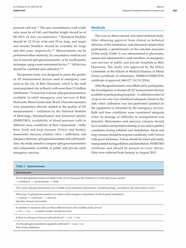
Table 1 – Questionnaire
Questionnaire
Do the sphygmomanometers available at the service belong to the institution or to the physicians/nurses?( ) institution ( ) professionals ( ) both
How many sphygmomanometers are available at the emergency department, considering triage, consultation and observation rooms?
What type of sphygmomanometer is available at the emergency department of this hospital/institution? ( ) aneroid ( ) electronic ( ) mercuryQuantity, brands and models:
In addition to standard cuffs, are there different sized cuffs available at the service? ( ) no ( ) yes – available number, brands and sizes
Is there an adequacy between cuff and device? ( ) yes ( ) no
Are the sphygmomanometers regularly calibrated? ( ) yes ( ) noDate of last calibration:
Maia et al.
Inadequacies of sphygmomanometers in emergency care
Int J Cardiovasc Sci. 2017;30(2):100-108
Original Article
pressure cuff size.1,7 The arm circumference/cuff width
ratio must be of 0.40, and bladder length should be of
80-100% of arm circumference.1.8 Standard bladder
should be 12-13 cm wide and 35 cm long, and larger
and smaller bladders should be available for large
and thin arms, respectively.9,10 Measurements can be
performed either indirectly, by auscultatory method and
use of aneroid sphygmomanometer, or by oscillometric
technique, using a semi-automated device.10,11 All devices
should be validated and calibrated.10,12
The present study was designed to assess the quality
of AP measurement devices used at emergency care
units in the city of Belo Horizonte, which is the sixth
most populated city in Brazil, with more than 2.5 million
inhabitants.13 It aimed to evaluate sphygmomanometers
available at adult emergency care services of Belo
Horizonte, Minas Gerais state, Brazil. Outcome measures
were parameters directly related to the quality of AP
measurements – validation by the National Institute
of Metrology, Standardization and Industrial Quality
(INMETRO), availability of blood pressure cuffs in
different sizes, conditions of their components – bulb,
hose, hook and loop closures (Velcro and hooks),
manometer, mercury column, valve – calibration, and
adequacy between sphygmomanometer and cuff brands.
Also, the study aimed to compare sphygmomanometers
and components available at public and private adult
emergency services.
Methods
This was an observational, non-interventional study. After obtaining approval from clinical or technical directors of the institutions, and informed consent from participants, a questionnaire on the outcome measures of this study (Table 1) was administered to physicians, nurses and administrative staff members of emergency care services of public and private hospitals in Belo Horizonte. The study was approved by the Ethics Committee of the School of Medical Sciences of Minas Gerais (certificate of submission: 35484614.9.0000.5134; certificate of approval: 846.017, 10/19/2014).
After the questionnaires were filled out by participants, the investigators evaluated all AP measurement devices available in participating hospitals. A calibration interval of up to one year was considered adequate, based on the date when calibration was last performed (printed on the equipment or informed by the emergency service). Bulb and hose conditions were considered adequate when no damage or difficulty in manipulation was detected. Manometers and mercury columns should have numbers and pointers starting at zero and in perfect conditions during inflation and disinflation. Hook and loop closures should be in good conditions, with Velcros with good stickiness. Valves should be intact and easily manipulated during inflation and disinflation. INMETRO certificate seal should be present in every device. Data were collected from January to August 2015.
102Maia et al.
Inadequacies of sphygmomanometers in emergency care
Int J Cardiovasc Sci. 2017;30(2):100-108
Original Article
Inclusion criteria
Both public and private hospitals offering emergency care services for the general population.
Exclusion criteria
Hospitals that did not accept to participate in the study, hospitals whose emergency services are not open for the general population, and specialized hospitals (maternity, pediatric, psychiatric hospitals, otolaryngology, ophthalmology and orthopedics emergencies).
Statistical analysis
According to information provided by the Medical Board of Minas Gerais, 16 public hospitals and 12 private hospitals that met the inclusion criteria were identified.
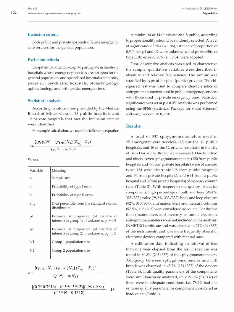
For sample calculation, we used the following equation
A minimum of 14 (6 private and 8 public, according to proportionality) should be randomly selected. A level of significance of 5% (z = 1.96), estimate of proportion of 0.5 (since p1 and p2 were unknown), and probability of type II (b) error of 20% (z = 0.84) were adopted.
First, descriptive analysis was used to characterize the sample; qualitative variables were described in absolute and relative frequencies. The sample was stratified by type of hospital (public/private). The chi-squared test was used to compare characteristics of sphygmomanometers used in public emergency services with those used in private emergency ones. Statistical significance was set at p < 0.05. Analysis was performed using the SPSS (Statistical Package for Social Sciences) software, version 20.0, 2012.
Results
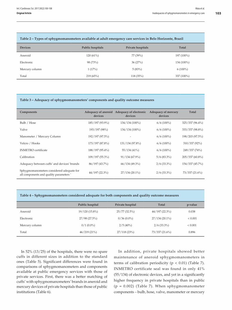
A total of 337 sphygmomanometers used in 25 emergency care services (15 out the 16 public hospitals, and 10 of the 12 private hospitals) in the city of Belo Horizonte, Brazil, were assessed. One hundred and ninety-seven sphygmomanometers (120 from public hospitals and 77 from private hospitals) were of aneroid type, 134 were electronic (98 from public hospitals and 36 from private hospitals), and 6 (1 from a public hospital and 5 from private hospitals) of mercury column type (Table 2). With respect to the quality of device components, high percentage of bulb and hose (96.4%, 325/337), valve (98.8%, 333/337), hook and loop closures (92%, 310/337), and manometers and mercury columns (97.5%, 198/203) were considered adequate. For the last item (manometers and mercury columns, electronic sphygmomanometers were not included in the analysis. INMETRO certificate seal was detected in 74% (49/337) of the instruments, and was more frequently absent in electronic devices compared with manual ones.
A calibration date indicating an interval of less than one year elapsed from the last inspection was found in 60.8% (205/337) of the sphygmomanometers. Adequacy between sphygmomanometer and cuff brands was observed in 45.7% (154/337) of the devices (Table 3). If all quality parameters of the components were simultaneously analyzed, only 21.6% (73/337) of them were in adequate conditions, i.e., 78.4% had one or more quality parameter or component considered as inadequate (Table 4).
Where:
Variable Meaning
n Sample size
a Probability of type I error
b Probability of type II error
za/2 (1-a) percentile from the standard normal distribution
p1 Estimate of proportion (of variable of interest) in group 1; if unknown, p1 = 0.5
p2 Estimate of proportion (of variable of interest) in group 2; if unknown, p2 = 0.5
N1
N2
Group 1 population size
Group 2 population size
103
Table 2 – Types of sphygmomanometers available at adult emergency care services in Belo Horizonte, Brazil
Devices Public hospitals Private hospitals Total
Aneroid 120 (61%) 77 (39%) 197 (100%)
Electronic 98 (73%) 36 (27%) 134 (100%)
Mercury column 1 (17%) 5 (83%) 6 (100%)
Total 219 (65%) 118 (35%) 337 (100%)
Table 3 – Adequacy of sphygmomanometers’ components and quality outcome measures
Components Adequacy of aneroid devices
Adequacy of electronic devices
Adequacy of mercury devices
Total
Bulb / Hose 185/197 (93.9%) 134/134 (100%) 6/6 (100%) 325/337 (96.4%)
Valve 193/197 (98%) 134/134 (100%) 6/6 (100%) 333/337 (98.8%)
Manometer / Mercury Column 192/197 (97.5%) - 6/6 (100%) 198/203 (97.5%)
Velcro / Hooks 173/197 (87.8%) 131/134 (97.8%) 6/6 (100%) 310/337 (92%)
INMETRO certificate 188/197 (95.4%) 55/134 (41%) 6/6 (100%) 249/337 (74%)
Calibration 109/197 (55.3%) 91/134 (67.9%) 5/6 (83.3%) 205/337 (60.8%)
Adequacy between cuffs’ and devices’ brands 86/197 (43.7%) 66/134 (49.3%) 2/6 (33.3%) 154/337 (45.7%)
Sphygmomanometers considered adequate for all components and quality parameters ’
44/197 (22.3%) 27/134 (20.1%) 2/6 (33.3%) 73/337 (21.6%)
Table 4 – Sphygmomanometers considered adequate for both components and quality outcome measures
Public hospital Private hospital Total p-value
Aneroid 19/120 (15.8%) 25/77 (32.5%) 44/197 (22.3%) 0.038
Electronic 27/98 (27.5%) 0/36 (0.0%) 27/134 (20.1%) < 0.001
Mercury column 0/1 (0.0%) 2/5 (40%) 2/6 (33.3%) < 0.001
Total 46/219 (21%) 27/118 (23%) 73/337 (21.6%) 0.896
Maia et al.
Inadequacies of sphygmomanometers in emergency care
Int J Cardiovasc Sci. 2017;30(2):100-108
Original Article
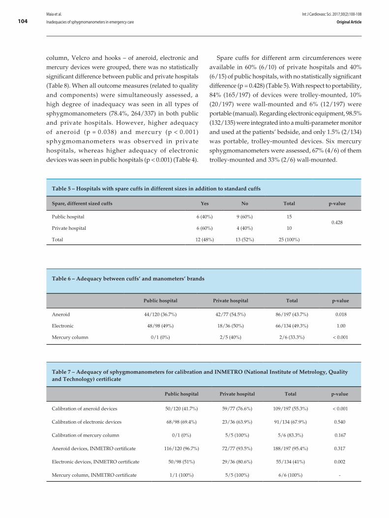
In 52% (13/25) of the hospitals, there were no spare cuffs in different sizes in addition to the standard ones (Table 5). Significant differences were found in comparisons of sphygmomanometers and components available at public emergency services with those of private services. First, there was a better matching of cuffs’ with sphygmomanometers’ brands in aneroid and mercury devices of private hospitals than those of public institutions (Table 6).
In addition, private hospitals showed better
maintenance of aneroid sphygmomanometers in
terms of calibration periodicity (p < 0.01) (Table 7).
INMETRO certificate seal was found in only 41%
(55/134) of electronic devices, and yet in a significantly
higher frequency in private hospitals than in public
(p = 0.002) (Table 7). When sphygmomanometer
components – bulb, hose, valve, manometer or mercury
104
Table 5 – Hospitals with spare cuffs in different sizes in addition to standard cuffs
Spare, different sized cuffs Yes No Total p-value
Public hospital 6 (40%) 9 (60%) 150.428
Private hospital 6 (60%) 4 (40%) 10
Total 12 (48%) 13 (52%) 25 (100%)
Table 6 – Adequacy between cuffs’ and manometers’ brands
Public hospital Private hospital Total p-value
Aneroid 44/120 (36.7%) 42/77 (54.5%) 86/197 (43.7%) 0.018
Electronic 48/98 (49%) 18/36 (50%) 66/134 (49.3%) 1.00
Mercury column 0/1 (0%) 2/5 (40%) 2/6 (33.3%) < 0.001
Table 7 – Adequacy of sphygmomanometers for calibration and INMETRO (National Institute of Metrology, Quality and Technology) certificate
Public hospital Private hospital Total p-value
Calibration of aneroid devices 50/120 (41.7%) 59/77 (76.6%) 109/197 (55.3%) < 0.001
Calibration of electronic devices 68/98 (69.4%) 23/36 (63.9%) 91/134 (67.9%) 0.540
Calibration of mercury column 0/1 (0%) 5/5 (100%) 5/6 (83.3%) 0.167
Aneroid devices, INMETRO certificate 116/120 (96.7%) 72/77 (93.5%) 188/197 (95.4%) 0.317
Electronic devices, INMETRO certificate 50/98 (51%) 29/36 (80.6%) 55/134 (41%) 0.002
Mercury column, INMETRO certificate 1/1 (100%) 5/5 (100%) 6/6 (100%) -
Maia et al.
Inadequacies of sphygmomanometers in emergency care
Int J Cardiovasc Sci. 2017;30(2):100-108
Original Article
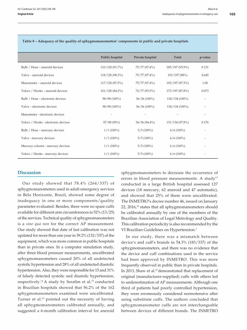
column, Velcro and hooks – of aneroid, electronic and mercury devices were grouped, there was no statistically significant difference between public and private hospitals (Table 8). When all outcome measures (related to quality and components) were simultaneously assessed, a high degree of inadequacy was seen in all types of sphygmomanometers (78.4%, 264/337) in both public and private hospitals. However, higher adequacy of aneroid (p = 0.038) and mercury (p < 0.001) sphygmomanometers was observed in private hospitals, whereas higher adequacy of electronic devices was seen in public hospitals (p < 0.001) (Table 4).
Spare cuffs for different arm circumferences were available in 60% (6/10) of private hospitals and 40% (6/15) of public hospitals, with no statistically significant difference (p = 0.428) (Table 5). With respect to portability, 84% (165/197) of devices were trolley-mounted, 10% (20/197) were wall-mounted and 6% (12/197) were portable (manual). Regarding electronic equipment, 98.5% (132/135) were integrated into a multi-parameter monitor and used at the patients’ bedside, and only 1.5% (2/134) was portable, trolley-mounted devices. Six mercury sphygmomanometers were assessed, 67% (4/6) of them trolley-mounted and 33% (2/6) wall-mounted.
105
Table 8 – Adequacy of the quality of sphygmomanometers’ components in public and private hospitals
Public hospital Private hospital Total p-value
Bulb / Hose – aneroid devices 110/120 (91.7%) 75/77 (97.4%) 185/197 (93.9%) 0.131
Valve – aneroid devices 118/120 (98.3%) 75/77 (97.4%) 193/197 (98%) 0.645
Manometer – aneroid devices 117/120 (97.5%) 75/77 (97.4%) 192/197 (97.5%) 1.00
Velcro / Hooks – aneroid devices 101/120 (84.2%) 72/77 (93.5%) 173/197 (87.8%) 0.073
Bulb / Hose – electronic devices 98/98 (100%) 36/36 (100%) 134/134 (100%) -
Valve – electronic devices 98/98 (100%) 36/36 (100%) 134/134 (100%) -
Manometer - electronic devices - - - -
Velcro / Hooks - electronic devices 97/98 (99%) 34/36 (94.4%) 131/134 (97.8%) 0.176
Bulb / Hose – mercury devices 1/1 (100%) 5/5 (100%) 6/6 (100%) -
Valve - mercury devices 1/1 (100%) 5/5 (100%) 6/6 (100%) -
Mercury column - mercury devices 1/1 (100%) 5/5 (100%) 6/6 (100%) -
Velcro / Hooks - mercury devices 1/1 (100%) 5/5 (100%) 6/6 (100%) -
Maia et al.
Inadequacies of sphygmomanometers in emergency care
Int J Cardiovasc Sci. 2017;30(2):100-108
Original Article
Discussion
Our study showed that 78.4% (264/337) of sphygmomanometers used in adult emergency services in Belo Horizonte, Brazil, showed some degree of inadequacy in one or more components/quality parameter evaluated. Besides, there were no spare cuffs available for different arm circumferences in 52% (13/25) of the services. Technical quality of sphygmomanometers is a sine qua non for the correct AP measurement. Our study showed that date of last calibration was not updated for more than one year in 39.2% (132/337) of the equipment, which was more common in public hospitals than in private ones. In a computer simulation study, after three blood pressure measurements, uncalibrated sphygmomanometers caused 20% of all undetected systolic hypertension and 28% of all undetected diastolic hypertension. Also, they were responsible for 15 and 31% of falsely detected systolic and diastolic hypertension, respectively.14 A study by Serafim et al.15 conducted in Brazilian hospitals showed that 56.2% of the 162 sphygmomanometers examined were uncalibrated. Turner et al.16 pointed out the necessity of having all sphygmomanometers calibrated annually, and suggested a 6-month calibration interval for aneroid
sphygmomanometers to decrease the occurrence of errors in blood pressure measurements. A study17 conducted in a large British hospital assessed 127 devices (18 mercury, 62 aneroid and 47 automatic), and showed that 25% of them were uncalibrated. The INMETRO’s decree number 46, issued on January 22, 2016,18 states that all sphygmomanometers should be calibrated annually by one of the members of the Brazilian Association of Legal Metrology and Quality. This calibration periodicity is also recommended by the VI Brazilian Guidelines on Hypertension.1
In our study, there was a mismatch between device’s and cuff’s brands in 54.3% (183/337) of the sphygmomanometers, and there was no evidence that the device and cuff combinations used in the service had been approved by INMETRO. This was more frequently observed in public than in private hospitals. In 2013, Shaw et al.19 demonstrated that replacement of original (manufacturer-supplied) cuffs with others led to underestimation of AP measurements. Although one third of patients had poorly controlled hypertension, they were erroneously considered normotensive after using substitute cuffs. The authors concluded that sphygmomanometer cuffs are not interchangeable between devices of different brands. The INMETRO
106Maia et al.
Inadequacies of sphygmomanometers in emergency care
Int J Cardiovasc Sci. 2017;30(2):100-108
Original Article
approves the use of specific cuffs for specific manometers regarding brands and models, and determines that, if a cuff of electronic devices had been previously used with equipment of different brands, this brand/model combination should be clearly informed.18,20 There has been a strong recommendation21 on replacement of mercury column sphygmomanometers with others due to high risk of toxicity and environmental contamination. However, its use is still approved by the Brazilian National Health Surveillance Agency. This type of sphygmomanometer accounted for 1.7% (6/337) of all equipment evaluated in this study.
Furthermore, in our study, nearly half of hospitals, including public and private ones, did not have spare, different size cuffs in the emergency rooms. The importance using correct cuffs for different arm circumferences has been shown by several authors,22-24 and is an essential prerequisite for proper measurement of AP.1,6,10 A cuff smaller than the arm circumference overestimates, whereas larger cuffs underestimates AP measurements.24,25 A study analyzing scientific papers published in Brazilian journals reported that 64% of the studies did not mention the sizes of the cuffs or their adequacies to arm circumferences.26 A study conducted in a teaching hospital in Sao Paulo state showed that using an arm circumference to cuff width ratio of 0.4, more than 50% of patients required a cuff smaller than 12 cm and 22% a larger one. The study showed that the standard sized cuff was adequate for only 17% of participants.27 Similarly, Freitas et al.28 found that only 50% of patients used adequately sized cuffs, since standard cuffs were the only available ones at public health centers. The unavailability of cuffs for different arm circumferences is still a challenge faced by healthcare providers, and this scenario was also found in emergency care units in Belo Horizonte.
The INMETRO also recommends that every cuff, even if not in use, should be inspected once a year, counting from the date of last acquisition.29 We found that 26% of the cuffs did not have the INMETRO seal, which is required by the technical metrology regulations.18,20,29 In Europe and the United States, devices are released for use after being submitted and approved by validation studies, according to the standards issued by the British Hypertension Society,30 the Association for the Advancement of Medical Instrumentation31 and the European Society of Hypertension,32 available at http://www.dableducational.org/sphygmomanometers/devices_2_sbpm. html) e http://www.bhsoc.org/bp_monitors/automatic.stm.
The INMETRO requires that every manufacturer presents a c l in ica l t r ia l on the use of the sphygmomanometer, conducted according to international guidelines, as a prerequisite for approval for use in Brazil.18,20 On the dableducational.org website, we did not find any review of the models available in our study. Taking into account all components and quality parameters, we found a high percentage of inadequacy (78%) of devices, especially due to lack of regular calibration or INMETRO certificate, and mismatching between cuff’s and device’s brands.
This study has some limitations. Due to the observational nature of the study, we and could not test and confirm the calibration status or the adequacy between cuffs and sphygmomanometers, which were verified by date of last calibration (or its absence) and the brands of components, respectively. Besides, the small number of mercury sphygmomanometers makes the comparison with other types difficult.
Conclusion
Most of sphygmomanometers available at adult emergency care services of the hospitals included in the study in Belo Horizonte, Brazil, were considered inadequate for use. Their general conditions should be improved, particularly in terms of regular calibration, availability of spare cuffs for different arm circumferences, and compatibility between cuffs and manometers.
Author contributions
Conception and design of the research: Maia KAP, Malachias MVB. Acquisition of data: Maia KAP, Paiva IV, Mariano RM, Paiva RV. Analysis and interpretation of the data: Maia KAP, Malachias MVB. Statistical analysis: Maia KAP. Obtaining financing: Maia KAP. Writing of the manuscript: Maia KAP, Malachias MVB. Critical revision of the manuscript for intellectual content: Maia KAP, Malachias MVB.
Potential Conflict of Interest
No potential conflict of interest relevant to this article was reported.
Sources of Funding
There were no external funding sources for this study.
Study Association This article is part of the thesis of master submitted
by Kleisson Antonio Pontes Maia, from Faculdade de Ciências Médicas de Minas Gerais.
107
1. Sociedade Brasileira de Cardiologia; Sociedade Brasileira de Hipertensão; Sociedade Brasileira de Nefrologia. VI Diretrizes Brasileiras de Hipertensão Arterial. Rev Bras Hipertens. 2010;17(1):4-64.
2. Cesarino CB, Cipullo JP, Martin JF, Ciorlia LA, Godoy MR, Cordeiro JA, et al. Prevalence and sociodemographic factors in a hypertensive population in São José do Rio Preto, São Paulo, Brazil. Arq Bras Cardiol. 2008;91(1):29-35.
3. Rosário TM, Scala LC, França GV, Pereira MR, Jardim PC. Prevalence, control and treatment of arterial hypertension in Nobres - MT. Arq Bras Cardiol. 2009;93(6):622-8, 672-8.
4. Mendis S, Puska P, Norrving B. Global atlas on cardiovascular disease prevention and control. Geneva: World Health Organization. (WHO); 2011.
5. Marik PE, Varon J. Hypertensive crises: challenges and management. Chest. 2007;131(6):1949-62. Erratum in: Chest. 2007;132(5):1721.
6. Stern RH. The new hypertension guidelines. J Clin Hypertens (Greenwich). 2013;15(10):748-51.
7. Puig AO, Dalfó-Pibernat A, Rosàs NJ, Isaac EM, Pérez-Romero L, Llorach EG, et al. Determination of arm circumference for correct measurement of blood pressure. Results of an intervention study. Hipertens Riesgo Vasc. 2015;32(1):6-11.
8. Pickering TG, Hall JE, Appel LJ, Falkner BE, Graves J, Hill MN, et al; Subcommittee of Professional and Public Education of the American Heart Association Council on High Blood Pressure Research. Recommendations for blood pressure measurement in humans and experimental animals part 1: blood pressure measurement in humans: a statement for professionals from the Subcommittee of Professional and Public Education of the American Heart Association Coun. Hypertension. 2005;45(1):142-61.
9. O'Brien E, Pickering T, Asmar R, Myers M, Parati G, Staessen J, et al; Working Group on Blood Pressure Monitoring of the European Society of Hypertension. Working Group on Blood Pressure Monitoring of the European Society of Hypertension International Protocol for validation of blood pressure measuring devices in adults. Blood Press Monit. 2002;7(1):3-17
10. Mancia G, Fagard R, Narkiewicz K, Redán J, Zanchetti A, Böhm M, et al; ESH/ESC Task Force for the Management of Arterial Hypertension. 2013 practice guidelines for the management of arterial hypertension of the European Society of Hypertension (ESH) and the European Society of Cardiology (ESC): ESH/ESC Task Force for the Management of Arterial Hypertension. J Hypertens. 2013;31(10):1925-38.
11. Parati G, Asmar R, Stergiou GS. Self blood pressure monitoring at home by wrist devices: a reliable approach? J Hypertens. 2002;20(4):573-8.
12. Weber MA, Schiffrin EL, White WB, Mann S, Lindholm LH, Kenerson JG, et al. Clinical practice guidelines for the management of hypertension in the community a statement by the American Society of Hypertension and the International Society of Hypertension. J Hypertens. 2014;32(1):3-15.
13. Instituto Brasileiro de Geografia e Estatística. (IBGE). [Internet]. [Citado em 2016 dez 10]. Disponível em: http://www.ibge.gov.br/home/estatistica/populacao/estimativa2015/estimativa_tcu.shtm
14. Turner MJ, Irwig L, Bune AJ, Kam PC, Baker AB. Lack of sphygmomanometer calibration causes over-and under-detection of hypertension: a computer simulation study. J Hypertens. 2006;24(10):1931-8.
15. Serafim TS, Toma GA, Gusmão JE, Colósimo FC, Silva SS, Pierin AM. Evaluation of the conditions of use of sphygmomanometers in hospital services. Acta Paul Enferm. 2012;25(6):940-6.
16. Turner MJ, Speechly C, Bignell N. Sphygmomanometer calibration: why, how and how often? Aust Fam Physician. 2007;36(10):834-8.
17. De Greeff A, Lorde I, Wilton A, Seed P, Coleman AJ, Shennan AH. Calibration accuracy of hospital-based non-invasive blood pressure measuring devices. J Hum Hypertens. 2010;24(1):58-63.
18. Ministério do Desenvolvimento, Indústria e Comércio Exterior. Instituto Nacional de Metrologia, Qualidade e Tecnologia. Portaria nº. 46, de 22 de janeiro de 2016. Diário Oficial da União. 26 jan 2016(17);Seção 1.
19. Shaw KC, McEniery CM, Wilkinson IB, Brown MJ. Unsafe health and safety: sphygmomanometer cuffs are not interchangeable. J Hum Hypertens. 2013;27(7):434-6.
20. Brasil. Ministério do Desenvolvimento, Indústria e Comércio Exterior. Instituto Nacional de Metrologia, Qualidade e Tecnologia. Considera que os esfignomanômetros de medição não invasiva devem atender às especificações metrológicas, de forma a garantir a sua confiabilidade. Diário Oficial da União, Brasília, de 26/01/2016 (nº 17, Seção 1, pag. 31).
21. Brasil. Ministério do Trabalho e Previdência Social. NR 15 - Norma Regulamentadora 15. Atividades e Operações Insalubres. Diário Oficial da União, Brasília, de 15 de setembro de 2015.
22. Tomlinson BU. Accurately measuring blood pressure: factors that contribute to false measurements. Medsurg Nurs. 2010;19(2):90-4.
23. Palatini P, Frick GN. Cuff and bladder: overlooked components of BP measurement devices in the modern era? Am J Hypertens. 2012;25(2):136-8.
24. McVicker JT. Blood pressure measurement-Does anyone do it right? an assessment of the reliability of equipment in use and the measurement techniques of clinicians. J Fam Plann Reprod Health Care. 2001;27(3):163-4.
25. Pickering TG, Hall JE, Appel LJ, Falkner BE, Graves JW, Hill MN, et al; Council on High Blood Pressure Research Professional and Public Education Subcommittee, American Heart Association. Recommendations for blood pressure measurement in humans: an AHA scientific statement from the Council on High Blood Pressure Research Professional and Public Education Subcommittee. J Clin Hypertens (Greenwich). 2005;7(2):102-9.
26. Holanda HE, Mion Junior D, Pierin AM. [Blood pressure measurement. Criteria employed in scientific articles published in Brazilian journals]. Arq Bras Cardiol. 1997;68(6):433-6.
27. Veiga EV, Arcuri EA, Cloutier L, Santos JL. Blood pressure measurement: arm circumference and cuff size availability. Rev Lat Am Enfermagem. 2009;17(4):455-61.
28. Freitas CC, Pantarotto RF, Costa LR. Relation arm circumference and the cuffs size used in Basic Health Units in São Paulo countryside. J Health Sci Inst. 2013;31(3):48-52.
29. Brasil. Ministério do Desenvolvimento, Indústria e Comércio Exterior. Instituto Nacional de Metrologia, Qualidade e Tecnologia. Portaria Inmetro nº 153, de 12 de agosto de 2005. Considera que os esfignomanômetros mecânicos, de medição não invasiva, devem atender as especificações de forma a garantir a sua confiabilidade metrológica. Diário Oficial da União de 12 de agosto de 2005.
30. O’Brien E, Petrie J, Littler W, de Swiet M, Padfield PL, Altman DG, et al. The British Hypertension Society protocol for the evaluation of blood measuring devices. J Hypertens.1993;11(Suppl 2):543-62.
31. Association for the Advancement of Medical Instrumentation. (AAMI). American National Standard for Electronic or Automated Sphygmomanometers (ANSI). Arlington, VA; 1993.
32. O'Brien E, Atkins N, Stergiou G, Karpettas N, Parati G, Asmar R, et al; Working Group on Blood Pressure Monitoring of the European Society of Hypertension. Society of Hypertension International Protocol revision 2010 for the validation of blood pressure measuring devices in adults. Blood Press Monit. 2010;15(1):23-38. Erratum in: Blood Press Monit. 2010;15(3):171-2.
References
Maia et al.
Inadequacies of sphygmomanometers in emergency care
Int J Cardiovasc Sci. 2017;30(2):100-108
Original Article

108Maia et al.
Inadequacies of sphygmomanometers in emergency care
Int J Cardiovasc Sci. 2017;30(2):100-108
Original Article
DOI: 10.5935/2359-4802.20170037
109International Journal of Cardiovascular Sciences. 2017;30(2):109-116
ORIGINAL ARTICLE
Mailing Address: Priscila Neri LacerdaRua Reitor Miguel Calmon, s/n. Postal Code: 40110-100. Vale do Canela, Salvador, BA – BrazilE-mail: [email protected]
Assessment of Right Ventricle Function and Myocardial Fibrosis by Cardiovascular Magnetic Resonance in Patients with Inferior Wall Myocardial InfarctionPriscila Neri Lacerda,1 Rafael Fernandes Almeida,2 Fernanda Gabriella Figueiredo Pinto,2 Adilson Machado Gomes Júnior,1 Jéssica Mendes Santos,1 Cristiano Ricardo Bastos de Macêdo,2 André Maurício Souza Fernandes,2 Roque Aras Júnior2
Universidade Federal da Bahia (UFBA);1 Hospital Ana Neri,2 Salvador, BA – Brazil
Manuscript received October 20, 2016; revised manuscript February 22, 2017; accepted February 28, 2017.
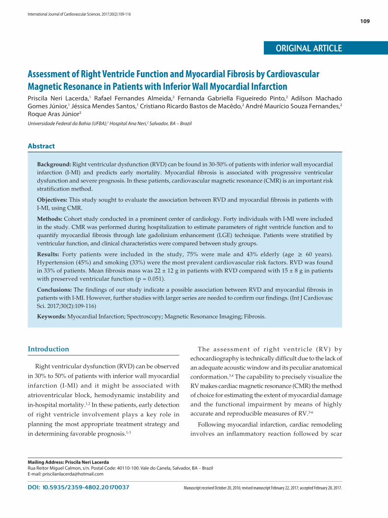
Abstract
Background: Right ventricular dysfunction (RVD) can be found in 30-50% of patients with inferior wall myocardial infarction (I-MI) and predicts early mortality. Myocardial fibrosis is associated with progressive ventricular dysfunction and severe prognosis. In these patients, cardiovascular magnetic resonance (CMR) is an important risk stratification method.
Objectives: This study sought to evaluate the association between RVD and myocardial fibrosis in patients with I-MI, using CMR.
Methods: Cohort study conducted in a prominent center of cardiology. Forty individuals with I-MI were included in the study. CMR was performed during hospitalization to estimate parameters of right ventricle function and to quantify myocardial fibrosis through late gadolinium enhancement (LGE) technique. Patients were stratified by ventricular function, and clinical characteristics were compared between study groups.
Results: Forty patients were included in the study, 75% were male and 43% elderly (age ≥ 60 years). Hypertension (45%) and smoking (33%) were the most prevalent cardiovascular risk factors. RVD was found in 33% of patients. Mean fibrosis mass was 22 ± 12 g in patients with RVD compared with 15 ± 8 g in patients with preserved ventricular function (p = 0.051).
Conclusions: The findings of our study indicate a possible association between RVD and myocardial fibrosis in patients with I-MI. However, further studies with larger series are needed to confirm our findings. (Int J Cardiovasc Sci. 2017;30(2):109-116)
Keywords: Myocardial Infarction; Spectroscopy; Magnetic Resonance Imaging; Fibrosis.
Introduction
Right ventricular dysfunction (RVD) can be observed
in 30% to 50% of patients with inferior wall myocardial
infarction (I-MI) and it might be associated with
atrioventricular block, hemodynamic instability and
in-hospital mortality.1,2 In these patients, early detection
of right ventricle involvement plays a key role in
planning the most appropriate treatment strategy and
in determining favorable prognosis.1-3
The assessment of r ight ventricle (RV) by echocardiography is technically difficult due to the lack of an adequate acoustic window and its peculiar anatomical conformation.3,4 The capability to precisely visualize the RV makes cardiac magnetic resonance (CMR) the method of choice for estimating the extent of myocardial damage and the functional impairment by means of highly accurate and reproducible measures of RV.3-6
Following myocardial infarction, cardiac remodeling involves an inflammatory reaction followed by scar
110Lacerda et al.
Right ventricle function and myocardial fibrosis
Int J Cardiovasc Sci. 2017;30(2):109-116
Original Article
formation at the site of infarction.7 However, sustained fibrotic activity results in stiffening of the myocardium and is associated with progressive ventricular dysfunction and severe prognosis.7,8 Late-gadolinium enhancement (LGE) CMR has been used extensively in a large number of studies as the technique of choice for detection and measurement of myocardial fibrosis.9-12
In patients with I-MI, therefore, CMR has been established as the gold standard imaging method for the assessment of RV function and myocardial fibrosis.9-12 This study aimed to evaluate the association between myocardial fibrosis and RVD in patients with I-MI, using CMR.
Methods
Study population
A total of fifty-seven patients with acute ST segment elevation myocardial infarction with inferior wall involvement (ST segment elevation in D2, D3 and aVF derivations on the electrocardiography) were prospectively recruited at Ana Neri Hospital, Brazil, between January and December 2014. Patients were excluded if they had metallic implants incompatible with CMR, glomerular filtration rate (GFR) < 30 ml/min, severe claustrophobia or gadolinium hypersensitivity.
Clinical data including age, sex, family history, comorbidities and cardiovascular risk factors were retrospectively collected from patients’ medical records. CMR was performed during hospitalization to estimate parameters of RV function and to quantify myocardial fibrosis. Right ventricular ejection fraction (RVEF), end-systolic volume and end-diastolic volume were measured to estimate ventricular function. LGE-CMR technique was used to measure myocardial fibrosis in the inferior wall. Patients were stratified by ventricular function, considering RVD if RVEF < 40%.
The study was approved by the ethical, institutional review board (Ana Nery Hospital Ethics Committee) and the National Ethics Committee and all patients provided written informed consent.
CMR acquisition
Patients were scanned in the supine position and CMR studies were performed using a 1.5 T whole-body scanner (Avanto, Siemens Medical Solutions, Germany).
An 8 channel cardiac coil was used for signal reception. Scout images were obtained to plan the four-chamber, three-chamber and two-chamber views, as well as short axis cine imaging. ECG-gated steady-state free precession (SSFP) short-axis images of the ventricles were acquired during breath holds with 20 image frames per cardiac cycle. Acquisition parameters were: 8-mm slice thickness, FOV 300, matrix 128 x 128.
A stack of images, using a minimum of 8 and a maximum of 12 slices in short-axis plane (slice thickness 8-mm; gap 2-mm) was acquired, allowing coverage of the entire cardiac volume. Every effort was made to obtain adequate images with a satisfactory right ventricle depiction.
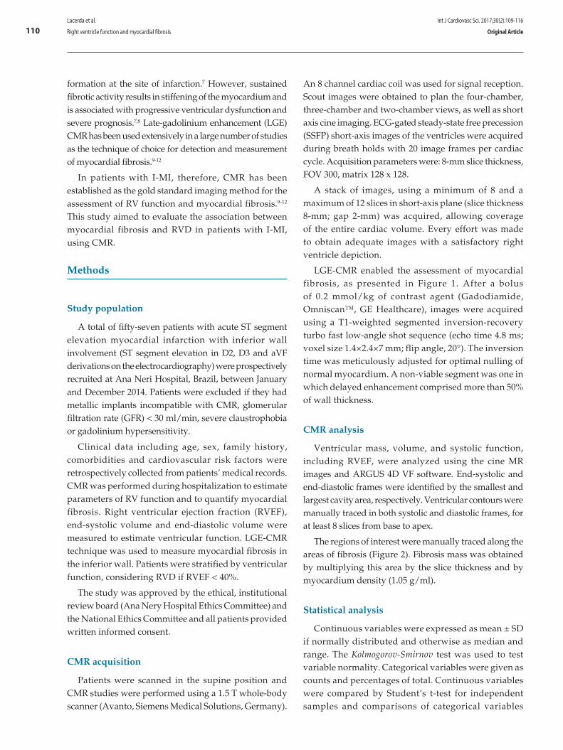
LGE-CMR enabled the assessment of myocardial fibrosis, as presented in Figure 1. After a bolus of 0.2 mmol/kg of contrast agent (Gadodiamide, Omniscan™, GE Healthcare), images were acquired using a T1-weighted segmented inversion-recovery turbo fast low-angle shot sequence (echo time 4.8 ms; voxel size 1.4×2.4×7 mm; flip angle, 20°). The inversion time was meticulously adjusted for optimal nulling of normal myocardium. A non-viable segment was one in which delayed enhancement comprised more than 50% of wall thickness.
CMR analysis
Ventricular mass, volume, and systolic function, including RVEF, were analyzed using the cine MR images and ARGUS 4D VF software. End-systolic and end-diastolic frames were identified by the smallest and largest cavity area, respectively. Ventricular contours were manually traced in both systolic and diastolic frames, for at least 8 slices from base to apex.
The regions of interest were manually traced along the areas of fibrosis (Figure 2). Fibrosis mass was obtained by multiplying this area by the slice thickness and by myocardium density (1.05 g/ml).
Statistical analysis
Continuous variables were expressed as mean ± SD if normally distributed and otherwise as median and range. The Kolmogorov-Smirnov test was used to test variable normality. Categorical variables were given as counts and percentages of total. Continuous variables were compared by Student’s t-test for independent samples and comparisons of categorical variables
111
Figure 2 – Measurement of fibrosis by late gadolinium enhancement (1 represents the area of fibrosis).
Figure 1 – Late gadolinium enhancement-cardiac magnetic resonance images from patients with inferior wall myocardial infarction (white arrows show myocardial fibrosis in the inferior wall).
Lacerda et al.
Right ventricle function and myocardial fibrosis
Int J Cardiovasc Sci. 2017;30(2):109-116
Original Article
were made using Fisher’s exact test. The Pearson’s correlation test was applied to examine the association between RVD and fibrosis. Multivariate logistic regression was performed to determine predictors
of RVD. P-values of less than 0.05 were considered significant. Statistical analysis was performed using Statistical Package for the Social Sciences software, version 17.0.
112
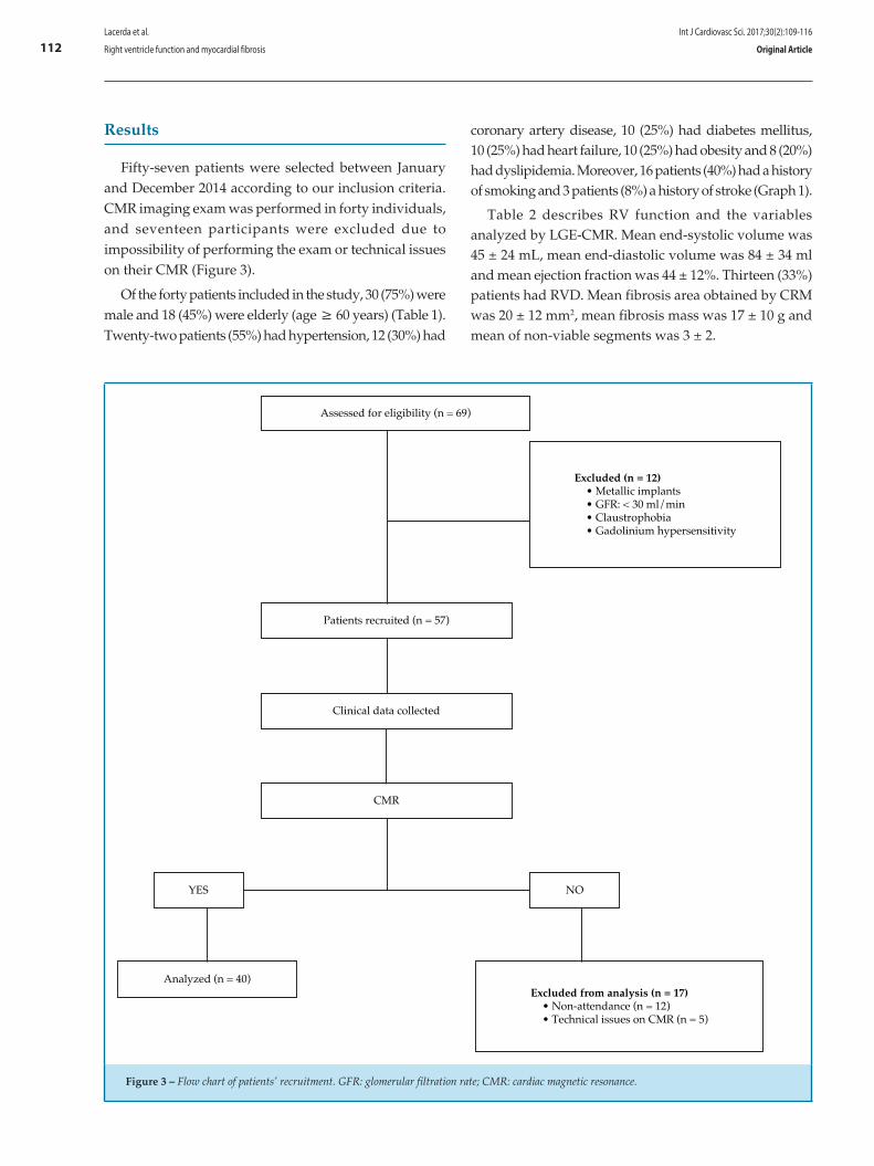
Assessed for eligibility (n = 69)
Excluded (n = 12)• Metallic implants• GFR: < 30 ml/min• Claustrophobia• Gadolinium hypersensitivity
Patients recruited (n = 57)
Clinical data collected
CMR
YES NO
Analyzed (n = 40)Excluded from analysis (n = 17)
• Non-attendance (n = 12)• Technical issues on CMR (n = 5)
Figure 3 – Flow chart of patients’ recruitment. GFR: glomerular filtration rate; CMR: cardiac magnetic resonance.
Lacerda et al.
Right ventricle function and myocardial fibrosis
Int J Cardiovasc Sci. 2017;30(2):109-116
Original Article
Results
Fifty-seven patients were selected between January and December 2014 according to our inclusion criteria. CMR imaging exam was performed in forty individuals, and seventeen participants were excluded due to impossibility of performing the exam or technical issues on their CMR (Figure 3).
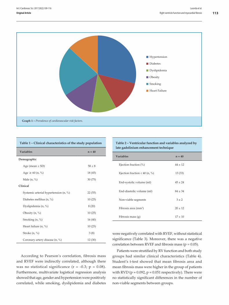
Of the forty patients included in the study, 30 (75%) were male and 18 (45%) were elderly (age ≥ 60 years) (Table 1). Twenty-two patients (55%) had hypertension, 12 (30%) had
coronary artery disease, 10 (25%) had diabetes mellitus, 10 (25%) had heart failure, 10 (25%) had obesity and 8 (20%) had dyslipidemia. Moreover, 16 patients (40%) had a history of smoking and 3 patients (8%) a history of stroke (Graph 1).
Table 2 describes RV function and the variables analyzed by LGE-CMR. Mean end-systolic volume was 45 ± 24 mL, mean end-diastolic volume was 84 ± 34 ml and mean ejection fraction was 44 ± 12%. Thirteen (33%) patients had RVD. Mean fibrosis area obtained by CRM was 20 ± 12 mm2, mean fibrosis mass was 17 ± 10 g and mean of non-viable segments was 3 ± 2.
113
Hypertension
Diabetes
Dyslipidemia
Obesity
Smoking
Heart Failure
Graph 1 – Prevalence of cardiovascular risk factors.
Lacerda et al.
Right ventricle function and myocardial fibrosis
Int J Cardiovasc Sci. 2017;30(2):109-116
Original Article
Table 1 – Clinical characteristics of the study population
Variables n = 40
Demographic
Age (mean ± SD) 58 ± 8
Age ≥ 60 (n, %) 18 (45)
Male (n, %) 30 (75)
Clinical
Systemic arterial hypertension (n, %) 22 (55)
Diabetes mellitus (n, %) 10 (25)
Dyslipidemia (n, %) 8 (20)
Obesity (n, %) 10 (25)
Smoking (n, %) 16 (40)
Heart failure (n, %) 10 (25)
Stroke (n, %) 3 (8)
Coronary artery disease (n, %) 12 (30)
Table 2 – Ventricular function and variables analyzed by late gadolinium enhancement technique
Variables n = 40
Ejection fraction (%) 44 ± 12
Ejection fraction < 40 (n, %) 13 (33)
End-systolic volume (ml) 45 ± 24
End-diastolic volume (ml) 84 ± 34
Non-viable segments 3 ± 2
Fibrosis area (mm2) 20 ± 12
Fibrosis mass (g) 17 ± 10
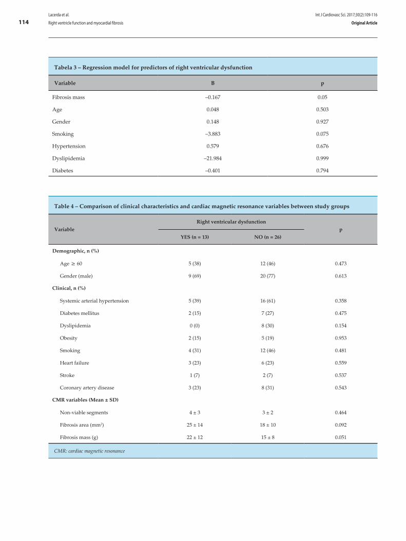
According to Pearson’s correlation, fibrosis mass and RVEF were indirectly correlated, although there was no statistical significance (r = -0.3; p = 0.08). Furthermore, multivariate logistical regression analysis showed that age, gender and hypertension were positively correlated, while smoking, dyslipidemia and diabetes
were negatively correlated with RVEF, without statistical significance (Table 3). Moreover, there was a negative correlation between RVEF and fibrosis mass (p = 0.05).
Patients were stratified by RV function and both study groups had similar clinical characteristics (Table 4). Student’s t-test showed that mean fibrosis area and mean fibrosis mass were higher in the group of patients with RVD (p = 0.092, p = 0.051 respectively). There were no statistically significant differences in the number of non-viable segments between groups.
114
Tabela 3 – Regression model for predictors of right ventricular dysfunction
Variable B p
Fibrosis mass –0.167 0.05
Age 0.048 0.503
Gender 0.148 0.927
Smoking –3.883 0.075
Hypertension 0.579 0.676
Dyslipidemia –21.984 0.999
Diabetes –0.401 0.794
Lacerda et al.
Right ventricle function and myocardial fibrosis
Int J Cardiovasc Sci. 2017;30(2):109-116
Original Article
Table 4 – Comparison of clinical characteristics and cardiac magnetic resonance variables between study groups
Variable
Right ventricular dysfunction
p
YES (n = 13) NO (n = 26)
Demographic, n (%)
Age ≥ 60 5 (38) 12 (46) 0.473
Gender (male) 9 (69) 20 (77) 0.613
Clinical, n (%)
Systemic arterial hypertension 5 (39) 16 (61) 0.358
Diabetes mellitus 2 (15) 7 (27) 0.475
Dyslipidemia 0 (0) 8 (30) 0.154
Obesity 2 (15) 5 (19) 0.953
Smoking 4 (31) 12 (46) 0.481
Heart failure 3 (23) 6 (23) 0.559
Stroke 1 (7) 2 (7) 0.537
Coronary artery disease 3 (23) 8 (31) 0.543
CMR variables (Mean ± SD)
Non-viable segments 4 ± 3 3 ± 2 0.464
Fibrosis area (mm2) 25 ± 14 18 ± 10 0.092
Fibrosis mass (g) 22 ± 12 15 ± 8 0.051
CMR: cardiac magnetic resonance
115Lacerda et al.
Right ventricle function and myocardial fibrosis
Int J Cardiovasc Sci. 2017;30(2):109-116
Original Article
Discussion
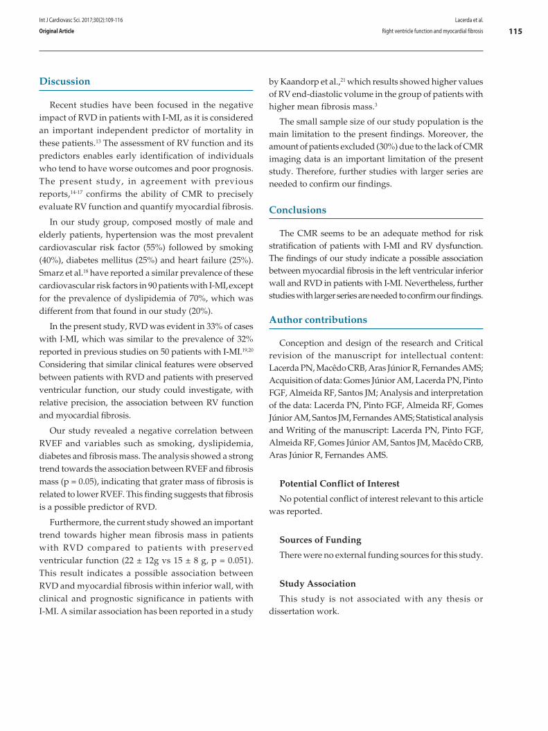
Recent studies have been focused in the negative impact of RVD in patients with I-MI, as it is considered an important independent predictor of mortality in these patients.13 The assessment of RV function and its predictors enables early identification of individuals who tend to have worse outcomes and poor prognosis. The present study, in agreement with previous reports,14-17 confirms the ability of CMR to precisely evaluate RV function and quantify myocardial fibrosis.
In our study group, composed mostly of male and elderly patients, hypertension was the most prevalent cardiovascular risk factor (55%) followed by smoking (40%), diabetes mellitus (25%) and heart failure (25%). Smarz et al.18 have reported a similar prevalence of these cardiovascular risk factors in 90 patients with I-MI, except for the prevalence of dyslipidemia of 70%, which was different from that found in our study (20%).
In the present study, RVD was evident in 33% of cases with I-MI, which was similar to the prevalence of 32% reported in previous studies on 50 patients with I-MI.19,20 Considering that similar clinical features were observed between patients with RVD and patients with preserved ventricular function, our study could investigate, with relative precision, the association between RV function and myocardial fibrosis.
Our study revealed a negative correlation between RVEF and variables such as smoking, dyslipidemia, diabetes and fibrosis mass. The analysis showed a strong trend towards the association between RVEF and fibrosis mass (p = 0.05), indicating that grater mass of fibrosis is related to lower RVEF. This finding suggests that fibrosis is a possible predictor of RVD.
Furthermore, the current study showed an important trend towards higher mean fibrosis mass in patients with RVD compared to patients with preserved ventricular function (22 ± 12g vs 15 ± 8 g, p = 0.051). This result indicates a possible association between RVD and myocardial fibrosis within inferior wall, with clinical and prognostic significance in patients with I-MI. A similar association has been reported in a study
by Kaandorp et al.,21 which results showed higher values of RV end-diastolic volume in the group of patients with higher mean fibrosis mass.3
The small sample size of our study population is the main limitation to the present findings. Moreover, the amount of patients excluded (30%) due to the lack of CMR imaging data is an important limitation of the present study. Therefore, further studies with larger series are needed to confirm our findings.
Conclusions
The CMR seems to be an adequate method for risk stratification of patients with I-MI and RV dysfunction. The findings of our study indicate a possible association between myocardial fibrosis in the left ventricular inferior wall and RVD in patients with I-MI. Nevertheless, further studies with larger series are needed to confirm our findings.
Author contributions
Conception and design of the research and Critical revision of the manuscript for intellectual content: Lacerda PN, Macêdo CRB, Aras Júnior R, Fernandes AMS; Acquisition of data: Gomes Júnior AM, Lacerda PN, Pinto FGF, Almeida RF, Santos JM; Analysis and interpretation of the data: Lacerda PN, Pinto FGF, Almeida RF, Gomes Júnior AM, Santos JM, Fernandes AMS; Statistical analysis and Writing of the manuscript: Lacerda PN, Pinto FGF, Almeida RF, Gomes Júnior AM, Santos JM, Macêdo CRB, Aras Júnior R, Fernandes AMS.
Potential Conflict of Interest
No potential conflict of interest relevant to this article was reported.
Sources of Funding
There were no external funding sources for this study.
Study Association
This study is not associated with any thesis or dissertation work.
116
1. Zehender M, Kasper W, Kauder E, Schonthaler M, Geibel A, Olschewski M, et al. Right ventricular infarction as an independent predictor of prognosis after acute inferior myocardial infarction. N Engl J Med. 1993;328(14):981-8.
2. Kakouros N, Cokkinos DV. Right ventricular myocardial infarction: pathophysiology, diagnosis, and management. Postgrad Med J. 2010;86(1022):719-28.
3. Galea N, Francone M, Carbone I, Cannata D, Vullo F, Galea R, et al. Utility of cardiac magnetic resonance in the evaluation of right ventricular involvement in patients with myocardial infarction. Radiol Med. 2013;119(5):309-17.
4. Rallidis LS, Makavos G, Nihoyannopoulos P. Right ventricular involvement in coronary artery disease: role of echocardiography for diagnosis and prognosis. J Am Soc Echocardiogr. 2014;27(3):223-9.
5. Rambihar S, Dokainish H. Right ventricular involvement in patients with coronary artery disease. Curr Opin Cardiol. 2010;25(5):456-63.
6. Todiere G, Aquaro GD, Piaggi P, Formisano F, Barison A, Masci PG, et al. Progression of myocardial fibrosis assessed with cardiac magnetic resonance in hypertrophic cardiomyopathy. J Am Coll Cardiol. 2012;60(10):922-9.
7. Ambale-Venkatesh B, Lima JA. Cardiac MRI: a central prognostic tool in myocardial fibrosis. Nat Rev Cardiol. 2015;12(1):18-29.
8. Allman KC, Shaw LJ, Hachamovitch R, Udelson JE. Myocardial viability testing and impact of revascularization on prognosis in patients with coronary artery disease and left ventricular dysfunction: a meta-analysis. J Am Coll Cardiol. 2002;39(7):1151-8.
9. Mewton N, Liu Chia Y, Croisille P, Bluemke D, Lima JA. Assessment of myocardial fibrosis with cardiac magnetic resonance. J Am Coll Cardiol. 2012;57(8):891-903.
10. Barranhas AD, Santos AS, Coelho-Filho OR, Marchiori E, Rochitte CE, Nacif MS. Cardiac magnetic resonance imaging in clinical practice. Radiol Bras. 2014;47(1):1-8.
11. Simonetti OP, Kim RJ, Fieno DS, Hillenbrand HB, Wu E, Bundy JM, et al. An improved MR imaging technique for the visualization of Myocardial infarction. Radiology 2001;218(1):215-23.
12. Manka R, Fleck E, Paetsch I. Silent inferior myocardial infarction with extensive right ventricular scarring. Int J Cardiol. 2008;127(3)e186-7.
13. Kinch JW, Ryan TJ. Right ventricular infarction. N Engl J Med. 1994;330(17):1211-7.
14. Masci PG, Francone M, Desmet W, Ganame J, Todiere G, Donato R, et al. Right ventricular ischemic injury in patients with acute ST-segment elevation myocardial infarction: characterization with cardiovascular magnetic resonance. Circulation. 2010;122(14):1405-12.
15. Kelle S, Roes SD, Klein C, Kokocinski T, de Roos A, Fleck E. Prognostic value of myocardial infarct size and contractile reserve using magnetic resonance imaging. J Am Coll Cardiol. 2009;54(19):1770-7.
16. Jensen CJ, Jochims M, Hunold P, Sabin GV, Schlosser T, Bruder O. Right ventricular involvement in acute left ventricular myocardial infarction: prognostic implications of MRI findings. AJR Am J Roentgenol. 2010;194(3):592-8.
17. Gerber BL, Rousseau MF, Ahn SA, le Polain de Waroux JB, Pouleur AC, Phlips T. Prognostic value of myocardial viability by delayed-enhanced magnetic resonance in patients with coronary artery disease and low ejection fraction: impact of revascularization therapy. J Am Coll Cardiol. 2012;59(9):825-35.
18. Smarz K, Zaborska B, Jaxa-Chamiec T, Maciejewski P, Budaj A. Right ventricular dysfunction and exercise capacity after inferior (posterior) wall acute myocardial infarction. Am J Cardiol. 2012;110(6):784-9.
19. Liaqat A, Asghar N, Rehman A. In hospital Outcome of acute inferior with right ventricular or posterior wall myocardial infarction. Ann Pak Inst Med Sci. 2013;9(4):219-24.
20. Iqbal A, Muddarangappa R, Shah SKD, Vidyasagar S. A study of right ventricular infarction in inferior wall myocardial infarction. J Clin Sci Res. 2013;2:66-71.
21. Kaandorp TA, Lamba HJ, Poldermansc D, Viergeverb EP, Boersmac E, van der Wallb EE, et al. Assessment of right ventricular infarction with contrast-enhanced magnetic resonance imaging. Coron Artery Dis. 2007;18(1):39-43.
References
Lacerda et al.
Right ventricle function and myocardial fibrosis
Int J Cardiovasc Sci. 2017;30(2):109-116
Original Article
DOI: 10.5935/2359-4802.20170024
117International Journal of Cardiovascular Sciences. 2017;30(2):117-122
ORIGINAL ARTICLE
Mailing Address: Carolina Campos TassiniRua Tiradentes, 1730. Postal Code 14400550, Centro, Franca, São Paulo, SP – BrazilE-mail: [email protected]; [email protected]
Assessment of the Lifestyle of University Students in the Healthcare Area Using the Fantastic QuestionnaireCarolina Campos Tassini,1 Gabriela Ribeiro do Val,1 Sarah da Silva Candido,2 Cynthia Kallás Bachur1,2
Universidade de Franca,1 Franca; Universidade de São Paulo,2 USP, Ribeirão Preto – Brazil
Manuscript received October 20, 2016; revised November 28, 2016; accepted March 01, 2017.
Abstract
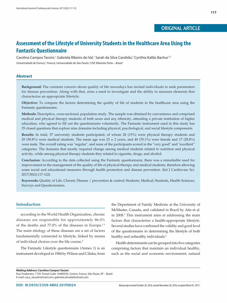
Background: The constant concern about quality of life nowadays has incited individuals to seek parameters for disease prevention. Along with that, arise a need to investigate and the ability to measure elements that characterize an appropriate lifestyle.
Objective: To compare the factors determining the quality of life of students in the healthcare area using the Fantastic questionnaire.
Methods: Descriptive, cross-sectional, population study. The sample was obtained by convenience and comprised medical and physical therapy students of both sexes and any ethnicity, attending a private institution of higher education, who agreed to fill out the questionnaire voluntarily. The Fantastic instrument used in this study has 25 closed questions that explore nine domains including physical, psychological, and social lifestyle components.
Results: In total, 57 university students participated, of whom 28 (15%) were physical therapy students and 29 (50.8%) were medical students. The mean age was 23 ± 2 years, and 40 (70.1%) were female and 17 (28.8%) were male. The overall rating was "regular", and none of the participants scored in the "very good" and "excellent” categories. The domains that mostly required change among medical students related to nutrition and physical activity, while among physical therapy students they related to cigarette, drugs, and alcohol.
Conclusion: According to the data collected using the Fantastic questionnaire, there was a remarkable need for improvement in the management of the quality of life of physical therapy and medical students, therefore allowing some social and educational measures through health promotion and disease prevention. (Int J Cardiovasc Sci. 2017;30(2):117-122)
Keywords: Quality of Life, Chronic Disease / prevention & control; Students, Medical; Students, Health Sciences; Surveys and Questionnaires.
Introduction
according to the World Health Organization, chronic diseases are responsible for approximately 86.0% of the deaths and 77.0% of the diseases in Europe.1-3 The main etiology of these diseases are a set of factors fundamentally connected to lifestyle, linked by means of individual choices over the life course.4
The Fantastic Lifestyle questionnaire (Annex 1) is an instrument developed in 1984 by Wilson and Ciliska, from
the Department of Family Medicine at the University of McMaster, Canada, and validated in Brazil by Añe et al. in 2008.5 This instrument aims at addressing the main factors that characterize a health-appropriate lifestyle. Several studies have confirmed the validity and good level of the questionnaire in determining the lifestyle of both healthy and unhealthy individuals.6
Health determinants can be grouped into five categories comprising factors that maintain an individual healthy, such as the social and economic environment, natural
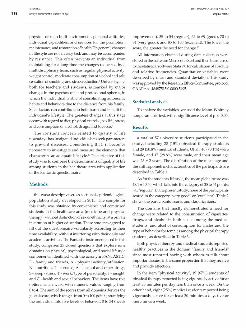
118Tassini et al.
Lifestyle assessement in students college
Int J Cardiovasc Sci. 2017;30(2):117-122
Original Article
physical or man-built environment, personal attitudes, individual capabilities, and services for the promotion, maintenance, and restoration of health.7 In general, changes in lifestyle are not an easy task and may be accompanied by resistance. This often prevents an individual from maintaining for a long time the changes requested by a multidisciplinary team, such as regular physical activity, weight control, moderate consumption of alcohol and salt, cessation of smoking, and stress reduction.8 University life, both for teachers and students, is marked by major changes in the psychosocial and professional spheres, in which the individual is able of consolidating autonomic habits and behaviors due to the distance from his family. Such factors can contribute to both harm and benefit the individual’s lifestyle. The greatest changes at this stage occur with regard to diet, physical exercise, sex life, stress, and consumption of alcohol, drugs, and tobacco.9
The constant concern related to quality of life nowadays has instigated individuals to seek parameters to prevent diseases. Considering that, it becomes necessary to investigate and measure the elements that characterize an adequate lifestyle.10 The objective of this study was to compare the determinants of quality of life among students in the healthcare area with application of the Fantastic questionnaire.
Methods
this was a descriptive, cross-sectional, epidemiological, population study developed in 2015. The sample for this study was obtained by convenience and comprised students in the healthcare area (medicine and physical therapy), without distinction of sex or ethnicity, at a private institution of higher education. These students agreed to fill out the questionnaire voluntarily according to their time availability, without interfering with their daily and academic activities. The Fantastic instrument, used in this study, comprises 25 closed questions that explore nine domains on physical, psychological, and social lifestyle components, identified with the acronym FANTASTIC: F - family and friends, A - physical activity/affiliation, N - nutrition, T - tobacco, A - alcohol and other drugs, S - sleep/stress, T - work/type of personality, I - insight, and C - health and sexual behaviors. The items have five options as answers, with numeric values ranging from 0 to 4. The sum of the scores from all domains derives the global score, which ranges from 0 to 100 points, stratifying the individual into five levels of behavior: 0 to 34 (needs
improvement), 35 to 54 (regular), 55 to 69 (good), 70 to 84 (very good), and 85 to 100 (excellent). The lower the score, the greater the need for change.10
All information obtained during data collection were stored in the software Microsoft Excel and then transferred to the statistical software Stata 9.0 for calculation of absolute and relative frequencies. Quantitative variables were described by mean and standard deviation. This study was approved by the Research Ethics Committee, protocol CAAE no.: 48487515.0.0000.5495.
Statistical analysis
To analyze the variables, we used the Mann-Whitney nonparametric test, with a significance level of p ≤ 0.05.
Results
a total of 57 university students participated in the study, including 28 (15%) physical therapy students and 29 (50.8%) medical students. Of all, 40 (70.1%) were female, and 17 (28.8%) were male, and their mean age was 23 ± 2 years. The distribution of the mean age and the anthropometric characteristics of the participants are described in Table 1.
As for the students' lifestyle, the mean global score was 48.1 ± 10.50, which falls into the category of 35 to 54 points, i.e., "regular". In the present study, none of the participants scored in the category "very good" or "excellent". Table 2 shows the participants' scores and classifications.
The domains that mostly demonstrated a need for change were related to the consumption of cigarettes, drugs, and alcohol in both sexes among the medical students, and alcohol consumption for males and the type of behavior for females among the physical therapy students, as described in Table 3.
Both physical therapy and medical students reported healthy practices in the domain "family and friends" since most reported having with whom to talk about important issues, in the same proportion that they receive and provide affection.
In the item "physical activity", 19 (67%) students of physical therapy reported being vigorously active for at least 30 minutes per day less than once a week. On the other hand, eight (25%) medical students reported being vigorously active for at least 30 minutes a day, five or more times a week.
119Tassini et al.
Lifestyle assessement in students college
Int J Cardiovasc Sci. 2017;30(2):117-122
Original Article
Table 1 – Anthropometric characteristics of the sample, with values presented as mean ± standard deviation. Franca, 2015
Male (n = 17) Female (n = 40)
Age (years) 24 ± 2.23 23 ± 2.34
Weight (kg) 80 ± 13.70 65 ± 15.2
Height (m) 1.78 ± 0.07 1.65 ± 0.05
BMI (kg/cm²) 25 ± 3.52 24 ± 5.11
n: number of participants; kg: kilogram; cm: centimeter; BMI: body mass index.
Table 2 – Students' scores obtained with the Fantastic Lifestyle questionnaire. Franca, 2015
Needs improvement 0 to 34 points Regular 35 to 54 points Good 55 to 69 points
Physical therapy 0 (0%) 25 (89.2%) 3 (10.7%)
Medicine 1 (0.29%) 22 (75.8%) 6 (20.6%)
Table 3 – Description (mean and standard deviation) of the domains of the Fantastic questionnaire, divided by sex and course. Franca, 2015
Medicine Physical Therapy
Domain Male (n = 15) Female (n = 14) Male (n = 2) Female (n = 26)
Family and friends 4 ± 0 4 ± 0.53 4 ± 0.62 4 ± 0.58
Physical activity 2.3 ± 1.4 2 ± 1.3 4 ± 1.15 4 ± 0.59
Nutrition 1.8 ± 1.17 1.8 ± 1.3 4 ± 1.83 4 ± 0.59
Cigarette and drugs 0.5 ± 0.8 0.2 ± 0.3 2 ± 2.31 4 ± 0.87
Alcohol 1.2 ± 1.0 0.8 ± 1.0 0 ± 1.26 4 ± 0.88
Sleep, seat belt, stress and safe sex 2.7 ± 1.1 2.8 ± 1.1 3 ± 1.05 4 ± 0.89
Type of behavior 2.4 ± 1.0 1.7 ± 1.1 3 ± 1.08 3 ± 0.91
Insight 2 ± 1.0 2.2 ± 1.1 3 ± 1.83 3 ± 0.93
Work 3 ± 0 3 ± 0.6 2 ± 2.00 4 ± 0.92
In regards to eating habits, this study showed in the "nutrition" domain that physical therapy students consume more servings of fruits and vegetables that medical students. On the other hand, medical students reported eating more sugar, salt, and foods with excess fat. In this dimension, the question "am I within a
range of... kilograms of a weight considered healthy for me?" was used to inferring the body perception that the students had of themselves. Within the results, we observed that most physical therapy and medical students considered themselves to be more than 2 kg above their ideal weight.
120Tassini et al.
Lifestyle assessement in students college
Int J Cardiovasc Sci. 2017;30(2):117-122
Original Article
In the domain "tobacco, alcohol, and drugs" among the main practices, we estimated that 23 (76%) medical students and 24 (85%) physical therapy students had not consumed cigarettes within the prior 5 years. In all, 21 (75%) physical therapy students and 20 (66%) medical students reported consuming less than seven doses of drinks during the week, while 13 (43%) medical students reported occasionally drinking more than four doses of alcoholic beverages on one occasion, with a similar number of physical therapy students reporting the same. Additionally, 23 (82%) physical therapy students reported never having driven after consuming alcoholic beverages; among the medical students, 20 (66%) reported having sometimes driven after consuming alcoholic beverages. None of the students in both courses reported consuming some type of drug, 25 (83%) medical students and 19 (67%) physical therapy students reported never having used drugs that could be purchased without a prescription. Among the physical therapy and medical students, 19 (67%) and 19 (63%), respectively, reported consuming drinks containing caffeine (coffee, tea, or colas) one to two times per day.
On the domain "sleep and stress", eight (28%) physical therapy students and 10 (33%) medical students reported rarely sleeping well or feeling relaxed. In all, 13 (43%) medical students indicated feeling able sometimes to cope with day-to-day stress, while 10 (35.7%) physical therapy students reported almost always knowing how to deal with daily stress. Additionally, 13 (46%) physical therapy students almost always relax and enjoy their free time, in contrast to only 10 (33%) medical students who reported being able to do so.
In the domain "work, type of behavior, and insight", the medical students reported feeling more in a hurry when compared with the physical therapy students. On the other hand, physical therapy students reported feeling less nervousness and hostility, and appeared to be more satisfied with the work and functions that they had compared with the medical students, who in turn reported feeling more disappointed and overwhelmed than the physical therapy students.
In relation to the last domain, "sexual behavior, health, and others", we observed that 19 (67%) physical therapy students reported always using condoms, while 15 (50%) medical students reported using condoms with relative frequency.
Discussion
As this study used a convenience sample comprising students in the healthcare area, we observed a limitation of the participants in terms of lack of time availability to fill out the questionnaire, especially among medical students.
The insertion of youths in the university is a period of change marked by the conquering of more autonomy and responsibility. The environmental transformation with the entry into the university world brings new interference in an environment in which students often take root or initiate unhealthy behaviors such as smoking or consuming alcoholic beverages and illicit drugs. Increased demands, responsibility, and stress of the studies imposed by the university have been responsible for the emergence of physical and emotional problems in these students.11
The emotional and personal demands bring great adversities with the progression of the academic life. Universities offer the skills necessary for a proper professional exercise on the offered course but may become an appropriate place for the acquisition of habits that make these students and future professionals susceptible to various types of diseases.12
The quantification of lifestyle is known to be a challenging and imprecise task because it comprises several dimensions that are naturally difficult to be measured directly in an objective manner. Based in the domains "habits and addictions addressed" assessed during the study, we found that medical and physical therapy students in general fit into the categories "needs improvement" (1.6%), "regular" (79.6%), and "good" (15.2%), and that none of the participants scored in the category "very good" or "excellent". These results differ from those obtained in the study by Rodriguez Añez et al.,6 in which a survey using the Fantastic questionnaire among graduate and postgraduate students with a mean age of 21.3 years showed that 82.3% had lifestyle levels between "good" and ''very good''.
In regards to medical students, the domain cigarette, drugs, and alcohol, examined with the Fantastic questionnaire, presented the worst scores of all fields. A study conducted by Silva et al.13 found that the consumption of alcohol over 12 months by surveyed students rated first, followed by tobacco.6
121Tassini et al.
Lifestyle assessement in students college
Int J Cardiovasc Sci. 2017;30(2):117-122
Original Article
Overall, the consumption of illicit substances and alcohol has increased among graduate students. In this sense, more and more alternatives have been sought out to help individuals cope with such impact, including activities and training to face the stress imposed by the university’s demands and measures to prevent the use of these substances harmful to health through early detection of this setback. The deficiency of effective policies against the use of drugs, alcohol, and tobacco by students has been observed in several studies. However, none of the Brazilian medical schools are heading actions against this problem, such as activities and training sessions enabling the best ways to deal with the stress imposed by the universities.14-16
In terms of "sleep" and "stress", many students had difficulty in reconciling leisure, sleep, free time, and activities demanded by the university, and reported feeling tired and stressed with the daily routine. Camargo and Bueno17 claimed that it is often difficult to find a balance between physiological and psychological needs and requirements of the work organization, and that this conflict may trigger an emotional change that is often not elaborated, resulting in effects on physical and mental health.
In the present study, many medical students reported poor eating habits in the "nutrition" domain. With the university routine, adolescents adopt practical and fast habits, such as a preference for industrialized products, low intake of fruits, vegetables and legumes, and omission of meals. The routine of irregular eating is considered a fundamental behavior to the development of obesity, diabetes, hypertension, and chronic diseases, which contribute to public spending in the health area.17,18
Medical students presented the worst scores in the "physical activity" domain. Similar to findings in the literature, a sedentary lifestyle is perpetuated in beginning students, as well as in those completing the studies.19 Silva et al.20 have reported that there is a need to guide students toward a healthy lifestyle and stimulate them to practice physical activity as a way of improving a sedentary lifestyle and its impact on health.
A study conducted by Vieira et al. has shown that the habit of practicing sports in Brazilian universities is deficient and needs encouragement to prevent a sedentary lifestyle. Less than half of the adolescents had the habit of practicing physical activity, and most of those who practiced it did not exceed 4.5 hours weekly.21 The incentive to sports is able to
indirectly improve other domains such as stress and consumption of alcohol, cigarettes, and drugs, as well as adequate eating habits, reducing the risk of chronic diseases. These observations emphasize the importance of this domain.
The results show that the questionnaire has a high-quality classification ability. Having no change in the items that determine lifestyle, the individuals are reclassified. This is important during intervention programs, which promote lifestyle in order to improve health and quality of life. Positive changes in the classification demonstrate that the questionnaire is reaching the expected results. The Fantastic lifestyle questionnaire is a tool to help different professionals get to know individuals evaluated in primary care, as well as to guide them toward a better quality of life.13
Conclusion
The data obtained in the present study using the Fantastic questionnaire suggest a need for intervention in the quality of life of medical and physical therapy students through educational programs, as well as the implementation of means to encourage the promotion of health and improvement of quality of life.
Author contributions
Conception and design of the research: Tassini CC, Val GR, Candido SS, Bachur CK. Acquisition of data: Tassini CC, Val GR, Candido SS, Bachur CK. Analysis and interpretation of the data: Tassini CC, Val GR, Candido SS, Bachur CK. Statistical analysis: Tassini CC, Val GR, Candido SS, Bachur CK. Writing of the manuscript: Tassini CC, Val GR, Candido SS, Bachur CK. Critical revision of the manuscript for intellectual content: Tassini CC, Val GR, Candido SS, Bachur CK.
Potential Conflict of Interest
No potential conflict of interest relevant to this article was reported.
Sources of Funding
There were no external funding sources for this study.
Study Association
This study is not associated with any thesis or dissertation work.
122
1. World Health Organization. (WHO). Global status report on alcohol and health. Geneva; 2011. [Cited in 2014 Aug 28]. Available from http://www.who.int/substance_abuse/publications/global_alcohol_report/msbgsruprofiles.pdf
2. World Health Organization. (WHO). Global strategy to reduce the harmful use of alcohol. Geneva; 2010. [Cited in 2014 Aug 28]. Available from: http://www.who.int/substance_abuse/activities/gsrhua/en/
3. World Health Organization. (WHO). Europe. A strategy to prevent chronic disease in Europe: a focus on public health action. The CINDI vision. 2004. [Cited in 2014 Aug 28]. Available from: http://www.who.int/en/health-topics/noncommunicable-diseases/cancer/publications/pre-2009/a-strategy-to-prevent-chronic-disease-in-Europe-a-focus-on-public-health-action-the-cindi-vision
4. Portugal. Ministério da Saúde. Direção-Geral da Saúde. Programa nacional de intervenção integrada sobre determinantes da saúde relacionados com os estilos de vida. 2003. [Acesso em 2016 jun 01]. Disponível em: http://static.publico.pt/docs/pesoemedida/DGS_Programa_Nacional_Intervencao_Integrada_Determinantes_Saude_Relacionados_Estilos_Vida_2003.pdf
5. Wilson DM, Nielsen E, Ciliska D. Lifestyle assessment: testing the FANTASTIC instrument. Can Fam Physician. 1984;30:1863-6.
6. Rodriguez Añez CR, Reis RS, Pretroski EL. Brazilian Version of a Lifestyle Questionnaire: Translation and Validation for Young Adults. Arq Bras Cardiol. 2008;91(2):92-8.
7. Canadian Society for Exercise Physiology. (CSEF). The Canadian Physical Activity, Fitness and Lifestyle Appraisal: CSEP's guide to health active living. 2nd ed. Ottawa; 2003.
8. Spinel LF, Püschel VA. Perfil de estilo de vida de pessoas com doença cardiovascular. Rev Gaúcha Enferm. 2007;28(4):534-41.
9. Dinger MK, Waigandt A. Dietary intake and physical activity behaviors of male and female college students. Am J Health Promot. 1997;11(5):360-2.
10. Elliott WJ. Secondary hypertension: renovascular hypertension. In: Black H, Elliott WG. (editors). Hypertension: a companion to Braunwald’s heart disease. Philadelphia: Elsevier; 2007. p. 93-105.
11. Grácio JC. Determinantes do consumo de bebidas alcoólicas nos estudantes do ensino superior de Coimbra. [Dissertação]. Coimbra: Universidade de Coimbra; 2009.
12. Loureiro E, McIntyre T, Mota-Cardoso R, Ferreira MA. [The relationship between stress and life-style of students at the Faculty of Medicine of Oporto]. Acta Med Port. 2008;21(3):209-14.
13. Silva LV, Malbergier A, Stempliuk Vde A, de Andrade AG. [Factors associated with drug and alcohol use among university students]. Rev Saúde Pública. 2006;40(2):280-8.
14. Ramos-Dias JC, Libardi MC, Zillo CM, Igarashi MH, Senger MH. Quality of life among 100 medical students at the Catholic University in Sorocaba, São Paulo. Rev Bras Educ Med. 2010;34(1):116-23.
15. Costa LF, Alfani AC, Del Nero TO, Chade MC, Greghl V, Tribist AL, et al. Comparação do uso de drogas entre acadêmicos do curso de medicina da Pontifícia Universidade Católica de São Paulo. Rev Fac Cienc Med Sorocaba. 2004;6(1):7-14.
16. err-Corrêa F, Andrade AG, Bassit AZ, Boccuto NM. Alcohol and drug use by Unesp medical students. Rev Bras Psiquiatr. 1999;21(2):95-100.
17. Camargo RA, Bueno SM. Leisure, life besides work for a soccer team of hospital workers. Rev Latino Am Enfermagem. 2003;11(4):490-8.
18. Cansian AC, Gollino L, Alves JB, Pereira EM. Assessment of intake of fruit and vegetables among college students. Nutrire. 2012;37(1):54-63.
19. Feitosa EP, Dantas CA, Andrade-Wartha ER, Marcellini PS, Mendes-Neto RS. Hábitos alimentares de estudantes de uma Universidade pública no Nordeste, Brasil. Alim Nutr Araraquara. 2010;21(2):225-30.
20. Santos JJ, Silva R. Estilos de vida de estudantes universitários: comparação entre ingressantes e concluintes. [Relatório Técnico-científico]. Porto: Universidade Fernando Pessoa; 2012.
21. Vieira VC, Priore SE, Ribeiro SM, Franceschini SC, Almeida LP. Socioeconomic, nutritional and health profile of adolescents recently admitted to a Brazilian public university. Rev Nutr. 2002;15(3):273-82.
References
Tassini et al.
Lifestyle assessement in students college
Int J Cardiovasc Sci. 2017;30(2):117-122
Original Article
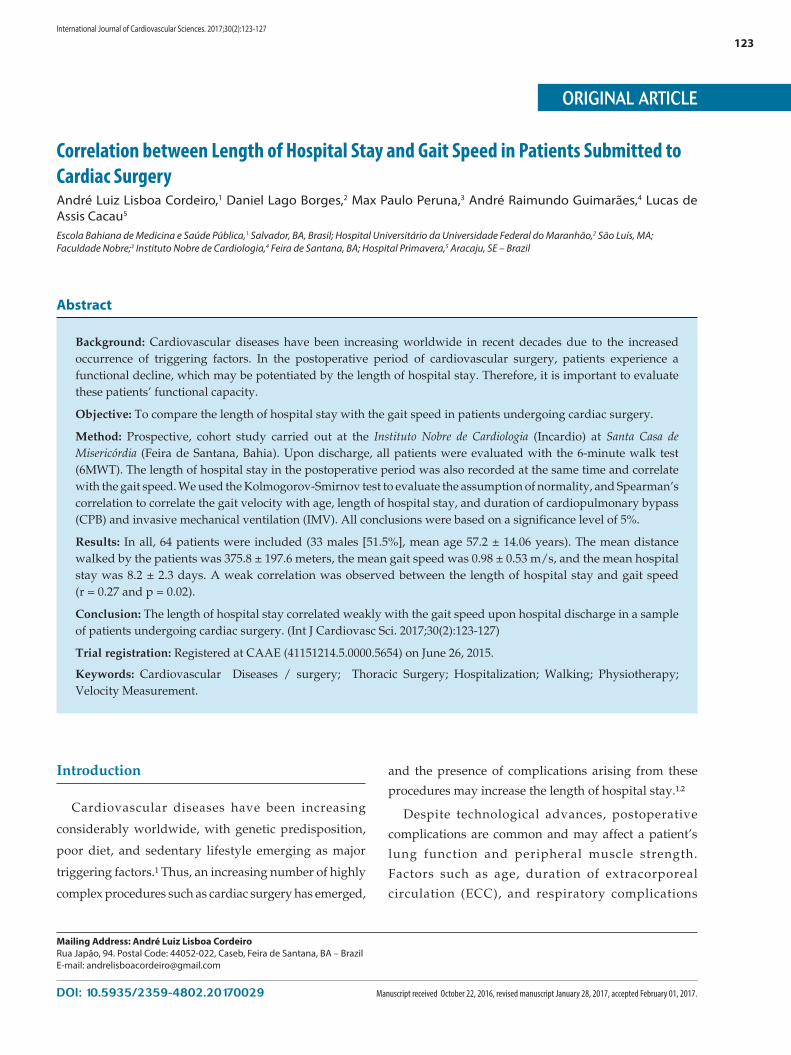
DOI: 10.5935/2359-4802.20170029
123International Journal of Cardiovascular Sciences. 2017;30(2):123-127
ORIGINAL ARTICLE
Mailing Address: André Luiz Lisboa CordeiroRua Japão, 94. Postal Code: 44052-022, Caseb, Feira de Santana, BA – BrazilE-mail: [email protected]
Correlation between Length of Hospital Stay and Gait Speed in Patients Submitted to Cardiac SurgeryAndré Luiz Lisboa Cordeiro,1 Daniel Lago Borges,2 Max Paulo Peruna,3 André Raimundo Guimarães,4 Lucas de Assis Cacau5
Escola Bahiana de Medicina e Saúde Pública,1 Salvador, BA, Brasil; Hospital Universitário da Universidade Federal do Maranhão,2 São Luís, MA; Faculdade Nobre;3 Instituto Nobre de Cardiologia,4 Feira de Santana, BA; Hospital Primavera,5 Aracaju, SE – Brazil
Manuscript received October 22, 2016, revised manuscript January 28, 2017, accepted February 01, 2017.
Abstract
Background: Cardiovascular diseases have been increasing worldwide in recent decades due to the increased occurrence of triggering factors. In the postoperative period of cardiovascular surgery, patients experience a functional decline, which may be potentiated by the length of hospital stay. Therefore, it is important to evaluate these patients’ functional capacity.
Objective: To compare the length of hospital stay with the gait speed in patients undergoing cardiac surgery.
Method: Prospective, cohort study carried out at the Instituto Nobre de Cardiologia (Incardio) at Santa Casa de Misericórdia (Feira de Santana, Bahia). Upon discharge, all patients were evaluated with the 6-minute walk test (6MWT). The length of hospital stay in the postoperative period was also recorded at the same time and correlate with the gait speed. We used the Kolmogorov-Smirnov test to evaluate the assumption of normality, and Spearman’s correlation to correlate the gait velocity with age, length of hospital stay, and duration of cardiopulmonary bypass (CPB) and invasive mechanical ventilation (IMV). All conclusions were based on a significance level of 5%.
Results: In all, 64 patients were included (33 males [51.5%], mean age 57.2 ± 14.06 years). The mean distance walked by the patients was 375.8 ± 197.6 meters, the mean gait speed was 0.98 ± 0.53 m/s, and the mean hospital stay was 8.2 ± 2.3 days. A weak correlation was observed between the length of hospital stay and gait speed (r = 0.27 and p = 0.02).
Conclusion: The length of hospital stay correlated weakly with the gait speed upon hospital discharge in a sample of patients undergoing cardiac surgery. (Int J Cardiovasc Sci. 2017;30(2):123-127)
Trial registration: Registered at CAAE (41151214.5.0000.5654) on June 26, 2015.
Keywords: Cardiovascular Diseases / surgery; Thoracic Surgery; Hospitalization; Walking; Physiotherapy; Velocity Measurement.
Introduction
Cardiovascular diseases have been increasing
considerably worldwide, with genetic predisposition,
poor diet, and sedentary lifestyle emerging as major
triggering factors.¹ Thus, an increasing number of highly
complex procedures such as cardiac surgery has emerged,
and the presence of complications arising from these
procedures may increase the length of hospital stay.¹,²
Despite technological advances, postoperative
complications are common and may affect a patient’s
lung function and peripheral muscle strength.
Factors such as age, duration of extracorporeal
circulation (ECC), and respiratory complications
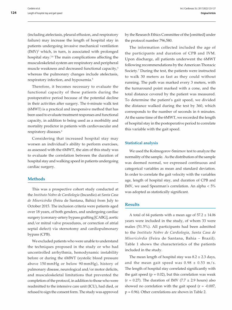
124Cordeiro et al.
Length of hospital stay and gait speed
Int J Cardiovasc Sci. 2017;30(2):123-127
Original Article
(including atelectasis, pleural effusion, and respiratory failure) may increase the length of hospital stay in patients undergoing invasive mechanical ventilation (IMV)3 which, in turn, is associated with prolonged hospital stay.3,4 The main complications affecting the musculoskeletal system are respiratory and peripheral muscle weakness and decreased functional capacity,4 whereas the pulmonary changes include atelectasis, respiratory infection, and hypoxemia.5
Therefore, it becomes necessary to evaluate the functional capacity of these patients during the postoperative period because of the potential decline in their activities after surgery. The 6-minute walk test (6MWT) is a practical and inexpensive method that has been used to evaluate treatment responses and functional capacity, in addition to being used as a morbidity and mortality predictor in patients with cardiovascular and respiratory diseases.6
Considering that increased hospital stay may worsen an individual’s ability to perform exercises, as assessed with the 6MWT, the aim of this study was to evaluate the correlation between the duration of hospital stay and walking speed in patients undergoing cardiac surgery.
Methods
This was a prospective cohort study conducted at the Instituto Nobre de Cardiologia (Incardio) at Santa Casa de Misericórdia (Feira de Santana, Bahia) from July to October 2015. The inclusion criteria were patients aged over 18 years, of both genders, and undergoing cardiac surgery (coronary-artery bypass grafting [CABG], aortic and/or mitral valve procedures, or correction of atrial septal defect) via sternotomy and cardiopulmonary bypass (CPB).
We excluded patients who were unable to understand the techniques proposed in the study or who had uncontrolled arrhythmia, hemodynamic instability before or during the 6MWT (systolic blood pressure above 150 mmHg or below 90 mmHg), history of pulmonary disease, neurological and/or motor deficits, and musculoskeletal limitations that prevented the completion of the protocol, in addition to those who were readmitted to the intensive care unit (ICU), had died, or refused to sign the consent form. The study was approved
by the Research Ethics Committee of the [omitted] under
the protocol number 796,580.
The information collected included the age of
the participants and duration of CPB and IVM.
Upon discharge, all patients underwent the 6MWT
following recommendations by the American Thoracic
Society.7 During the test, the patients were instructed
to walk 30 meters as fast as they could without
running. The path was marked every 3 meters, with
the turnaround point marked with a cone, and the
total distance covered by the patient was measured.
To determine the patient’s gait speed, we divided
the distance walked during the test by 360, which
corresponds to the number of seconds in 6 minutes.
At the same time of the 6MWT, we recorded the length
of hospital stay in the postoperative period to correlate
this variable with the gait speed.
Statistical analysis
We used the Kolmogorov-Smirnov test to analyze the
normality of the sample. As the distribution of the sample
was deemed normal, we expressed continuous and
categorical variables as mean and standard deviation.
In order to correlate the gait velocity with the variables
age, length of hospital stay, and duration of CPB and
IMV, we used Spearman’s correlation. An alpha < 5%
was adopted as statistically significant.
Results
A total of 64 patients with a mean age of 57.2 ± 14.06
years were included in the study, of whom 33 were
males (51.5%). All participants had been admitted
to the Instituto Nobre de Cardiologia, Santa Casa de
Misericórdia (Feira de Santana, Bahia – Brazil).
Table 1 shows the characteristics of the patients
included in the study.
The mean length of hospital stay was 8.2 ± 2.3 days,
and the mean gait speed was 0.98 ± 0.53 m/s.
The length of hospital stay correlated significantly with
the gait speed (p = 0.02), but this correlation was weak
(r = 0.27). The duration of IMV (7.7 ± 2.9 hours) also
showed no correlation with the gait speed (r = -0.007,
p = 0.96). Other correlations are shown in Table 2.
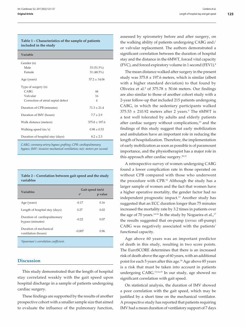
125
Table 1 – Characteristics of the sample of patients included in the study
Variable
Gender (n) Male Female
33 (51.5%)31 (48.5%)
Age (years) 57.2 ± 14.06
Type of surgery (n) CABG Valvular Correction of atrial septal defect
44164
Duration of CPB (minutes) 71.3 ± 21.4
Duration of IMV (hours) 7.7 ± 2.9
Walk distance (meters) 375.8 ± 197.6
Walking speed (m/s) 0.98 ± 0.53
Duration of hospital stay (days) 8.2 ± 2.3
CABG: coronary-artery bypass grafting; CPB: cardiopulmonary bypass; IMV: invasive mechanical ventilation; m/s: meters per second.
Table 2 – Correlation between gait speed and the study variables
Variables Gait speed (m/s)ra p value
Age (years) -0.17 0.16
Length of hospital stay (days) 0.27 0.02
Duration of cardiopulmonary bypass (minutes)
-0.22 0.07
Duration of mechanical ventilation (hours)
-0.007 0.96
aSpearman's correlation coefficient.
Cordeiro et al.
Length of hospital stay and gait speed
Int J Cardiovasc Sci. 2017;30(2):123-127
Original Article
Discussion
This study demonstrated that the length of hospital stay correlated weakly with the gait speed upon hospital discharge in a sample of patients undergoing cardiac surgery.
These findings are supported by the results of another prospective cohort with a smaller sample size that aimed to evaluate the influence of the pulmonary function,
assessed by spirometry before and after surgery, on the walking ability of patients undergoing CABG and/or valvular replacement. The authors demonstrated a significant correlation between the duration of hospital stay and the distance in the 6MWT, forced vital capacity (FVC), and forced expiratory volume in 1 second (FEV1).8
The mean distance walked after surgery in the present study was 375.8 ± 197.6 meters, which is similar (albeit with a higher standard deviation) to that found by Oliveira et al.8 of 375.78 ± 50.66 meters. Our findings are also similar to those of another cohort study with a 2-year follow-up that included 215 patients undergoing CABG, in which the sedentary participants walked 375.53 ± 210.92 meters after 2 years.9 The 6MWT is a test well tolerated by adults and elderly patients after cardiac surgery without complications,10 and the findings of this study suggest that early mobilization and ambulation have an important role in reducing the length of hospitalization. Therefore, the implementation of early mobilization as soon as possible is of paramount importance, and the physiotherapist has a major role in this approach after cardiac surgery.10,11
A retrospective survey of women undergoing CABG found a lower complication rate in those operated on without CPB compared with those who underwent the procedure with CPB.12 Although the study has a larger sample of women and the fact that women have a higher operative mortality, the gender factor had no independent prognostic impact.13 Another study has suggested that an ECC duration longer than 75 minutes increased the mortality rate by 3.2 times in patients over the age of 70 years.14-16 In the study by Nogueira et al.,17 the results suggested that on-pump (versus off-pump) CABG was negatively associated with the patients’ functional capacity.
Age above 60 years was an important predictor of death in this study, resulting in two score points. The EuroSCORE determines that there is an increased risk of death above the age of 60 years, with an additional point for each 5 years after this age.18 Age above 85 years is a risk that must be taken into account in patients undergoing CABG.15,16,19 In our study, age showed no significant correlation with gait speed.
On statistical analysis, the duration of IMV showed a poor correlation with the gait speed, which may be justified by a short time on the mechanical ventilator. A prospective study has reported that patients requiring IMV had a mean duration of ventilatory support of 7 days
126
1. Leguisamo CP, Kalil RA, Furlani AP. Effetiveness of a preoperative physiotherapeutic approach in myocardial revascularization. Braz J Cardiovasc Surg. 2005;20(2):134-41.
2. Shakouri SK, Salekzamani Y, Taghizadieh A, Sabbagh-Jadid H, Soleymani J, Sahebi L, et al. Effect of respiratory rehabilitation before open cardiac surgery on respiratory function: a randomized clinical trial. J Cardiovasc Thorac Res. 2015;7(1):13-7.
3. Pinheiro AR, Christofoletti G. Fisioterapia motora em pacientes internados na unidade de terapia intensiva: uma revisão sistemática. Rev Bras Ter Intensiva. 2012;24(2):188-96.
4. Fonseca L, Vieira FN, Azzolin KO. Fatores associados ao tempo de ventilação mecânica no pós-operatório de cirurgia cardíaca. Rev Gaúcha Enf. 2014;35(2):67-72.
5. Preisig A, Lagni VB, Almeida VL, Vieira FN, Lucio EA, Santos LJ, et al. Ventilação não invasiva após cirurgia cardiovascular: um ensaio clínico randomizado. Rev Bras Cardiol. 2014;27(1):539-48.
6. Laizo A, Delgado FE, Rocha GM. Complicações que aumentam o tempo de permanência na unidade de terapia intensiva na cirurgia cardíaca. Rev Bras Cir Cardiovasc. 2010;25(2):166-71.
7. ATS Committee on Proficiency Standards for Clinical Pulmonary Function Laboratories. ATS statement guidelines for the six-minute walk test. Am J Respir Crit Care Med. 2002;166(1):111-7.
8. Oliveira EK, Silva VZ, Turquetto AL. Relationship on walk test and pulmonary function tests with the length of hospitalization in cardiac surgery patients. Rev Bras Cir Cardiovasc. 2009;24(4):478-84.
9. Nery RM, Martini MR, Vidor CD, Mahmud MI, Zanini M, Loureiro A, et al. Alterações na capacidade funcional de pacientes após dois anos da cirurgia de revascularização do miocárdio. Rev Bras Cir Cardiovasc. 2010;25(2):224-8.
10. Fiorina C, Vizzardi E, Lorusso R, Maggio M, De Cicco G, Nodari S, et al. The 6-min walking test early after cardiac surgery. Reference values and the effects of rehabilitation programme. Eur J Cardiothorac Surg. 2007;32(5):724-9.
11. Cordeiro AL, Silva AA, Santana MI. Impacto hemodinâmico da deambulação nos pacientes submetidos a cirurgia cardíaca. Rev DERC. 2015;21(2):54-7.
12. Escobar RD. Estudo comparativo entre cirurgia de revascularização miocárdica com e sem circulação extracorpórea em mulheres. Rev Bras Cir Cardiovasc. 2010;25(2):238-44.
13. Amato VL, Timerman A, Paes ÂT, Baltar VT, Farsky PS, Farran JA, et al. Immediate results of myocardial revascularization: comparison between men and women. Arq Bras Cardiol. 2004;83 Spec No:14-20.
14. Anderson AJ, Barros Neto FX, Costa MD, Dantas LD, Hueb AC, Prata MF. Preditores de mortalidade em pacientes acima de 70 anos na revascularização miocárdica ou troca valvar com circulação extracorpórea. Rev Bras Cir Cardiovasc. 2011;26(1):69-75.
15. Alves Junior L, Rodrigues AJ, Evora PR, Basseto S, Scorzoni Filho A, Luciano PM, et al. Fatores de risco em septuagenários ou mais idosos submetidos a revascularização do miocárdio e ou operações valvares. Rev Bras Cir Cardiovasc. 2008;23(4):550-5.
References
Cordeiro et al.
Length of hospital stay and gait speed
Int J Cardiovasc Sci. 2017;30(2):123-127
Original Article
and mean length of ICU stay of 13 days. This study found
that half of the patients who were not extubated within
24 hours remained in the ICU for more than one week.20
One of the hypotheses to explain the weak correlation
between the variables in the present study is the fact that
often, clinical factors such as international normalized
ratio (INR) value and echocardiographic alterations
may increase the length of hospital stay, but these
variables do not correlate with the patient’s functional
capacity. Therefore, a longer length of hospitalization
may be sufficient to help the patients return to their
preoperative functionality and perform well during
the tests.
The limitations of this study include the small sample
size and the lack of information regarding the patients’
pulmonary function and body mass index, which
can have an impact on the completion of the 6MWT.
Moreover, the study did not include a preoperative
evaluation to demonstrate the progression of the gait
speed during hospital stay.
Conclusion
We conclude that the length of hospital stay correlated weakly with the gait speed upon hospital
discharge in a sample of patients undergoing cardiac surgery.
Author contributions
Conception and design of the research: Cordeiro ALL. Acquisition of data: Cordeiro ALL, Peruna MP, Guimarães AR. Analysis and interpretation of the data: Borges DL, Cacau LA. Statistical analysis: Borges DL, Cacau LA. Writing of the manuscript: Cordeiro ALL, Peruna MP, Guimarães AR. Critical revision of the manuscript for intellectual content: Cordeiro ALL, Borges DL, Cacau LA.
Potential Conflict of Interest
No potential conflict of interest relevant to this article was reported.
Sources of Funding
There were no external funding sources for this study.
Study Association
This article is part of the thesis of master submitted by André Luiz Lisboa Cordeiro, from Escola Bahiana de Medicina e Saúde Pública.

127
16. Silva AM, Campagnucci VP, Pereira WL, Rosa RF, Franken RA, Gandra SM, et al. Revascularização do miocárdio sem circulação extracorpórea em idosos: análise da morbidade e mortalidade. Rev Bras Cir Cardiovasc. 2008;23(1):40-5.
17. Nogueira CR, Hueb W, Takiuti ME, Girardi PB, Nakano T, Fernandes F, et al. Quality of life after on-pump and off-pump coronary artery bypass grafting surgery. Arq Bras Cardiol. 2008;91(4):238-44.
18. Cadore MP, Guaragna JC, Anacker JF, Albuquerque LC, Bodanese LC, Piccoli JD, et al. Score proposal to evaluate surgical risk in patients
submitted to myocardial revascularization surgery. Rev Bras Cir Cardiovasc. 2010;25(4):447-56.
19. De Bacco MW, Sartori AP, Sant'Anna JR, Santos MF, Prates PR, Kalil RA, et al. Risk factors for hospital mortality in valve replacement with mechanical prosthesis. Rev Bras Cir Cardiovasc. 2009;24(3):334-40.
20. Esteban A, Anzueto A, Frutos F, Alía I, Brochard L, Stewart TE, et al; Mechanical Ventilation International Study Group. Characteristics and outcomes in adult patients receiving mechanical ventilation: a 28-day international study. JAMA. 2002;287(3):345-55.
Cordeiro et al.
Length of hospital stay and gait speed
Int J Cardiovasc Sci. 2017;30(2):123-127
Original Article
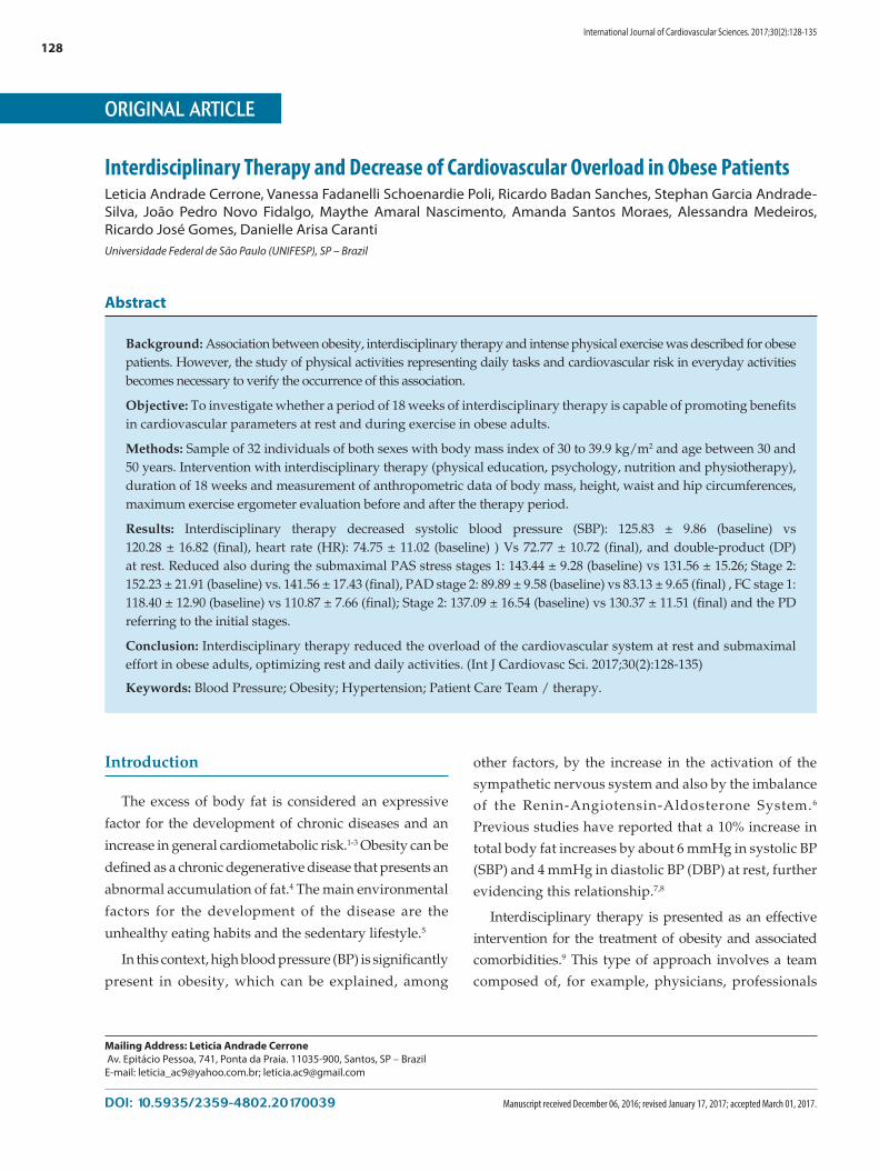
DOI: 10.5935/2359-4802.20170039
Introduction
The excess of body fat is considered an expressive
factor for the development of chronic diseases and an
increase in general cardiometabolic risk.1-3 Obesity can be
defined as a chronic degenerative disease that presents an
abnormal accumulation of fat.4 The main environmental
factors for the development of the disease are the
unhealthy eating habits and the sedentary lifestyle.5
In this context, high blood pressure (BP) is significantly
present in obesity, which can be explained, among
other factors, by the increase in the activation of the
sympathetic nervous system and also by the imbalance
of the Renin-Angiotensin-Aldosterone System.6
Previous studies have reported that a 10% increase in
total body fat increases by about 6 mmHg in systolic BP
(SBP) and 4 mmHg in diastolic BP (DBP) at rest, further
evidencing this relationship.7,8
Interdisciplinary therapy is presented as an effective
intervention for the treatment of obesity and associated
comorbidities.9 This type of approach involves a team
composed of, for example, physicians, professionals
128International Journal of Cardiovascular Sciences. 2017;30(2):128-135
ORIGINAL ARTICLE
Mailing Address: Leticia Andrade Cerrone Av. Epitácio Pessoa, 741, Ponta da Praia. 11035-900, Santos, SP – BrazilE-mail: [email protected]; [email protected]
Interdisciplinary Therapy and Decrease of Cardiovascular Overload in Obese PatientsLeticia Andrade Cerrone, Vanessa Fadanelli Schoenardie Poli, Ricardo Badan Sanches, Stephan Garcia Andrade-Silva, João Pedro Novo Fidalgo, Maythe Amaral Nascimento, Amanda Santos Moraes, Alessandra Medeiros, Ricardo José Gomes, Danielle Arisa CarantiUniversidade Federal de São Paulo (UNIFESP), SP – Brazil
Manuscript received December 06, 2016; revised January 17, 2017; accepted March 01, 2017.
Abstract
Background: Association between obesity, interdisciplinary therapy and intense physical exercise was described for obese patients. However, the study of physical activities representing daily tasks and cardiovascular risk in everyday activities becomes necessary to verify the occurrence of this association.
Objective: To investigate whether a period of 18 weeks of interdisciplinary therapy is capable of promoting benefits in cardiovascular parameters at rest and during exercise in obese adults.
Methods: Sample of 32 individuals of both sexes with body mass index of 30 to 39.9 kg/m2 and age between 30 and 50 years. Intervention with interdisciplinary therapy (physical education, psychology, nutrition and physiotherapy), duration of 18 weeks and measurement of anthropometric data of body mass, height, waist and hip circumferences, maximum exercise ergometer evaluation before and after the therapy period.
Results: Interdisciplinary therapy decreased systolic blood pressure (SBP): 125.83 ± 9.86 (baseline) vs 120.28 ± 16.82 (final), heart rate (HR): 74.75 ± 11.02 (baseline) ) Vs 72.77 ± 10.72 (final), and double-product (DP) at rest. Reduced also during the submaximal PAS stress stages 1: 143.44 ± 9.28 (baseline) vs 131.56 ± 15.26; Stage 2: 152.23 ± 21.91 (baseline) vs. 141.56 ± 17.43 (final), PAD stage 2: 89.89 ± 9.58 (baseline) vs 83.13 ± 9.65 (final) , FC stage 1: 118.40 ± 12.90 (baseline) vs 110.87 ± 7.66 (final); Stage 2: 137.09 ± 16.54 (baseline) vs 130.37 ± 11.51 (final) and the PD referring to the initial stages.
Conclusion: Interdisciplinary therapy reduced the overload of the cardiovascular system at rest and submaximal effort in obese adults, optimizing rest and daily activities. (Int J Cardiovasc Sci. 2017;30(2):128-135)
Keywords: Blood Pressure; Obesity; Hypertension; Patient Care Team / therapy.
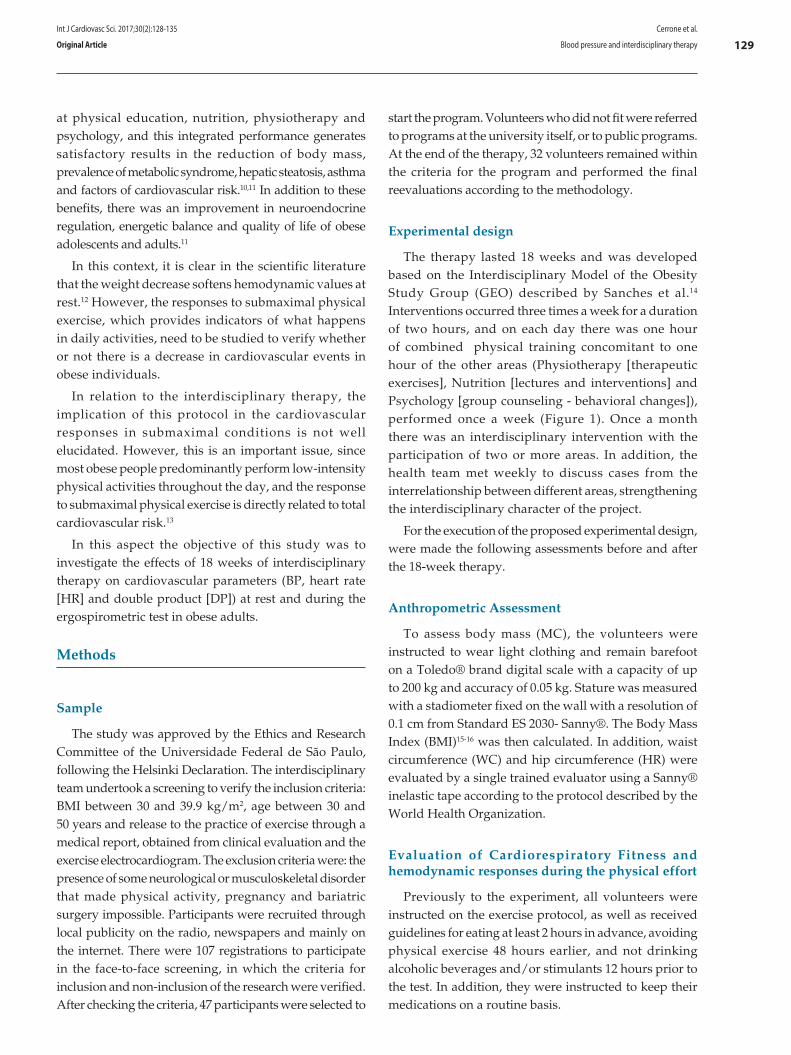
129
at physical education, nutrition, physiotherapy and psychology, and this integrated performance generates satisfactory results in the reduction of body mass, prevalence of metabolic syndrome, hepatic steatosis, asthma and factors of cardiovascular risk.10,11 In addition to these benefits, there was an improvement in neuroendocrine regulation, energetic balance and quality of life of obese adolescents and adults.11
In this context, it is clear in the scientific literature that the weight decrease softens hemodynamic values at rest.12 However, the responses to submaximal physical exercise, which provides indicators of what happens in daily activities, need to be studied to verify whether or not there is a decrease in cardiovascular events in obese individuals.
In relation to the interdisciplinary therapy, the implication of this protocol in the cardiovascular responses in submaximal conditions is not well elucidated. However, this is an important issue, since most obese people predominantly perform low-intensity physical activities throughout the day, and the response to submaximal physical exercise is directly related to total cardiovascular risk.13
In this aspect the objective of this study was to investigate the effects of 18 weeks of interdisciplinary therapy on cardiovascular parameters (BP, heart rate [HR] and double product [DP]) at rest and during the ergospirometric test in obese adults.
Methods
Sample
The study was approved by the Ethics and Research Committee of the Universidade Federal de São Paulo, following the Helsinki Declaration. The interdisciplinary team undertook a screening to verify the inclusion criteria: BMI between 30 and 39.9 kg/m2, age between 30 and 50 years and release to the practice of exercise through a medical report, obtained from clinical evaluation and the exercise electrocardiogram. The exclusion criteria were: the presence of some neurological or musculoskeletal disorder that made physical activity, pregnancy and bariatric surgery impossible. Participants were recruited through local publicity on the radio, newspapers and mainly on the internet. There were 107 registrations to participate in the face-to-face screening, in which the criteria for inclusion and non-inclusion of the research were verified. After checking the criteria, 47 participants were selected to
start the program. Volunteers who did not fit were referred to programs at the university itself, or to public programs. At the end of the therapy, 32 volunteers remained within the criteria for the program and performed the final reevaluations according to the methodology.
Experimental design
The therapy lasted 18 weeks and was developed based on the Interdisciplinary Model of the Obesity Study Group (GEO) described by Sanches et al.14 Interventions occurred three times a week for a duration of two hours, and on each day there was one hour of combined physical training concomitant to one hour of the other areas (Physiotherapy [therapeutic exercises], Nutrition [lectures and interventions] and Psychology [group counseling - behavioral changes]), performed once a week (Figure 1). Once a month there was an interdisciplinary intervention with the participation of two or more areas. In addition, the health team met weekly to discuss cases from the interrelationship between different areas, strengthening the interdisciplinary character of the project.
For the execution of the proposed experimental design, were made the following assessments before and after the 18-week therapy.
Anthropometric Assessment
To assess body mass (MC), the volunteers were instructed to wear light clothing and remain barefoot on a Toledo® brand digital scale with a capacity of up to 200 kg and accuracy of 0.05 kg. Stature was measured with a stadiometer fixed on the wall with a resolution of 0.1 cm from Standard ES 2030- Sanny®. The Body Mass Index (BMI)15-16 was then calculated. In addition, waist circumference (WC) and hip circumference (HR) were evaluated by a single trained evaluator using a Sanny® inelastic tape according to the protocol described by the World Health Organization.
Evaluation of Cardiorespiratory Fitness and hemodynamic responses during the physical effort
Previously to the experiment, all volunteers were instructed on the exercise protocol, as well as received guidelines for eating at least 2 hours in advance, avoiding physical exercise 48 hours earlier, and not drinking alcoholic beverages and/or stimulants 12 hours prior to the test. In addition, they were instructed to keep their medications on a routine basis.
Cerrone et al.
Blood pressure and interdisciplinary therapy
Int J Cardiovasc Sci. 2017;30(2):128-135
Original Article
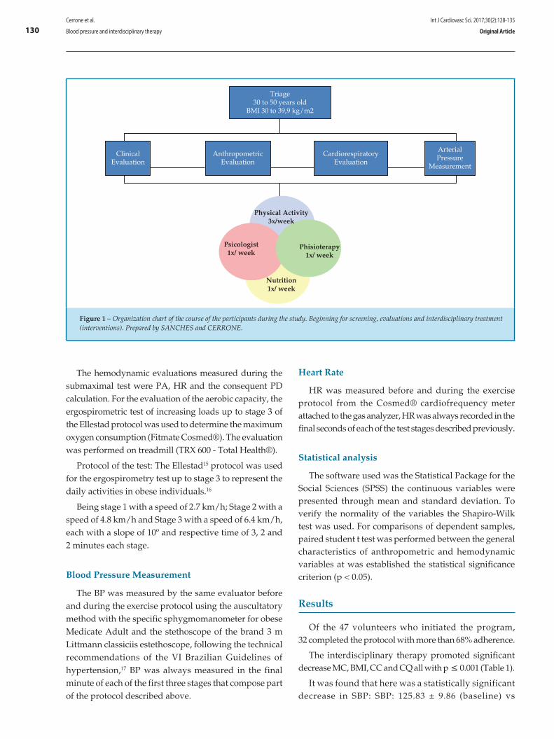
130
Triage30 to 50 years old
BMI 30 to 39,9 kg/m2
ClinicalEvaluation
AnthropometricEvaluation
CardiorespiratoryEvaluation
ArterialPressure
Measurement
Physical Activity3x/week
Psicologist1x/ week
Phisioterapy1x/ week
Nutrition1x/ week
Figure 1 – Organization chart of the course of the participants during the study. Beginning for screening, evaluations and interdisciplinary treatment (interventions). Prepared by SANCHES and CERRONE.
The hemodynamic evaluations measured during the submaximal test were PA, HR and the consequent PD calculation. For the evaluation of the aerobic capacity, the ergospirometric test of increasing loads up to stage 3 of the Ellestad protocol was used to determine the maximum oxygen consumption (Fitmate Cosmed®). The evaluation was performed on treadmill (TRX 600 - Total Health®).
Protocol of the test: The Ellestad15 protocol was used for the ergospirometry test up to stage 3 to represent the daily activities in obese individuals.16
Being stage 1 with a speed of 2.7 km/h; Stage 2 with a speed of 4.8 km/h and Stage 3 with a speed of 6.4 km/h, each with a slope of 10º and respective time of 3, 2 and 2 minutes each stage.
Blood Pressure Measurement
The BP was measured by the same evaluator before and during the exercise protocol using the auscultatory method with the specific sphygmomanometer for obese Medicate Adult and the stethoscope of the brand 3 m Littmann classiciis estethoscope, following the technical recommendations of the VI Brazilian Guidelines of hypertension,17 BP was always measured in the final minute of each of the first three stages that compose part of the protocol described above.
Heart Rate
HR was measured before and during the exercise protocol from the Cosmed® cardiofrequency meter attached to the gas analyzer, HR was always recorded in the final seconds of each of the test stages described previously.
Statistical analysis
The software used was the Statistical Package for the Social Sciences (SPSS) the continuous variables were presented through mean and standard deviation. To verify the normality of the variables the Shapiro-Wilk test was used. For comparisons of dependent samples, paired student t test was performed between the general characteristics of anthropometric and hemodynamic variables at was established the statistical significance criterion (p < 0.05).
Results
Of the 47 volunteers who initiated the program, 32 completed the protocol with more than 68% adherence.
The interdisciplinary therapy promoted significant decrease MC, BMI, CC and CQ all with p ≤ 0.001 (Table 1).
It was found that here was a statistically significant decrease in SBP: SBP: 125.83 ± 9.86 (baseline) vs
Cerrone et al.
Blood pressure and interdisciplinary therapy
Int J Cardiovasc Sci. 2017;30(2):128-135
Original Article
131
120.28 ± 16.82 (final); Heart rate (HR): 74.75 ± 11.02 (baseline) vs 72.77 ± 10.72 (final) and double-product (PD): 9139.06 ± 1739.162 (baseline) vs. 8464.37 ± 2481.76 (final) at rest after 18 weeks of interdisciplinary therapy in both sexes with p ≤ 0.01. (Figure 2).
The hemodynamic variables (SBP, DBP, HR and SD) during exercise, pre and post-therapy are presented in figure 3. Our results showed a decrease in the values of PAS stages 1: 143.44 ± 9.28 (baseline) vs 131.56 ± 15.26; Stage 2: 152.23 ± 21.91 (baseline) vs. 141.56 ± 17.43 (final), PAD stage 2: 89.89 ± 9.58 (baseline) vs 83.13 ± 9.65 (final), FC stage 1: 118.40 ± 12.90 (baseline) vs 110.87 ± 7.66 (final); Stage 2: 137.09 ± 16.54 (baseline) vs 130.37 ± 11.51 (final) and the DP stage 1: 13856.14 ± 3858.45 (baseline) vs 9091.45 ± 7249.90 (final ) And stage 2: 19691.14 ± 6906.31 (baseline) vs 11577.50 ± 9259.98 (final).
Discussion
Interdisciplinary therapy was efficient in reducing MC, BMI, CC, CQ and SBP at rest and SBP, FC and DP at submaximal intensities in obese individuals. Regarding the anthropometric data (MC, BMI, CC and QC), our results corroborate with those presented by Sanches et al.10,14 and Franz et al.12 regarding the decrease in values when compared to values before and after interdisciplinary therapy.
Submaximal hemodynamic responses reflect the energetic demand for cardiovascular effort imposed on everyday activities, such as walking from one place to another, cycling, cleaning the house and even jogging to reach a bus.18 In this regard, it is important to emphasize that these elevated cardiovascular variables represent a public health problem that can silently affect several systems such as: renal, circulatory, nervous and visual.19,20
The expected behavior of BP in a stress test is an increase in SBP according to increased cardiac output and the maintenance or a slight decrease in DBP. This decrease depends mainly on the peripheral vascular resistance and will represent the efficiency of the vasodilator mechanisms.8,21,22 Corroborating the studies of Franz et al.12 we observed that changes in lifestyle are closely related to improvements in the cardiovascular system.
Decreased cardiac overload at rest indicates an improvement in the mechanisms responsible for cardiovascular control, emphasizing the importance of baroreceptor control, but also emphasizing the contribution of the mechanisms of the renin-angiotensin-aldosterone system, peripheral vasodilation and possible inhibition of the sympathetic nervous system.8,21,22 It is important to note that decreases in BP, HR and DP even in small magnitudes, promote large reductions in the rate of cardiovascular complications and general mortality.23
The scientific literature shows that the physical exercise, present in the interdisciplinary therapy, promotes a decrease of the BP values in a chronic way. In this context, we discussed several factors for this improvement, such as the increase of the parasympathetic tonus in the myocardium, a decrease in peripheral vascular resistance or an increase in the vasodilatory effect of endothelial function.24
In this area, and corroborating previous studies, interdisciplinary therapy was efficient in improving cardiorespiratory parameters in the face of submaximal and maximal exertion.10 Therefore, we understand that interdisciplinary therapy provides the individual with comprehensive care, including benefits to biological systems, such as the cardiovascular system, cited above. With a didactic purpose we explain these systems in isolation, but in practice they are interrelated in a holistic way and offer a complex improvement with benefits to individuals who still need to be studied.3,24
Table 1 – Anthropometric changes before and after 18 weeks of Interdisciplinary Intervention
Variables Basal Mean ± SD After therapy Mean ± SD P
BM (kg) 97.00 ± 12.80 94.09 ± 12.19 0.001*
Height (m) 1.66 ± 0.13 1.66 ± 0.13 0.813
BMI (kg/m²) 34.96 ± 2.93 33.68 ± 2.73 0.001*
WC (cm) 110.05 ± 8.96 107.20 ± 8.99 0.001*
HC (cm) 120.92 ± 7.97 117.01 ± 7.75 0.001*
Anthropometric variables before and after 18 weeks of intervention. BM: body mass; BMI: body mass index; WC: waist circumference; HC: hip circumference, P (significance level adopted at 0.05 (*), n (32). Presented in Mean and Standard Deviation.
Cerrone et al.
Blood pressure and interdisciplinary therapy
Int J Cardiovasc Sci. 2017;30(2):128-135
Original Article
132
In addition to physical exercise, dietary intake has a great influence on BP control. Excessive sodium intake (greater than 2000 mg/day) and excessive dietary intake are associated with an increased risk of BP elevation, which is considered a risk factor for death.25 Thus, we know that the negative energy balance, with consequent loss of body mass, has a great contribution in reducing food intake and improving the overall quality of food.
The interdisciplinary approach in the treatment of obesity has the proposal to go beyond weight loss, seeking to achieve improvements in the health of the individual as a whole, including its psychosocial dimension.26 Interventions that cover these different dimensions are fundamental even for adherence to therapy, since stress27 and symptoms of depression,28 for example, have already been evidenced as factors that interfere in the performance of physical activity.
According to the results and the evidence cited, it is important to highlight the limitations of the present study, such as: absence of a control group, follow-up of the menstrual cycle of the volunteers, and hypotensive drugs.
Conclusions
Interdisciplinary therapy allowed the reduction of SBP, HR and rest DP and in the submaximal intensities, also preserving the lower use of the heart in daily efforts. The decrease in these variables at rest leads to a lower overload on the cardiovascular system of individuals in their daily life and the decrease during exercise preserves the heart in daily activities. Therefore, we understand that interdisciplinary therapy is an important intervention strategy to increase the level of daily physical activity, aiming at the daily cardioprotective effect, especially for individuals who need to decrease the overload to the cardiovascular system such as obese individuals.
Acknowledge
Thanks to Prof. Dr Raphael Ritti for the advice and follow-up of the writing process and to Prof. Bruno Remígio for the elaboration of the graphs.
A
140
130
120
110
100
0Basal Final
Basal Final Basal Final
Basal Final
PAS
(mm
Hg)
PAS
(mm
Hg)
*
**
B
100
90
80
70
60
50
40
0
100
90
80
70
60
50
40
0
C D
FC (b
pm
)
12000
10000
8000
6000
0
DP
(mm
Hg*
bp
m)
Figure 2 – Resting hemodynamic variables before and after 18 weeks of interdisciplinary intervention. SBP (systolic blood pressure), DBP (diastolic blood pressure), HR (heart rate) and DP (double-product). Statistically significant results (*), n (32). The data is displayed in Average and Standard Deviation.
Cerrone et al.
Blood pressure and interdisciplinary therapy
Int J Cardiovasc Sci. 2017;30(2):128-135
Original Article
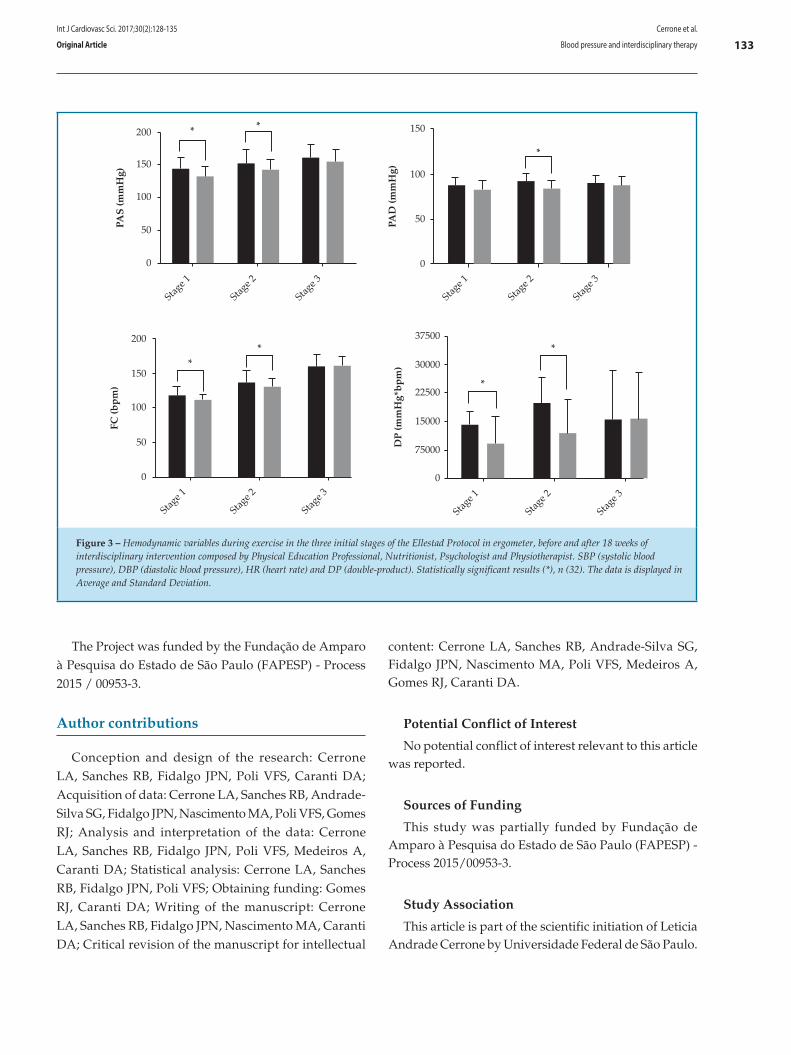
133
200
150
100
50
0
200
150
100
50
0
150
100
50
0
PAS
(mm
Hg)
PAD
(mm
Hg)
FC (b
pm
)
DP
(mm
Hg*
bp
m)
Stag
e 1
Stag
e 2
Stag
e 3
Stag
e 1
Stag
e 2
Stag
e 3
Stag
e 1
Stag
e 2
Stag
e 3
Stag
e 1
Stag
e 2
Stag
e 3
37500
30000
22500
15000
75000
0
*
*
* *
*
*
*
Figure 3 – Hemodynamic variables during exercise in the three initial stages of the Ellestad Protocol in ergometer, before and after 18 weeks of interdisciplinary intervention composed by Physical Education Professional, Nutritionist, Psychologist and Physiotherapist. SBP (systolic blood pressure), DBP (diastolic blood pressure), HR (heart rate) and DP (double-product). Statistically significant results (*), n (32). The data is displayed in Average and Standard Deviation.
The Project was funded by the Fundação de Amparo à Pesquisa do Estado de São Paulo (FAPESP) - Process 2015 / 00953-3.
Author contributions
Conception and design of the research: Cerrone LA, Sanches RB, Fidalgo JPN, Poli VFS, Caranti DA; Acquisition of data: Cerrone LA, Sanches RB, Andrade-Silva SG, Fidalgo JPN, Nascimento MA, Poli VFS, Gomes RJ; Analysis and interpretation of the data: Cerrone LA, Sanches RB, Fidalgo JPN, Poli VFS, Medeiros A, Caranti DA; Statistical analysis: Cerrone LA, Sanches RB, Fidalgo JPN, Poli VFS; Obtaining funding: Gomes RJ, Caranti DA; Writing of the manuscript: Cerrone LA, Sanches RB, Fidalgo JPN, Nascimento MA, Caranti DA; Critical revision of the manuscript for intellectual
content: Cerrone LA, Sanches RB, Andrade-Silva SG, Fidalgo JPN, Nascimento MA, Poli VFS, Medeiros A, Gomes RJ, Caranti DA.
Potential Conflict of Interest
No potential conflict of interest relevant to this article was reported.
Sources of Funding
This study was partially funded by Fundação de Amparo à Pesquisa do Estado de São Paulo (FAPESP) - Process 2015/00953-3.
Study Association
This article is part of the scientific initiation of Leticia Andrade Cerrone by Universidade Federal de São Paulo.
Cerrone et al.
Blood pressure and interdisciplinary therapy
Int J Cardiovasc Sci. 2017;30(2):128-135
Original Article
134
1. Calvo SS, Egan JM. The endocrinology of taste receptors. Nat Rev Endocrinol. 2015;11(4):213-27.
2. Trayhurn P, Bing C, Wood IS. Adipose tissue and adipokines--energy regulation from the human perspective. J Nutr. 2006;136(7 Suppl):1935S-9S.
3. César M, Tenório C, Kennedy C, Sá CK, Freitas MM, Ruas GR, et al. Continuous or interval training and inflammatory response in obese women. Int J Cardiovasc Sci. 2015;28(4):282-9.
4. Conde WL, Monteiro CA. Nutrition transition and double burden of undernutrition and excess of weight in Brazil. Am J Clin Nutr. 2014;100(6):1617S-22S.
5. Ng M, Fleming T, Robinson M, Thomson B, Graetz N, Margono C, et al. Global, regional, and national prevalence of overweight and obesity in children and adults during 1980–2013: a systematic analysis for the Global Burden of Disease Study 2013. Lancet. 2014;384(9945):766-781. Erratum in: Lancet. 2014;384(9945):746.
6. Rahmouni K, Correia ML, Haynes WG, Mark AL. Obesity-associated hypertension: new insights into mechanisms. Hypertension. 2005;45(1):9-14.
7. Cauduro A, Bergmann M, Bergmann G. Physical activity, overweight and blood pressure: independent and combined association in adolescents. Rev Bras Ativ Fis Saúde. 2016;20:483.
8. Pieruzzi F, Antolini L, Salerno FR, Giussani M, Brambilla P, Galbiati S, et al. The role of blood pressure, body weight and fat distribution on left ventricular mass, diastolic function and cardiac geometry in children. J Hypertens. 2015;33(6):1182-92.
9. Damaso AR, de Piano A, Campos RM, Corgosinho FC, Siegfried W, Caranti DA, et al. Multidisciplinary approach to the treatment of obese adolescents: effects on cardiovascular risk factors, inflammatory profile, and neuroendocrine regulation of energy balance. Int J Endocrinol. 2013;2013:541032.
10. Sanches RB, Silva SG, Rossi S, Fidalgo JP, Moraes AS, Jamar G, et al. Body composition and aerobic fitness in obese women: beneficial effects of interdisciplinary therapy. Rev Bras Ativ Fis Saúde. 2013;18(3):354-62.
11. Bischoff SC, Damms-Machado A, Betz C, Herpertz S, Legenbauer T, Low T, et al. Multicenter evaluation of an interdisciplinary 52-week weight loss program for obesity with regard to body weight, comorbidities and quality of life--a prospective study. Int J Obes (Lond). 2012;36(4):614-24.
12. Franz MJ, Boucher JL, Rutten-Ramos S, VanWormer JJ. Lifestyle weight-loss intervention outcomes in overweight and obese adults with type 2 diabetes: a systematic review and meta-analysis of randomized clinical trials. J Acad Nutr Diet. 2015;115(9):1447-63.
13. Fagard RH. Exercise characteristics and the blood pressure response to dynamic physical training. Med Sci Sports Exerc. 2001;33(6 Suppl):S484-92.
14. Sanches RB, Andrade-Silva SG, Novo Fidalgo JP, Moraes AS, Poli VF, Oyama LM, et al. Interdisciplinary therapy improves cardiorespiratory fitness and inflammatory markers in obese adult women. Obes Med. 2016;2:1-7.
15. Ellestad MH, Allen W, Wan MC, Kemp GL. Maximal treadmill stress testing for cardiovascular evaluation. Circulation. 1969;39(4):517-22.
16. Borg G. Borg’s perceived exertion and pain scales. Champaign: Hum Kinetics; 1998. p. 43-6.
17. Sociedade Brasileira de Cardiologia; Sociedade Brasileira de Hipertensão; Sociedade Brasileira de Nefrologia. [VI Brazilian Guidelines on Hypertension]. Arq Bras Cardiol. 2010;95(1 Suppl):1-51. Erratum in: Arq Bras Cardiol. 2010;95(4):553.
18. Monteiro MF, Sobral Filho DC. Exercício físico e o controle da pressão arterial. Rev Bras Med Esporte. 2004;10(6):513-6.
19. Lovato NS, Loch MR, González AD, Lopes ML. Adherence to physical activity programs: characteristics of participants and very assiduous and less assiduous. 2015;20(2):184-92.
20. Weber C, Costa J, Olinto MT, Manenti ER, Henn R, Paniz VM, et al. Physical activity after hospital for circulatory diseases. Rev Bras Ativ Fis Saúde. 2016;20(5):492-502.
21. Fall T, Hagg S, Ploner A, Magi R, Fischer K, Draisma HH, et al; ENGAGE Consortium. Age- and sex-specific causal effects of adiposity on cardiovascular risk factors. Diabetes. 2015;64(5):1841-52.
22. Javorka M, Turianikova Z, Tonhajzerova I, Lazarova Z, Czippelova B, Javorka K. Heart rate and blood pressure control in obesity - how to detect early dysregulation? Clin Physiol Funct Imaging. 2016;36(5):337-45.
23. Duschek S, Worsching J, Reyes Del Paso GA. Autonomic cardiovascular regulation and cortical tone. Clin Physiol Funct Imaging. 2015;35(5):383-92.
24. Beck DT, Casey DP, Martin JS, Emerson BD, Braith RW. Exercise training improves endothelial function in young prehypertensives. Exp Biol Med (Maywood). 2013;238(4):433-41.
25. Thuesen BH, Toft U, Buhelt LP, Linneberg A, Friedrich N, Nauck M, et al. Estimated daily salt intake in relation to blood pressure and blood lipids: the role of obesity. Eur J Prev Cardiol. 2015;22(12):1567-74.
26. Nutter S, Russell-Mayhew S, Alberga AS, Arthur N, Kassan A, Lund DE, et al. Positioning of weight bias: moving towards social justice. J Obes. 2016;2016:3753650.
27. Stults-Kolehmainen MA, Sinha R. The effects of stress on physical activity and exercise. Sports Med. 2014;44(1):81-121.
28. Mazzeschi C, Pazzagli C, Buratta L, Reboldi GP, Battistini D, Piana N, et al. Mutual interactions between depression/quality of life and adherence to a multidisciplinary lifestyle intervention in obesity. J Clin Endocrinol Metab. 2012;97(12):E2261-5.
References
Cerrone et al.
Blood pressure and interdisciplinary therapy
Int J Cardiovasc Sci. 2017;30(2):128-135
Original Article

135Cerrone et al.
Blood pressure and interdisciplinary therapy
Int J Cardiovasc Sci. 2017;30(2):128-135
Original Article
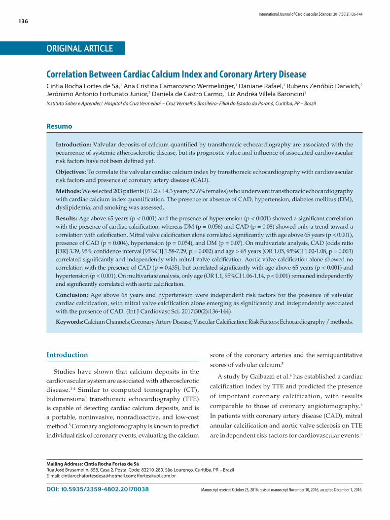
DOI: 10.5935/2359-4802.20170038
Introduction
Studies have shown that calcium deposits in the cardiovascular system are associated with atherosclerotic disease.1-4 Similar to computed tomography (CT), bidimensional transthoracic echocardiography (TTE) is capable of detecting cardiac calcium deposits, and is a portable, noninvasive, nonradioactive, and low-cost method.5 Coronary angiotomography is known to predict individual risk of coronary events, evaluating the calcium
score of the coronary arteries and the semiquantitative
scores of valvular calcium.5
A study by Gaibazzi et al.6 has established a cardiac
calcification index by TTE and predicted the presence
of important coronary calcification, with results
comparable to those of coronary angiotomography.6
In patients with coronary artery disease (CAD), mitral
annular calcification and aortic valve sclerosis on TTE
are independent risk factors for cardiovascular events.7
136International Journal of Cardiovascular Sciences. 2017;30(2):136-144
ORIGINAL ARTICLE
Mailing Address: Cintia Rocha Fortes de SáRua José Brusamolin, 658, Casa 2. Postal Code: 82210-280. São Lourenço, Curitiba, PR – BrazilE-mail: [email protected]; [email protected]
Correlation Between Cardiac Calcium Index and Coronary Artery DiseaseCintia Rocha Fortes de Sá,1 Ana Cristina Camarozano Wermelinger,1 Daniane Rafael,1 Rubens Zenóbio Darwich,2 Jerônimo Antonio Fortunato Junior,2 Daniela de Castro Carmo,1 Liz Andréa Villela Baroncini1
Instituto Saber e Aprender;1 Hospital da Cruz Vermelha2 – Cruz Vermelha Brasileira- Filial do Estado do Paraná, Curitiba, PR – Brazil
Manuscript received October 23, 2016; revised manuscript November 10, 2016; accepted December 1, 2016.
Resumo
Introduction: Valvular deposits of calcium quantified by transthoracic echocardiography are associated with the occurrence of systemic atherosclerotic disease, but its prognostic value and influence of associated cardiovascular risk factors have not been defined yet.
Objectives: To correlate the valvular cardiac calcium index by transthoracic echocardiography with cardiovascular risk factors and presence of coronary artery disease (CAD).
Methods: We selected 203 patients (61.2 ± 14.3 years; 57.6% females) who underwent transthoracic echocardiography with cardiac calcium index quantification. The presence or absence of CAD, hypertension, diabetes mellitus (DM), dyslipidemia, and smoking was assessed.
Results: Age above 65 years (p < 0.001) and the presence of hypertension (p < 0.001) showed a significant correlation with the presence of cardiac calcification, whereas DM (p = 0.056) and CAD (p = 0.08) showed only a trend toward a correlation with calcification. Mitral valve calcification alone correlated significantly with age above 65 years (p < 0.001), presence of CAD (p = 0.004), hypertension (p = 0.054), and DM (p = 0.07). On multivariate analysis, CAD (odds ratio [OR] 3.39, 95% confidence interval [95%CI] 1.58-7.29, p = 0.002) and age > 65 years (OR 1.05, 95%CI 1.02-1.08, p = 0.003) correlated significantly and independently with mitral valve calcification. Aortic valve calcification alone showed no correlation with the presence of CAD (p = 0.435), but correlated significantly with age above 65 years (p < 0.001) and hypertension (p < 0.001). On multivariate analysis, only age (OR 1.1, 95%CI 1.06-1.14, p < 0.001) remained independently and significantly correlated with aortic calcification.
Conclusion: Age above 65 years and hypertension were independent risk factors for the presence of valvular cardiac calcification, with mitral valve calcification alone emerging as significantly and independently associated with the presence of CAD. (Int J Cardiovasc Sci. 2017;30(2):136-144)
Keywords: Calcium Channels; Coronary Artery Disease; Vascular Calcification; Risk Factors; Echocardiography / methods.
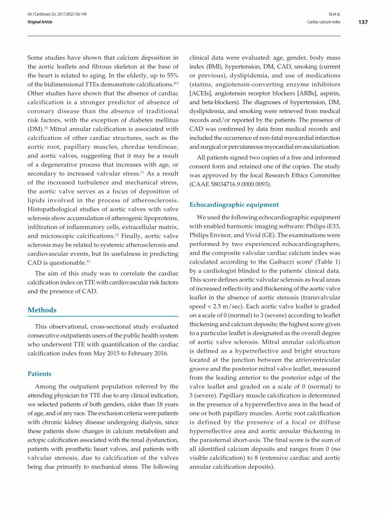
137
Some studies have shown that calcium deposition in the aortic leaflets and fibrous skeleton at the base of the heart is related to aging. In the elderly, up to 55% of the bidimensional TTEs demonstrate calcifications.8,9 Other studies have shown that the absence of cardiac calcification is a stronger predictor of absence of coronary disease than the absence of traditional risk factors, with the exception of diabetes mellitus (DM).10 Mitral annular calcification is associated with calcification of other cardiac structures, such as the aortic root, papillary muscles, chordae tendineae, and aortic valves, suggesting that it may be a result of a degenerative process that increases with age, or secondary to increased valvular stress.11 As a result of the increased turbulence and mechanical stress, the aortic valve serves as a focus of deposition of lipids involved in the process of atherosclerosis. Histopathological studies of aortic valves with valve sclerosis show accumulation of atherogenic lipoproteins, infiltration of inflammatory cells, extracellular matrix, and microscopic calcifications.12 Finally, aortic valve sclerosis may be related to systemic atherosclerosis and cardiovascular events, but its usefulness in predicting CAD is questionable.13
The aim of this study was to correlate the cardiac calcification index on TTE with cardiovascular risk factors and the presence of CAD.
Methods
This observational, cross-sectional study evaluated consecutive outpatients users of the public health system who underwent TTE with quantification of the cardiac calcification index from May 2015 to February 2016.
Patients
Among the outpatient population referred by the attending physician for TTE due to any clinical indication, we selected patients of both genders, older than 18 years of age, and of any race. The exclusion criteria were patients with chronic kidney disease undergoing dialysis, since these patients show changes in calcium metabolism and ectopic calcification associated with the renal dysfunction, patients with prosthetic heart valves, and patients with valvular stenosis, due to calcification of the valves being due primarily to mechanical stress. The following
clinical data were evaluated: age, gender, body mass index (BMI), hypertension, DM, CAD, smoking (current or previous), dyslipidemia, and use of medications (statins, angiotensin-converting enzyme inhibitors [ACEIs], angiotensin receptor blockers [ARBs], aspirin, and beta-blockers). The diagnoses of hypertension, DM, dyslipidemia, and smoking were retrieved from medical records and/or reported by the patients. The presence of CAD was confirmed by data from medical records and included the occurrence of non-fatal myocardial infarction and surgical or percutaneous myocardial revascularization.
All patients signed two copies of a free and informed consent form and retained one of the copies. The study was approved by the local Research Ethics Committee (CAAE 58034716.9.0000.0093).
Echocardiographic equipment
We used the following echocardiographic equipment with enabled harmonic imaging software: Philips iE33, Philips Envisor, and Vivid (GE). The examinations were performed by two experienced echocardiographers, and the composite valvular cardiac calcium index was calculated according to the Gaibazzi score6 (Table 1) by a cardiologist blinded to the patients' clinical data. This score defines aortic valvular sclerosis as focal areas of increased reflectivity and thickening of the aortic valve leaflet in the absence of aortic stenosis (transvalvular speed < 2.5 m/sec). Each aortic valve leaflet is graded on a scale of 0 (normal) to 3 (severe) according to leaflet thickening and calcium deposits; the highest score given to a particular leaflet is designated as the overall degree of aortic valve sclerosis. Mitral annular calcification is defined as a hyperreflective and bright structure located at the junction between the atrioventricular groove and the posterior mitral valve leaflet, measured from the leading anterior to the posterior edge of the valve leaflet and graded on a scale of 0 (normal) to 3 (severe). Papillary muscle calcification is determined in the presence of a hyperreflective area in the head of one or both papillary muscles. Aortic root calcification is defined by the presence of a focal or diffuse hyperreflective area and aortic annular thickening in the parasternal short-axis. The final score is the sum of all identified calcium deposits and ranges from 0 (no visible calcification) to 8 (extensive cardiac and aortic annular calcification deposits).
Sá et al.
Cardiac calcium index
Int J Cardiovasc Sci. 2017;30(2):136-144
Original Article
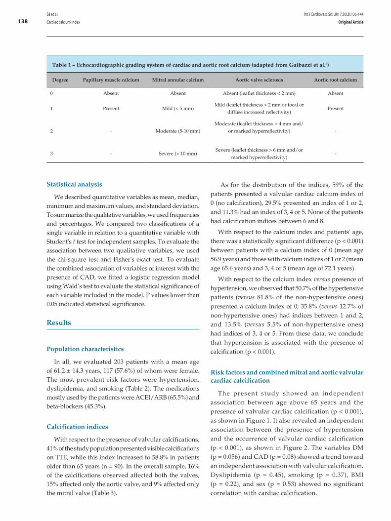
138
Table 1 – Echocardiographic grading system of cardiac and aortic root calcium (adapted from Gaibazzi et al.6)
Degree Papillary muscle calcium Mitral annular calcium Aortic valve sclerosis Aortic root calcium
0 Absent Absent Absent (leaflet thickness < 2 mm) Absent
1 Present Mild (< 5 mm)Mild (leaflet thickness > 2 mm or focal or
diffuse increased reflectivity)Present
2 - Moderate (5-10 mm)Moderate (leaflet thickness > 4 mm and/
or marked hyperreflectivity) -
3 - Severe (> 10 mm)Severe (leaflet thickness > 6 mm and/or
marked hyperreflectivity)-
Statistical analysis
We described quantitative variables as mean, median, minimum and maximum values, and standard deviation. To summarize the qualitative variables, we used frequencies and percentages. We compared two classifications of a single variable in relation to a quantitative variable with Student's t test for independent samples. To evaluate the association between two qualitative variables, we used the chi-square test and Fisher's exact test. To evaluate the combined association of variables of interest with the presence of CAD, we fitted a logistic regression model using Wald’s test to evaluate the statistical significance of each variable included in the model. P values lower than 0.05 indicated statistical significance.
Results
Population characteristics
In all, we evaluated 203 patients with a mean age of 61.2 ± 14.3 years, 117 (57.6%) of whom were female. The most prevalent risk factors were hypertension, dyslipidemia, and smoking (Table 2). The medications mostly used by the patients were ACEI/ARB (65.5%) and beta-blockers (45.3%).
Calcification indices
With respect to the presence of valvular calcifications, 41% of the study population presented visible calcifications on TTE, while this index increased to 58.8% in patients older than 65 years (n = 90). In the overall sample, 16% of the calcifications observed affected both the valves, 15% affected only the aortic valve, and 9% affected only the mitral valve (Table 3).
As for the distribution of the indices, 59% of the patients presented a valvular cardiac calcium index of 0 (no calcification), 29.5% presented an index of 1 or 2, and 11.3% had an index of 3, 4 or 5. None of the patients had calcification indices between 6 and 8.
With respect to the calcium index and patients' age, there was a statistically significant difference (p < 0.001) between patients with a calcium index of 0 (mean age 56.9 years) and those with calcium indices of 1 or 2 (mean age 65.6 years) and 3, 4 or 5 (mean age of 72.1 years).
With respect to the calcium index versus presence of hypertension, we observed that 50.7% of the hypertensive patients (versus 81.8% of the non-hypertensive ones) presented a calcium index of 0; 35.8% (versus 12.7% of non-hypertensive ones) had indices between 1 and 2; and 13.5% (versus 5.5% of non-hypertensive ones) had indices of 3, 4 or 5. From these data, we conclude that hypertension is associated with the presence of calcification (p < 0.001).
Risk factors and combined mitral and aortic valvular cardiac calcification
The present study showed an independent association between age above 65 years and the presence of valvular cardiac calcification (p < 0.001), as shown in Figure 1. It also revealed an independent association between the presence of hypertension and the occurrence of valvular cardiac calcification (p < 0.001), as shown in Figure 2. The variables DM (p = 0.056) and CAD (p = 0.08) showed a trend toward an independent association with valvular calcification. Dyslipidemia (p = 0.45), smoking (p = 0.37), BMI (p = 0.22), and sex (p = 0.53) showed no significant correlation with cardiac calcification.
Sá et al.
Cardiac calcium index
Int J Cardiovasc Sci. 2017;30(2):136-144
Original Article
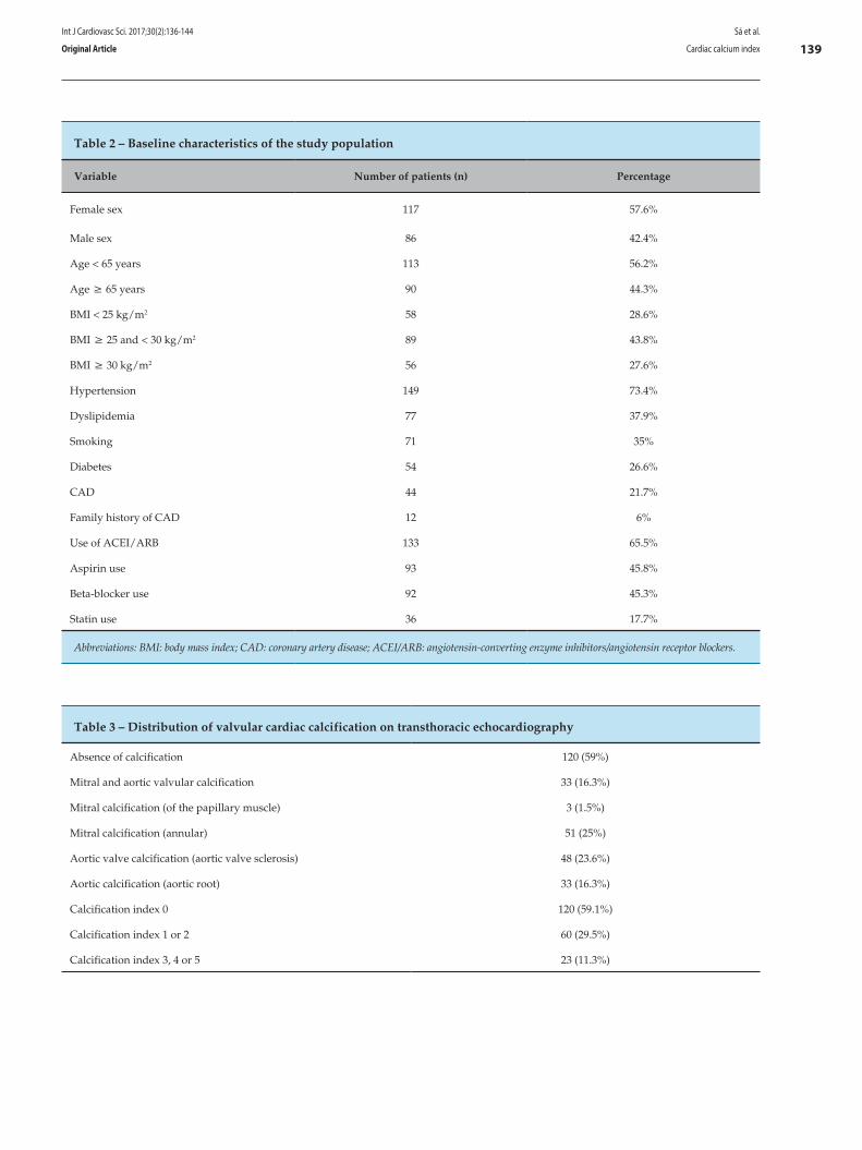
139
Table 2 – Baseline characteristics of the study population
Variable Number of patients (n) Percentage
Female sex 117 57.6%
Male sex 86 42.4%
Age < 65 years 113 56.2%
Age ≥ 65 years 90 44.3%
BMI < 25 kg/m2 58 28.6%
BMI ≥ 25 and < 30 kg/m2 89 43.8%
BMI ≥ 30 kg/m2 56 27.6%
Hypertension 149 73.4%
Dyslipidemia 77 37.9%
Smoking 71 35%
Diabetes 54 26.6%
CAD 44 21.7%
Family history of CAD 12 6%
Use of ACEI/ARB 133 65.5%
Aspirin use 93 45.8%
Beta-blocker use 92 45.3%
Statin use 36 17.7%
Abbreviations: BMI: body mass index; CAD: coronary artery disease; ACEI/ARB: angiotensin-converting enzyme inhibitors/angiotensin receptor blockers.
Table 3 – Distribution of valvular cardiac calcification on transthoracic echocardiography
Absence of calcification 120 (59%)
Mitral and aortic valvular calcification 33 (16.3%)
Mitral calcification (of the papillary muscle) 3 (1.5%)
Mitral calcification (annular) 51 (25%)
Aortic valve calcification (aortic valve sclerosis) 48 (23.6%)
Aortic calcification (aortic root) 33 (16.3%)
Calcification index 0 120 (59.1%)
Calcification index 1 or 2 60 (29.5%)
Calcification index 3, 4 or 5 23 (11.3%)
Sá et al.
Cardiac calcium index
Int J Cardiovasc Sci. 2017;30(2):136-144
Original Article
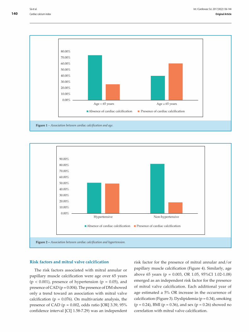
140
80.00%
70.00%
60.00%
50.00%
40.00%
30.00%
20.00%
10.00%
0.00%Age < 65 years Age ≥ 65 years
Absence of cardiac calcification Presence of cardiac calcification
Figure 1 – Association between cardiac calcification and age.
90.00%
80.00%
70.00%
60.00%
50.00%
40.00%
30.00%
20.00%
10.00%
0.00%Hypertensive Non-hypertensive
Absence of cardiac calcification Presence of cardiac calcification
Figure 2 – Association between cardiac calcification and hypertension.
Risk factors and mitral valve calcification
The risk factors associated with mitral annular or papillary muscle calcification were age over 65 years (p < 0.001), presence of hypertension (p = 0.05), and presence of CAD (p = 0.004). The presence of DM showed only a trend toward an association with mitral valve calcification (p = 0.076). On multivariate analysis, the presence of CAD (p = 0.002, odds ratio [OR] 3.39, 95% confidence interval [CI] 1.58-7.29) was an independent
risk factor for the presence of mitral annular and/or
papillary muscle calcification (Figure 4). Similarly, age
above 65 years (p = 0.003, OR 1.05, 95%CI 1.02-1.08)
emerged as an independent risk factor for the presence
of mitral valve calcification. Each additional year of
age estimated a 5% OR increase in the occurrence of
calcification (Figure 3). Dyslipidemia (p = 0.34), smoking
(p = 0.24), BMI (p = 0.36), and sex (p = 0.26) showed no
correlation with mitral valve calcification.
Sá et al.
Cardiac calcium index
Int J Cardiovasc Sci. 2017;30(2):136-144
Original Article
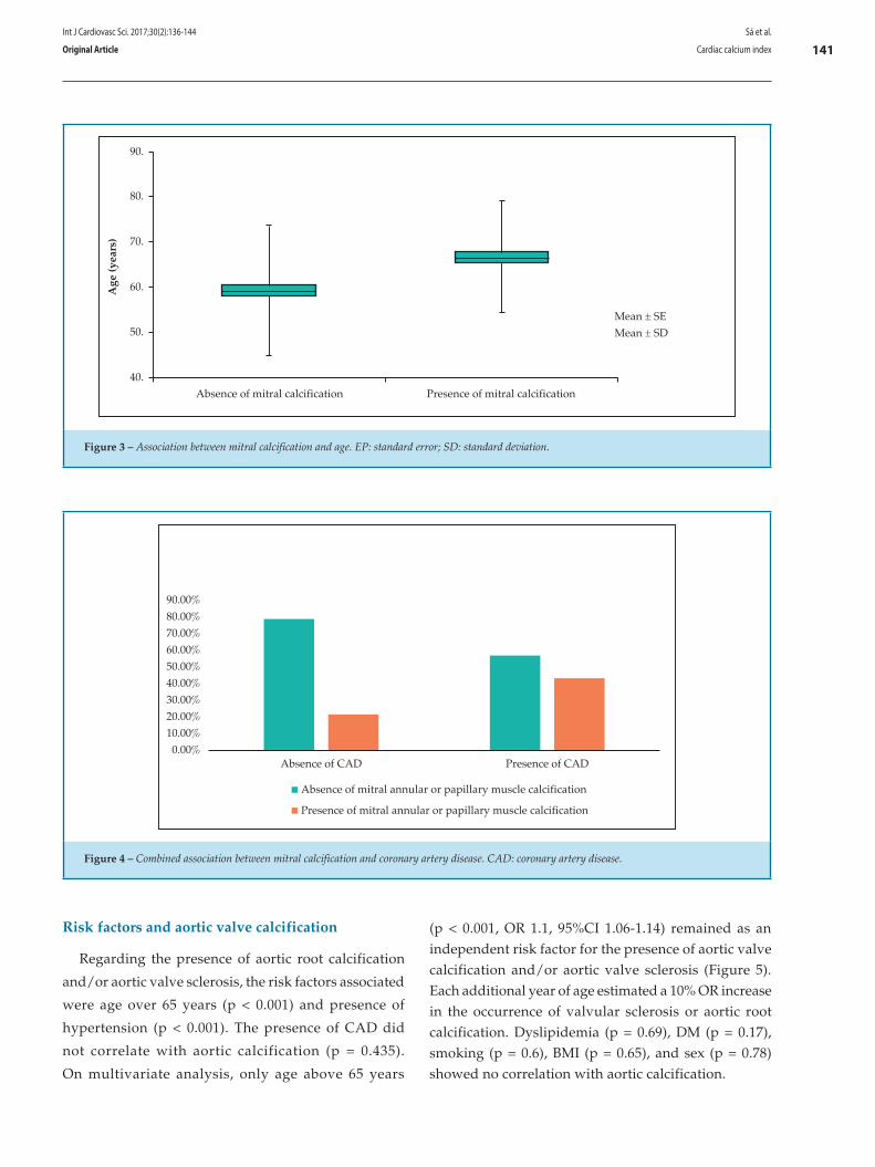
141
90.
80.
70.
60.
50.
40.
Age
(yea
rs)
Absence of mitral calcification Presence of mitral calcification
Mean ± SE
Mean ± SD
Figure 3 – Association between mitral calcification and age. EP: standard error; SD: standard deviation.
90.00%
80.00%
70.00%
60.00%
50.00%
40.00%
30.00%
20.00%
10.00%
0.00%Absence of CAD Presence of CAD
Absence of mitral annular or papillary muscle calcification
Presence of mitral annular or papillary muscle calcification
Figure 4 – Combined association between mitral calcification and coronary artery disease. CAD: coronary artery disease.
Risk factors and aortic valve calcification
Regarding the presence of aortic root calcification
and/or aortic valve sclerosis, the risk factors associated
were age over 65 years (p < 0.001) and presence of
hypertension (p < 0.001). The presence of CAD did
not correlate with aortic calcification (p = 0.435).
On multivariate analysis, only age above 65 years
(p < 0.001, OR 1.1, 95%CI 1.06-1.14) remained as an independent risk factor for the presence of aortic valve calcification and/or aortic valve sclerosis (Figure 5). Each additional year of age estimated a 10% OR increase in the occurrence of valvular sclerosis or aortic root calcification. Dyslipidemia (p = 0.69), DM (p = 0.17), smoking (p = 0.6), BMI (p = 0.65), and sex (p = 0.78) showed no correlation with aortic calcification.
Sá et al.
Cardiac calcium index
Int J Cardiovasc Sci. 2017;30(2):136-144
Original Article
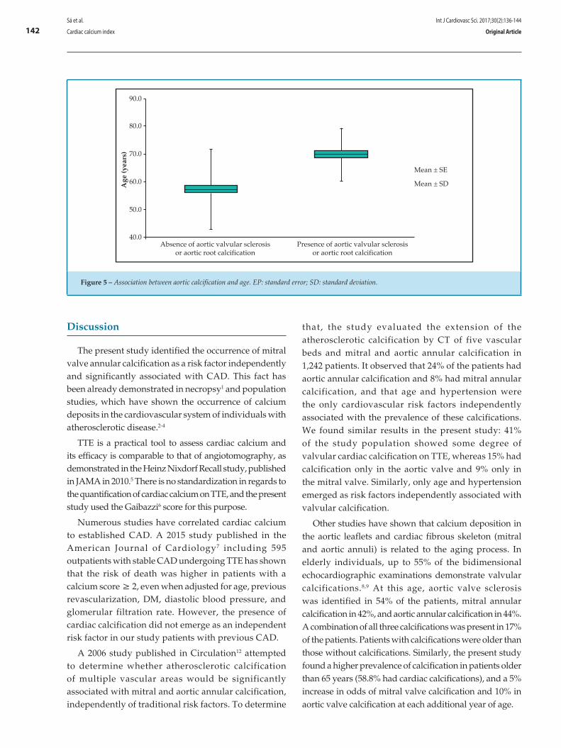
142
90.0
80.0
70.0
60.0
50.0
40.0
Mean ± SE
Mean ± SD
Absence of aortic valvular sclerosisor aortic root calcification
Presence of aortic valvular sclerosisor aortic root calcification
Age
(yea
rs)
Figure 5 – Association between aortic calcification and age. EP: standard error; SD: standard deviation.
Discussion
The present study identified the occurrence of mitral valve annular calcification as a risk factor independently and significantly associated with CAD. This fact has been already demonstrated in necropsy1 and population studies, which have shown the occurrence of calcium deposits in the cardiovascular system of individuals with atherosclerotic disease.2-4
TTE is a practical tool to assess cardiac calcium and its efficacy is comparable to that of angiotomography, as demonstrated in the Heinz Nixdorf Recall study, published in JAMA in 2010.5 There is no standardization in regards to the quantification of cardiac calcium on TTE, and the present study used the Gaibazzi6 score for this purpose.
Numerous studies have correlated cardiac calcium to established CAD. A 2015 study published in the American Journal of Cardiology7 including 595 outpatients with stable CAD undergoing TTE has shown that the risk of death was higher in patients with a calcium score ≥ 2, even when adjusted for age, previous revascularization, DM, diastolic blood pressure, and glomerular filtration rate. However, the presence of cardiac calcification did not emerge as an independent risk factor in our study patients with previous CAD.
A 2006 study published in Circulation12 attempted to determine whether atherosclerotic calcification of multiple vascular areas would be significantly associated with mitral and aortic annular calcification, independently of traditional risk factors. To determine
that, the study evaluated the extension of the atherosclerotic calcification by CT of five vascular beds and mitral and aortic annular calcification in 1,242 patients. It observed that 24% of the patients had aortic annular calcification and 8% had mitral annular calcification, and that age and hypertension were the only cardiovascular risk factors independently associated with the prevalence of these calcifications. We found similar results in the present study: 41% of the study population showed some degree of valvular cardiac calcification on TTE, whereas 15% had calcification only in the aortic valve and 9% only in the mitral valve. Similarly, only age and hypertension emerged as risk factors independently associated with valvular calcification.
Other studies have shown that calcium deposition in the aortic leaflets and cardiac fibrous skeleton (mitral and aortic annuli) is related to the aging process. In elderly individuals, up to 55% of the bidimensional echocardiographic examinations demonstrate valvular calcifications.8,9 At this age, aortic valve sclerosis was identified in 54% of the patients, mitral annular calcification in 42%, and aortic annular calcification in 44%. A combination of all three calcifications was present in 17% of the patients. Patients with calcifications were older than those without calcifications. Similarly, the present study found a higher prevalence of calcification in patients older than 65 years (58.8% had cardiac calcifications), and a 5% increase in odds of mitral valve calcification and 10% in aortic valve calcification at each additional year of age.
Sá et al.
Cardiac calcium index
Int J Cardiovasc Sci. 2017;30(2):136-144
Original Article
143
A study has shown that patients with mitral annular calcification have cardiovascular, renal, metabolic, and functional profiles worse than those with aortic ring calcification or aortic valve sclerosis. The same study also demonstrated that the more intense the calcification, the greater is the relationship with cardiovascular disease. When the risk was adjusted for the presence of DM, hypertension, previous CAD, ankle-brachial index (ABI) ≤ 0.9, serum creatinine ≥ 1.5 mg/dL, carotid stenosis ≥ 25%, left ventricular ejection fraction (LVEF), and myocardial mass (g), only mitral annular calcification was associated with cardiovascular disease.14 Similarly, the present study revealed in the multivariate analysis that the presence of CAD and age above 65 years are independent risk factors for the presence of mitral annular and/or papillary muscle calcification.
Although several studies have shown a connection between aortic valve sclerosis and risk factors for atherosclerosis and cardiovascular morbidity, the data are still controversial. A 2015 study by Bhatt et al.13 attempted to determine the association between the presence or absence of aortic valve sclerosis and cardiovascular risk factors, CAD extent, and severity of coronary lesions. After allocating 482 patients evaluated with coronary cineangiography into two groups, with and without valvular sclerosis, age was the only independent predictor of aortic valve sclerosis. Aortic valve sclerosis was also not independently associated with the number of obstructed vessels or degree of obstruction, showing that it is probably a benign marker of senile degenerative cardiac changes, regardless of the severity and complexity of the CAD. Finally, aortic valve sclerosis may be related to systemic atherosclerosis and cardiovascular events, but its usefulness in predicting CAD is questionable. In the present study, multivariate analysis revealed that only age was an independent risk factor for the presence of aortic valve calcification. In contrast, some studies have considered the absence of aortic root calcification and mitral annular calcification as a stronger predictor of absence of coronary disease than the absence of traditional risk factors, with the exception of DM.15
The use of ACEIs, aspirin, beta-blockers, and statins was frequent among the patients in this study due to the prevalence of hypertension and dyslipidemia in these individuals. There is no treatment based on evidence to reduce or prevent mitral annular calcification, and it unknown whether commonly used medications such as aspirin, beta-blockers, and statins may prevent its pathogenesis. Several studies suggest that warfarin is associated with increased valvular calcification.11
Although Bhatt et al.13 found no association between hypercholesterolemia and mitral annular calcification, further studies are needed to determine whether the use of statins would affect prognosis in these patients. Although many patients in the present study used drugs such as ACEIs (65.5% of the patients), aspirin (45.8%), and statin (17.7%), the study was not delineated to correlate the use of drugs and the presence of cardiac calcification.
Some factors have limited this study. First, the study population consisted mainly of elderly and hypertensive patients, since these patients were referred to TTE from an outpatient cardiology clinic. Second, the clinical diagnoses of the risk factors were established by self-report by the patients or through information obtained by review of their medical records. Another limiting factor of the study was the relative subjectivity in the quantification of cardiac calcification. Finally, the medications used by the patients were not included in the multivariate analysis.
Conclusion
The present study concluded that age above 65 years and hypertension were independent risk factors for the presence of valvular cardiac calcification, with mitral valvular calcification alone significantly and independently associated with the presence of CAD.
Author contributions
Conception and design of the research and Critical revision of the manuscript for intellectual content: Sá CRF, Baroncini LAV, Wermelinger ACC; Acquisition of data: Sá CRF, Rafael D, Darwich RZ, Fortunato Junior JA, Carmo DC; Analysis and interpretation of the data and Statistical analysis: Sá CRF, Baroncini LAV; Obtaining funding and Writing of the manuscript: Sá CRF.
Potential Conflict of Interest
No potential conflict of interest relevant to this article was reported.
Sources of Funding
There were no external funding sources for this study.
Study Association
This article is part of the thesis of specialization submitted by Cintia Rocha Fortes de Sá, from Hospital da Cruz Vermelha - Filial Paraná.
Sá et al.
Cardiac calcium index
Int J Cardiovasc Sci. 2017;30(2):136-144
Original Article
144
1. Roberts WC. Morphologic features of the normal and abnormal mitral valve. Am J Cardiol. 1983;51(6):1005-28.
2. Rossi A, Faggiano P, Amado AE, Cicoira M, Bonapace S, Franceschini L, et al. Mitral and aortic valve sclerosis/calcification and carotid atherosclerosis: results from 1065 patients. Heart Vessels. 2014;29(6):776-83.
3. Agno FS, Chinali M, Bella JN, Liu JE, Arnett DK, Kitzman DW, et al. Aortic valve sclerosis is associated with preclinical cardiovascular disease in hypertensive adults: the Hypertension Genetic Epidemiology Network study. J Hypertens. 2005;23(4):867-73.
4. Agmon Y, Khandheria BK, Meissner I, Sicks JR, O Fallon WM, Wiebers DO, et al. Aortic valve sclerosis and aortic atherosclerosis different manifestations of the same disease?Insights from a population-based study. J Am Coll Cardiol. 2001;38(3):827-34.
5. Erbel R, Möhlenkamp S, Moebus S, Schmermund A, Lehmann N, Stang A, et al; Heinz Nixdorf Recall Study Investigative Group. Coronary risk stratification, distribution, and reclassification improvement based on quantification of subclinical coronary atherosclerosis: the Heinz Nixdorf recall study. J Am Coll Cardiol. 2010;56(17):1397-406.
6. Gaibazzi N, Baldari C, Faggiano P, Albertini L, Faden G, Pigazzani F, et al. Cardiac calcium score on 2D echo: correlations with cardiac and coronary calcium at multi-detector computed tomography. Cardiovasc Ultrasound. 2014;12:43.
7. Saha AS, Beatty AL, Mishra RK, Whooley MA, Schiller NB. Usefulness of an echocardiographic composite cardiac calcium score to predict death in patients with stable coronary artery disease (from the Heart and Soul Study) Am J Cardiol. 2015;116(1):50-8.
8. Koulaouzidis G, Nicoll R, MacArthur T, Jenkins PJ, Henein MY. Coronary artery calcification correlates with the presence and severity of valve calcification. Int J Cardiol. 2013;168(6):5263-6.
9. Utsunomiya H, Yamamoto H, Kunita E, Kitagawa T, Ohashi N, Oka T, et al. Combined presence of aortic valve calcification and mitral anular calcification as a marker of the extent and vulnerable characteristics of coronary artery plaque assessed by 64 multidetector computed tomography. Atherosclerosis. 2010;213(1):166-72.
10. Acartürk E, Bozkurt A, Cayli M, Demir M. Mitral annular calcification and aortic valve calcification may help in predicting significant coronary artery disease. Angiology. 2003;54(5):561-567.
11. Sherif HM. Calcification of left-sided valvular structures: evidence of a proinflammatory milieu. J Heart Valve Dis. 2009;18(1):52-60.
12. Freeman RV, Otto CM. Spectrum of calcific aortic valve disease. Pathogenesis, disease progression, and treatment strategies. Circulation. 2005;111(24):3316-26.
13. Bhatt H, Sanghani D, Julliar K, Fernaine G. Does aortic valve sclerosis predicts the severity and complexity of coronary artery disease? Indian Heart J. 2015;67(3):239-44.
14. Jeon DS, Atar S, Brasch AV, Luo H, Mirocha J, Navqi TZ, et al. Association of mitral annulus calcification, aortic valve sclerosis and aortic root calcification with abnormal myocardial perfusion single photon emission tomography in subjects age < or =65 years old. J Am Coll Cardiol. 2001;38(7):1988-93.
15. Acartürk E, Bozkurt A, Cayli M, Demir M. Mitral annular calcification and aortic valve calcification may help in predicting significant coronary artery disease. Angiology. 2003;54(5):561-7.
References
Sá et al.
Cardiac calcium index
Int J Cardiovasc Sci. 2017;30(2):136-144
Original Article
DOI: 10.5935/2359-4802.20170033
145International Journal of Cardiovascular Sciences. 2017;30(2):145-156
ORIGINAL ARTICLE
Mailing Address: Rafael Bonfante Cabarcas Av. libertador con Av. Andrés Bello, Decanato de Ciencias de la Salud, Universidad Centroccidental "Lisandro Alvarado". Postal Code: 3001, Barquisimeto – Venezuela E-mail: [email protected]
Rescue Therapy with Nifurtimox and Dipyridamole for Severe Acute Chagas Myocarditis with Congestive Heart Failure in NMRI Albino MiceDaniela Yustiz Aparicio,1 María González-Hernández,1 Greybis Hernández-Forero,1 María Guédez-Ortiz,1 Sonia Santeliz,1 Loredana Goncalves,2 Rafael Bonfante Cabarcas1
Biochemistry Research Unit, Faculty of Health Sciences, Universidad Centroccidental "Lisandro Alvarado" (UCLA),1 Barquisimeto, Venezuela; Universidad de los Andes,2 Mérida – Venezuela
Manuscript received October 16, 2016; revised November 10, 2016; accepted February 02, 2017.
Abstract
Background: Chagas disease is a global health problem; therefore, the development of new therapeutic protocols is necessary. Our group recently demonstrated that nifurtimox associated with dipyridamole has curative effects in mice with acute Chagas disease. In this study, we assess the effect of this therapeutic protocol in chagasic mice with heart failure.
Objective: To evaluate whether nifurtimox and dipyridamole are useful to rescue mice with severe acute chagasic myocarditis with heart failure.
Methods: 42 mice with acute chagasic myocarditis and congestive heart failure were divided into three groups: control chagas (n = 11), Nif-Dip treated with nifurtimox and dipyridamole (n = 14) and Nif-Dip-heart failure treated with nifurtimox and dipyridamole associated with digoxin, furosemide, and captopril (n = 17). Nifurtimox and dipyridamole doses were 40 and 30 mg/kg/day, respectively, for 6 weeks. Mice underwent clinical, electrocardiographic, hemoparasitological and histopathological assessments.
Results: Lower mortality in Nif-Dip (28.57%; n = 4) compared to control chagas (54.54%; n = 6) and Nif-Dip-heart failure (52.9%; n = 9) was observed. Clinically, nifurtimox and dipyridamole-treated mice increased body weight and improved heart failure without splenomegaly. In these groups, parasitemia and tissue parasites were eradicated; fibrosis, myocytolysis, inflammatory cell infiltrate and mast cells decreased. Repolarization disorders, prolonged QRS and QT intervals, increase of S wave amplitude and atrioventricular dissociation were reversed by the treatment.
Conclusion: Nifurtimox with dipyridamole can rescue NMRI mice from severe acute chagas disease, as nifurtimox showed trypanocidal activity and dipyridamole potentiated its effect. Dipyridamole would be useful in chagasic heart failure. (Int J Cardiovasc Sci. 2017;30(2):145-156)
Keywords: Chagas Disease; Chagas Cardiomyopathy; Heart Failure; Mice; Nifurtimox; Dipyridamole.
Introduction
Chagas Disease (ChD) is a global public health problem due to a high residual 1.06% prevalence in endemic countries, where reemergence has been reported as oral transmission outbreaks and because of the disease globalization, as a product of the migration of people from endemic countries to developed countries; it has
resulted in a 4.2% prevalence in immigrant populations
in Europe.1-4
Symptomatic acute ChD cases have been associated
with patent parasitemia and severe acute myocarditis,
with high morbidity and mortality.5 Severe heart damage
would reflect parasite pathogenic action; therefore, it
would be mandatory to conduct aggressive trypanocidal
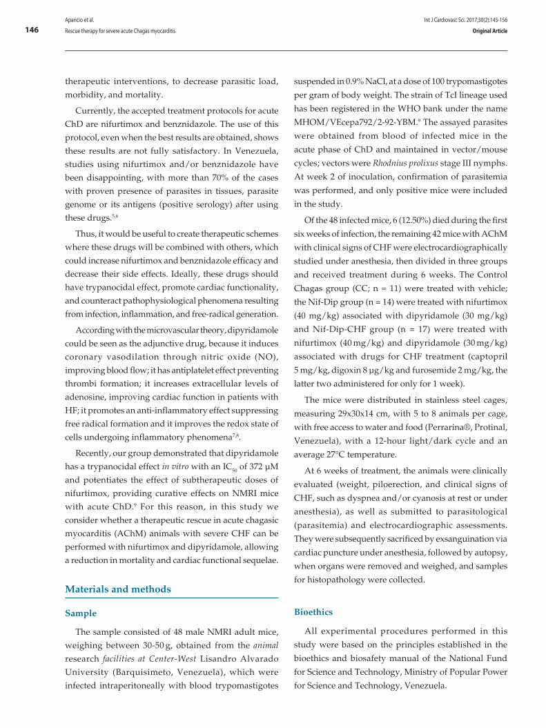
146Aparicio et al.
Rescue therapy for severe acute Chagas myocarditis
Int J Cardiovasc Sci. 2017;30(2):145-156
Original Article
therapeutic interventions, to decrease parasitic load,
morbidity, and mortality.
Currently, the accepted treatment protocols for acute
ChD are nifurtimox and benznidazole. The use of this
protocol, even when the best results are obtained, shows
these results are not fully satisfactory. In Venezuela,
studies using nifurtimox and/or benznidazole have
been disappointing, with more than 70% of the cases
with proven presence of parasites in tissues, parasite
genome or its antigens (positive serology) after using
these drugs.5,6
Thus, it would be useful to create therapeutic schemes
where these drugs will be combined with others, which
could increase nifurtimox and benznidazole efficacy and
decrease their side effects. Ideally, these drugs should
have trypanocidal effect, promote cardiac functionality,
and counteract pathophysiological phenomena resulting
from infection, inflammation, and free-radical generation.
According with the microvascular theory, dipyridamole
could be seen as the adjunctive drug, because it induces
coronary vasodilation through nitric oxide (NO),
improving blood flow; it has antiplatelet effect preventing
thrombi formation; it increases extracellular levels of
adenosine, improving cardiac function in patients with
HF; it promotes an anti-inflammatory effect suppressing
free radical formation and it improves the redox state of
cells undergoing inflammatory phenomena7,8.
Recently, our group demonstrated that dipyridamole
has a trypanocidal effect in vitro with an IC50 of 372 µM
and potentiates the effect of subtherapeutic doses of
nifurtimox, providing curative effects on NMRI mice
with acute ChD.9 For this reason, in this study we
consider whether a therapeutic rescue in acute chagasic
myocarditis (AChM) animals with severe CHF can be
performed with nifurtimox and dipyridamole, allowing
a reduction in mortality and cardiac functional sequelae.
Materials and methods
Sample
The sample consisted of 48 male NMRI adult mice,
weighing between 30-50 g, obtained from the animal
research facilities at Center-West Lisandro Alvarado
University (Barquisimeto, Venezuela), which were
infected intraperitoneally with blood trypomastigotes
suspended in 0.9% NaCl, at a dose of 100 trypomastigotes
per gram of body weight. The strain of TcI lineage used
has been registered in the WHO bank under the name
MHOM/VEcepa792/2-92-YBM.9 The assayed parasites
were obtained from blood of infected mice in the
acute phase of ChD and maintained in vector/mouse
cycles; vectors were Rhodnius prolixus stage III nymphs.
At week 2 of inoculation, confirmation of parasitemia
was performed, and only positive mice were included
in the study.
Of the 48 infected mice, 6 (12.50%) died during the first
six weeks of infection, the remaining 42 mice with AChM
with clinical signs of CHF were electrocardiographically
studied under anesthesia, then divided in three groups
and received treatment during 6 weeks. The Control
Chagas group (CC; n = 11) were treated with vehicle;
the Nif-Dip group (n = 14) were treated with nifurtimox
(40 mg/kg) associated with dipyridamole (30 mg/kg)
and Nif-Dip-CHF group (n = 17) were treated with
nifurtimox (40 mg/kg) and dipyridamole (30 mg/kg)
associated with drugs for CHF treatment (captopril
5 mg/kg, digoxin 8 µg/kg and furosemide 2 mg/kg, the
latter two administered for only for 1 week).
The mice were distributed in stainless steel cages,
measuring 29x30x14 cm, with 5 to 8 animals per cage,
with free access to water and food (Perrarina®, Protinal,
Venezuela), with a 12-hour light/dark cycle and an
average 27°C temperature.
At 6 weeks of treatment, the animals were clinically
evaluated (weight, piloerection, and clinical signs of
CHF, such as dyspnea and/or cyanosis at rest or under
anesthesia), as well as submitted to parasitological
(parasitemia) and electrocardiographic assessments.
They were subsequently sacrificed by exsanguination via
cardiac puncture under anesthesia, followed by autopsy,
when organs were removed and weighed, and samples
for histopathology were collected.
Bioethics
All experimental procedures performed in this
study were based on the principles established in the
bioethics and biosafety manual of the National Fund
for Science and Technology, Ministry of Popular Power
for Science and Technology, Venezuela.
147Aparicio et al.
Rescue therapy for severe acute Chagas myocarditis
Int J Cardiovasc Sci. 2017;30(2):145-156
Original Article
Electrocardiography
Electrocardiographic studies were performed under anesthesia with sodium pentobarbital 25 mg/kg and ketamine 25 mg/kg of body weight ip, in bipolar configuration, using 3 needle-type electrodes (one positive, one negative and one neutral or reference), positioned in the subcutaneous tissue. We worked with 4 lead-ECG configurations: in DI, a positive electrode was placed together with the reference electrode on the left shoulder joint, while the negative electrode was positioned on the right shoulder joint; in DII, a positive electrode was positioned on the xiphoid process, while a negative one was placed on the right shoulder joint and the reference on the left shoulder joint; in DIII, both the negative electrode and the reference electrode were placed on the left shoulder joint and a positive electrode was maintained on the xiphoid process, and in AVF lead, a negative electrode was positioned on the cervical midline above the suprasternal fossa, while a positive electrode was placed on the xiphoid process and a reference one remained on the left shoulder joint.
Electrodes were connected to a BioAmp amplifier (AD Instruments, New Zealand), and analog signals were converted to digital signals through a Powerlab/8sp interphase system (AD Instruments, New Zealand), displayed, recorded, and analyzed on a personal computer using the Chart v4.2.1 software (AD Instruments New Zealand). The signal capture was performed at a 1000 events/sec frequency and filtered at 60 Hz.
Histopathology
Right ventricular and cardiac apex samples were fixed with 4% paraformaldehyde solution in PBS, pH 7.4 for 2 hours. Fixed tissue samples were embedded successively in 10, 15, 20, 25 and 30% sucrose solutions until samples migrated to the container bottom in each solution; then samples were stored at -70°C until used. Samples were thawed and embedded in Optimal Cutting Temperature (OCT) compound, and again frozen with liquid nitrogen, cryostat-cut obtaining 5 µm-sections, put on microscope slides and stained with hematoxylin-eosin or toluidine blue stains. Additionally, another group of samples was fixed with 10% formalin in PBS pH 7.4, embedded in paraffin, cut with a microtome and stained with hematoxylin-eosin.
Data analysis
Data is expressed as absolute values, percentages or mean ± SEM. Paired or unpaired Student’s t test was
used to analyze the significance of observed differences before and after treatment in a group or to compare two unrelated groups, respectively; while ANOVA followed by Tukey’s post-test was used to analyze significance of observed differences between the three experimental groups. A value of p < 0.05 was considered significant. GraphPad Prism Software 5.0 was used for the statistical analysis.
Results
All mice became infected with an average parasitemia of 4.19 ± 0.74 x 10⁶ parasites/mL on the 6th week post-infection before treatment protocol began. In all experimental treated groups, parasites were not detected at the 7th week, while in the CC group, parasitemia disappeared between the 8th and 9th week; all groups had 0 parasitemia at sacrifice.
During the post-anesthetic period (0-48 hours), the CC group had 45.45% (n = 5), Nif-Dip 21.42% (n = 3) and Nif-Dip-CHF 41.17% (n = 7) of mortality rate, respectively. During the following six weeks, 1 animal of each CC and Nif-Dip groups and 2 from Nif-Dip-CHF group died, resulting in a total mortality of 54.54% (n = 6), 28.57% (n = 4) and 52.9% (n = 9), respectively.
In Figure 1, clinical parameters observed at the end of the experimental protocol are shown. We can observe that treated groups significantly increased body weight; while spleen weight and CHF clinical signs decreased significantly in these groups as compared with the CC group.
Quantitative and qualitative electrocardiographic parameters are shown in Tables I and II, respectively, before and after starting the therapeutic protocol. Altered electrocardiographic signs found before treatment included decreased heart rate, cardiac conduction disturbances, characterized as an increase in QRS and QT intervals values; alterations in ventricular repolarization, characterized by disorders in amplitude, axis direction and T wave decay (see Table I and Figures 2 and 3). These signs were reversed by the therapeutic protocol; however, R and S wave amplitudes decreased (See Table I and Figures 2 and 3). Regarding qualitative data disorders, nifurtimox-dipyridamole-based therapeutic protocol tended to decrease atrioventricular dissociation and post-depolarization disorders; however, rhythm disorders (nodal and ventricular extrasystoles) tended
to increase (see Table II).
148
Figure 1 – Clinical parameters quantified in mice with AChD treated or not with nifurtimox and dipyridamole. In A, body weight is shown; mice submitted to the treatment protocol increased weight. In B, normalized spleen weight based on body weight is shown; splenomegaly decreased in mice submitted to the treatment protocol. In C, a summative clinical analysis based on the presence (1) or absence (0) of the following clinical signs were performed in each mouse: dyspnea and cyanosis at rest or under anesthesia and piloerection; mice submitted to the treatment protocol had decreased signs of CHF or infection. * means p < 0.05 when comparing treated groups in relation to Control Chagas non-treated group, using ANOVA test followed by Tukey’s post-test.
Aparicio et al.
Rescue therapy for severe acute Chagas myocarditis
Int J Cardiovasc Sci. 2017;30(2):145-156
Original Article
Histopathological analysis (see Table III) of the right
ventricular sections from the CC mice group displayed
thickened epicardium, myocytolysis and diffuse
mononuclear inflammatory infiltrate; while in the
Nif-Dip mice group, only reduced focal inflammatory
infiltrate was observed and epicardial thickness was
diminished; fibrosis degree was similar in both groups.
In the cardiac apex sections, fibrosis, myocytolysis,
epicardium thickness and inflammatory infiltrate were
lower in mice treated with nifurtimox-dipyridamole
when compared to the CC group. In both sections, in
60% of untreated mice, intact and/or broken parasitic
nests were present, while in sections of nifurtimox-
dipyridamole treated mice amastigotes and/or parasite
nests were not observed (see Figure 4). Additionally, in
60% (n = 3) of CC and in 50% (n = 2) of Nif-Dip group
mice, mast cells were observed in cardiac apex sections;
however, the number of mast cells per field was higher
in the CC group (10.60 ± 7.53) than in the Nif-Dip group
(1.00 ± 0.82); (see Figure 5 and Table III).
Discussion
In this study, we demonstrate that the combination of
nifurtimox with dipyridamole was therapeutically useful
in mice with AChM with CHF, since, compared with
untreated control mice, it reduced mortality, decreased
or eradicated infection and, furthermore clinical, and
histopathological signs of severe AChM were reversed.
To our knowledge, this is the first study carried out in
animal models to evaluate nifurtimox-dipyridamole-
based therapeutic rescue in mice with severe AChM.
The trypanocidal effect of nifurtimox treatment was
tested in the present study by demonstrating absence
of parasitemia after the first week of treatment and
the absence of amastigotes or parasite nests in cardiac
right ventricular and apex histopathology sections.
These results concur with those reported by Santeliz
et al.9 who demonstrated that the nifurtimox (40 mg/kg)
and dipyridamole (30 mg/kg) combination eradicates
tissue and hematic parasites in mice with acute ChD
without CHF. The trypanocidal effect shown in the
present work could depend solely on the effect of
nifurtimox at a 40 mg/kg dose, as this dose has been
reported to be curative (16); however, several studies
in murine models about the use of nifurtimox in ChD
treatment have been controversial (see below).
Studies on nifurtimox effect in experimental animals
have used high doses of the drug, obtaining conflicting
results; while Bustamante et al.10 working with C57BL/6
(Ly5.2 +) mice obtained a 95-100% cure rate with
doses of 100 mg/kg, during 40 days of continuous
or 13 days of intermittent treatment, Wong-Baeza
et al.11 working with NIH albino mice, obtained a
slight parasitemia decrease, with similar doses using a
50-day therapeutic protocol. This discordance observed
between the aforementioned studies and our work can be explained by mouse strains used; however,
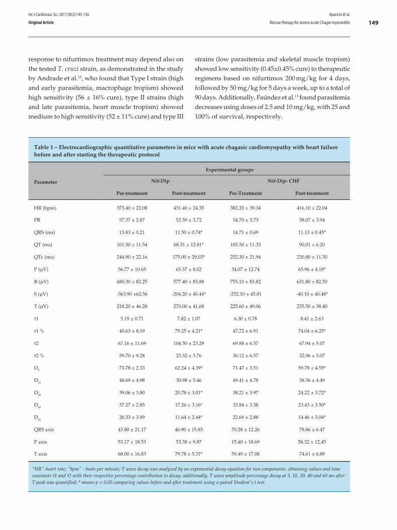
149Aparicio et al.
Rescue therapy for severe acute Chagas myocarditis
Int J Cardiovasc Sci. 2017;30(2):145-156
Original Article
Table 1 – Electrocardiographic quantitative parameters in mice with acute chagasic cardiomyopathy with heart failure before and after starting the therapeutic protocol
Parameter
Experimental groups
Nif-Dip Nif-Dip- CHF
Pre-treatment Post-treatment Pre-Treatment Post-treatment
HR (bpm) 373.40 ± 22.08 431.40 ± 24.35 382.20 ± 39.34 416.10 ± 22.04
PR 57.37 ± 2.87 52.59 ± 3.72 54.70 ± 3.73 58.07 ± 3.94
QRS (ms) 13.83 ± 0.21 11.50 ± 0.74* 14.71 ± 0.69 11.13 ± 0.45*
QT (ms) 101.50 ± 11.54 68.31 ± 12.81* 105.50 ± 11.33 90.01 ± 6.20
QTc (ms) 244.90 ± 22.16 175.00 ± 29.03* 252.30 ± 21.94 230.80 ± 11.50
P (µV) 56.77 ± 10.65 65.57 ± 8.02 34.07 ± 12.74 65.96 ± 4.18*
R (µV) 680.30 ± 82.25 577.40 ± 83.88 755.10 ± 83.82 631.80 ± 82.50
S (µV) -363.90 ±62.56 -204.20 ± 40.44* -252.10 ± 45.81 -40.10 ± 40.48*
T (µV) 218.20 ± 46.28 270.00 ± 41.68 225.60 ± 49.06 255.50 ± 38.40
τ1 5.19 ± 0.71 7.82 ± 1.07 6.30 ± 0.78 8.41 ± 2.63
τ1 % 40.63 ± 8.19 79.25 ± 4.21* 47.72 ± 6.91 74.04 ± 6.25*
τ2 67.16 ± 11.69 104.50 ± 23.29 69.88 ± 6.57 67.94 ± 5.07
τ2 % 59.70 ± 9.28 23.32 ± 3.76 30.12 ± 6.57 32.06 ± 5.07
D5 73.78 ± 2.33 62.24 ± 4.39* 71.47 ± 3.51 59.78 ± 4.55*
D10 48.69 ± 4.98 30.98 ± 5.46 49.41 ± 4.78 38.56 ± 4.49
D20 39.06 ± 3.80 20.78 ± 3.01* 38.21 ± 3.97 24.22 ± 3.72*
D40 37.27 ± 2.85 17.26 ± 3.16* 33.84 ± 3.38 23.43 ± 3.50*
D60 20.33 ± 3.09 11.64 ± 2.44* 22.69 ± 2.88 14.46 ± 3.04*
QRS axis 43.80 ± 21.17 46.90 ± 15.85 70.28 ± 12.26 78.86 ± 6.47
P axis 53.17 ± 18.53 53.38 ± 9.87 15.40 ± 18.69 58.32 ± 12.45
T axis 68.00 ± 16.83 79.78 ± 5.31* 59.49 ± 17.08 74.61 ± 6.89
“HR”-heart rate; “bpm” - beats per minute; T wave decay was analyzed by an exponential decay equation for two components. obtaining values and time constants τ1 and τ2 with their respective percentage contribution to decay; additionally, T wave amplitude percentage decay at 5. 10. 20. 40 and 60 ms after T peak was quantified; * means p < 0.05 comparing values before and after treatment using a paired Student’s t test.
response to nifurtimox treatment may depend also on the tested T. cruzi strain, as demonstrated in the study by Andrade et al.12, who found that Type I strain (high and early parasitemia, macrophage tropism) showed high sensitivity (56 ± 16% cure), type II strains (high and late parasitemia, heart muscle tropism) showed medium to high sensitivity (52 ± 11% cure) and type III
strains (low parasitemia and skeletal muscle tropism) showed low sensitivity (0.45±0.45% cure) to therapeutic regimens based on nifurtimox 200 mg/kg for 4 days, followed by 50 mg/kg for 5 days a week, up to a total of 90 days. Additionally, Faúndez et al.13 found parasitemia decreases using doses of 2.5 and 10 mg/kg, with 25 and 100% of survival, respectively.
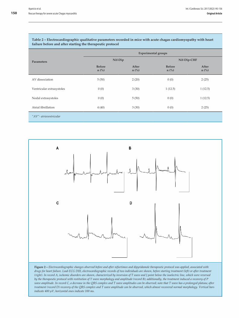
150
Figure 2 – Electrocardiographic changes observed before and after nifurtimox and dipyridamole therapeutic protocol was applied, associated with drugs for heart failure. Lead-ECG DIII, electrocardiographic records of two individuals are shown, before starting treatment (left) or after treatment (right). In record A, ischemic disorders are shown, characterized by inversion of T wave and J point below the isoelectric line, which were reversed by the therapeutic protocol with restitution of T wave morphology and amplitude (record B); additionally, the treatment induced a recovery of P wave amplitude. In record C, a decrease in the QRS complex and T wave amplitudes can be observed; note that T wave has a prolonged plateau; after treatment (record D) recovery of the QRS complex and T wave amplitude can be observed, which almost recovered normal morphology. Vertical bars indicate 400 µV, horizontal ones indicate 100 ms.
Table 2 – Electrocardiographic qualitative parameters recorded in mice with acute chagas cardiomyopathy with heart failure before and after starting the therapeutic protocol
Parameters
Experimental groups
Nif-Dip Nif-Dip-CHF
Beforen (%)
Aftern (%)
Beforen (%)
Aftern (%)
AV dissociation 5 (50) 2 (20) 0 (0) 2 (25)
Ventricular extrasystoles 0 (0) 3 (30) 1 (12.5) 1 (12.5)
Nodal extrasystoles 0 (0) 5 (50) 0 (0) 1 (12.5)
Atrial fibrillation 4 (40) 3 (30) 0 (0) 2 (25)
“AV”- atrioventricular
Aparicio et al.
Rescue therapy for severe acute Chagas myocarditis
Int J Cardiovasc Sci. 2017;30(2):145-156
Original Article
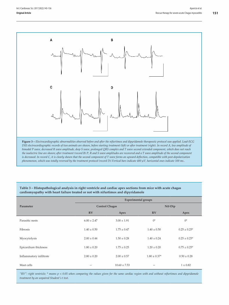
151
Table 3 – Histopathological analysis in right ventricle and cardiac apex sections from mice with acute chagas cardiomyopathy with heart failure treated or not with nifurtimox and dipyridamole
Parameter
Experimental groups
Control Chagas Nif-Dip
RV Apex RV Apex
Parasitic nests 6.00 ± 2.47 3.00 ± 1.91 0* 0*
Fibrosis 1.40 ± 0.50 1.75 ± 0.47 1.40 ± 0.50 0.25 ± 0.25*
Myocytolysis 2.00 ± 0.44 1.50 ± 0.28 1.40 ± 0.24 0.25 ± 0.25*
Epicardium thickness 1.80 ± 0.20 1.75 ± 0.25 1.20 ± 0.20 0.75 ± 0.25*
Inflammatory infiltrate 2.80 ± 0.20 2.00 ± 0.57 1.80 ± 0.37* 0.50 ± 0.28
Mast cells -- 10.60 ± 7.53 -- 1 ± 0.82
“RV”: right ventricle; * means p < 0.05 when comparing the values given for the same cardiac region with and without nifurtimox and dipyridamole treatment by an unpaired Student's t test.
Figure 3 – Electrocardiographic abnormalities observed before and after the nifurtimox and dipyridamole therapeutic protocol was applied. Lead-ECG DIII electrocardiographic records of two animals are shown, before starting treatment (left) or after treatment (right). In record A, low amplitude of bimodal P wave, decreased R wave amplitude, deep S wave, prolonged QRS complex and T wave second extended component, which does not reach the isoelectric line are shown; after treatment (record B) P, R and S wave amplitudes are recovered and a T wave amplitude of the second component is decreased. In record C, it is clearly shown that the second component of T wave forms an upward deflection, compatible with post-depolarization phenomenon, which was totally reversed by the treatment protocol (record D).Vertical bars indicate 400 µV, horizontal ones indicate 100 ms..
Aparicio et al.
Rescue therapy for severe acute Chagas myocarditis
Int J Cardiovasc Sci. 2017;30(2):145-156
Original Article
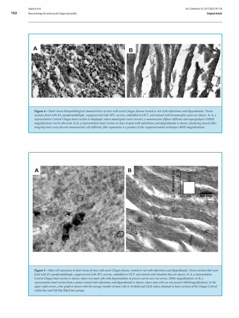
152
Figure 4 – Heart tissue histopathological characteristics of mice with acute Chagas disease treated or not with nifurtimox and dipyridamole. Tissue sections fixed with 4% paraformaldehyde, cryopreserved with 30% sucrose, embedded in OCT, and stained with hematoxylin-eosin are shown. In A, a representative Control Chagas heart section is displayed, where amastigotes nests (arrow), a mononuclear diffuse infiltrate and myocytolysis (1000X magnification) can be observed. In B, a representative heart section in mice treated with nifurtimox and dipyridamole is shown, disclosing muscle fiber integrity and a very discrete mononuclear cell infiltrate; fiber separation is a product of the cryopreservation technique (400X magnification).
Figure 5 – Mast cell expression in heart tissue of mice with acute Chagas disease, treated or not with nifurtimox and dipyridamole. Tissue sections that were fixed with 4% paraformaldehyde, cryopreserved with 30% sucrose, embedded in OCT, and stained with toluidine blue are shown. In A, a representative Control Chagas heart section is shown, where two mast cells with degranulation in process can be seen (see arrow, 1000x magnification). In B, a representative heart section from a mouse treated with nifurtimox and dipyridamole is shown, where mast cells are not present (400Xmagnification). In the upper right corner, a bar graph is shown with the average number of mast cells in 10 fields and SEM values obtained in heart sections of the Chagas Control (white bar) and Nif-Dip (black bar) groups.
Aparicio et al.
Rescue therapy for severe acute Chagas myocarditis
Int J Cardiovasc Sci. 2017;30(2):145-156
Original Article
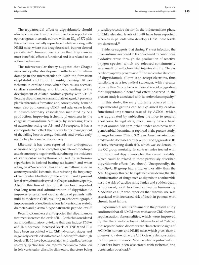
153Aparicio et al.
Rescue therapy for severe acute Chagas myocarditis
Int J Cardiovasc Sci. 2017;30(2):145-156
Original Article
The trypanocidal effect of dipyridamole should also be considered, as this effect has been reported on epimastigotes in axenic culture with an IC50 of 372 µM; this effect was partially reproduced while working with NMRI mice, where this drug decreased, but not cleared parasitemia.9 However, we propose that dipyridamole most beneficial effect is functional and it is related to its action mechanism.
The microvascular theory suggests that Chagas myocardiopathy development reflects endothelium damage in the microcirculation, with the formation of platelet and blood thrombi, causing diffuse ischemia in cardiac tissue, which then causes necrosis, cardiac remodeling, and fibrosis, leading to the development of dilated cardiomyopathy with CHF.14 Because dipyridamole is an antiplatelet agent, it prevents platelet thrombus formation and, consequently, hematic ones; also by increasing cGMP and adenosine levels, it induces coronary vasodilation mediated by NO production, improving ischemic phenomena in the chagasic myocardium. Similarly, by increasing levels of adenosine acting on A1- and A3-receptors, it has a cardioprotective effect that allows better management of the failing heart’s energy demands and avoids early apoptotic phenomena, respectively.15
Likewise, it has been reported that endogenous adenosine acting on A1-receptors generate a chronotropic and dromotropic negative effect, reducing the incidence of ventricular arrhythmias caused by ischemia-reperfusion in isolated beating rat hearts,16 and when acting on A2-receptors it has an antiarrhythmic effect in acute myocardial ischemia, thus reducing the frequency of ventricular fibrillation;17 therefore it could prevent lethal arrhythmias observed in Chagas cardiomyopathy. Also in this line of thought, it has been reported that long-term oral administration of dipyridamole improves physical and cardiac status of patients with mild to moderate CHF, resulting in echocardiographic improvements of ejection fraction, left ventricular systolic diameter, and plasma B-type natriuretic peptide level.18
Recently, Ramakers et al.19 reported that dipyridamole treatment increases the levels of IL-10, which is considered an anti-inflammatory cytokine that can induce TNF-α and IL-6 decrease. Increased levels of TNF-α and IL-6 have been associated with ChD advanced stages and negatively correlated with cardiac function;20,21 while high levels of IL-10 have been associated with cardiac function recovery, ejection fraction improvement and a reduction in left ventricular diastolic diameters, therefore being
a cardioprotective factor. In the indeterminate phase of ChD, elevated levels of IL-10 have been reported, whereas in patients who develop CChM these levels are decreased.22
Evidence suggests that during T. cruzi infection, the myocardium is exposed to lesions caused by continuous oxidative stress through the production of reactive oxygen species, which are released continuously as a result of mitochondrial injuries during Chagas cardiomyopathy progression.23 The molecular structure of dipyridamole allows it to accept electrons, thus functioning as a free radical scavenger, with a greater capacity than α-tocopherol and ascorbic acid, suggesting that dipyridamole beneficial effect observed in the present study is associated with its antioxidant capacity.7
In this study, the early mortality observed in all experimental groups can be explained by cardiac functional impairment caused by AChM, which was aggravated by subjecting the mice to general anesthesia. In vigil state, mice usually have a heart rate of around 580 bpm, while under anesthesia with pentobarbital-ketamine, as reported in the present study, it ranges between 373 and 382 bpm. Anesthesia-induced bradycardia decreases cardiac output and aggravates HF, thereby increasing death risk, which was evidenced in the CC group mortality. In contrast, mice treated with nifurtimox and dipyridamole had the lowest mortality, which could be related to those previously described dipyridamole effects (see above). Unexpectedly, the Nif-Dip-CHF group had a higher mortality than the Nif-Dip group; this can be explained considering that the administration of drugs such as digoxin to a vulnerable host, the risk of cardiac arrhythmias and sudden death is increased, as it has been shown in humans by Madelaire et al.,24 who reported that digoxin use was associated with increased risk of death in patients with chronic heart failure.
Experimental results obtained in the present study confirmed that all NMRI mice with acute ChD showed repolarization abnormalities, which were improved by the therapeutic scheme. Alvarado et al.25 stated that repolarization disorders are characteristic signs of AChM in humans and NMRI mice, which gives them a diagnostic value for acute ChD, clearly demonstrated in the present work. Ventricular repolarization disorders have been associated with ischemia and ventricular overload.26
154Aparicio et al.
Rescue therapy for severe acute Chagas myocarditis
Int J Cardiovasc Sci. 2017;30(2):145-156
Original Article
As nifurtimox has a trypanocidal effect potentiated by dipyridamole, it would quickly eradicate the infection, which would reverse the inflammatory phenomena related to parasitism, thus improving cardiac function; however, the adjuvant effect of dipyridamole must be crucial, because it reverses the functional ischemic phenomena, causing coronary vasodilation and improving the energy balance of cardiomyocytes, allowing the transmembrane ionic equilibrium to be restored and thus normalizing the repolarizing electrical phenomena.
Coincident with repolarization disorders, an increase in the S wave amplitude was observed in the present study, which was reversed with the therapeutic scheme. Few studies have defined the S wave electrogenic substrate; however, in humans, the S wave reflects a late depolarization of the diaphragmatic surface or Purkinje cell depolarization.27 Nonetheless, in mice, given the high density of early repolarizing currents, it is proposed that S wave also reflects an early repolarizing component,28 and thus, as repolarization disorders improves, the S wave amplitude decreases, as shown in the present study.
On the other hand, slowed intraventricular conduction reflected in QRS and QT values improved significantly after the therapeutic scheme was applied; however, atrioventricular (AV) conduction values reflected by PR length did not show improvement. Improvement of intraventricular conduction may reflect an inflammation and blood flow restoration in a reversible morphofunctional substrate, while irreversible AV conduction disorders, including arrhythmias such as atrial fibrillation, which seems to be a reflection of an irreversible damage, product of intense inflammation, remodeling and fibrosis of the atria, usually observed in ChD.29
Likewise, the R wave amplitude was unchanged, which might be due to irreversible damage to the myocardial structure or it could reflect a net pharmacological effect due to dipyridamole-induced negative inotropic effect mediated by adenosine acting on A1 receptors.9
Mast cell density in heart tissue histological sections was higher in the CC group when compared to the Nif-Dip group. Mast cells are inflammatory cells that increase and trigger a series of responses to inflammatory and infectious stimuli involved in myocarditis and dilated cardiomyopathy pathogenesis; a correlation between the degree of fibrosis and mast cell density in hearts of patients with dilated cardiomyopathy with CHF has been reported.30 In this regard, Rork et al.31 (2008) reported that mast cell degranulation contributes to myocardial injury observed in ischemia-reperfusion
protocols and adenosine receptors (A2A) activation reduced the infarct area.
Conclusion
In conclusion, this study showed that treatment with nifurtimox (40 mg/kg) and dipyridamole (30 mg/kg) has a beneficial effect on NMRI albino mice with acute ChD and CHF. The effect of nifurtimox is related to their trypanocidal activity, while the effect of dipyridamole can be related to its ability to potentiate nifurtimox trypanocidal effect and to its capacity to reverse the pathophysiological phenomena related to AChM, such as microvascular ischemia, free radical generation, immunomodulation of inflammation and cardiac overload. Dipyridamole safety, experience based on its use and wide therapeutic window constitute a valuable advantage for its inclusion in future ChD therapeutic protocols in humans.
Author contributions
Conception and design of the research: Cabarcas RB. Acquisition of data: Aparicio DY, González-Hernández M, Hernández-Forero G, Guédez-Ortiz M, Santeliz S, Goncalves L, Cabarcas RB. Analysis and interpretation of the data: Aparicio DY, González-Hernández M, Santeliz S, Goncalves L, Cabarcas RB. Statistical analysis: Aparicio DY, González-Hernández M, Santeliz S, Cabarcas RB. Obtaining financing: Cabarcas RB. Writing of the manuscript: Aparicio DY, González-Hernández M, Hernández-Forero G, Guédez-Ortiz M, Santeliz S, Goncalves L, Cabarcas RB. Critical revision of the manuscript for intellectual content: Aparicio DY, González-Hernández M, Hernández-Forero G, Guédez-Ortiz M, Santeliz S, Goncalves L, Cabarcas RB. Supervision: Santeliz S, Goncalves L, Cabarcas RB.
Potential Conflict of Interest
No potential conflict of interest relevant to this article was reported.
Sources of Funding
This study was funded by Fondo Nacional para la Ciencia y Tecnología (FONACIT), number: 2007001425, Ministerio del Poder Popular para la Ciencia y Tecnología, Venezuela.
Study Association
This study is not associated with any thesis or dissertation work.
155
References
1. World Health Organization. (WHO). Chagas disease in Latin America: An epidemiological update based on 2010 estimates. Wkly Epidemiol Rec. 2015;90(6):33-43.
2. Requena A, Aldasoro E, de Lazzari E, Sicuri E, Brown M, Moore D, et al. Prevalence of Chagas disease in Latin-American migrants living in Europe: a systematic review and meta-analysis. PLoS Negl Trop Dis. 2015;9(2):1-15.
3. Forsyth C, Hernandez S, Olmedo W, Abuhamidah A, raina M, Sanchez D, et al. Safety Profile of Nifurtimox for Treatment of Chagas Disease in the United States. Clin Infect Dis. 2016;63(8):1056-62.
4. Toso A, Vial F, Galanti N. Oral transmission of Chagas’ disease. Rev Méd Chile. 2011;139(2):258-66.
5. Noya B, Díaz Z, Colmenares C, Ruiz R, Mauriello L, Muñoz A, Noya O. Update on oral Chagas disease outbreaks in Venezuela: epidemiological, clinical and diagnostic approaches. Mem Inst Oswaldo Cruz. 2015;110(3):377-86.
6. Inglessis I, Carrasco H, Añez N, Fuenmayor C, Parada H, Pacheco J, et al. Clinical, parasitological and histopathologic follow-up studies of acute Chagas patients treanted with benznidazole. Arch Inst Cardiol Mex. 1998;68(5):405-10.
7. Kim H, Liao J. Translational therapeutics of dipyridamole. Arterioscler Thromb Vascular Biol. 2008;28(3):e39-e42.
8. Schaper W. Dipyridamole, an underestimated vascular protective drug. Cardiovasc Drugs Ther. 2005;19(5):357-63.
9. Santeliz S, Caicedo P, Giraldo E, Alvarez C, Yustiz MD, Rodriguez-Bonfante C, et al. Dipyridamole potentiated trypanocidal effect of nifurtimox and improved cardiac function in NMRI mice with acute chagasic myocarditis. Mem Inst Oswaldo Cruz. 2016 [In Press].
10. Bustamante JM, Craft JM, Crowe BD, Ketchie SA, Tarleton RL. New, combined, and reduced dosing treatment protocols cure Trypanosoma cruzi infection in mice. J Infect Dis. 2014;209(1):150-62.
11. Wong-Baeza C, Nogueda-Torres B, Serna M, Meza-Toledo S, Baeza I, Wong C. Trypanocidal effect of the benzyl ester of N-propyl oxamate: a bi-potential prodrug for the treatment of experimental Chagas disease. BMC Pharmacol Toxicol. 2015;16:10.
12. Andrade SG, Magalhães JB, Pontes A. Evaluation of chemotherapy with benznidazole and nifurtimox in mice infected with Trypanosoma cruzi strains of different types. Bull World Health Organ.1985;63(4):721-6.
13. Faúndez M, López-Muñoz R, Torres G, Morello A, Ferreira J, Kemmerling U, et al. Buthionine sulfoximine has anti-Trypanosoma cruzi activity in a murine model of acute Chagas’ disease and enhances the efficacy of nifurtimox. Antimicrob Agents Chemother. 2008;52(5):1837-9.
14. Rossi M, Tanowitz H, Malvestio L, Celes M, Campos E, Blefari V, et al. Coronary microvascular disease in chronic Chagas cardiomyopathy including an overview on history, pathology, and other proposed pathogenic mechanisms. PLoS Negl Trop Dis. 2010;4(8):e674.
15. Hussain A, Gharanei A, Nagra A, Maddock H. Caspase inhibition via A3 adenosine receptors: a new cardioprotective mechanism against myocardial infarction. Cardiovasc Drugs Ther. 2014;28(1):19-32.
16. Lee Y, Chern J, Yen M. Antiarrhythmic effects of BN-063, a newly synthesized adenosine A1 agonist, on myocardial ischaemia in rats. Br J Pharmacol. 1994;112(4):1031-6.
17. Schreieck J, Richardt G. Endogenous adenosine reduces the occurrence of ischemia-induced ventricular fibrillation in rat heart. J Mol Cell Cardiol. 1999;31(1):123-34.
18. Sanada S, Asanuma H, Koretsune Y, Watanabe K, Nanto S, Awata N, et al., Long-term oral administration of dipyridamole improves both cardiac and physical status in patients with mild to moderate chronic heart failure: a prospective open-randomized study. Hypertens Res. 2007;30(10):913-9.
19. Ramakers B, Riksen N, Stal T, Heemskerk S, van den Broek P, Peters W, et al. Dipyridamole augments the antiinflammatory response during human endotoxemia. Critical Care. 2011;15(6):R289.
20. López L, Arai K, Giménez E, Jiménez M, Pascuzo C, Rodríguez- Bonfante C, C-reactive protein and interleukin-6 serum levels increase as Chagas disease progresses towards cardiac failure. Rev Española Cardiol. 2006;59(1):50-6.
21. D’Angelo Mendoza E, Rodríguez Bonfante C, Camacho I, Martínez J, Perdomo T, Cabrera A, et al. [Patients suffering dilated chagasic cardiopathy or non chagasic cardiopathy show an increased levels of tumor necrosis factor alpha]. Invest Clin. 2005;46(3):229-40.
22. Sousa GR, Gomes JA, Fares RC, Damásio MP, Chaves AT, Ferreira KS, et al. Plasma cytokine expression is associated with cardiac morbidity in Chagas disease. PLoS One.2014;9(3):e87082.
23. Vyatkina G, Bhatia V, Gerstner A, Papaconstantinou J, Garg N. Impaired mitochondrial respiratory chain and bioenergetics during chagasic cardiomyopathy development. Biochim Biophys Acta. 2004;1689(2):162-73.
24. Madelaire C, Schou M, Nelveg-Kristensen K, Schmiegelow M, Torp-Pedersen C, Gustafsson F, et al. Use of digoxin and risk of death or readmission for heart failure and sinus rhythm: a nationwide propensity score matched study. Int J Cardiol. 2016;221:944-50.
25. Alvarado-Tapias E, Miranda-Pacheco M, Rodríguez-Bonfante C, Velásquez G, Loyo J, Gil-Oviedo M, et al. Electrocardiography repolarization abnormalities are characteristic signs of acute chagasic cardiomyopathy. Invest Clin. 2012;53(4):378-94.
26. De Micheli A, Medrano G. On the electrophysiopathological concept and ECG manifestations of ischemia, injury and necrosis. Arch Cardiol Mex. 2009;79(1):2-4.
27. Ashley E, Niebauer J. Cardiology explained. London: Remedica; 2004. Chap. 4. [Cited in 2016 Feb 10]. Available from: http://www.ncbi.nlm.nih.gov/books/NBK2204/
28. Boukens B, Rivaud M, Rentschler S, Coronel R. Misinterpretation of the mouse ECG: ‘‘musing the waves of Mus musculus’’. J Physiol. 2014;592(21):4613-26.
29. Novaes R, Penitente A, Gonçalves R, Talvani A, Peluzio M, Neves C, et al. Trypanosoma cruzi infection induces morphological reorganization of the myocardium parenchyma and stroma, and modifies the mechanical properties of atrial and ventricular cardiomyocytes in rats. Cardiovasc Pathol. 2013;22(4):270-9.
30. Batlle M, Pérez-Villa F, Lázaro A, Garcia-Pras E, Ramirez J, Ortiz J, et al. Correlation between mast cell density and myocardial fibrosis in congestive heart failure patients. Transplant Proc. 2007;39(7):2347-9.
31. Rork T, Wallace K, Kennedy D, Marshall M, Lankford A, Linden J. Adenosine A2A receptor activation reduces infarct size in the isolated, perfused mouse heart by inhibiting resident cardiac mast cell degranulation. Am J Physiol Heart Circ Physiol. 2008;295(5):H1825-33.
Aparicio et al.
Rescue therapy for severe acute Chagas myocarditis
Int J Cardiovasc Sci. 2017;30(2):145-156
Original Article

156Aparicio et al.
Rescue therapy for severe acute Chagas myocarditis
Int J Cardiovasc Sci. 2017;30(2):145-156
Original Article
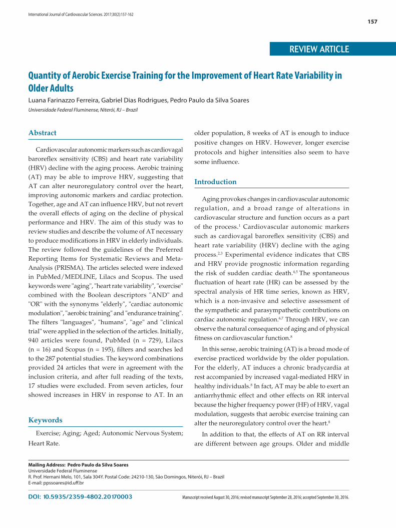
DOI: 10.5935/2359-4802.20170003
Abstract
Cardiovascular autonomic markers such as cardiovagal baroreflex sensitivity (CBS) and heart rate variability (HRV) decline with the aging process. Aerobic training (AT) may be able to improve HRV, suggesting that AT can alter neuroregulatory control over the heart, improving autonomic markers and cardiac protection. Together, age and AT can influence HRV, but not revert the overall effects of aging on the decline of physical performance and HRV. The aim of this study was to review studies and describe the volume of AT necessary to produce modifications in HRV in elderly individuals. The review followed the guidelines of the Preferred Reporting Items for Systematic Reviews and Meta-Analysis (PRISMA). The articles selected were indexed in PubMed/MEDLINE, Lilacs and Scopus. The used keywords were "aging", "heart rate variability", "exercise" combined with the Boolean descriptors "AND" and "OR" with the synonyms "elderly", "cardiac autonomic modulation", "aerobic training" and "endurance training". The filters "languages", "humans", "age" and "clinical trial" were applied in the selection of the articles. Initially, 940 articles were found, PubMed (n = 729), Lilacs (n = 16) and Scopus (n = 195), filters and searches led to the 287 potential studies. The keyword combinations provided 24 articles that were in agreement with the inclusion criteria, and after full reading of the texts, 17 studies were excluded. From seven articles, four showed increases in HRV in response to AT. In an
older population, 8 weeks of AT is enough to induce positive changes on HRV. However, longer exercise protocols and higher intensities also seem to have some influence.
Introduction
Aging provokes changes in cardiovascular autonomic regulation, and a broad range of alterations in cardiovascular structure and function occurs as a part of the process.1 Cardiovascular autonomic markers such as cardiovagal baroreflex sensitivity (CBS) and heart rate variability (HRV) decline with the aging process.2,3 Experimental evidence indicates that CBS and HRV provide prognostic information regarding the risk of sudden cardiac death.4,5 The spontaneous fluctuation of heart rate (HR) can be assessed by the spectral analysis of HR time series, known as HRV, which is a non-invasive and selective assessment of the sympathetic and parasympathetic contributions on cardiac autonomic regulation.6,7 Through HRV, we can observe the natural consequence of aging and of physical fitness on cardiovascular function.8
In this sense, aerobic training (AT) is a broad mode of exercise practiced worldwide by the older population. For the elderly, AT induces a chronic bradycardia at rest accompanied by increased vagal-mediated HRV in healthy individuals.8 In fact, AT may be able to exert an antiarrhythmic effect and other effects on RR interval because the higher frequency power (HF) of HRV, vagal modulation, suggests that aerobic exercise training can alter the neuroregulatory control over the heart.8
In addition to that, the effects of AT on RR interval are different between age groups. Older and middle
157International Journal of Cardiovascular Sciences. 2017;30(2):157-162
REVIEW ARTICLE
Mailing Address: Pedro Paulo da Silva SoaresUniversidade Federal FluminenseR. Prof. Hernani Melo, 101, Sala 304Y. Postal Code: 24210-130, São Domingos, Niterói, RJ – BrazilE-mail: [email protected]
Quantity of Aerobic Exercise Training for the Improvement of Heart Rate Variability in Older AdultsLuana Farinazzo Ferreira, Gabriel Dias Rodrigues, Pedro Paulo da Silva SoaresUniversidade Federal Fluminense, Niterói, RJ – Brazil
Manuscript received August 30, 2016; revised manuscript September 28, 2016; accepted September 30, 2016.
Exercise; Aging; Aged; Autonomic Nervous System;
Heart Rate.
Keywords
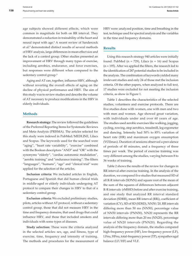
158
age subjects showed different effects, which were common in magnitude for both on RR interval. They demonstrated a reduction in trainability of the heart and neural input with age.8 A recent review of Sandercock et al.8 demonstrated distinct results of several methods of HRV analysis, large differences in mean effect size and the lack of a control group. Other studies presented an improvement of HRV through many types of exercise, including aerobics, endurance, and force exercises, but responses were different when compared to the sedentary control group.9
Aging and AT can, together, influence HRV, although without reverting the overall effects of aging on the decline of physical performance and HRV. The aim of this study was to review studies and describe the volume of AT necessary to produce modifications in the HRV in elderly individuals.
Methods
Research strategy: The review followed the guidelines of the Preferred Reporting Items for Systematic Reviews and Meta-Analysis (PRISMA). The articles selected for this study were indexed in PubMed/MEDLINE, Lilacs and Scopus. The keywords used for the searched were “aging”, “heart rate variability”, “exercise” combined with the Boolean descriptors “AND” and “OR” with the synonyms “elderly“, “cardiac autonomic modulation”, “aerobic training” and “endurance training”. The filters “languages”, “humans”, “age” and “clinical trial” were applied for the selection of the articles.
Inclusion criteria: We included articles in English, Portuguese and Spanish that did human clinical trials in middle-aged or elderly individuals undergoing AT protocol to compare their changes in HRV to that of a sedentary control group.
Exclusion criteria: We excluded preliminary studies, pilots, articles without AT protocol, without a sedentary control group, those that did not measure HRV in the time and frequency domains, that used drugs that could influence HRV, and those that included smokers and individuals with some types of diseases.
Study selection: These were the criteria analyzed in the selected articles: sex, age, and fitness, type of exercise, time, frequency and intensity of training. The methods and procedures for the measurement of
HRV were: analyzed position, time and breathing in the
test, technique used for spectral analysis and the variables
in the time and frequency domains.
Results
Using this research strategy 940 articles were initially
found: PubMed (n = 729), Lilacs (n = 16) and Scopus
(n = 195). After we applied the filters, the research led to
the identification of 287 potential studies to be included in
the analysis. The combination of keywords yielded many
irrelevant studies and only 24 of those met the inclusion
criteria. Of the other papers, when analyzed to full text,
17 studies were excluded for not meeting the inclusion
criteria, as show in Figure 1.
Table 1 describes the characteristics of the selected
studies, volunteers and exercise protocols. There are
five studies done with women, one with men and two
with men and women. Age showed great variation,
with individuals under and over 60 years of age.
The studies used aerobic exercises like walking, jogging,
cycling, rowing, step aerobics, treadmill, leg ergometer
and dancing. Intensity had 50% to 85% variation of
heart rate (HR) and maximum oxygen onsumption
(VO2max). Duration of sessions observed a prevalence
of periods of 40 minutes, and a frequency of three
times per week. The period of exercise training was
very different among the studies, varying between 8 to
36 weeks of training.
Table 2 shows the results of the review for changes in
RR interval after exercise training. In the analysis of the
duration, we compared five studies that measured SD of
all R-R intervals (SDNN) and square root of the mean of
the sum of the squares of differences between adjacent
R-R intervals (rMSSD) before and after exercise training,
and one study that analyzed RR interval standard
deviation (SDRR), mean RR interval (RRi), coefficient of
variation (CV), SD of SD (SDSD), NN50/20, RR intervals
differing more than 50 ms (NN50), percentage value
of NN50 intervals (PNN50), NN20 represents the RR
intervals differing more than 20 ms (NN20), percentage
value of NN20 intervals (PNN20). Regarding the
analysis of the frequency domain, the studies compared
high-frequency power (HF), low-frequency power (LF),
LFnu, HFnu, total-frequency power (TP), sympathovagal
balance (LF/HF) and VLF.
Ferreira et al.
Physical training and heart rate variability
Int J Cardiovasc Sci. 2017;30(2):157-162
Review Article
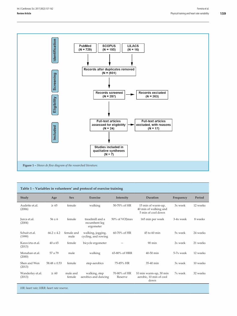
159
Figure 1 – Shows de flow diagram of the researched literature.
Table 1 – Variables in volunteers’ and protocol of exercise training
Study Age Sex Exercise Intensity Duration Frequency Period
Audette et.al. (2006)
≥ 65 female walking 50-70% of HR 15 min of warm-up, 40 min of walking and
5 min of cool down
3x week 12 weeks
Jurca et.al. (2004)
56 ± 6 female treadmill and a recumbent leg
ergometer
50% of VO2max 165 min per week 3-4x week 8 weeks
Schuit et.al. (1999)
66.2 ± 4.2 female and male
walking, jogging, cycling, and rowing
60-70% of HR 45 to 60 min 5x week 24 weeks
Karavirta et.al.(2013)
40 a 65 female bicycle ergometer -- 90 min 2x week 21 weeks
Monahan et.al.(2000)
57 a 79 male walking 65-80% of HRR 40-50 min 5-7x week 12 weeks
Shen and Wen(2013)
58.48 ± 0.53 female step-aerobics 75-85% HR 35-40 min 3x week 10 weeks
Wanderley et.al.(2013)
≥ 60 male and female
walking, step aerobics and dancing
70-80% of HR Reserve
10 min warm-up, 30 min aerobic, 10 min of cool
down
7x week 32 weeks
HR: heart rate; HRR: heart rate reserve.
Ferreira et al.
Physical training and heart rate variability
Int J Cardiovasc Sci. 2017;30(2):157-162
Review Article
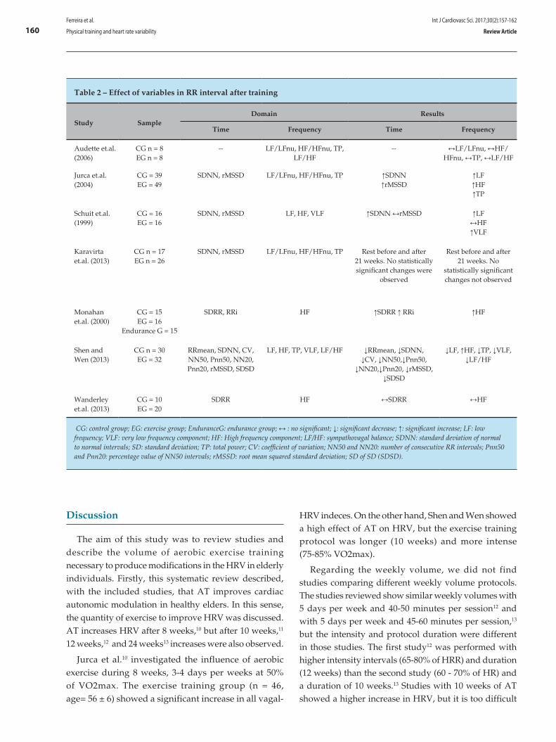
160
Table 2 – Effect of variables in RR interval after training
Study SampleDomain Results
Time Frequency Time Frequency
Audette et.al. (2006)
CG n = 8EG n = 8
-- LF/LFnu, HF/HFnu, TP, LF/HF
-- ↔LF/LFnu, ↔HF/HFnu, ↔TP, ↔LF/HF
Jurca et.al. (2004)
CG = 39 EG = 49
SDNN, rMSSD LF/LFnu, HF/HFnu, TP ↑SDNN↑rMSSD
↑LF↑HF↑TP
Schuit et.al.(1999)
CG = 16EG = 16
SDNN, rMSSD LF, HF, VLF ↑SDNN ↔rMSSD ↑LF↔HF↑VLF
Karavirta et.al. (2013)
CG n = 17EG n = 26
SDNN, rMSSD LF/LFnu, HF/HFnu, TP Rest before and after 21 weeks. No statistically significant changes were
observed
Rest before and after 21 weeks. No
statistically significant changes not observed
Monahan et.al. (2000)
CG = 15EG = 16
Endurance G = 15
SDRR, RRi HF ↑SDRR ↑ RRi ↑HF
Shen and Wen (2013)
CG n = 30EG = 32
RRmean, SDNN, CV, NN50, Pnn50, NN20, Pnn20, rMSSD, SDSD
LF, HF, TP, VLF, LF/HF ↓RRmean, ↓SDNN, ↓CV, ↓NN50,↓Pnn50,
↓NN20,↓Pnn20, ↓rMSSD, ↓SDSD
↓LF, ↑HF, ↓TP, ↓VLF, ↓LF/HF
Wanderley et.al. (2013)
CG = 10EG = 20
SDRR HF ↔SDRR ↔HF
CG: control group; EG: exercise group; EnduranceG: endurance group; ↔ : no significant; ↓: significant decrease; ↑: significant increase; LF: low frequency; VLF: very low frequency component; HF: High frequency component; LF/HF: sympathovagal balance; SDNN: standard deviation of normal to normal intervals; SD: standard deviation; TP: total power; CV: coefficient of variation; NN50 and NN20: number of consecutive RR intervals; Pnn50 and Pnn20: percentage value of NN50 intervals; rMSSD: root mean squared standard deviation; SD of SD (SDSD).
Discussion
The aim of this study was to review studies and describe the volume of aerobic exercise training necessary to produce modifications in the HRV in elderly individuals. Firstly, this systematic review described, with the included studies, that AT improves cardiac autonomic modulation in healthy elders. In this sense, the quantity of exercise to improve HRV was discussed. AT increases HRV after 8 weeks,10 but after 10 weeks,11 12 weeks,12 and 24 weeks13 increases were also observed.
Jurca et al.10 investigated the influence of aerobic exercise during 8 weeks, 3-4 days per weeks at 50% of VO2max. The exercise training group (n = 46, age= 56 ± 6) showed a significant increase in all vagal-
HRV indeces. On the other hand, Shen and Wen showed a high effect of AT on HRV, but the exercise training protocol was longer (10 weeks) and more intense (75-85% VO2max).
Regarding the weekly volume, we did not find studies comparing different weekly volume protocols. The studies reviewed show similar weekly volumes with 5 days per week and 40-50 minutes per session12 and with 5 days per week and 45-60 minutes per session,13 but the intensity and protocol duration were different in those studies. The first study12 was performed with higher intensity intervals (65-80% of HRR) and duration (12 weeks) than the second study (60 - 70% of HR) and a duration of 10 weeks.13 Studies with 10 weeks of AT showed a higher increase in HRV, but it is too difficult
Ferreira et al.
Physical training and heart rate variability
Int J Cardiovasc Sci. 2017;30(2):157-162
Review Article
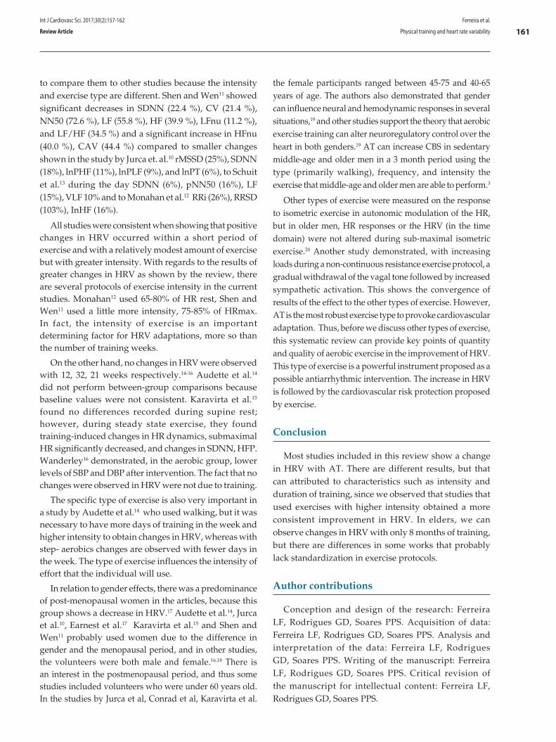
161
to compare them to other studies because the intensity and exercise type are different. Shen and Wen11 showed significant decreases in SDNN (22.4 %), CV (21.4 %), NN50 (72.6 %), LF (55.8 %), HF (39.9 %), LFnu (11.2 %), and LF/HF (34.5 %) and a significant increase in HFnu (40.0 %), CAV (44.4 %) compared to smaller changes shown in the study by Jurca et. al.10 rMSSD (25%), SDNN (18%), lnPHF (11%), lnPLF (9%), and lnPT (6%), to Schuit et al.13 during the day SDNN (6%), pNN50 (16%), LF (15%), VLF 10% and to Monahan et al.12 RRi (26%), RRSD (103%), InHF (16%).
All studies were consistent when showing that positive changes in HRV occurred within a short period of exercise and with a relatively modest amount of exercise but with greater intensity. With regards to the results of greater changes in HRV as shown by the review, there are several protocols of exercise intensity in the current studies. Monahan12 used 65-80% of HR rest, Shen and Wen11 used a little more intensity, 75-85% of HRmax. In fact, the intensity of exercise is an important determining factor for HRV adaptations, more so than the number of training weeks.
On the other hand, no changes in HRV were observed with 12, 32, 21 weeks respectively.14-16 Audette et al.14 did not perform between-group comparisons because baseline values were not consistent. Karavirta et al.15 found no differences recorded during supine rest; however, during steady state exercise, they found training-induced changes in HR dynamics, submaximal HR significantly decreased, and changes in SDNN, HFP. Wanderley16 demonstrated, in the aerobic group, lower levels of SBP and DBP after intervention. The fact that no changes were observed in HRV were not due to training.
The specific type of exercise is also very important in a study by Audette et al.14 who used walking, but it was necessary to have more days of training in the week and higher intensity to obtain changes in HRV, whereas with step- aerobics changes are observed with fewer days in the week. The type of exercise influences the intensity of effort that the individual will use.
In relation to gender effects, there was a predominance of post-menopausal women in the articles, because this group shows a decrease in HRV.17 Audette et al.14, Jurca et al.10, Earnest et al.17 Karavirta et al.15 and Shen and Wen11 probably used women due to the difference in gender and the menopausal period, and in other studies, the volunteers were both male and female.16,18 There is an interest in the postmenopausal period, and thus some studies included volunteers who were under 60 years old. In the studies by Jurca et al, Conrad et al, Karavirta et al.
the female participants ranged between 45-75 and 40-65 years of age. The authors also demonstrated that gender can influence neural and hemodynamic responses in several situations,19 and other studies support the theory that aerobic exercise training can alter neuroregulatory control over the heart in both genders.19 AT can increase CBS in sedentary middle-age and older men in a 3 month period using the type (primarily walking), frequency, and intensity the exercise that middle-age and older men are able to perform.3
Other types of exercise were measured on the response to isometric exercise in autonomic modulation of the HR, but in older men, HR responses or the HRV (in the time domain) were not altered during sub-maximal isometric exercise.20 Another study demonstrated, with increasing loads during a non-continuous resistance exercise protocol, a gradual withdrawal of the vagal tone followed by increased sympathetic activation. This shows the convergence of results of the effect to the other types of exercise. However, AT is the most robust exercise type to provoke cardiovascular adaptation. Thus, before we discuss other types of exercise, this systematic review can provide key points of quantity and quality of aerobic exercise in the improvement of HRV. This type of exercise is a powerful instrument proposed as a possible antiarrhythmic intervention. The increase in HRV is followed by the cardiovascular risk protection proposed by exercise.
Conclusion
Most studies included in this review show a change in HRV with AT. There are different results, but that can attributed to characteristics such as intensity and duration of training, since we observed that studies that used exercises with higher intensity obtained a more consistent improvement in HRV. In elders, we can observe changes in HRV with only 8 months of training, but there are differences in some works that probably lack standardization in exercise protocols.
Author contributions
Conception and design of the research: Ferreira LF, Rodrigues GD, Soares PPS. Acquisition of data: Ferreira LF, Rodrigues GD, Soares PPS. Analysis and interpretation of the data: Ferreira LF, Rodrigues GD, Soares PPS. Writing of the manuscript: Ferreira LF, Rodrigues GD, Soares PPS. Critical revision of the manuscript for intellectual content: Ferreira LF, Rodrigues GD, Soares PPS.
Ferreira et al.
Physical training and heart rate variability
Int J Cardiovasc Sci. 2017;30(2):157-162
Review Article
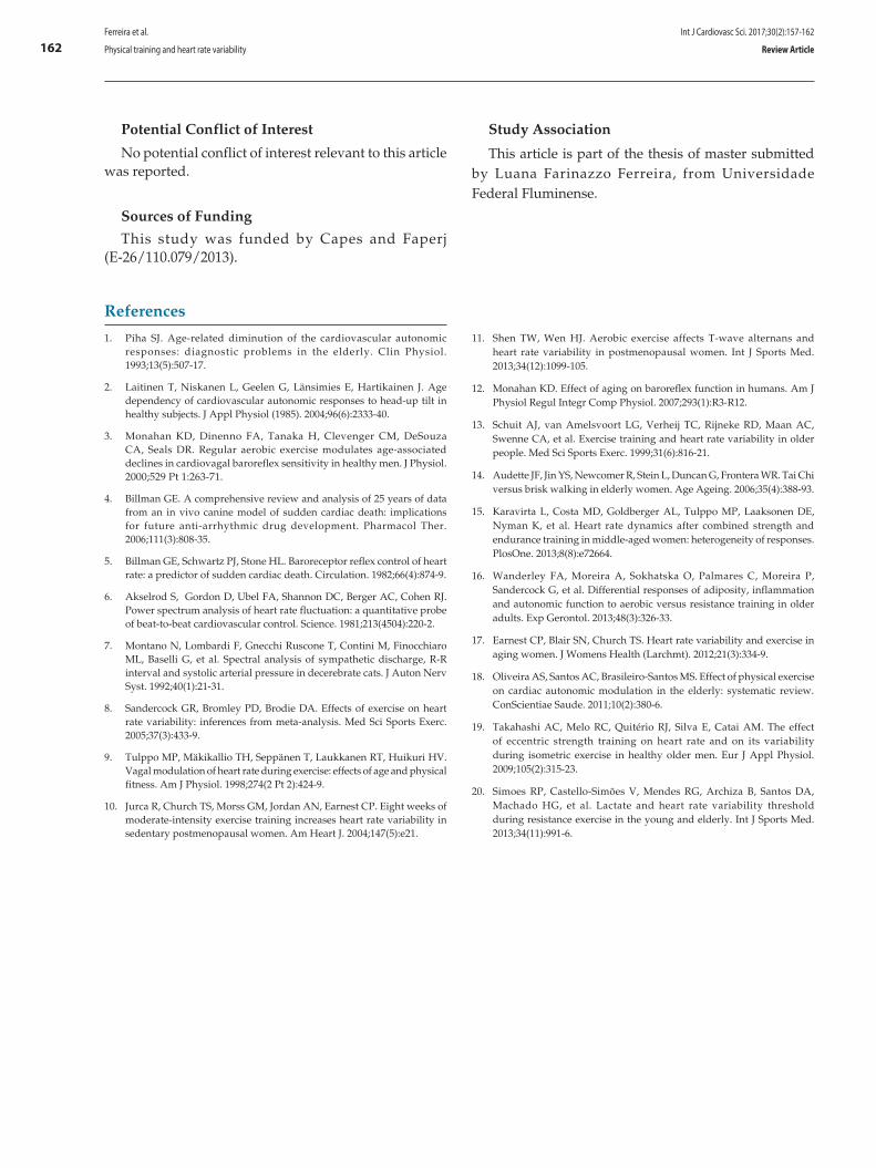
162
1. Piha SJ. Age-related diminution of the cardiovascular autonomic responses: diagnostic problems in the elderly. Clin Physiol. 1993;13(5):507-17.
2. Laitinen T, Niskanen L, Geelen G, Länsimies E, Hartikainen J. Age dependency of cardiovascular autonomic responses to head-up tilt in healthy subjects. J Appl Physiol (1985). 2004;96(6):2333-40.
3. Monahan KD, Dinenno FA, Tanaka H, Clevenger CM, DeSouza CA, Seals DR. Regular aerobic exercise modulates age-associated declines in cardiovagal baroreflex sensitivity in healthy men. J Physiol. 2000;529 Pt 1:263-71.
4. Billman GE. A comprehensive review and analysis of 25 years of data from an in vivo canine model of sudden cardiac death: implications for future anti-arrhythmic drug development. Pharmacol Ther. 2006;111(3):808-35.
5. Billman GE, Schwartz PJ, Stone HL. Baroreceptor reflex control of heart rate: a predictor of sudden cardiac death. Circulation. 1982;66(4):874-9.
6. Akselrod S, Gordon D, Ubel FA, Shannon DC, Berger AC, Cohen RJ. Power spectrum analysis of heart rate fluctuation: a quantitative probe of beat-to-beat cardiovascular control. Science. 1981;213(4504):220-2.
7. Montano N, Lombardi F, Gnecchi Ruscone T, Contini M, Finocchiaro ML, Baselli G, et al. Spectral analysis of sympathetic discharge, R-R interval and systolic arterial pressure in decerebrate cats. J Auton Nerv Syst. 1992;40(1):21-31.
8. Sandercock GR, Bromley PD, Brodie DA. Effects of exercise on heart rate variability: inferences from meta-analysis. Med Sci Sports Exerc. 2005;37(3):433-9.
9. Tulppo MP, Mäkikallio TH, Seppänen T, Laukkanen RT, Huikuri HV. Vagal modulation of heart rate during exercise: effects of age and physical fitness. Am J Physiol. 1998;274(2 Pt 2):424-9.
10. Jurca R, Church TS, Morss GM, Jordan AN, Earnest CP. Eight weeks of moderate-intensity exercise training increases heart rate variability in sedentary postmenopausal women. Am Heart J. 2004;147(5):e21.
11. Shen TW, Wen HJ. Aerobic exercise affects T-wave alternans and heart rate variability in postmenopausal women. Int J Sports Med. 2013;34(12):1099-105.
12. Monahan KD. Effect of aging on baroreflex function in humans. Am J Physiol Regul Integr Comp Physiol. 2007;293(1):R3-R12.
13. Schuit AJ, van Amelsvoort LG, Verheij TC, Rijneke RD, Maan AC, Swenne CA, et al. Exercise training and heart rate variability in older people. Med Sci Sports Exerc. 1999;31(6):816-21.
14. Audette JF, Jin YS, Newcomer R, Stein L, Duncan G, Frontera WR. Tai Chi versus brisk walking in elderly women. Age Ageing. 2006;35(4):388-93.
15. Karavirta L, Costa MD, Goldberger AL, Tulppo MP, Laaksonen DE, Nyman K, et al. Heart rate dynamics after combined strength and endurance training in middle-aged women: heterogeneity of responses. PlosOne. 2013;8(8):e72664.
16. Wanderley FA, Moreira A, Sokhatska O, Palmares C, Moreira P, Sandercock G, et al. Differential responses of adiposity, inflammation and autonomic function to aerobic versus resistance training in older adults. Exp Gerontol. 2013;48(3):326-33.
17. Earnest CP, Blair SN, Church TS. Heart rate variability and exercise in aging women. J Womens Health (Larchmt). 2012;21(3):334-9.
18. Oliveira AS, Santos AC, Brasileiro-Santos MS. Effect of physical exercise on cardiac autonomic modulation in the elderly: systematic review. ConScientiae Saude. 2011;10(2):380-6.
19. Takahashi AC, Melo RC, Quitério RJ, Silva E, Catai AM. The effect of eccentric strength training on heart rate and on its variability during isometric exercise in healthy older men. Eur J Appl Physiol. 2009;105(2):315-23.
20. Simoes RP, Castello-Simões V, Mendes RG, Archiza B, Santos DA, Machado HG, et al. Lactate and heart rate variability threshold during resistance exercise in the young and elderly. Int J Sports Med. 2013;34(11):991-6.
References
Potential Conflict of Interest
No potential conflict of interest relevant to this article was reported.
Sources of Funding
This study was funded by Capes and Faperj (E-26/110.079/2013).
Study Association
This article is part of the thesis of master submitted by Luana Farinazzo Ferreira, from Universidade Federal Fluminense.
Ferreira et al.
Physical training and heart rate variability
Int J Cardiovasc Sci. 2017;30(2):157-162
Review Article
DOI: 10.5935/2359-4802.20170014
Abstract
The objective of this study is to make a review of the narrative of coronary artery tortuosity (CAT) approaching several situations in clinical practice where tortuosity can have a relevant role, and also evaluate if tortuosity can be related to the presence of myocardial ischemia in patients without coronary obstruction using scientific evidences in medical literature. Textbook of applied Physiology in Cardiology with study of coronary circulation, theoretical articles with studies of Hemodynamics, Fluid and Mechanical Dynamic, and experimental articles with simulation in computers were used as support to answer this last question.
Introduction
Coronary circulation
There are two basic types of coronary vessels: conductance and resistance vessels. The epicardial arteries, right and left, and its main and major branches that emerge in acute angle of relative large caliber work as vessels of conductance offering in the diastole minimum resistance to the blood flow. The deep perforators that originate in a right angle of the epicardial arteries penetrate deeply in the myocardial walls and nurture sub endocardial layers offering great resistance to the flow mainly in the ventricular systole. They are responsible for the coronary flow autoregulation maintaining it adequately in broad spectrum of the pressure variation and increasing the flow in exercising situations mainly through the
local metabolic regulation. Microcirculation is part of the coronary circulation constituted by arterioles and capillaries responsible for regulating the oxygen supply to the myocardium.
The heart is a highly aerobic organ, but it depends almost exclusively on the oxidation of the substrates to generate energy which will move it and has almost no oxygen reserve. It receives about 5% of cardiac output and it is a little perfused organ, but it is the organ that has the highest oxygen extraction of the organism.
Taking into consideration Fick’s equation (oxygen consumption = coronary flow X arteriovenous difference of oxygen), we verified that the physiological determinants of the coronary flow are the same that command the demand and consumption of oxygen: blood pressure, heart rate, ventricular wall tension, dP/dt maximum.
According to Poiseuille’s law, the flow in any vessel system is directly proportional to the difference of pressure in its extremities, and inversely proportional to the resistance of the system, which is in turn proportional to the length of the tube, viscosity of the fluid and inversely proportional to the fourth power of the radius (most important factor). Influences of vasomotility of the autonomous nervous system, of drugs, mainly of local autoregulation, determine variations in the flow. Due to cyclical pressure and variations and myocardium tension, the coronary flow, in the systole represents from 25% to 30% of the total, and from 70% to 75% in the diastole.
Factors that can affect myocardial consumption of oxygen and consequently the coronary flow can be divided into 3 groups:
a) Factors that affect consumption (demand): intraventricular tension, heart rate, myocardial contractile state and electrical activation, and cardiac metabolism.
163International Journal of Cardiovascular Sciences. 2017;30(2):163-170
REVIEW ARTICLE
Mailing Address: André Pereira Duque EstradaRua Coronel Moreira César, 376/602. Postal Code: 24230064. Niterói, Rio de Janeiro, RJ – BrazilE-mail: [email protected]
Coronary tortuosity and its role in myocardial ischemia in patients with no coronary obstructionsAndré Pereira Duque Estrada,1 Rosane de Oliveira Lopes2 Humberto Villacorta Junior2
Hospital Federal dos Servidores do Estado1; Universidade Federal Fluminense2, Niterói, RJ – Brazil
Manuscript received August26, 2016; revised manuscript September 30, 2016; accepted November 21, 2016.
Coronary Vessels; Coronary Circulation; Myocardial Ischemia; Hypertension; Aging.
Keywords
164
b) Factors that affect oxygen supply (circulation): perfusion pressure (aortic pressure minus left atrial pressure), anatomical condition of arterial bed, arterial PaO2 and %HbO2, dissociation of O2 from hemoglobin, transmyocardial distribution of flow with capillary opening and microcirculation alterations.
c) Factors that compromise oxygen supply to the myocardium (arterial caliber): neurovegetative and humoral action, local regulation (nitric oxide, adenosine, pH and PCO2), coronary flow reserve and drugs.1
Coronary flow regulation, coronary flow reserve, and myocardial ischemia
Regardless of extra-coronary factors, the contraction or relaxation of the arteries and arterioles are influenced by muscular, neurovegetative, and humoral factors acting on vessel walls. There are, at least, four main systems: the myogenic control reflects intrinsic property of the vascular muscle to react to pressure distension on the wall vessel (possibly by channels activated by distension), autonomous control with catecholamine, adrenaline, and noradrenaline, endothelial control with major importance of nitric oxide- a potent vasodilator released by mechanical forces of flow friction on the endothelium – and the main – the metabolic control by the partial drop of the pressure of the local oxygen.
Small partial pressure variation of oxygen (decrease) can be sufficient to cause vasodilatation and flow increase, balancing the demand and supply until a new imbalance occurs.
The mechanism of this active dilatation results from a direct effect of hypoxia on coronary artery smooth muscle, and/or the increase of metabolic vasodilators by the effect of hypoxia in the cells, mainly the adenosine.
The concept of the coronary flow reserve is related to the maximum ability of coronary vessels to increase flow in response to myocardial demand and this capacity is 5 times greater in relation to the flow at rest.
When compensation mechanisms are exhausted the process of myocardial ischemia occurs, with metabolic contractility and electrocardiographic changes in the moment that the coronary flow decreases from 40 ml/min.1
Development
Coronary arteries tend to be more tortuous than other arteries and accompany repetitive movements
of flexion and relaxation that occur during the cardiac cycle. Intra luminal traction and pressure are two forces that try to stretch the vessel and opposing to this there is the force of retraction that depends basically of elastin. The degeneration of elastin in the coronary wall can occur due to pathological processes or related to old age and lead to aneurysmal dilatations and to tortuosity.2
Postmortem angiographies of 145 patients without coronary obstruction showed that coronary tortuosity is positively related to age and negatively related to the heart weight in equal importance, and, in a smaller degree, positively to the caliber of the vessel.
Moreover coronary tortuosity is positively related to arterial hypertension and to the female gender, and negatively related to the process of atherosclerosis.3
In Turkey, in 2013, 148 male patients that underwent coronariography in a period of three months were selected. Exclusion criteria included patients with a history of revascularization surgery or coronary angioplasty. These patients were divided into two groups: with and without coronary tortuosity, and the relation between of tortuosity with coronary obstruction was studied. A negative correlation between the employed tortuosity score and severe obstructive coronary disease was found. It was questioned if coronary tortuosity could represent a genetic geometric factor of protection against obstructive coronary disease or a mechanism of coronary remodeling.4
Patients with stable angina and no coronary obstruction, but with coronary tortuosity, present an increased calcium score in relation to those without tortuosity, suggesting an association with subclinical atherosclerosis. A similar finding was found in another study that evaluated patients with retinal arteries tortuosity, and with coronary tortuosity that presented an increase of the mid-intimal thickness in the carotids. In this last study, it was verified an association with females and individuals of short stature. They are more commonly found in the circumflex followed by the anterior descending artery and lastly right coronary.5 The latter can be described as S or C shaped and the S shape presents less frequent obstructive atherosclerotic disease.6,7
A statistical analysis of an angiographic study with 52 patients in which were selected 32 left coronary arteriography with no obstruction, and 35 right coronary arteriography with no obstruction showed that, in the left coronary, tortuosity was larger in its distal portion and, in the right coronary, it was smaller in its middle segment.8
Estrada et al.
Coronary tortuosity
Int J Cardiovasc Sci. 2017;30(2):163-170
Review Article
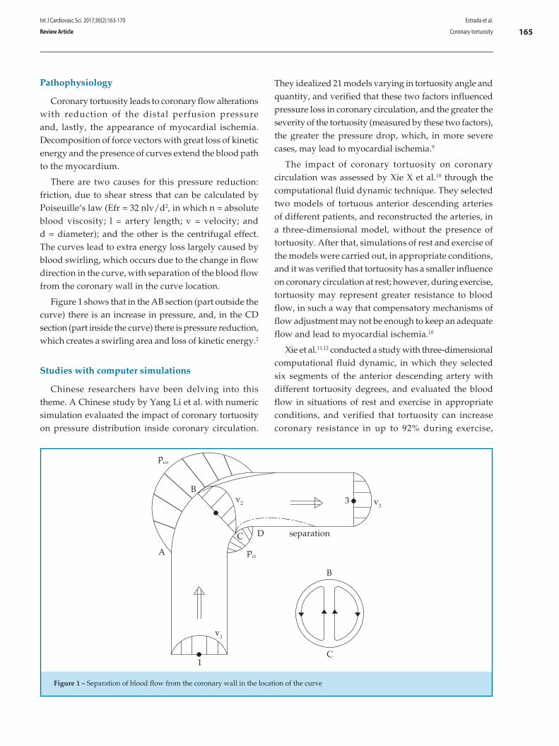
165
pci
DC separation
B
C
v1
1
v2 3 v3
pco
B
A
Figure 1 – Separation of blood flow from the coronary wall in the location of the curve
Pathophysiology
Coronary tortuosity leads to coronary flow alterations with reduction of the distal perfusion pressure and, lastly, the appearance of myocardial ischemia. Decomposition of force vectors with great loss of kinetic energy and the presence of curves extend the blood path to the myocardium.
There are two causes for this pressure reduction: friction, due to shear stress that can be calculated by Poiseuille’s law (Efr = 32 nlv/d2, in which n = absolute blood viscosity; l = artery length; v = velocity; and d = diameter); and the other is the centrifugal effect. The curves lead to extra energy loss largely caused by blood swirling, which occurs due to the change in flow direction in the curve, with separation of the blood flow from the coronary wall in the curve location.
Figure 1 shows that in the AB section (part outside the curve) there is an increase in pressure, and, in the CD section (part inside the curve) there is pressure reduction, which creates a swirling area and loss of kinetic energy.2
Studies with computer simulations
Chinese researchers have been delving into this theme. A Chinese study by Yang Li et al. with numeric simulation evaluated the impact of coronary tortuosity on pressure distribution inside coronary circulation.
They idealized 21 models varying in tortuosity angle and quantity, and verified that these two factors influenced pressure loss in coronary circulation, and the greater the severity of the tortuosity (measured by these two factors), the greater the pressure drop, which, in more severe cases, may lead to myocardial ischemia.9
The impact of coronary tortuosity on coronary circulation was assessed by Xie X et al.10 through the computational fluid dynamic technique. They selected two models of tortuous anterior descending arteries of different patients, and reconstructed the arteries, in a three-dimensional model, without the presence of tortuosity. After that, simulations of rest and exercise of the models were carried out, in appropriate conditions, and it was verified that tortuosity has a smaller influence on coronary circulation at rest; however, during exercise, tortuosity may represent greater resistance to blood flow, in such a way that compensatory mechanisms of flow adjustment may not be enough to keep an adequate flow and lead to myocardial ischemia.10
Xie et al.11,12 conducted a study with three-dimensional computational fluid dynamic, in which they selected six segments of the anterior descending artery with different tortuosity degrees, and evaluated the blood flow in situations of rest and exercise in appropriate conditions, and verified that tortuosity can increase coronary resistance in up to 92% during exercise,
Estrada et al.
Coronary tortuosity
Int J Cardiovasc Sci. 2017;30(2):163-170
Review Article
166
causing coronary autoregulation to fail. This study also suggests that tortuosity may constitute a risk factor for atherosclerosis, since it can lead to the appearance of a region with low, wavering shear stress in the internal wall of the curve’s descent, when the angle of the curve is greater than 120 degrees.11,12 Outro estudo recente confirma que a tortuosidade leva a queda da pressão e pode levar a déficit de perfusão.13
Prevalence
A retrospective study from 2009 carried out in West Virginia with 1221 patients, who had undergone catheterization in the previous 8 months, identified 12.45% of patients with coronary tortuosity and showed a higher occurrence in women and lower incidence of obstructive coronary disease, but it did not find predictors of coronary tortuosity among the following conditions: systemic arterial hypertension, diabetes mellitus, advanced age, dyslipidemia, smoking, and family history of obstructive coronary disease.14
Another retrospective study carried out at the Zhongda Hospital of Southeast University in Nanjing, China, with 1010 patients who had undergone coronarography due to anginal complaints, separated these patients into four groups according to the presence or absence of coronary tortuosity and presence or absence of coronary obstructions, and did a 2 to 4-year follow-up with these patients. The prevalence of tortuosity was 39.1% and was significantly higher in women and in patients with systemic arterial hypertension. It was negatively related to the presence of coronary obstructions.5
Age-related alterations
Among the modifications in coronary circulation that occur with the aging process, are the increase of coronary tortuosity with minimal atherosclerotic lesions, calcification and,3,15 at least in animals,16 an imbalance between the extension of the capillary network and myocyte hypertrophy. Maximum oxygen consumption is progressively reduced. In advanced age rats, it has also been demonstrated a higher deviation angle of secondary branches in relation to the main branch.
Studies with hypertensive individuals
There is a strong correlation between the presence of hypertension and coronary tortuosity.17 In 1981, Sanchez Torres G. et al.17 studied, in Mexico, a group
of 46 hypertensive patients, who were divided into three subgroups: Group 1 – angina pectoris; Group 2 – left ventricular hypertrophy in the electrocardiogram; and Group 3 – asymptomatic and without left ventricular hypertrophy in the electrocardiogram. In the coronarography, Group 1 presented coronary obstructions in 28% and tortuosity in 94.8%; Group 2 presented tortuosity in 74.9%; and Group 3 in 69.1%. Groups 2 and 3 did not present cases of coronary obstruction.18
In 1982, Sanchez Torres G. et al.18 continued to study the presence of myocardial ischemia in hypertensive patients, this time with patients who met the criteria of left ventricular hypertrophy in the electrocardiogram. In a group of 70 patients who all underwent ergometric test, coronarography, and left ventriculography, and 10 patients who also underwent a study with myocardial scintigraphy, describe "corkscrew tortuosity" in coronary angiography (without associated obstructions) in 83.7% of patients and suggest that subendocardial ischemia may be related to the increase of ventricular mass with a lower coronary reserve.19
Relation to ventricular relaxation
An echocardiographic study with 104 patients (50 with coronary tortuosity and 54 without) has shown that coronary tortuosity is related to the worsening of ventricular relaxation20 evaluated by the decrease of the E/A relation of the transmitral flow, increase of the E-wave deceleration time, increase of isovolumetric relaxation time, and greater thickness of the interventricular septum and of the left ventricular posterior wall.21
Relation to coronary dissection
A known cause of non-atherosclerotic acute coronary syndrome is the spontaneous dissection of the coronary associated to the presence of fibromuscular dysplasia in non-coronary arteries. A case control study with 246 patients with spontaneous coronary dissection and 313 control patients without coronary obstructions submitted to coronarography showed a significantly higher prevalence of tortuosity in patients who had the first episode of spontaneous dissection (78% vs 17%, p < 0.0001), even though these patients have a low prevalence of arterial hypertension (34%). The recurrence of spontaneous dissection occurred in tortuous segments in 80% of cases. The presence of severe tortuosity was related
Estrada et al.
Coronary tortuosity
Int J Cardiovasc Sci. 2017;30(2):163-170
Review Article
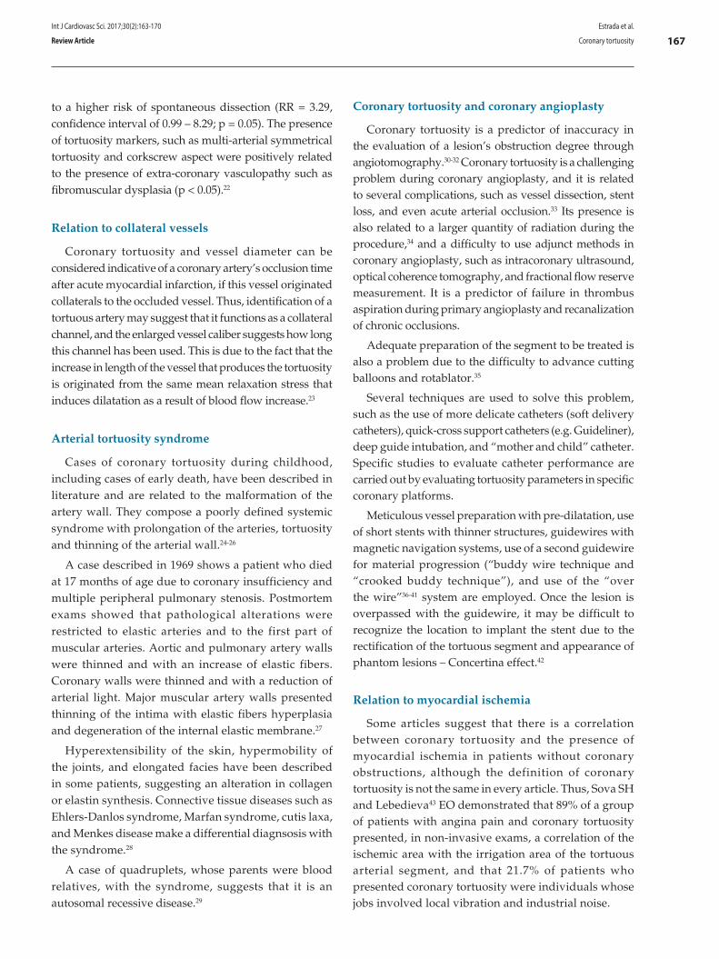
167
to a higher risk of spontaneous dissection (RR = 3.29, confidence interval of 0.99 – 8.29; p = 0.05). The presence of tortuosity markers, such as multi-arterial symmetrical tortuosity and corkscrew aspect were positively related to the presence of extra-coronary vasculopathy such as fibromuscular dysplasia (p < 0.05).22
Relation to collateral vessels
Coronary tortuosity and vessel diameter can be considered indicative of a coronary artery’s occlusion time after acute myocardial infarction, if this vessel originated collaterals to the occluded vessel. Thus, identification of a tortuous artery may suggest that it functions as a collateral channel, and the enlarged vessel caliber suggests how long this channel has been used. This is due to the fact that the increase in length of the vessel that produces the tortuosity is originated from the same mean relaxation stress that induces dilatation as a result of blood flow increase.23
Arterial tortuosity syndrome
Cases of coronary tortuosity during childhood, including cases of early death, have been described in literature and are related to the malformation of the artery wall. They compose a poorly defined systemic syndrome with prolongation of the arteries, tortuosity and thinning of the arterial wall.24-26
A case described in 1969 shows a patient who died at 17 months of age due to coronary insufficiency and multiple peripheral pulmonary stenosis. Postmortem exams showed that pathological alterations were restricted to elastic arteries and to the first part of muscular arteries. Aortic and pulmonary artery walls were thinned and with an increase of elastic fibers. Coronary walls were thinned and with a reduction of arterial light. Major muscular artery walls presented thinning of the intima with elastic fibers hyperplasia and degeneration of the internal elastic membrane.27
Hyperextensibility of the skin, hypermobility of the joints, and elongated facies have been described in some patients, suggesting an alteration in collagen or elastin synthesis. Connective tissue diseases such as Ehlers-Danlos syndrome, Marfan syndrome, cutis laxa, and Menkes disease make a differential diagnsosis with the syndrome.28
A case of quadruplets, whose parents were blood relatives, with the syndrome, suggests that it is an autosomal recessive disease.29
Coronary tortuosity and coronary angioplasty
Coronary tortuosity is a predictor of inaccuracy in the evaluation of a lesion’s obstruction degree through angiotomography.30-32 Coronary tortuosity is a challenging problem during coronary angioplasty, and it is related to several complications, such as vessel dissection, stent loss, and even acute arterial occlusion.33 Its presence is also related to a larger quantity of radiation during the procedure,34 and a difficulty to use adjunct methods in coronary angioplasty, such as intracoronary ultrasound, optical coherence tomography, and fractional flow reserve measurement. It is a predictor of failure in thrombus aspiration during primary angioplasty and recanalization of chronic occlusions.
Adequate preparation of the segment to be treated is also a problem due to the difficulty to advance cutting balloons and rotablator.35
Several techniques are used to solve this problem, such as the use of more delicate catheters (soft delivery catheters), quick-cross support catheters (e.g. Guideliner), deep guide intubation, and “mother and child” catheter. Specific studies to evaluate catheter performance are carried out by evaluating tortuosity parameters in specific coronary platforms.
Meticulous vessel preparation with pre-dilatation, use of short stents with thinner structures, guidewires with magnetic navigation systems, use of a second guidewire for material progression (“buddy wire technique and “crooked buddy technique”), and use of the “over the wire”36-41 system are employed. Once the lesion is overpassed with the guidewire, it may be difficult to recognize the location to implant the stent due to the rectification of the tortuous segment and appearance of phantom lesions – Concertina effect.42
Relation to myocardial ischemia
Some articles suggest that there is a correlation between coronary tortuosity and the presence of myocardial ischemia in patients without coronary obstructions, although the definition of coronary tortuosity is not the same in every article. Thus, Sova SH and Lebedieva43 EO demonstrated that 89% of a group of patients with angina pain and coronary tortuosity presented, in non-invasive exams, a correlation of the ischemic area with the irrigation area of the tortuous arterial segment, and that 21.7% of patients who presented coronary tortuosity were individuals whose jobs involved local vibration and industrial noise.
Estrada et al.
Coronary tortuosity
Int J Cardiovasc Sci. 2017;30(2):163-170
Review Article
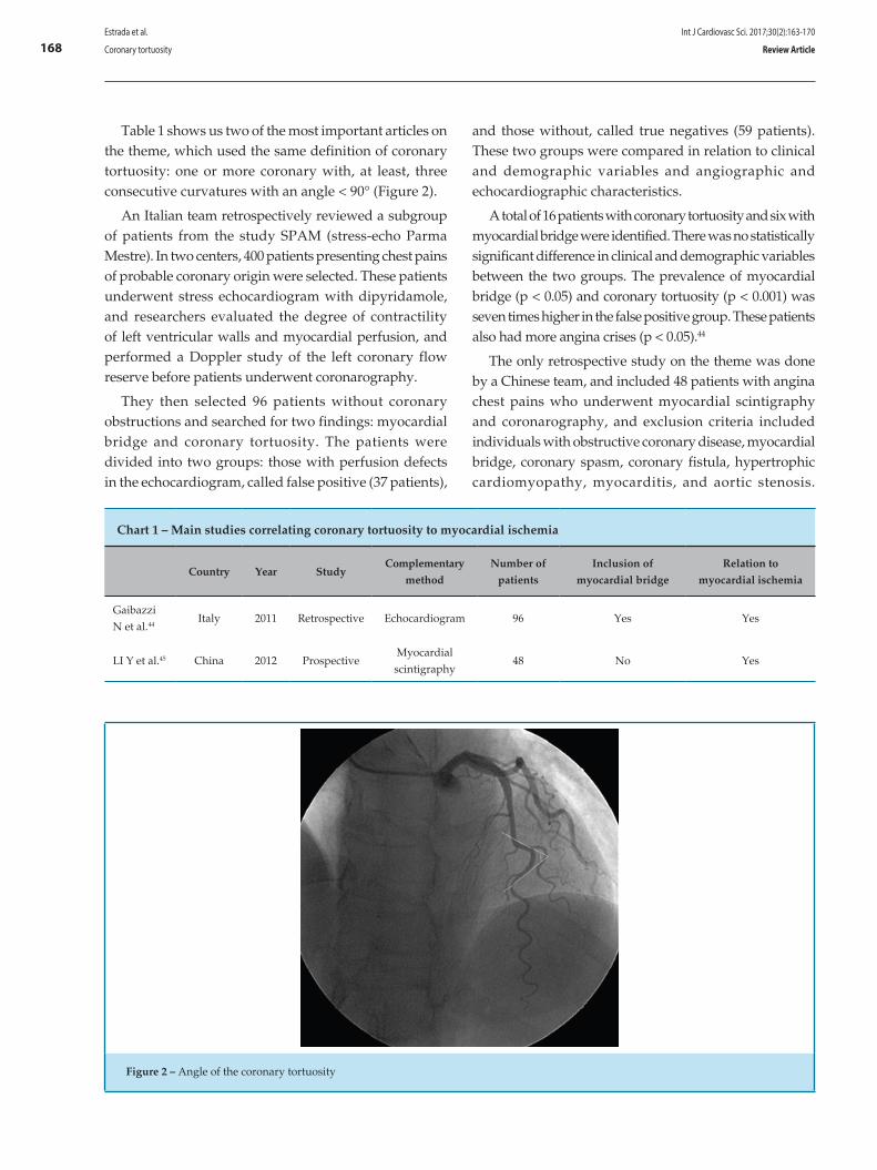
168
Figure 2 – Angle of the coronary tortuosity
Chart 1 – Main studies correlating coronary tortuosity to myocardial ischemia
Country Year StudyComplementary
method
Number of
patients
Inclusion of
myocardial bridge
Relation to
myocardial ischemia
Gaibazzi N et al.44
Italy 2011 Retrospective Echocardiogram 96 Yes Yes
LI Y et al.45 China 2012 ProspectiveMyocardial scintigraphy
48 No Yes
Table 1 shows us two of the most important articles on the theme, which used the same definition of coronary tortuosity: one or more coronary with, at least, three consecutive curvatures with an angle < 90° (Figure 2).
An Italian team retrospectively reviewed a subgroup of patients from the study SPAM (stress-echo Parma Mestre). In two centers, 400 patients presenting chest pains of probable coronary origin were selected. These patients underwent stress echocardiogram with dipyridamole, and researchers evaluated the degree of contractility of left ventricular walls and myocardial perfusion, and performed a Doppler study of the left coronary flow reserve before patients underwent coronarography.
They then selected 96 patients without coronary obstructions and searched for two findings: myocardial bridge and coronary tortuosity. The patients were divided into two groups: those with perfusion defects in the echocardiogram, called false positive (37 patients),
and those without, called true negatives (59 patients). These two groups were compared in relation to clinical and demographic variables and angiographic and echocardiographic characteristics.
A total of 16 patients with coronary tortuosity and six with myocardial bridge were identified. There was no statistically significant difference in clinical and demographic variables between the two groups. The prevalence of myocardial bridge (p < 0.05) and coronary tortuosity (p < 0.001) was seven times higher in the false positive group. These patients also had more angina crises (p < 0.05).44
The only retrospective study on the theme was done by a Chinese team, and included 48 patients with angina chest pains who underwent myocardial scintigraphy and coronarography, and exclusion criteria included individuals with obstructive coronary disease, myocardial bridge, coronary spasm, coronary fistula, hypertrophic cardiomyopathy, myocarditis, and aortic stenosis.
Estrada et al.
Coronary tortuosity
Int J Cardiovasc Sci. 2017;30(2):163-170
Review Article
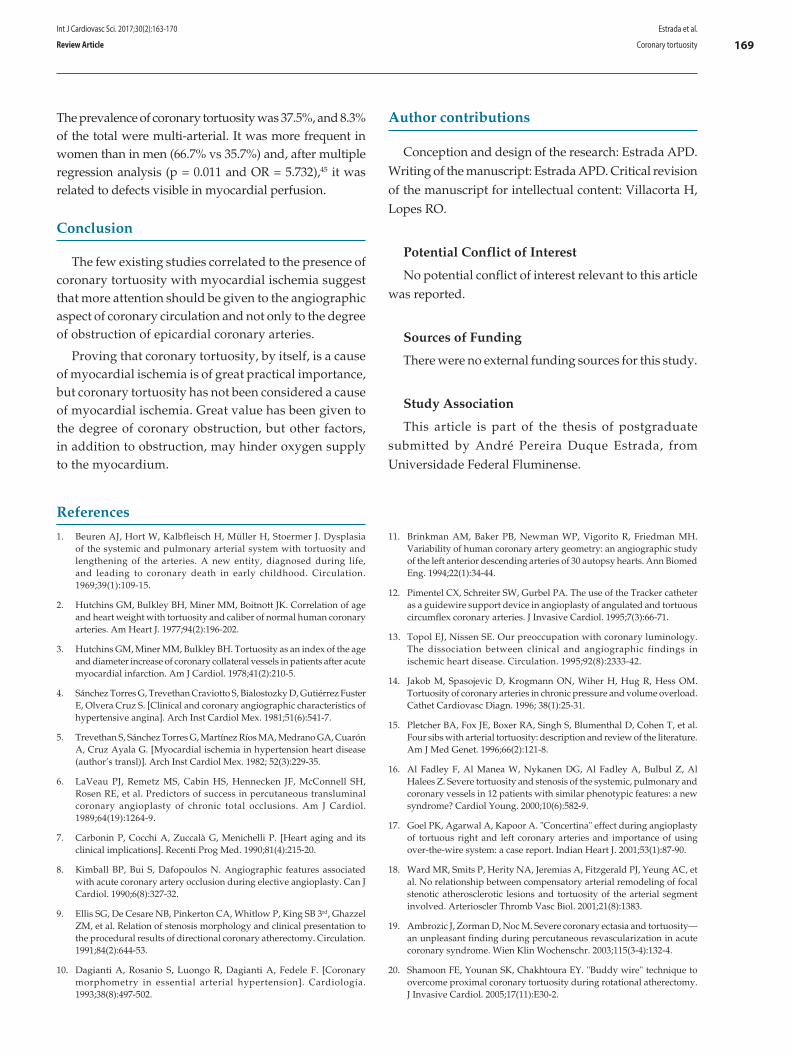
169
1. Beuren AJ, Hort W, Kalbfleisch H, Müller H, Stoermer J. Dysplasia of the systemic and pulmonary arterial system with tortuosity and lengthening of the arteries. A new entity, diagnosed during life, and leading to coronary death in early childhood. Circulation. 1969;39(1):109-15.
2. Hutchins GM, Bulkley BH, Miner MM, Boitnott JK. Correlation of age and heart weight with tortuosity and caliber of normal human coronary arteries. Am Heart J. 1977;94(2):196-202.
3. Hutchins GM, Miner MM, Bulkley BH. Tortuosity as an index of the age and diameter increase of coronary collateral vessels in patients after acute myocardial infarction. Am J Cardiol. 1978;41(2):210-5.
4. Sánchez Torres G, Trevethan Craviotto S, Bialostozky D, Gutiérrez Fuster E, Olvera Cruz S. [Clinical and coronary angiographic characteristics of hypertensive angina]. Arch Inst Cardiol Mex. 1981;51(6):541-7.
5. Trevethan S, Sánchez Torres G, Martínez Ríos MA, Medrano GA, Cuarón A, Cruz Ayala G. [Myocardial ischemia in hypertension heart disease (author’s transl)]. Arch Inst Cardiol Mex. 1982; 52(3):229-35.
6. LaVeau PJ, Remetz MS, Cabin HS, Hennecken JF, McConnell SH, Rosen RE, et al. Predictors of success in percutaneous transluminal coronary angioplasty of chronic total occlusions. Am J Cardiol. 1989;64(19):1264-9.
7. Carbonin P, Cocchi A, Zuccalà G, Menichelli P. [Heart aging and its clinical implications]. Recenti Prog Med. 1990;81(4):215-20.
8. Kimball BP, Bui S, Dafopoulos N. Angiographic features associated with acute coronary artery occlusion during elective angioplasty. Can J Cardiol. 1990;6(8):327-32.
9. Ellis SG, De Cesare NB, Pinkerton CA, Whitlow P, King SB 3rd, Ghazzel ZM, et al. Relation of stenosis morphology and clinical presentation to the procedural results of directional coronary atherectomy. Circulation. 1991;84(2):644-53.
10. Dagianti A, Rosanio S, Luongo R, Dagianti A, Fedele F. [Coronary morphometry in essential arterial hypertension]. Cardiologia. 1993;38(8):497-502.
11. Brinkman AM, Baker PB, Newman WP, Vigorito R, Friedman MH. Variability of human coronary artery geometry: an angiographic study of the left anterior descending arteries of 30 autopsy hearts. Ann Biomed Eng. 1994;22(1):34-44.
12. Pimentel CX, Schreiter SW, Gurbel PA. The use of the Tracker catheter as a guidewire support device in angioplasty of angulated and tortuous circumflex coronary arteries. J Invasive Cardiol. 1995;7(3):66-71.
13. Topol EJ, Nissen SE. Our preoccupation with coronary luminology. The dissociation between clinical and angiographic findings in ischemic heart disease. Circulation. 1995;92(8):2333-42.
14. Jakob M, Spasojevic D, Krogmann ON, Wiher H, Hug R, Hess OM. Tortuosity of coronary arteries in chronic pressure and volume overload. Cathet Cardiovasc Diagn. 1996; 38(1):25-31.
15. Pletcher BA, Fox JE, Boxer RA, Singh S, Blumenthal D, Cohen T, et al. Four sibs with arterial tortuosity: description and review of the literature. Am J Med Genet. 1996;66(2):121-8.
16. Al Fadley F, Al Manea W, Nykanen DG, Al Fadley A, Bulbul Z, Al Halees Z. Severe tortuosity and stenosis of the systemic, pulmonary and coronary vessels in 12 patients with similar phenotypic features: a new syndrome? Cardiol Young. 2000;10(6):582-9.
17. Goel PK, Agarwal A, Kapoor A. "Concertina" effect during angioplasty of tortuous right and left coronary arteries and importance of using over-the-wire system: a case report. Indian Heart J. 2001;53(1):87-90.
18. Ward MR, Smits P, Herity NA, Jeremias A, Fitzgerald PJ, Yeung AC, et al. No relationship between compensatory arterial remodeling of focal stenotic atherosclerotic lesions and tortuosity of the arterial segment involved. Arterioscler Thromb Vasc Biol. 2001;21(8):1383.
19. Ambrozic J, Zorman D, Noc M. Severe coronary ectasia and tortuosity—an unpleasant finding during percutaneous revascularization in acute coronary syndrome. Wien Klin Wochenschr. 2003;115(3-4):132-4.
20. Shamoon FE, Younan SK, Chakhtoura EY. "Buddy wire" technique to overcome proximal coronary tortuosity during rotational atherectomy. J Invasive Cardiol. 2005;17(11):E30-2.
References
The prevalence of coronary tortuosity was 37.5%, and 8.3% of the total were multi-arterial. It was more frequent in women than in men (66.7% vs 35.7%) and, after multiple regression analysis (p = 0.011 and OR = 5.732),45 it was related to defects visible in myocardial perfusion.
Conclusion
The few existing studies correlated to the presence of coronary tortuosity with myocardial ischemia suggest that more attention should be given to the angiographic aspect of coronary circulation and not only to the degree of obstruction of epicardial coronary arteries.
Proving that coronary tortuosity, by itself, is a cause of myocardial ischemia is of great practical importance, but coronary tortuosity has not been considered a cause of myocardial ischemia. Great value has been given to the degree of coronary obstruction, but other factors, in addition to obstruction, may hinder oxygen supply to the myocardium.
Author contributions
Conception and design of the research: Estrada APD. Writing of the manuscript: Estrada APD. Critical revision of the manuscript for intellectual content: Villacorta H, Lopes RO.
Potential Conflict of Interest
No potential conflict of interest relevant to this article was reported.
Sources of Funding
There were no external funding sources for this study.
Study Association
This article is part of the thesis of postgraduate submitted by André Pereira Duque Estrada, from Universidade Federal Fluminense.
Estrada et al.
Coronary tortuosity
Int J Cardiovasc Sci. 2017;30(2):163-170
Review Article
170
21. Tsapaki V, Magginas A, Vano E, Kottou S, Papadakis E, Dafnomili P, et al. Factors that influence radiation dose in percutaneous coronary intervention. J Interv Cardiol. 2006;19(3):237-44.
22. Hoop R, Steinmann B, Valsangiacomo Buechel ER. Cardiovascular findings in arterial tortuosity syndrome. Eur Heart J. 2006;27(17):2045.
23. Subramanyan R, Narayan R, Maskeri SA. Familial arterial tortuosity syndrome. Indian Heart J. 2007;59(2):178-80.
24. Zegers ES, Meursing BT, Zegers EB, Oude Ophuis AJ. Coronary tortuosity: a long and winding road. Neth Heart J. 2007;15(5):191-5.
25. Ramcharitar S, Patterson MS, van Geuns RJ, van der Ent M, Sianos G, Welten GM, et al. A randomised controlled study comparing conventional and magnetic guidewires in a two-dimensional branching tortuous phantom simulating angulated coronary vessels. Cathet Cardiovasc Interv. 2007;70(5):662-8.
26. Turgut O, Yilmaz A, Yalta K, Yilmaz BM, Ozyol A, Kendirlioglu O, et al. Tortuosity of coronary arteries: an indicator for impaired left ventricular relaxation? Int J Cardiovasc Imaging. 2007;23(6):671-7.
27. Saeed B, Banerjee S, Brilakis ES. Percutaneous coronary intervention in tortuous coronary arteries: associated complications and strategies to improve success. J Intervent Cardiol. 2008; 21(6):504-11.
28. Subramanyan R, Sridhar A, Cherian K. Arterial tortuosity syndrome. Pediatr Cardiol. 2009;30(4):555-6.
29. Zhu H, Ding Z, Piana RN, Gehrig TR, Friedman MH. Cataloguing the geometry of the human coronary arteries: a potential tool for predicting risk of coronary artery disease. Int J Cardiol. 2009;135(1):43-52.
30. Groves SS, Jain AC, Warden BE, Gharib W, Beto RJ 2nd. Severe coronary tortuosity and the relationship to significant coronary artery disease. W V Med J. 2009; 105(4):14-7.
31. Turgut O, Tandogan I, Yalta K, Yilmaz MB, Dizman R. Geodesic pattern of coronary arteries as a predictor for cardiovascular risk: clinical perspectives. Int J Cardiol. 2010;142(3):e38-9.
32. Chism BS, Lee RW, Sweeney JP, Fortuin FD. The crooked buddy technique: use of a Wiggle wire alongside an extra support wire to improve device deliverability. J Invasive Cardiol. 2010;22(8):377-81.
33. Li Y, Shen C, Ji Y, Feng Y, Ma G, Liu N. Clinical implication of coronary tortuosity in patients with coronary artery disease. PloS One. 2011;6(8):e24232.
34. Gaibazzi N, Rigo F, Reverberi C. Severe coronary tortuosity or myocardial bridging in patients with chest pain, normal coronary arteries, and reversible myocardial perfusion defects. Am J Cardiol. 2011;108(7):973-8.
35. Li Y, Shi Z, Cai Y, Feng Y, Ma G, Shen C, et al. Impact of coronary tortuosity on coronary pressure: numerical simulation study. PloS One. 2012;7(8):e42558.
36. Dahdouh ZS, Roule V, Sabatier R, Grollier G. An alternative approach in tortuous coronary artery and distal stenosis during transradial percutaneous coronary intervention: deep engagement by a 5-Fr guiding catheter. Turk Kardiyol Dern Ars. 2012;40(2):159-61.
37. Li Y, Liu NF, Gu ZZ, Chen Y, Lu J, Feng Y, et al. Coronary tortuosity is associated with reversible myocardial perfusion defects in patients without coronary artery disease. Chin Med J (Engl). 2012;125(19):3581-3.
38. Xie X, Wang Y, Zhu H, Zhou H, Zhou J. Impact of coronary tortuosity on coronary blood supply: a patient-specific study. PloS One. 2013;8(5):e64564.
39. Sova SH, Lebedieva EO. [The coronary arterial tortuosity in workers of vibro-noisy professions and its role in the ischemic damage of the myocardium]. Lik Sprava. 2013;(5):121-7.
40. Xie X, Wang Y, Zhou H. Impact of coronary tortuosity on the coronary blood flow: a 3D computational study. J Biomech. 2013;46(11):1833-41.
41. Helmberger M, Pienn M, Urschler M, Kullnig P, Stollberger R, Kovacs G, et al. Quantification of tortuosity and fractal dimension of the lung vessels in pulmonary hypertension patients. PloS One. 2014;9(1):e87515.
42. Parekh P, Agrawal N, Vasavada A, Vinchurkar M. Extreme coronary artery tortuosity in association with tortuosity of the systemic arteries: a rare and challenging situation for the interventionist. BMJ Case Rep. 2014;2014.
43. Eleid MF, Guddeti RR, Tweet MS, Lerman A, Singh M, Best PJ, et al. Coronary artery tortuosity in spontaneous coronary artery dissection: angiographic characteristics and clinical implications. Circ Cardiovasc Interv. 2014;7(5):656-62.
44. Xie X, Wang Y, Zhu H, Zhou J. Computation of hemodynamics in tortuous left coronary artery: a morphological parametric study. J Biomech Eng. 2014;136(10):101006.
45. Lebedeva EO, Lazoryshynets VV, Beshliaga VM, Grusha MM. [Diagnosis of ischemic heart disease caused by tortuosity of coronary arteries]. Lik Sprava. 2015;(1-2):38-43.
46. Christopoulos G, Kandzari DE, Yeh RW, Jaffer FA, Karmpaliotis D, Wyman MR, et al. Development and Validation of a Novel Scoring System for Predicting Technical Success of Chronic Total Occlusion Percutaneous Coronary Interventions: The PROGRESS CTO (Prospective Global Registry for the Study of Chronic Total Occlusion Intervention) Score. JACC Cardiovasc Interv. 2016;9(1):1-9.
47. Finn R, Morris L. An experimental assessment of catheter trackability forces with tortuosity parameters along patient-specific coronary phantoms. Proc Inst Mech Eng H. 2016;230(2):153-65.
48. El Tahlawi M, Sakrana A, Elmurr A, Gouda M, Tharwat M. The relation between coronary tortuosity and calcium score in patients with chronic stable angina and normal coronaries by CT angiography. Atherosclerosis. 2016;246:334-7.
49. Vorobtsova N, Chiastra C, Stremler MA, Sane DC, Migliavacca F, Vlachos P. Effects of vessel tortuosity on coronary hemodynamics: an idealized and patient-specific computational study. Ann Biomed Eng. 2016;44(7):2228-39.
Estrada et al.
Coronary tortuosity
Int J Cardiovasc Sci. 2017;30(2):163-170
Review Article
DOI: 10.5935/2359-4802.20170032
171International Journal of Cardiovascular Sciences. 2017;30(2):171-176
BRIEF COMMUNICATION
Mailing Address: Luiz Rodrigo Augustemak de LimaCampus Reitor João David Ferreira Lima, Centro de Desportos. Postal Code: 88040-900, Trindade, Florianópolis, SC – Brazil E-mail: [email protected], [email protected]
Exercise Improves Cardiovascular Risk Factors, Fitness, and Quality Of Life in Hiv+ Children and Adolescents: Pilot StudyLuiz Rodrigo Augustemak de Lima,1,3 Isabela de Carlos Back,1 Carmem Cristina Beck,1 Bruno Caramelli2
Universidade Federal de Santa Catarina (UFSC);1 Instituto do Coração (Incor) – Faculdade de Medicina da Universidade de São Paulo,2 São Paulo, SP;Núcleo de Pesquisa em Cineantropometria e Desempenho Humano (NuCiDH),3 UFSC, Florianópolis, SC – Brazil
Manuscript received September 27, 2016, revised mansucript November 30, 2016, accepted February 01, 2017.
Cardiovascular Diseases / physiopathology; Exercise;
Physical Fitness; Life Style; Child; Adolescent;
Atherosclerosis; Carotid Intima-Media Thickness.
Keywords
Abstract
Children and adolescents infected by HIV through mother-to-child transmission are at high risk of developing premature cardiovascular diseases due to dyslipidemia, insulin resistance and low-grade chronic inflammation. The aim of the pilot study was to verify the effect of a playful exercise program on cardiovascular, morphological, metabolic, fitness, and quality of life outcomes. A non-randomized clinical trial consisting of 24 sessions of playful aerobic and resistive exercises was applied to 10 children and adolescents living with HIV from Florianopolis, Brazil. The following variables were obtained before and after the program: fasting total cholesterol, HDL-c, LDL-c, triglycerides, glucose, C-reactive protein, blood pressure, common carotid artery intima-media thickness (CCA-IMT), flexibility, muscular endurance, aerobic fitness, anthropometry, and measured quality of life. After the intervention, a decrease in systolic blood pressure (-6.8 mmHg, 6.6%; p = 0.019) and CCA-IMT (-60.0 µm, 12.2%; p = 0.002) was observed after 24 sessions. There was an increase in upper-limb muscular endurance (+3.3 rep.min-1, 63.5%; p = 0.002), flexibility (+5.7 cm, 26.0%; p = 0.001), and quality of life (+10.4 points, 27.5%; p = 0.003). In our sample of children and adolescents living with HIV, a short-term exercise program was associated with improvement in cardiovascular risk, fitness, and quality of life.
Introduction
Exercise is a non-pharmacological treatment for
adults living with human immunodeficiency virus
(HIV), because it can reduce HIV-associated symptoms,
cardiovascular, morphological, metabolic abnormalities
and improve fitness, anxiety and depression.1 In children
and adolescents living with HIV, only one study has
demonstrated the feasibility, safety and efficacy of
aerobic and resistive exercises to improve muscle
strength and endurance, aerobic fitness and fat free
mass.2 This is important, since fitness is reduced in
several pediatric pathological conditions and might be
associated with premature mortality..3 However, no effect
on lipids was observed and cardiovascular variables
were not analyzed.2
Although the effect of exercise on the health of children
and adolescents is evident, the magnitude of the effect
depends on the intervention characteristics (e.g. intensity
and volume of sessions) and health status at the baseline
intervention (e.g. normal values of cardiovascular
and lipid profiles).4 Since the long-term exposure to
HIV infection and highly-active antiretroviral therapy
(HAART) are associated with dyslipidemia, insulin
resistance and low-grade inflammation that increase
the risk of cardiovascular diseases,5-7exercise could
mitigate unfavorable conditions of HIV-infected children.
This study reports preliminary data on the effect of a
short-term playful exercise program on cardiometabolic
risk factors, our primary outcome. Fitness and quality of
life were also tested as our secondary outcomes.
172Lima et al.
Exercise and cardiovascular risk factors in hiv+
Int J Cardiovasc Sci. 2017;30(2):171-176
Brief Communication
Methods
Study design and patient population
This was a non-randomized clinical trial that evaluated a sample of children and adolescents before and after 8 weeks of aerobic and resistive exercises, conducted in the second half of 2008 at the Rehabilitation Center in Florianopolis, Brazil. This pilot study was carried out with 10 children and adolescents infected by HIV-through mother-to-child transmission and follow at a reference hospital for the treatment of pediatric HIV infection. Before inclusion, the patients were evaluated regarding the risk of exercise. The study was approved by the Ethics Committee of the Hospital (063/2007).
Intervention
The program consisted of 24 exercise sessions, with each session lasting 90 minutes. There was a gradual increase in the duration of aerobic and muscle resistive exercises from 40 to 60 minutes (every two weeks). A 48-hour interval was used between sessions for recovery. Each session consisted of warm-up/stretching (15 min), playful aerobic and muscle-resistance activities, such as dancing, and recreational and pre-sports games (40-60 minutes), and cool-down (10 min). Playful activities appropriate for the patients’ ages were selected. Most activities were organized in a circuit system to permit the session to be more dynamic. The intensity of each session was monitored with a heart rate monitor, thus permitting to determine the exercise time in the previously calculated target zone as 50-85% of the heart rate reserve.8
Outcomes
Fasting serum total cholesterol, triglycerides, high-density (HDL-c) and low-density (LDL-c) lipoprotein cholesterol, glucose and ultrasensitive C-reactive protein were assayed using standard procedures.5 Blood pressure and resting heart rate were measured as previously described.5 Intima-media thickness of the right common carotid artery (CCA-IMT) was measured using the Vivid i system (GE, Horten, Norway) with a 12.5-MHz linear transducer. The three best images of the carotid bulb segment close to the bifurcation were analyzed.5
Fitness was assessed by the Fitnessgram®.9 Flexibility was evaluated by the sit-and-reach test. Muscular endurance was assessed by the abdominal curl-up test and flexed
arm hang test. Aerobic fitness was measured in a submaximal exercise test performed on a treadmill and the peak oxygen consumption was estimated.10 Anthropometric measurements were performed using standard procedures.11 Body mass index, trunk-extremity skinfold ratio, and upper arm muscle area were calculated. Quality of life was evaluated using the “Autoquestionnaire Qualité de vie Enfant Imagé”.12
Statistical analysis
The Shapiro-Wilk test was used to verify Gaussian distribution. Descriptive analyses were presented as mean and standard deviation for data before and after the intervention. The after – before intervention differences (Δº) were calculated to describe effects. Paired Student t-test and Mann–Whitney U test were performed, adopting a p-value ≤ 0.05 in two-tailed analyses. Statistical analyses were performed using the STATA 11.0 (Stata Corporation, College Station, TX, USA) and GraphPad Prism 5.0 (GraphPad Software, Inc., San Diego, CA, USA) packages.
Results
The sample included nine girls and one boy aged 13.0 years (interquartile range [IQR]= 11.5 to 15.5 years); 4/10 participants were white and 6/10 were in moderate to severe stages of HIV infection. Half the participants used protease inhibitors, 8/10 used nucleoside reverse transcriptase inhibitors (NRTIs) and non-nucleoside reverse transcriptase inhibitors (NNRTIs), and 2/10 were undergoing HAART. The median CD4+ T-lymphocyte count was 722.0 cells.ml-1 (IQR= 647.5 and 914.2) and the median viral load was 17,750 copies.ml-1 (IQR= 368 and 26.000). Eight of the 10 subjects were pubertal, one was prepubertal, and the other was post-pubertal. Figure 1 shows the time spent in the target heart rate zone (50-85%) during each session.
Table 1 shows the changes in outcomes after the exercise program. A decrease was observed in systolic blood pressure (6.6%) and CCA-IMT (12.2%), as well as an increase in muscular endurance (63.5%), flexibility (26.0%) and quality of life (27.5%). The CD4+ T-lymphocyte count and viral load remained unchanged after the intervention. No dropout from the exercise program or intercurrence during the program was observed.
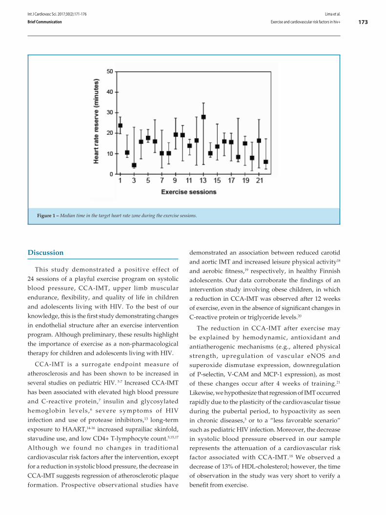
173
Figure 1 – Median time in the target heart rate zone during the exercise sessions.
Lima et al.
Exercise and cardiovascular risk factors in hiv+
Int J Cardiovasc Sci. 2017;30(2):171-176
Brief Communication
Discussion
This study demonstrated a positive effect of 24 sessions of a playful exercise program on systolic blood pressure, CCA-IMT, upper limb muscular endurance, flexibility, and quality of life in children and adolescents living with HIV. To the best of our knowledge, this is the first study demonstrating changes in endothelial structure after an exercise intervention program. Although preliminary, these results highlight the importance of exercise as a non-pharmacological therapy for children and adolescents living with HIV.
CCA-IMT is a surrogate endpoint measure of atherosclerosis and has been shown to be increased in several studies on pediatric HIV. 5-7 Increased CCA-IMT has been associated with elevated high blood pressure and C-reactive protein,7 insulin and glycosylated hemoglobin levels,6 severe symptoms of HIV infection and use of protease inhibitors,13 long-term exposure to HAART,14-16 increased suprailiac skinfold, stavudine use, and low CD4+ T-lymphocyte count.5,15,17 Although we found no changes in traditional cardiovascular risk factors after the intervention, except for a reduction in systolic blood pressure, the decrease in CCA-IMT suggests regression of atherosclerotic plaque formation. Prospective observational studies have
demonstrated an association between reduced carotid and aortic IMT and increased leisure physical activity18 and aerobic fitness,19 respectively, in healthy Finnish adolescents. Our data corroborate the findings of an intervention study involving obese children, in which a reduction in CCA-IMT was observed after 12 weeks of exercise, even in the absence of significant changes in C-reactive protein or triglyceride levels.20
The reduction in CCA-IMT after exercise may be explained by hemodynamic, antioxidant and antiatherogenic mechanisms (e.g., altered physical strength, upregulation of vascular eNOS and superoxide dismutase expression, downregulation of P-selectin, V-CAM and MCP-1 expression), as most of these changes occur after 4 weeks of training.21 Likewise, we hypothesize that regression of IMT occurred rapidly due to the plasticity of the cardiovascular tissue during the pubertal period, to hypoactivity as seen in chronic diseases,3 or to a “less favorable scenario” such as pediatric HIV infection. Moreover, the decrease in systolic blood pressure observed in our sample represents the attenuation of a cardiovascular risk factor associated with CCA-IMT.18 We observed a decrease of 13% of HDL-cholesterol; however, the time of observation in the study was very short to verify a benefit from exercise.
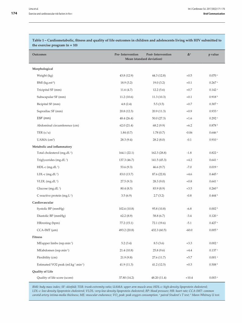
174Lima et al.
Exercise and cardiovascular risk factors in hiv+
Int J Cardiovasc Sci. 2017;30(2):171-176
Brief Communication
Table 1 – Cardiometabolic, fitness and quality of life outcomes in children and adolescents living with HIV submitted to the exercise program (n = 10)
Outcomes Pre- Intervention Post- Intervention Δ° p valueMean (standard deviation)
Morphological
Weight (kg) 43.8 (12.9) 44.3 (12.8) +0.5 0.070 a
BMI (kg.est-2) 18.9 (3.2) 19.0 (3.2) +0.1 0.267 a
Tricipital SF (mm) 11.6 (4.7) 12.2 (5.6) +0.7 0.142 a
Subscapular SF (mm) 11.2 (10.6) 11.3 (10.3) +0.1 0.918 b
Bicipital SF (mm) 4.8 (2.4) 5.5 (3.5) +0.7 0.307 a
Suprailiac SF (mm) 20.8 (12.3) 20.9 (11.3) +0.9 0.933 a
ΣSF (mm) 48.4 (26.4) 50.0 (27.3) +1.6 0.292 a
Abdominal circumference (cm) 62.0 (21.4) 68.2 (9.9) +6.2 0.878 b
TER (s/u) 1.84 (0.7) 1.78 (0.7) -0.06 0.646 b
UAMA (cm2) 28.3 (9.4) 28.2 (8.0) -0.1 0.910 a
Metabolic and inflammatory
Total cholesterol (mg.dL-1) 164.1 (22.1) 162.3 (28.8) -1.8 0.822 a
Triglycerides (mg.dL-1) 137.3 (46.7) 141.5 (45.3) +4.2 0.641 a
HDL-c (mg.dL-1) 53.6 (9.3) 46.6 (9.7) -7.0 0.019 a
LDL-c (mg.dL-1) 83.0 (13.7) 87.6 (22.8) +4.6 0.445 a
VLDL (mg.dL-1) 27.5 (9.3) 28.3 (9.0) +0.8 0.641 a
Glucose (mg.dL-1) 80.4 (8.5) 83.9 (8.9) +3.5 0.260 b
C-reactive protein (mg.L-1) 3.5 (6.9) 2.7 (3.2) -0.8 0.444 b
Cardiovascular
Systolic BP (mmHg) 102.6 (10.8) 95.8 (10.8) -6.8 0.002 b
Diastolic BP (mmHg) 62.2 (8.9) 58.8 (6.7) -3.4 0.120 a
HRresting (bpm) 77.2 (15.1) 72.1 (19.6) -5.1 0.427 a
CCA-IMT (µm) 493.2 (20.8) 432.3 (60.5) -60.0 0.005 b
Fitness
MEupper limbs (rep.min-1) 5.2 (3.4) 8.5 (3.6) +3.3 0.002 a
MEabdomen (rep.min-1) 21.4 (10.8) 25.8 (9.6) +4.4 0.137 a
Flexibility (cm) 21.9 (9.8) 27.6 (11.7) +5.7 0.001 a
Estimated VO2 peak (ml.kg-1.min-1) 41.9 (11.3) 41.2 (12.5) +0.3 0.508 b
Quality of Life
Quality of life score (score) 37.80 (14.2) 48.20 (11.4) +10.4 0.003 a
BMI: body mass index; SF: skinfold; TER: trunk-extremity ratio; UAMA: upper arm muscle area; HDL-c: high-density lipoprotein cholesterol; LDL-c: low-density lipoprotein cholesterol; VLDL: very-low-density lipoprotein cholesterol; BP: blood pressure; HR: heart rate; CCA-IMT: common carotid artery intima-media thickness; ME: muscular endurance; VO2 peak: peak oxygen consumption. a paired Student’s T test; b Mann Whitney U test.
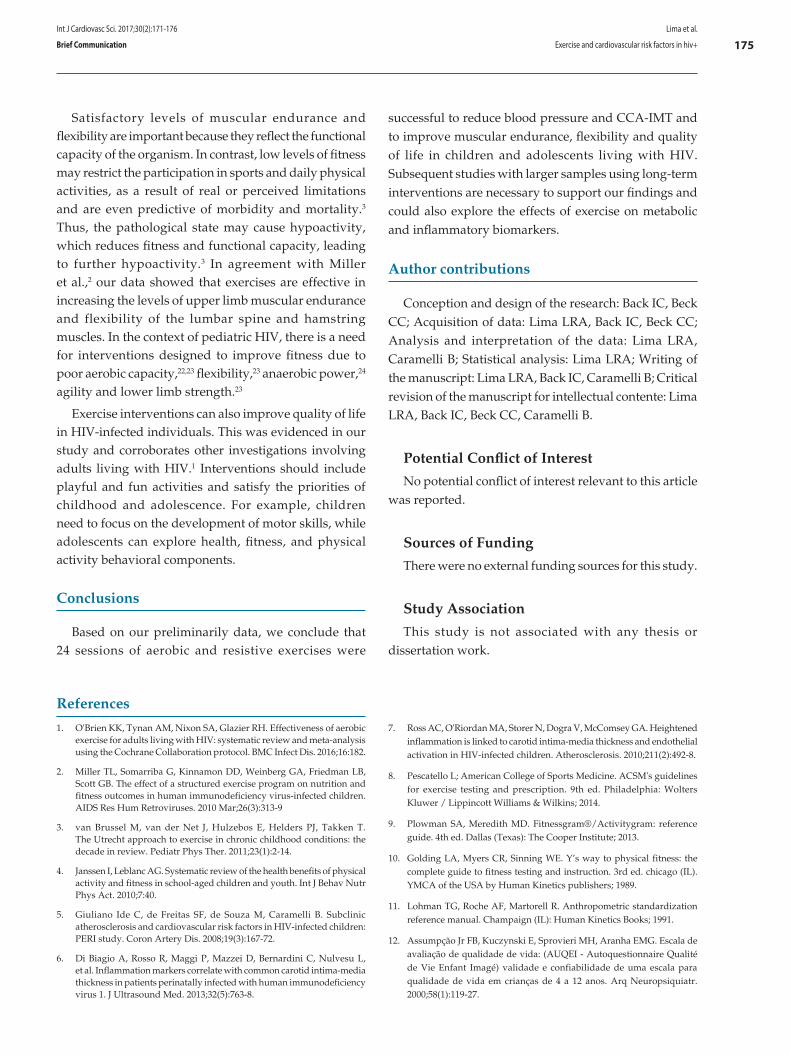
175
1. O'Brien KK, Tynan AM, Nixon SA, Glazier RH. Effectiveness of aerobic exercise for adults living with HIV: systematic review and meta-analysis using the Cochrane Collaboration protocol. BMC Infect Dis. 2016;16:182.
2. Miller TL, Somarriba G, Kinnamon DD, Weinberg GA, Friedman LB, Scott GB. The effect of a structured exercise program on nutrition and fitness outcomes in human immunodeficiency virus-infected children. AIDS Res Hum Retroviruses. 2010 Mar;26(3):313-9
3. van Brussel M, van der Net J, Hulzebos E, Helders PJ, Takken T. The Utrecht approach to exercise in chronic childhood conditions: the decade in review. Pediatr Phys Ther. 2011;23(1):2-14.
4. Janssen I, Leblanc AG. Systematic review of the health benefits of physical activity and fitness in school-aged children and youth. Int J Behav Nutr Phys Act. 2010;7:40.
5. Giuliano Ide C, de Freitas SF, de Souza M, Caramelli B. Subclinic atherosclerosis and cardiovascular risk factors in HIV-infected children: PERI study. Coron Artery Dis. 2008;19(3):167-72.
6. Di Biagio A, Rosso R, Maggi P, Mazzei D, Bernardini C, Nulvesu L, et al. Inflammation markers correlate with common carotid intima-media thickness in patients perinatally infected with human immunodeficiency virus 1. J Ultrasound Med. 2013;32(5):763-8.
7. Ross AC, O'Riordan MA, Storer N, Dogra V, McComsey GA. Heightened inflammation is linked to carotid intima-media thickness and endothelial activation in HIV-infected children. Atherosclerosis. 2010;211(2):492-8.
8. Pescatello L; American College of Sports Medicine. ACSM's guidelines for exercise testing and prescription. 9th ed. Philadelphia: Wolters Kluwer / Lippincott Williams & Wilkins; 2014.
9. Plowman SA, Meredith MD. Fitnessgram®/Activitygram: reference guide. 4th ed. Dallas (Texas): The Cooper Institute; 2013.
10. Golding LA, Myers CR, Sinning WE. Y’s way to physical fitness: the complete guide to fitness testing and instruction. 3rd ed. chicago (IL). YMCA of the USA by Human Kinetics publishers; 1989.
11. Lohman TG, Roche AF, Martorell R. Anthropometric standardization reference manual. Champaign (IL): Human Kinetics Books; 1991.
12. Assumpção Jr FB, Kuczynski E, Sprovieri MH, Aranha EMG. Escala de avaliação de qualidade de vida: (AUQEI - Autoquestionnaire Qualité de Vie Enfant Imagé) validade e confiabilidade de uma escala para qualidade de vida em crianças de 4 a 12 anos. Arq Neuropsiquiatr. 2000;58(1):119-27.
References
Lima et al.
Exercise and cardiovascular risk factors in hiv+
Int J Cardiovasc Sci. 2017;30(2):171-176
Brief Communication
Satisfactory levels of muscular endurance and flexibility are important because they reflect the functional capacity of the organism. In contrast, low levels of fitness may restrict the participation in sports and daily physical activities, as a result of real or perceived limitations and are even predictive of morbidity and mortality.3 Thus, the pathological state may cause hypoactivity, which reduces fitness and functional capacity, leading to further hypoactivity.3 In agreement with Miller et al.,2 our data showed that exercises are effective in increasing the levels of upper limb muscular endurance and flexibility of the lumbar spine and hamstring muscles. In the context of pediatric HIV, there is a need for interventions designed to improve fitness due to poor aerobic capacity,22,23 flexibility,23 anaerobic power,24 agility and lower limb strength.23
Exercise interventions can also improve quality of life in HIV-infected individuals. This was evidenced in our study and corroborates other investigations involving adults living with HIV.1 Interventions should include playful and fun activities and satisfy the priorities of childhood and adolescence. For example, children need to focus on the development of motor skills, while adolescents can explore health, fitness, and physical activity behavioral components.
Conclusions
Based on our preliminarily data, we conclude that 24 sessions of aerobic and resistive exercises were
successful to reduce blood pressure and CCA-IMT and to improve muscular endurance, flexibility and quality of life in children and adolescents living with HIV. Subsequent studies with larger samples using long-term interventions are necessary to support our findings and could also explore the effects of exercise on metabolic and inflammatory biomarkers.
Author contributions
Conception and design of the research: Back IC, Beck CC; Acquisition of data: Lima LRA, Back IC, Beck CC; Analysis and interpretation of the data: Lima LRA, Caramelli B; Statistical analysis: Lima LRA; Writing of the manuscript: Lima LRA, Back IC, Caramelli B; Critical revision of the manuscript for intellectual contente: Lima LRA, Back IC, Beck CC, Caramelli B.
Potential Conflict of Interest
No potential conflict of interest relevant to this article was reported.
Sources of Funding
There were no external funding sources for this study.
Study Association
This study is not associated with any thesis or dissertation work.
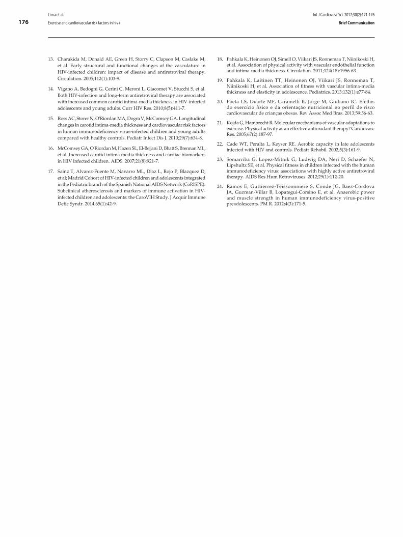
176
13. Charakida M, Donald AE, Green H, Storry C, Clapson M, Caslake M, et al. Early structural and functional changes of the vasculature in HIV-infected children: impact of disease and antiretroviral therapy. Circulation. 2005;112(1):103-9.
14. Vigano A, Bedogni G, Cerini C, Meroni L, Giacomet V, Stucchi S, et al. Both HIV-infection and long-term antiretroviral therapy are associated with increased common carotid intima-media thickness in HIV-infected adolescents and young adults. Curr HIV Res. 2010;8(5):411-7.
15. Ross AC, Storer N, O'Riordan MA, Dogra V, McComsey GA. Longitudinal changes in carotid intima-media thickness and cardiovascular risk factors in human immunodeficiency virus-infected children and young adults compared with healthy controls. Pediatr Infect Dis J. 2010;29(7):634-8.
16. McComsey GA, O'Riordan M, Hazen SL, El-Bejjani D, Bhatt S, Brennan ML, et al. Increased carotid intima media thickness and cardiac biomarkers in HIV infected children. AIDS. 2007;21(8):921-7.
17. Sainz T, Alvarez-Fuente M, Navarro ML, Diaz L, Rojo P, Blazquez D, et al; Madrid Cohort of HIV-infected children and adolescents integrated in the Pediatric branch of the Spanish National AIDS Network (CoRISPE). Subclinical atherosclerosis and markers of immune activation in HIV-infected children and adolescents: the CaroVIH Study. J Acquir Immune Defic Syndr. 2014;65(1):42-9.
18. Pahkala K, Heinonen OJ, Simell O, Viikari JS, Ronnemaa T, Niinikoski H, et al. Association of physical activity with vascular endothelial function and intima-media thickness. Circulation. 2011;124(18):1956-63.
19. Pahkala K, Laitinen TT, Heinonen OJ, Viikari JS, Ronnemaa T, Niinikoski H, et al. Association of fitness with vascular intima-media thickness and elasticity in adolescence. Pediatrics. 2013;132(1):e77-84.
20. Poeta LS, Duarte MF, Caramelli B, Jorge M, Giuliano IC. Efeitos do exercício físico e da orientação nutricional no perfil de risco cardiovascular de crianças obesas. Rev Assoc Med Bras. 2013;59:56-63.
21. Kojda G, Hambrecht R. Molecular mechanisms of vascular adaptations to exercise. Physical activity as an effective antioxidant therapy? Cardiovasc Res. 2005;67(2):187-97.
22. Cade WT, Peralta L, Keyser RE. Aerobic capacity in late adolescents infected with HIV and controls. Pediatr Rehabil. 2002;5(3):161-9.
23. Somarriba G, Lopez-Mitnik G, Ludwig DA, Neri D, Schaefer N, Lipshultz SE, et al. Physical fitness in children infected with the human immunodeficiency virus: associations with highly active antiretroviral therapy. AIDS Res Hum Retroviruses. 2012;29(1):112-20.
24. Ramos E, Guttierrez-Teissoonniere S, Conde JG, Baez-Cordova JA, Guzman-Villar B, Lopategui-Corsino E, et al. Anaerobic power and muscle strength in human immunodeficiency virus-positive preadolescents. PM R. 2012;4(3):171-5.
Lima et al.
Exercise and cardiovascular risk factors in hiv+
Int J Cardiovasc Sci. 2017;30(2):171-176
Brief Communication
DOI: 10.5935/2359-4802.20170021
177International Journal of Cardiovascular Sciences. 2017;30(2):177-180
CASE REPORT
Mailing Address: Rodrigo de Moura JoaquimRua São Francisco, 217, apt. 701, Centro. Postal Code: 88015-140, Florianópolis, SC – BrazilE-mail [email protected]
Caseous Necrosis of the Mitral Valve: Imaging Methods Allow the Diagnosis and Prevent SurgeryRodrigo de Moura Joaquim, Edileide de Barros Correia, Suéllen Lacerda Bezerra, Ibraim Masciarelli Francisco Pinto, Tiago Senra Garcia dos Santos, Fabiano de Castro AlbrechtInstituto Dante Pazzanese de Cardiologia, São Paulo, SP – Brazil
Manuscript received July 01, 2016; revised manuscript January 17, 2017; accepted January 23, 2017.
Calcinosis; Mitral Valve; Heart Neoplasms; Diagnostic Imaging; Magnetic Resonance Imaging; Multidetector Computed Tomography.
Keywords
Introduction
Calcification of the mitral annulus (CMA) is defined as a chronic degeneration of the mitral annulus fibrous involving mainly the posterior annulus.1 It is a common condition in the elderly, especially in women, and is associated with hypertension.2 Caseous necrosis of the mitral valve (CNMV) is a rare and less known CMA variant with a prevalence of 0.64%, which can reach up to 2.7% in autopsy studies of patients with CMA.1-3
On echocardiography, CNMV appears as a large rounded or semilunar echodense mass, with a liquid filling and central echolucency, similar to a periannular tumor.4,5 The mechanism of liquefaction and caseification are still not well understood, but it is believed to occur due to changes in calcium metabolism.1
Case Report
A 66-year-old female patient with a history of hypertension and chronic renal disease with an atrophic right kidney was referred to the emergency department of our institution after presenting a left atrial tumor mass with atypical characteristics, evidenced on echocardiography at another institution. She reported on the occasion a retrosternal and epigastric pressure-type pain associated with gastric fullness, without relation to physical effort, variable duration from minutes to hours, and without other associated symptoms. She also presented dyspnea on exertion.
The pat ient was hospi ta l ized to undergo complementary tests and for a possible surgical intervention. The electrocardiogram showed a sinus rhythm and presented criteria for left ventricular overload. Transthoracic echocardiography showed an enlarged left atrium (46 mm) with an indexed volume of 63 mL/m2, moderate concentric left ventricular hypertrophy, a 76% left ventricular ejection fraction, mild tricuspid insufficiency, thickened aortic valve without a significant gradient, and a sessile heterogeneous image with areas of calcification adhered to the ventricular face of the posterior leaflet of the mitral valve, restricting its mobility and opening, measuring 25 x 18 mm and causing mild reflux, with a valvular area of 1.9 cm2 (pressure half-time [PHT]) and a maximum transvalvular gradient of up to 12 and a mean of 4.
A magnetic resonance imaging of the heart (Figure 1) showed dilatation of the left atrium, mitral insufficiency, concentric and asymmetric left ventricular hypertrophy predominantly in the septum, measuring up to 23 mm in the middle and basal regions, and a cystic mass with well-defined borders adhered to the posterior leaflet of the mitral valve, with a heterogeneous content and a halo on late enhancement.
Coronary computed tomographic angiography (Figure 2) revealed no significant coronary obstructions and identified a well-delineated mass in the inferolateral portion of the mitral annulus, with regular borders and a fibrotic thin layer with high-intensity interior attenuation and focal coarse calcifications measuring 17 x 26 x 22 mm in the anteroposterior, transversal, and longitudinal axis, respectively.
Considering the association of the finding of important calcifications and the cystic image seen on noninvasive tests, the diagnosis of CNMV was established.
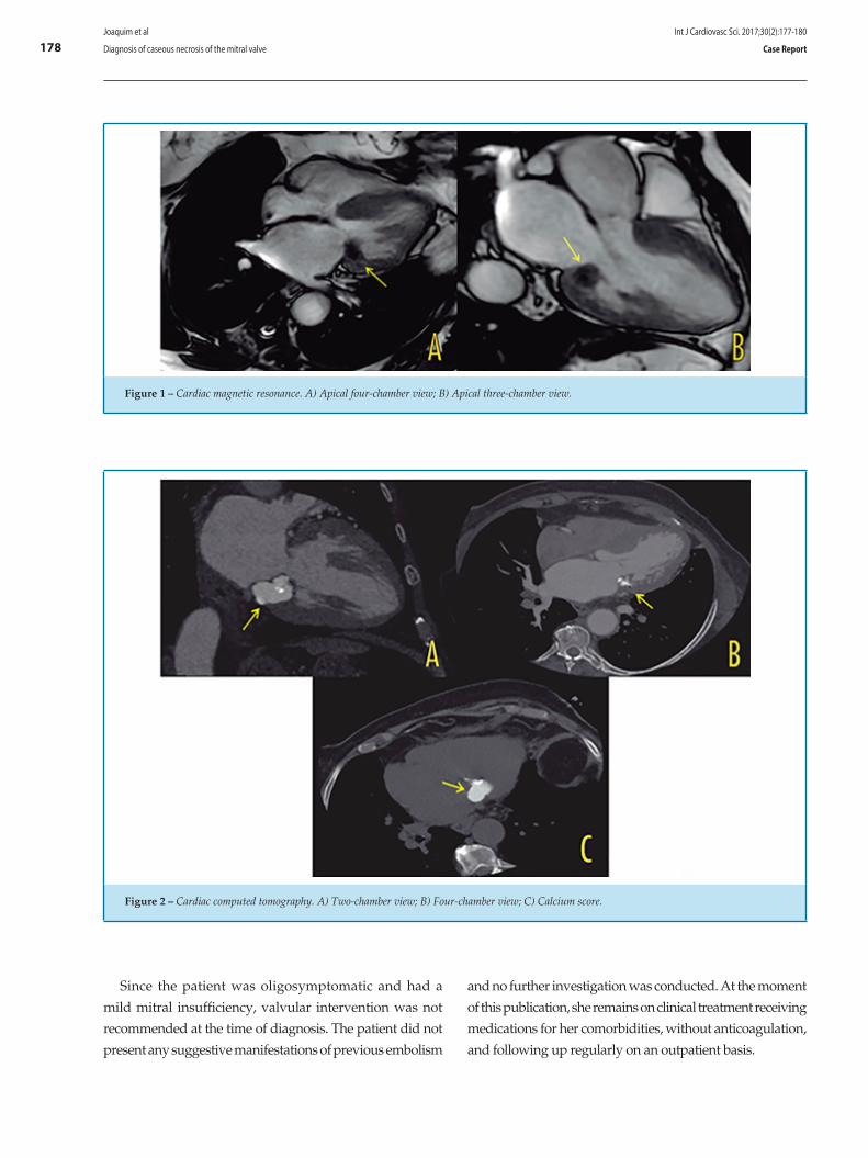
178
Figure 1 – Cardiac magnetic resonance. A) Apical four-chamber view; B) Apical three-chamber view.
Figure 2 – Cardiac computed tomography. A) Two-chamber view; B) Four-chamber view; C) Calcium score.
Joaquim et al
Diagnosis of caseous necrosis of the mitral valve
Int J Cardiovasc Sci. 2017;30(2):177-180
Case Report
Since the patient was oligosymptomatic and had a
mild mitral insufficiency, valvular intervention was not
recommended at the time of diagnosis. The patient did not
present any suggestive manifestations of previous embolism
and no further investigation was conducted. At the moment
of this publication, she remains on clinical treatment receiving
medications for her comorbidities, without anticoagulation,
and following up regularly on an outpatient basis.
179
1. Elgendy IY, Conti CR. Caseous calcification of the mitral annulus: a review. Clin Cardiol. 2013;36(10):E27-31.
2. Deluca G, Correale M, Ieva R, Del Salvatore B, Gramenzi S, Di Biase M. The incidence and clinical course of caseous calcification of the mitral annulus: a prospective echocardiographic study. J Am Soc Echocardiogr. 2008;21(7):828-33.
3. Pomerance A. Pathological and clinical study of calcification of the mitral valve ring. J Clin Pathol. 1970;23(4):354-61.
4. Stamou SC, Braverman AC, Kouchoukos NT. Caseous calcification of the anterior mitral valve annulus presenting as intracardiac mass. J Thorac Cardiovasc Surg. 2010;140(1):e9-10.
5. Harpaz D, Auerbach I, Vered Z, Motro M, Tobar A, Rosenblatt S. Caseous calcification of the mitral annulus: a neglected, unrecognized diagnosis. J Am Soc Echocardiogr. 2001;14(8):825-31.
6. Corre J, Leroux L, Coste P. Caseous necrosis of mitral annulus: a rare cause of stroke. Case Rep Cardiol. 2013;2013:748241.
References
Joaquim et al
Diagnosis of caseous necrosis of the mitral valve
Int J Cardiovasc Sci. 2017;30(2):177-180
Case Report
Discussion
The most common presentation of CNMV is as an intracardiac mass, and its diagnosis is incidental. When clinical manifestation occurs, it is usually with palpitations and dyspnea, and rarely with syncope due to atrioventricular blockade. These patients may present several other clinical findings related to the valvular lesion, with stenosis or insufficiency and, eventually, findings of systemic embolism.1,2,5
Harpaz et al.5 evaluated a series of 19 patients of whom 18 had isolated involvement of the posterior leaflet of the mitral annulus and one presented associated involvement of the anterior and posterior leaflets. In the same series, these authors observed that five patients (23%) presented some type of cerebrovascular event and only three patients underwent mitral valve replacement, while 84% of them were treated conservatively during a mean follow-up of 3.8 years.5 Several other cases of CNMV presenting as a stroke have already been described in the literature, demonstrating the importance of such type of event in this population.6-8 The mechanisms for the occurrence of stroke are unclear; it is unknown whether they occur due to their association with atherosclerosis, age and other risk factors related to the patients, or embolization of fragments of the mass.6
The differential diagnoses include intracardiac tumors, especially atrial myxoma, thrombosis of the coronary sinus, circumflex artery anomalies, and vegetations and abscesses of the mitral annulus.1,9,10 Multiple modalities of cardiovascular imaging should be used to differentiate between the various lesions and, in these cases, are capable of establishing a definitive diagnosis without requiring pathological analysis of the mass, eliminating invasive surgical procedures.9,10 Follow-up after diagnostic definition may be performed only with echocardiography at the discretion of the attending physician.10
CNMV is a dynamic process that, in some cases, may resolve spontaneously and even recur after surgically excised, depending on the valvular degeneration. Surgery is recommended upon dysfunction of the mitral valve with important stenosis or insufficiency, embolic events or when other types of tumors cannot be excluded regardless of the imaging method used.1 Anticoagulation may be prescribed in cases of embolic events, but no consensus has been established in this regard.8
Even though CNMV is a rare condition, it is important for cardiologists performing imaging tests to be familiar with this diagnosis, in order to avoid confusion with other masses or abscesses, thus preventing unnecessary surgery.1
Author contributions
Conception and design of the research: Joaquim RM, Bezerra SL. Acquisition of data: Joaquim RM, Bezerra SL. Analysis and interpretation of the data: Pinto IMF, Santos TSG. Writing of the manuscript: Joaquim RM, Correia EB, Albrecht FC. Critical revision of the manuscript for intellectual content: : Joaquim RM, Correia EB, Albrecht FC.
Potential Conflict of Interest
No potential conflict of interest relevant to this article was reported.
Sources of Funding
There were no external funding sources for this study.
Study Association
This study is not associated with any thesis or dissertation work.

180
7. Chevalier B, Reant P, Laffite S, Barandon L. Spontaneous fistulization of a caseous calcification of the mitral annulus: an exceptional cause of stroke. Eur J Cardiothorac Surg. 2011;39(6):e184-5.
8. Higashi H, Ohara T, Nakatani S, Hashimoto S, Torii T, Miyashita K, et al. A case of caseous calcification of the mitral annulus: a potential source of embolic stroke. J Cardiol Cases. 2010;2:e141-3.
9. Kydd AC, Gopalan D, Clarke SC, Rusk RA. Mitral annular caseous necrosis: insights from multimodality imaging. Eur Heart J. 2013;34(13):971.
10. Ribeiro S, Salgado A, Salomé N, Bettencourt N, Azevedo P, Costeira A, et al. Caseous calcification of the mitral annulus: A multi-modality imaging perspective. Rev Port Cardiol. 2012;31(4):313-6.
Joaquim et al
Diagnosis of caseous necrosis of the mitral valve
Int J Cardiovasc Sci. 2017;30(2):177-180
Case Report
DOI: 10.5935/2359-4802.20170036
181International Journal of Cardiovascular Sciences. 2017;30(2):181-184
CASE REPORT
Mailing Address: Sabrina Weiss StiesRua Pascoal Simone, 358, Coqueiros. Postal Code: 88080-350, Florianópolis, SC – BrazilE-mail [email protected]
Differential Diagnosis of Marfan Syndrome in a Teenage Volleyball AthleteFabrissio Portelinha Graffunder,1 Sabrina Weiss Sties,2 Ana Inês Gonzáles,1 Tales de Carvalho3
Universidade Federal de Santa Catarina (UFSC);1 Faculdade Avantis;2 Universidade do Estado de Santa Catarina (UDESC),3 Florianópolis, SC – Brazil
Manuscript received August 08, 2016, revised manuscript January 23, 2017, accepted January 31, 2017.
Marfan Syndrome; Aortic Rupture; Cardiovascular Diseases; Athletes; Exercise; Aortic Diseases.
Keywords
Introduction
Marfan syndrome (MFS) is an autosomal dominant hereditary disease mainly caused by mutations in the fibrillin-1 (FBN1) gene. It is characterized by the occurrence of thoracic aortic aneurysm and/or dissection, ectopia lentis, and systemic abnormalities.1
The highest risk of death associated with the syndrome is attributed to cardiovascular abnormalities, in particular, progressive aortic root aneurysm, leading to aortic dissection and rupture if not corrected surgically.2 The clinical diagnosis of MFS may be established by the revised Ghent nosology,3 although this can be challenging, since many characteristics of this disease are dependent on the age of the patient, while others are frequently seen in the general population, with substantial phenotypic variability. In addition, MFS has considerable overlap with other connective tissue diseases.2,4
Even though MFS is a rare condition (1:5,000),1 its prevalence is speculated to be much higher among individuals participating in sports, especially those sports in which tall stature and long limbs are advantageous. One such example is volleyball,5 which is classified as a moderate dynamic and low static sport.6
Studies on the practice of competitive sports by individuals with borderline or evident aortic root dilation are limited. Therefore, it becomes necessary to evaluate the differential diagnosis of MFS or any evident systemic disease.
In this context, the present study describes the case of a volleyball athlete with a possible diagnosis of MFS.
The athlete and his guardian signed both agreement and informed consent forms. The research was approved by the Research Ethics Committee at Universidade do Estado de Santa Catarina (UDESC).
Case Report
We report the case of a 16-year-old black male athlete with a height of 2.08 meters and weighing 80.9 kg. He was summoned to the Brazilian National Junior Volleyball Team in October 2013. During pre-joining assessment procedures, the patient was asymptomatic. He underwent clinical examination, ophthalmologic evaluation by a specialist, electrocardiography at rest, cardiopulmonary testing, and laboratory analysis, which showed no significant findings. While undergoing unidimensional and bidimensional echocardiography with color Doppler, a 45-mm diameter dilatation was observed on the thoracic ascendant aorta involving the sinuses of Valsalva. The athlete was removed from practice and underwent magnetic resonance angiography of the thoracic aorta, which evidenced a dilatation at the valvular plane with maximum diameters measured at this topography of 48 X 46 mm.
Based on the findings described above and the international literature, particularly the 36th Bethesda Conference, which contraindicates volleyball practice by individuals with ectasia at this level greater than 40 mm, the medical team recommended the athlete to be removed from practicing the sport. The patient was born with 3.8 kg and 51 cm, and his parents had normal height and weight. His family medical history was negative for MFS or sudden death, and he had no cognitive or sociability problems, showing excellent motor coordination and exercising with great ability the tasks of middle hitter, libero, and outside hitter in volleyball.
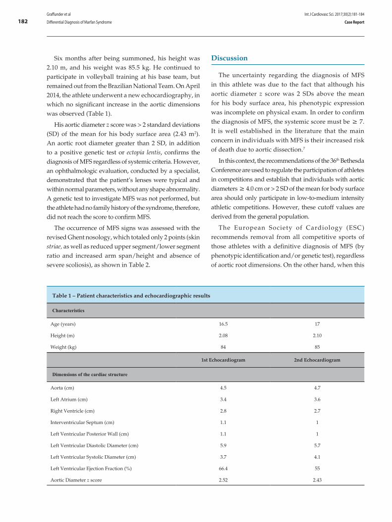
182Graffunder et al
Differential Diagnosis of Marfan Syndrome
Int J Cardiovasc Sci. 2017;30(2):181-184
Case Report
Table 1 – Patient characteristics and echocardiographic results
Characteristics
Age (years) 16.5 17
Height (m) 2.08 2.10
Weight (kg) 84 85
1st Echocardiogram 2nd Echocardiogram
Dimensions of the cardiac structure
Aorta (cm) 4.5 4.7
Left Atrium (cm) 3.4 3.6
Right Ventricle (cm) 2.8 2.7
Interventricular Septum (cm) 1.1 1
Left Ventricular Posterior Wall (cm) 1.1 1
Left Ventricular Diastolic Diameter (cm) 5.9 5.7
Left Ventricular Systolic Diameter (cm) 3.7 4.1
Left Ventricular Ejection Fraction (%) 66.4 55
Aortic Diameter z score 2.52 2.43
Six months after being summoned, his height was 2.10 m, and his weight was 85.5 kg. He continued to participate in volleyball training at his base team, but remained out from the Brazilian National Team. On April 2014, the athlete underwent a new echocardiography, in which no significant increase in the aortic dimensions was observed (Table 1).
His aortic diameter z score was > 2 standard deviations (SD) of the mean for his body surface area (2.43 m2). An aortic root diameter greater than 2 SD, in addition to a positive genetic test or ectopia lentis, confirms the diagnosis of MFS regardless of systemic criteria. However, an ophthalmologic evaluation, conducted by a specialist, demonstrated that the patient’s lenses were typical and within normal parameters, without any shape abnormality. A genetic test to investigate MFS was not performed, but the athlete had no family history of the syndrome, therefore, did not reach the score to confirm MFS.
The occurrence of MFS signs was assessed with the revised Ghent nosology, which totaled only 2 points (skin striae, as well as reduced upper segment/lower segment ratio and increased arm span/height and absence of severe scoliosis), as shown in Table 2.
Discussion
The uncertainty regarding the diagnosis of MFS in this athlete was due to the fact that although his aortic diameter z score was 2 SDs above the mean for his body surface area, his phenotypic expression was incomplete on physical exam. In order to confirm the diagnosis of MFS, the systemic score must be ≥ 7. It is well established in the literature that the main concern in individuals with MFS is their increased risk of death due to aortic dissection.7
In this context, the recommendations of the 36th Bethesda Conference are used to regulate the participation of athletes in competitions and establish that individuals with aortic diameters ≥ 4.0 cm or > 2 SD of the mean for body surface area should only participate in low-to-medium intensity athletic competitions. However, these cutoff values are derived from the general population.
The European Society of Cardiology (ESC) recommends removal from all competitive sports of those athletes with a definitive diagnosis of MFS (by phenotypic identification and/or genetic test), regardless of aortic root dimensions. On the other hand, when this
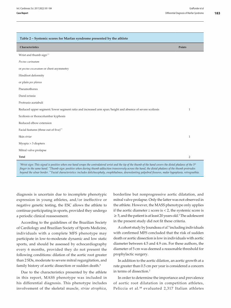
183Graffunder et al
Differential Diagnosis of Marfan Syndrome
Int J Cardiovasc Sci. 2017;30(2):181-184
Case Report
diagnosis is uncertain due to incomplete phenotypic expression in young athletes, and/or ineffective or negative genetic testing, the ESC allows the athlete to continue participating in sports, provided they undergo a periodic clinical reassessment.
According to the guidelines of the Brazilian Society of Cardiology and Brazilian Society of Sports Medicine, individuals with a complete MFS phenotype may participate in low-to-moderate dynamic and low static sports, and should be assessed by echocardiography every 6 months, provided they do not present the following conditions: dilation of the aortic root greater than 2 SDs, moderate to severe mitral regurgitation, and family history of aortic dissection or sudden death.8
Due to the characteristics presented by the athlete in this report, MASS phenotype was included in his differential diagnosis. This phenotype includes involvement of the skeletal muscle, striae atrophica,
Table 2 – Systemic scores for Marfan syndrome presented by the athlete
Characteristics Points
Wrist and thumb sign*,**
Pectus carinatum
or pectus excavatum or chest asymmetry
Hindfoot deformity
or plain pes planus
Pneumothorax
Dural ectasia
Protrusio acetabuli
Reduced upper segment/lower segment ratio and increased arm span/height and absence of severe scoliosis 1
Scoliosis or thoracolumbar kyphosis
Reduced elbow extension
Facial features (three out of five)***
Skin striae 1
Myopia > 3 diopters
Mitral valve prolapse
Total 2
*Wrist sign: This signal is positive when one hand wraps the contralateral wrist and the tip of the thumb of the hand covers the distal phalanx of the 5th finger in the same hand. **Thumb sign: positive when during thumb adduction transversely across the hand, the distal phalanx of the thumb protrudes beyond the ulnar border. ***Facial characteristics: includes dolichocephaly, enophthalmos, downslanting palpebral fissures, malar hypoplasia, retrognathia.
borderline but nonprogressive aortic dilatation, and mitral valve prolapse. Only the latter was not observed in the athlete. However, the MASS phenotype only applies if the aortic diameter z score is < 2, the systemic score is ≥ 5, and the patient is at least 20 years old.3 The adolescent in the present study did not fit these criteria.
A cohort study by Joundeau et al.9 including individuals with confirmed MFS concluded that the risk of sudden death or aortic dissection is low in individuals with aortic diameter between 4.5 and 4.9 cm. For these authors, the diameter of 5 cm was deemed a reasonable threshold for prophylactic surgery.
In addition to the aortic dilation, an aortic growth at a rate greater than 0.5 cm per year is considered a concern in terms of dissection.2
In order to determine the importance and prevalence of aortic root dilatation in competition athletes, Peliccia et al.10 evaluated 2,317 Italian athletes
184
1. Pepe G, Giusti B, Sticchi E, Abbate R, Gensini GF, Nistri S. Marfan syndrome: current perspectives. Appl Clin Genet. 2016;9:55-65.
2. Romaniello F, Mazzaglia D, Pellegrino A, Grego S, Fiorito R, Ferlosio A, et al. Aortopathy in Marfan Syndrome: an update. Cardiovasc Pathol. 2014;23(5):261-6.
3. Loeys BL, Dietz HC, Braverman AC, Callewaert BL, De Backer J, Devereux RB, et al. The revised Ghent nosology for the Marfan Syndrome. J Med Genet. 2010;47(7):476-85.
4. Tsang AK, Taverne A, Holcombe T. Marfan syndrome: a review of the literature and case report. Spec Care Dentist. 2013;33(5):248-54.
5. D’Andrea A. Cocchia R, Riegler L, Scarafile R, Salerno G, Gravino R, et al. Aortic root dimensions in elite athletes. Am J Cardiol. 2010;105(11):1629-34.
6. Mitchell JH, Haskell W, Snell P, Van Camp SP. Task force 8: classification of sports. J Am Coll Cardiol. 2005;45(8):1364-7.
7. Castellano JM, Silvay G, Castillo JG. Marfan Syndrome: clinical, surgical, and anesthetic considerations. Semin Cardiothorac Vasc Anesth. 2014;18(3):260-71.
8. Ghorayeb N, Costa RV, Castro I, Daher DJ, Oliveira Filho JA, Oliveira MA, et al; Sociedade Brasileira de Cardiologia. [Guidelines on exercise and sports cardiology from the Brazilian Society of Cardiology and the Brazilian Society of Sports Medicine]. Arq Bras Cardiol. 2013;100(1 Suppl. 2):1-41.
9. Joundeau G, Detaint D, Tubach F, Arnoult F, Milleron O, Raoux F, et al. Aortic event rate in the Marfan Population: a cohort study. Circulation. 2012;125(2):226-32.
10. Pelliccia A, DI Paolo FM, Quatrinni FM. Aortic root dilatation in athletic population. Prog Cardiovasc Dis. 2012;54(5):432-7.
References
Graffunder et al
Differential Diagnosis of Marfan Syndrome
Int J Cardiovasc Sci. 2017;30(2):181-184
Case Report
without cardiovascular disease, including men and women participating in Olympic Games and world championships, and competitors in different sports at national and regional levels. The authors found that the 99th percentile value for the aortic root diameter was 40 mm in men and 34 mm in women. Overall, 17 male athletes showed larger aortic diameters than the set threshold, including young athletes practicing rowing, basketball or volleyball who were tall and had a large body surface area, but none satisfied the revised Ghent criteria for the diagnosis of MFS. They were allowed to practice sports and were followed up every 6 months. A significant increase in the size of the aorta was observed, which in two of these athletes exceeded 5 cm. However, they had no cardiovascular complications, remained without any Ghent criterion, and had a negative test for genetic FBN1 mutations.
Conclusion
Changes in the aortic diameter pattern should be taken into account the individual’s body surface area and progressive aortic dilation.
There is a paucity of information on the practice of competitive sports by individuals with borderline or obvious aortic root dilation but without diagnostic criteria for MFS or any evident systemic disease.
The athlete in this report had a possible differential diagnosis of MFS that could be confirmed after periodic
evaluations over time. It is important to highlight that trainers in sports in which tall stature is advantageous and, thus common, should become acquainted with MFS, in order to identify through known signs potential athletes who have the disease and help them seek appropriate guidance.
Author contributions
Conception and design of the research: Graffunder FP, Sties SW, Gonzáles AI, Carvalho T. Acquisition of data: Graffunder FP, Sties SW, Gonzáles AI, Carvalho T. Analysis and interpretation of the data: Graffunder FP, Sties SW, Gonzáles AI, Carvalho T. Writing of the manuscript: Graffunder FP, Sties SW, Gonzáles AI, Carvalho T. Critical revision of the manuscript for intellectual content: Graffunder FP, Sties SW, Gonzáles AI, Carvalho T.
Potential Conflict of Interest
No potential conflict of interest relevant to this article was reported.
Sources of Funding
There were no external funding sources for this study.
Study Association
This study is not associated with any thesis or dissertation work.