Embed Size (px)
Citation preview
Relationship between hearing threshold at the affected and unaffected ear in unilateral Meniere’s disease Roberto Albera • Andrea Canale • Claudia Cassandro • Andrea Albera • Azia Maria Sammartano • �Federico Dagna
Università degli Studi di Torino, Città della Salute e della Scienza di Torino, ENT Department
INTRODUCTION
In MD hearing loss has been described to affect above all the low frequencies (upward curve) but tends to become irreversible and nonfluctuating at the higher frequencies over time (peaked curve) [5,6]. However other patterns have been described such as downward sloping curve, flat curve and bell curve [6]. Hearing impairment occurs within the first years of the disease and after 5-10 years hearing loss stabilizes at a mean level of 50-60 dB and rarely it causes a profound hearing loss [7]. The audiometric slope in MD is usually evaluated without taking in consideration the hearing threshold at the unaffected ear. It is our opinion that, at least in unilateral cases without other pathologies at the safe side, the comparison between the thresholds at the affected and unaffected side could be the only way to determine the true damage caused by the disease on hearing function; in fact by means of this approach it is possible to avoid the conditioning effects of the basal hearing threshold, in our hypothesis described by the hearing threshold at the unaffected side). The aim of the study was to determine the effects of MD on hearing function on the basis of the differences existing between the affected and the unaffected ear in a group of patients with definite unilateral MD and whose contralateral ear was not affected by any disease other than age related hearing loss (ARHL). Following this procedure we have also evaluated the possible effects of age and disease duration on hearing loss in MD.
MATERIAL AND METHODS The study group consisted of 86 subjects affected by definite unilateral MD according to the AAO-HNS 1995 guidelines [4] and analysed retrospectively. The second inclusion criterion was the absence of pathologies at the unaffected ear other than ARHL. Therefore the sample was limited to subjects whose pure-tone audiometry (PTA) threshold at the unaffected ear was better than 25 dB at each frequency tested or better than values expected at the 50th percentile in relation to age, according to ISO 7029-2000; in both cases PTA threshold evaluation was extended at all the frequencies tested (0.25-8 kHz). In each case we have evaluated: age, duration of the disease and contralateral ear status. Age of the sample ranged from 16 to 85 years (mean age 56 years, Standard Deviation 16); 42 (49%) were males and 44 (51%) females. Average MD duration was 59 months (ranging from 1 to 250, SD 58). The right ear was involved in 41cases (48%) and the left ear in 45 (52%). Each patient was submitted to pure-tone audiometry (PTA) in a sound proof chamber at frequencies between 0.25 and 8 kHz. We considered the best audiogram among at least three audiograms repeated in few weeks and carried out in a non-critic phase of the disease (no vertigo and hearing worsening, tinnitus and ear fullness judged at the lower level by the patients). Encephalic MRI was carried out in all the patients of the study group. In no case we have found lesions at the CNS or at the ponto-cerebellar angle. The statistical analysis was carried out by means of SPSS statistical and PRISM packages. Significancy level was set at 0.05. Among the parameters considered in the study only disease duration did not showed a Gaussian distribution at the Kolomogorov-Smirnoff’s test.
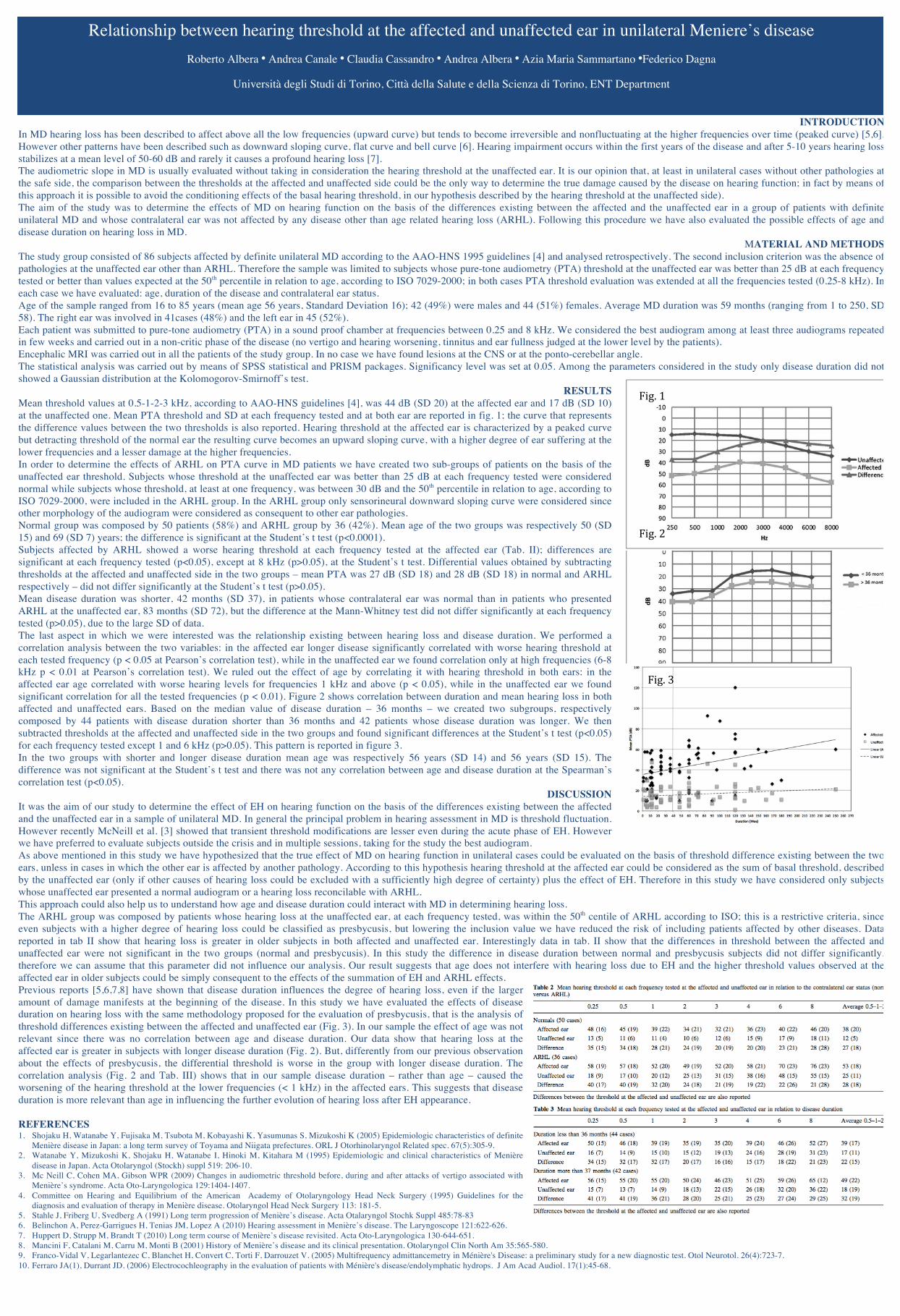
RESULTS Mean threshold values at 0.5-1-2-3 kHz, according to AAO-HNS guidelines [4], was 44 dB (SD 20) at the affected ear and 17 dB (SD 10) at the unaffected one. Mean PTA threshold and SD at each frequency tested and at both ear are reported in fig. 1; the curve that represents the difference values between the two thresholds is also reported. Hearing threshold at the affected ear is characterized by a peaked curve but detracting threshold of the normal ear the resulting curve becomes an upward sloping curve, with a higher degree of ear suffering at the lower frequencies and a lesser damage at the higher frequencies. In order to determine the effects of ARHL on PTA curve in MD patients we have created two sub-groups of patients on the basis of the unaffected ear threshold. Subjects whose threshold at the unaffected ear was better than 25 dB at each frequency tested were considered normal while subjects whose threshold, at least at one frequency, was between 30 dB and the 50th percentile in relation to age, according to ISO 7029-2000, were included in the ARHL group. In the ARHL group only sensorineural downward sloping curve were considered since other morphology of the audiogram were considered as consequent to other ear pathologies. Normal group was composed by 50 patients (58%) and ARHL group by 36 (42%). Mean age of the two groups was respectively 50 (SD 15) and 69 (SD 7) years; the difference is significant at the Student’s t test (p<0.0001). Subjects affected by ARHL showed a worse hearing threshold at each frequency tested at the affected ear (Tab. II); differences are significant at each frequency tested (p<0.05), except at 8 kHz (p>0.05), at the Student’s t test. Differential values obtained by subtracting thresholds at the affected and unaffected side in the two groups – mean PTA was 27 dB (SD 18) and 28 dB (SD 18) in normal and ARHL respectively – did not differ significantly at the Student’s t test (p>0.05). Mean disease duration was shorter, 42 months (SD 37), in patients whose contralateral ear was normal than in patients who presented ARHL at the unaffected ear, 83 months (SD 72), but the difference at the Mann-Whitney test did not differ significantly at each frequency tested (p>0.05), due to the large SD of data. The last aspect in which we were interested was the relationship existing between hearing loss and disease duration. We performed a correlation analysis between the two variables: in the affected ear longer disease significantly correlated with worse hearing threshold at each tested frequency (p < 0.05 at Pearson’s correlation test), while in the unaffected ear we found correlation only at high frequencies (6-8 kHz p < 0.01 at Pearson’s correlation test). We ruled out the effect of age by correlating it with hearing threshold in both ears: in the affected ear age correlated with worse hearing levels for frequencies 1 kHz and above (p < 0.05), while in the unaffected ear we found significant correlation for all the tested frequencies (p < 0.01). Figure 2 shows correlation between duration and mean hearing loss in both affected and unaffected ears. Based on the median value of disease duration – 36 months – we created two subgroups, respectively composed by 44 patients with disease duration shorter than 36 months and 42 patients whose disease duration was longer. We then subtracted thresholds at the affected and unaffected side in the two groups and found significant differences at the Student’s t test (p<0.05) for each frequency tested except 1 and 6 kHz (p>0.05). This pattern is reported in figure 3. In the two groups with shorter and longer disease duration mean age was respectively 56 years (SD 14) and 56 years (SD 15). The difference was not significant at the Student’s t test and there was not any correlation between age and disease duration at the Spearman’s correlation test (p<0.05).
DISCUSSION It was the aim of our study to determine the effect of EH on hearing function on the basis of the differences existing between the affected and the unaffected ear in a sample of unilateral MD. In general the principal problem in hearing assessment in MD is threshold fluctuation. However recently McNeill et al. [3] showed that transient threshold modifications are lesser even during the acute phase of EH. However we have preferred to evaluate subjects outside the crisis and in multiple sessions, taking for the study the best audiogram. As above mentioned in this study we have hypothesized that the true effect of MD on hearing function in unilateral cases could be evaluated on the basis of threshold difference existing between the two ears, unless in cases in which the other ear is affected by another pathology. According to this hypothesis hearing threshold at the affected ear could be considered as the sum of basal threshold, described by the unaffected ear (only if other causes of hearing loss could be excluded with a sufficiently high degree of certainty) plus the effect of EH. Therefore in this study we have considered only subjects whose unaffected ear presented a normal audiogram or a hearing loss reconcilable with ARHL. This approach could also help us to understand how age and disease duration could interact with MD in determining hearing loss. The ARHL group was composed by patients whose hearing loss at the unaffected ear, at each frequency tested, was within the 50th centile of ARHL according to ISO; this is a restrictive criteria, since even subjects with a higher degree of hearing loss could be classified as presbycusis, but lowering the inclusion value we have reduced the risk of including patients affected by other diseases. Data reported in tab II show that hearing loss is greater in older subjects in both affected and unaffected ear. Interestingly data in tab. II show that the differences in threshold between the affected and unaffected ear were not significant in the two groups (normal and presbycusis). In this study the difference in disease duration between normal and presbycusis subjects did not differ significantly, therefore we can assume that this parameter did not influence our analysis. Our result suggests that age does not interfere with hearing loss due to EH and the higher threshold values observed at the affected ear in older subjects could be simply consequent to the effects of the summation of EH and ARHL effects. Previous reports [5,6,7,8] have shown that disease duration influences the degree of hearing loss, even if the larger amount of damage manifests at the beginning of the disease. In this study we have evaluated the effects of disease duration on hearing loss with the same methodology proposed for the evaluation of presbycusis, that is the analysis of threshold differences existing between the affected and unaffected ear (Fig. 3). In our sample the effect of age was not relevant since there was no correlation between age and disease duration. Our data show that hearing loss at the affected ear is greater in subjects with longer disease duration (Fig. 2). But, differently from our previous observation about the effects of presbycusis, the differential threshold is worse in the group with longer disease duration. The correlation analysis (Fig. 2 and Tab. III) shows that in our sample disease duration – rather than age – caused the worsening of the hearing threshold at the lower frequencies (< 1 kHz) in the affected ears. This suggests that disease duration is more relevant than age in influencing the further evolution of hearing loss after EH appearance. REFERENCES 1. Shojaku H, Watanabe Y, Fujisaka M, Tsubota M, Kobayashi K, Yasumunas S, Mizukoshi K (2005) Epidemiologic characteristics of definite
Menière disease in Japan: a long term survey of Toyama and Niigata prefectures. ORL J Otorhinolaryngol Related spec. 67(5):305-9. 2. Watanabe Y, Mizukoshi K, Shojaku H, Watanabe I, Hinoki M, Kitahara M (1995) Epidemiologic and clinical characteristics of Menière
disease in Japan. Acta Otolaryngol (Stockh) suppl 519: 206-10. 3. Mc Neill C, Cohen MA, Gibson WPR (2009) Changes in audiometric threshold before, during and after attacks of vertigo associated with
Menière’s syndrome. Acta Oto-Laryngologica 129:1404-1407. 4. Committee on Hearing and Equilibrium of the American Academy of Otolaryngology Head Neck Surgery (1995) Guidelines for the
diagnosis and evaluation of therapy in Menière disease. Otolaryngol Head Neck Surgery 113: 181-5. 5. Stahle J, Friberg U, Svedberg A (1991) Long term progression of Menière’s disease. Acta Otalaryngol Stochk Suppl 485:78-83 6. Belinchon A, Perez-Garrigues H, Tenias JM, Lopez A (2010) Hearing assessment in Menière’s disease. The Laryngoscope 121:622-626. 7. Huppert D, Strupp M, Brandt T (2010) Long term course of Menière’s disease revisited. Acta Oto-Laryngologica 130-644-651. 8. Mancini F, Catalani M, Carru M, Monti B (2001) History of Menière’s disease and its clinical presentation. Otolaryngol Clin North Am 35:565-580. 9. Franco-Vidal V, Legarlantezec C, Blanchet H, Convert C, Torti F, Darrouzet V. (2005) Multifrequency admittancemetry in Ménière's Disease: a preliminary study for a new diagnostic test. Otol Neurotol. 26(4):723-7. 10. Ferraro JA(1), Durrant JD. (2006) Electrocochleography in the evaluation of patients with Ménière's disease/endolymphatic hydrops. J Am Acad Audiol. 17(1):45-68.
Fig.1
Fig.2
Fig.3


![Hunterston Jetty · TTS/PTS threshold [dB SEL-24] Impulsive threshold [dB SEL-24] Impulsive threshold [dB z-p] TTS 185 185 197 PTS 213 213 206 1.2.4 APPLICATION OF HEARING THRESHOLDS](https://img.pdfslide.us/doc/110x75/5d1376cc88c993b4258bccb1/hunterston-jetty-ttspts-threshold-db-sel-24-impulsive-threshold-db-sel-24.jpg)


![[756] HEARING IN CERTAIN ORTHOPTERA · Hearing in certain orthoptera 759 Fig. 1 shows graphs relating the threshold intensity in decibels to frequency of a pure tone stimulus for](https://img.pdfslide.us/doc/110x75/5e86810f7ace8239892ef11c/756-hearing-in-certain-orthoptera-hearing-in-certain-orthoptera-759-fig-1-shows.jpg)


![CLIA-RELATED HEARING DECISIONS - CMS...2017/08/14 · CLIA certificate revoked. 42 CFR 498.40 Request for Hearing. An affected party entitled to a hearing. 2/16/1999 [CR576] BAN Laboratories](https://img.pdfslide.us/doc/110x75/6021e719054fed4834707c8d/clia-related-hearing-decisions-cms-20170814-clia-certificate-revoked.jpg)