Embed Size (px)
Citation preview

Relation between Cell Activity
and the Distribution of Cytoplasmic Actin and Myosin
The discovery of actin and myosin in nonmuscle cells stimu-lated speculation regarding their role in cell motility; but, withthe exception of cytokinesis (18, 33, 35), there is little directevidence to support this notion. In the present work we havebrought together two of the methods most widely used to studycell movements : time-lapse recording of movements and flu-orescent antibody staining of actin and myosin . Time-lapsecinematography (1-4, 6, 7, 38) has been essential for docu-menting the relatively slow movements ofcultured cells includ-ing membrane ruffling (2, 7), cell spreading (38), and locomo-tion over glass surfaces (1-4) . Fluorescent antibody staininghas been used to localize actin and myosin (8, 16, 19, 23, 30-32) in regions ofcytoplasm presumably undergoing movementsat the time of fixation . Though one could reasonably inferfrom morphology alone that there was motion of the mitoticspindle, cleavage furrow, and membrane ruffles ofcells stainedwith fluorescent antibodies, these movements have not beenpreviously documented in specific stained cells. Furthermore,subtle cellular movements such as shape changes and celllocomotion cannot be deduced from the inspection of fixedcells .
84
IRA M. HERMAN, NANCY 1 . CRISONA, and THOMAS D. POLLARDDepartment of Cell Biology and Anatomy, The Johns Hopkins Medical School, Baltimore, Maryland21205 . Dr. Crisona's present address is the Department of Zoology, University of California, Berkeley,California 94720.
ABSTRACT We documented the activity of cultured cells on time-lapse videotapes and thenstained these identified cells with antibodies to actin and myosin . This experimental approachenabled us to directly correlate cellular activity with the distribution of cytoplasmic actin andmyosin . When trypsinized HeLa cells spread onto a glass surface, the cortical cytoplasm wasthe most actively motile and random, bleb-like extensions (0.5-4.0 Jtm wide, 2-5 p,m long)occurred over the entire surface until the cells started to spread . During spreading, rufflingmembranes were found at the cell perimeter. The actin staining was found alone in the surfaceblebs and ruffles and together with myosin staining in the cortical cytoplasm at the bases ofthe blebs and ruffles. In well-spread, stationary HeLa cells most of the actin and myosin wasfound in stress fibers but there was also diffuse antiactin fluorescence in areas of motilecytoplasm such as leading lamellae and ruffling membranes. Similarly, all 22 of the rapidlytranslocating embryonic chick cells had only diffuse actin staining . Between these extremeswere slow-moving HeLa cells, which had combinations of diffuse and fibrous antiactin andantimyosin staining . These results suggest that large actomyosin filament bundles are associatedwith nonmotile cytoplasm and that actively motile cytoplasm has a more diffuse distributionof these proteins .
In this study we documented the motility of several differenttypes of living cells on time-lapse videotapes before fixationand staining of the same cells for actin and myosin withfluorescent antibodies . With this unique approach we wereable to correlate directly the activities of living cells with thedistribution of cytoplasmic actin and myosin .
MATERIALS AND METHODS
Antibody Preparation
ANTtACTIN: Rabbit antibodies were prepared against chicken gizzard(smooth muscle) actin, labeled with fluorescein and purified by affinity chro-matography (22) .
ANTTMYOSIN : Dr . Keigi Fujiwara immunized a goat with the purifiedplatelet myosin rod exactly as described for immunizing rabbits (15). Immune Igwas labeled with rhodamine, and antimyosin IgG was purified by affinitychromatography (l5).
Cell Preparation
LIVING CELLS
HELA CELL CULTURES :
HeLa cells were grown on glass microscope coverslips exactly as described previously (2l) .
THE JOURNAL Of CELL BIOLOGY " VOLUME 90 JULY 1981 84-91©The Rockefeller University Press " 0021-9525/81/07/0084/08$1 .00

EMBRYONIC CULTURES :
Cells were obtained from the skin and wing budsof I1-d-old chick embryos. Embryonic tissue was minced and digested for 10 minat 37°C in 2.5 mg/mltrypsin in phosphate-buffered saline (PBS ; 0.015 Msodiumphosphate, 0.15 M NaCl, pH 7.3). Dissociated cells were grown for 2-3 d on 18x 18 mm glass cover slips in Dulbecco's minimum essential medium (DMEM)supplemented with 1% chick serum and 1% fetal calf serum.
TIME-LAPSE VIDEOTAPE RECORDS:
Living cells were observed while at-tached to cover slips submerged in growth medium in a 35-mm petri dish sealedwith parafilm to maintain a 5% COz atmosphere. The petri dish, containing cellsattached to glass cover slips, was placed in a 37°C air curtain on a stage of anNikon inverted light microscope equipped with phase-contrast optics. Time-lapsevideotapes were recorded with a Panasonic NV-8030 VTR. Although photo-graphs of the TV screen do not reproduce well, these videotapes allowed us toevaluate the activity of the cells in some detail . Locomotion,pseudopod extensionand retraction, mitosis, cytokinesis, and membrane ruffling were all easily ob-served . Intracellular movements of small organelles could not be evaluated.CELL SPREADING:
Confluent cultures of HeLa cells were briefly washed ina Ca"/Mg++-free PBS and trypsinized for 1.5 min at 37'C with 1 mg/mltrypsinin PBS. Trypsinized cells were shaken from the growth substrate, suspended infresh DMEM with 10% fetal calf serum (DMEM/10), pelleted by centrifugation,resuspended in DMEM/10, plated onto glass cover slips, and enclosed in a petridish that was sealed with Parafilm to maintain a 5% CO, atmosphere for time-lapse videotaping of cell spreading at 37°C.
FIXED CELLS
CELL FIXATION : HeLa and embryonic chicken cells were prepared forfluorescent antibody staining as described by Herman and Pollard (22) . Livingcells used for time-lapse videotaping were fixed for fluorescence microscopywithin 60 s after taping.
ANTIBODY STAINING OF FIXED CELLS: We directly stained fixed andacetone-treated HeLa cells for actin and/or myosin and embryonic chicken cellsforactin (the myosin antibodies did notcross-react) with 60-75,ug/mlof affinity-purified, fluorescein-labeled rabbit anti-chicken gizzard actin and 20-40 pg/mlaffinity-purified, rhodamine-labeled goat anti-human platelet myosin . Controlexperiments for actin and myosin staining were performed by incubating cells in1 00,ug/ml unlabeled antiactin or antimyosin before reaction with either 65-70Ag/ml fluorescein-antiactin or 20-40 pg/ml rhodamine-antimyosin. Labeledpreimmune IgG as well as labeled immune IgG that did not bind to theappropriate affinity columns were also used for control staining experiments . Asin our previous work (15, 16, 22), no fluorescence staining could be detected inthe controls .
Evaluation of Polypeptide Extraction duringFixation and Simulated Staining.
Confluent cultures of HeLa and PtK2 cells were washed in Ca"/Mg--freePBS before treatment with a l mg/ml solution of trypsin in PBS for 2 min at37°C . The trypsinized cells were suspended in DMEM/10, pelleted by centrifu-gation for 3 min at 1000 g, resuspended in PBS three times, and counted with ahemocytometer. Roughly, 3 x 10⁶cells (in duplicate or triplicate) were transferredwith siliconized pipettes into 1.5-ml siliconized Eppendorf Microcentrifuge tubes(Brinkmann Instruments, Inc., Westbury, N. Y.) and treated in one of thefollowing four ways :
(a) Cell pellets were suspended in 1 .0 ml of 4% formaldehyde in PBS for 5min at 37°C, pelleted by centrifugation at 10,000 g at 25°C, and washed with 1 .0ml of PBS at room temperature for 5 min. Samples were then pelleted bycentrifugation before suspension in absolute acetone cooled to -20'C for 1 min,washed three timeswith 1 ml of PBS at room temperature before a 1-h incubationin PBS. This simulates our standard fixation and antibody incubation procedure .
(b) Cells were handled exactly as in a but were dehydrated for I min with95% ethanol cooled to -20°C instead of acetone.
(c) Living cells were washed with PBS and then fixed with absolute methanolcooled to -20°C for 5 min instead of formaldehyde and acetone .
(d) Living cell pellets were dissolved directly into boiling SDS gel samplebuffer.The final cell pellets and the aqueous supernates from each step were
lyophilized. The organic extracts were dried under N2. All of these samples weresolubilized in 2% SDS and 10% ,B-mercaptoethanol at 100'C for 3-5 min inpreparation forpolyacrylamide gel electrophoresis. The gel samples were clarifiedby centrifugation at 10,000 g for 2 min at 25°C and 30 pl was applied in eachlane . Tube or slab SDS gel electrophoresis was carried out in a 14% polyacryl-amide resolving gel or a 7.5-15% polyacrylamide resolving gradient gel (29) . Thegels were stained with Coomassie Blue R-250 and scanned at 600 nm. Areasunder peaks were extrapolated to the baseline and measured by cutting andweighing. The mean weight of the duplicate or triplicate samples was calculated
with the standard deviation from the mean values . To detect myosin in these gelswe used a modification of the antibody overlay technique of Adair et al . (5). Gelswere fixed with 10% acetic acid, 25% isopropanol in water overnight . Gels werewashed and equilibrated with 0.15 M sodium chloride, 0.05 M Tris, pH 7.8, lmg/ml Triton X-100, and l mg/ml BSA(TTX-BSA) overnight . They were thenincubated with 100 jig/ml rabbit antimyosin Ig fraction (12) for 4 h. Gels werewashed with several changes of l liter of TTX-BSA and then incubated with 20ml of ' 25 1-labeled protein A in TTX-BSA (0.6 x 10⁶ cpm/ml) for 3 h (sp act,-10' cpm/pg). Gels were washed with TTX-BSA and dried on filter paper.Radioautograms were exposed on x-ray film (Cronex 2DC) at -70°C with anintensifying screen for 4 d.
RESULTS
Preparation of Cells for Antibody StainingOur objective was to correlate the activity ofliving cells with
the distribution ofactin and myosin in the cytoplasm, so it wasnecessary to establish that neither cellular morphology norprotein composition was altered by fixation, dehydration, orincubation with fluorescent antibody . We show that the grossmorphology of the living cultured cells was preserved duringantibody staining . Furthermore, the same fixation-dehydrationprocedure preserved the ultrastructure of these cells as well(23) . To establish that the actin and myosin were retained inthe cells carried through the fluorescent antibody procedure,we examined by gel electrophoresis the polypeptide composi-tion ofthe fixed cells and the various solutions used to preparethese cells (Fig. 1). Remarkably, the polypeptide compositionof the cells was altered very little by formaldehyde-acetone,formaldehyde-ethanol, or methanol fixation/permeabilization .With methanol, there was some aggregated material at the topof the gel . None of the fixation or wash solutions containedmore than traces ofpolypeptides. In the case ofPtK2 cells, 92%of the stained actin band was retained in the cell pellet byformaldehyde-acetone, 88% by formaldehyde-ethanol, and 82%by methanol. Because myosin accounts for <1% of HeLaprotein (41), the myosin heavy chain was difficult to identifywith certainty on the gels . A faint 200,000 mol wt band wasfound in the fixed cell pellets, but, to be certain that it wasmyosin and that no myosin was extracted, we stained the gelswith antimyosin . In the fixed cell pellet, both the 200,000 molwt band and some higher molecular weight material, presum-ably aggregated myosin, bound the antimyosin, but no myosinwas detected in this way in the buffer wash of these cells(Fig. 1) .
Antibody StainingCELL SPREADING:
We investigated HeLa cell spreading ona glass surface by correlating motile activity and fluorescentstaining for actin and myosin in the same cells . In the first fewminutes during which spherical HeLa cells began to settle onthe glass surface, many transient and rounded blebs 0.5-4 .0gm wide and 2-5 gm long formed and retracted at the cellsurface . These surface blebs were first described by Holtfreter(24), and later Taylor (38) described this behavior as stage I inthe spreading of conjunctiva cells onto a glass surface . Whenspeeded up by time-lapse these hyaline blebs of cytoplasmappeared to protrude and retract while moving around the cellperimeter, in an almost circular, wave-like motion . This surfaceblebbing continued until the cells began to attach more firmlyonto the glass. This coincides wth stage II of Taylor's schemeand occurs
20-40 min after contact with the glass substrate .Later, as these cells continued to flatten and spread out, thenuclei of the cells also flattened and became visible in phasecontrast . This corresponded to stage III in Taylor's scheme .
HERMAN ET AL . Cell Activity and Distribution of Actin and Myosin
85

FIGURE 1
Proteins present in fixed HeLa cells . Samples were boiledin 2% SDS and 10%,B-mercaptoethanol before electrophoresis in a7.5-15% polyacrylamide gradient slab gel . Lanes A-H, CoomassieBlue staining; lanes I and J, radioautography . (A) Molecular weightstandards. (B) Unfixed HeLa cells . (C) Polypeptides lost from form-aldehyde-fixed and acetone-treated HeLa cells during a 1-h incu-bation with PBS to simulate antibody staining . (D) Final cell pelletof formaldehyde-fixed and acetone-treated HeLa cells . (E) Polypep-tides lost from formaldehyde-fixed and ethanol-treated HeLa cellsduring a 1-h incubation with PBS to simulate antibody staining . ( F )
Final cell pellet of formaldehyde-fixed and ethanol-treated HeLacells . (G) Polypeptides lost from methanol-fixed HeLa cells duringa 1-h incubation with PBS to simulate antibody staining . (H) Finalcell pellet of methanol-fixed HeLa cells . (I and 1) Identical to C andD but treated with antimyosin and "'I-labeled protein A ratherthan being stained . (1) No radioactivity is seen, indicating thatmyosin is not lost during simulated antibody staining of cells . (J )Radioactivity is present in the fixed HeLa cells at a mobility corre-sponding to the heavy chain of myosin . High molecular weightmaterial at the top of the gel may be aggregated myosin .
In 24 HeLa cells with documented surface blebbing, themost intense fluorescent staining with both antiactin and anti-myosin was in the cell periphery, but the distribution andtexture of staining for actin and myosin were distinctive (Figs .2 and 3). The blebs themselves were outlined with an intensering of antiactin fluorescence juxtaposed to the plasma mem-brane (Fig. 2A). The antimyosin fluorescence was weak in the
86
THE JOURNAL OF CELL BIOLOGY " VOLUME 90, 1981
surface blebs but was intensely localized in a subcortical beltthat also stained intensely with antiactin (Figs . 2 and 3) .
Later, as spreading onto the glass surface continued, mem-brane ruffling was always associated with discrete regions ofthe cell perimeter, especially in the direction ofmovement (Fig .3, cell .3 ; Fig . 3, cells 4 and S, arrows) . As in other well-documented cases (1-3), the ruffling membrane of spreadingHeLa cells appears to build up at the margins and then retreattoward the cell center. In all cells with documented activity,ruffles were stained intensely with antiactin (Figs . 3-5) . Othershave also shown antiactin staining of membrane ruffles inspreading cells fixed during stage III ofthe Taylor scheme (25,31, 38) . On the other hand, antimyosin either did not stain oronly weakly stained these regions of membrane ruffling (Fig .3) . Punctate anitmyosin staining was concentrated in a circularsubcortical zone together with antiactin staining (Fig . 3, cell 4) .The antimyosin fluorescence remained radially arranged untilvery fine punctate fibers could be seen traversing the cell
FIGURE 2
Double antibody staining of spreading HeLa cells. Flu-orescent antibody staining of six spreading HeLa cells stained si-multaneously with 60 gg/ml fluorescein-antiactin (A, C, E, G) and20 üg/ml rhodamine-antimyosin (B, D, F, H) . The cells appearing inA -F were fixed <20 min after plating onto glass cover slips . Noticethe prominent surface blebbing outlined by actin fluorescence in Aand E. Antimyosin weakly stains the surface blebs but is concen-trated in a zone juxtaposed to the blebbing . The cell in G and H
was fixed nearly 2 h after trypsinization, and it is considerably flatterthan cells in A, C, and E. Bar, 4 tLm .

FIGURE 3 Correlation of cellular activity with actomyosin staining during the spreading of five HeLa cells . (A and B) Phase-contrast images of living cells taken 34 min and 1 min, respectively, before fixation . (C) Phase-contrast image of the same cellsafter fluorescent antibody staining . (D and E) Fluorescence images of the HeLa cells as stained with (D) 60 flg/ml fluorescein-antiactin and (E) 20 f~g/ml rhodamine-antimyosin . Bar, 4.0 Am .
interior (Fig . 3, cell 4) . This occurred 1-2 h after the cellssettled onto the glass during stage III of Taylor's scheme . Thisbright, peripheral staining in the spreading cells was particu-larly impressive because this region of the cytoplasm wasthinnest, making it unlikely that this intense staining ofthe cellperiphery was the result of a superimposed signal through along path length .
INTERPHASE CELLS:
That interphase HeLa cells were quiteheterogeneous in size, shape, motility, and contractile proteindistribution gave us the opportunity to analyze a variety ofcells . Because of this diversity, cell morphology alone was aninadequate indicator of motile activity, and the time-lapserecords of the living cells were necessary for the interpretationof the fluorescence micrographs .
At one extreme, -10% of interphase HeLa cells were highlyflattened and contained prominent stress fibers. All nine of theflat cells that we videotaped failed to move in 0.9-1.2 h (Figs.4 and 5) . Virtually all of the antiactin fluorescence in these flatnonmotile cells was confined to stress fibers, although therewas also some intense diffuse staining of membrane ruffles atthe cell periphery (Fig. 4 and cell I in Fig . 5) as in the spreadingcells . Myosin was also localized in the same stress fibers ofthese cells (not shown) .
At the other extreme, there were a number of small, wedge-shaped HeLa cells that moved rapidly (-0.5-3 Am/min) . Thesecells were usually lost during fixation and staining, whichaccounts for their absence in previous studies. In general theywere similar to the actively motile chick embryo cells describedbelow, because the antiactin and antimyosin gave diffuse stain-ing .Between these extremes there were many medium-sized cells
that were not highly motile but slowly changed their shapessuch as cells 2, 4, and 5 in Fig . 5 . These cells usually had acombination of fibrous and diffuse staining with antiactin andantimyosin . The diffuse component was usually spreadthroughout the cytoplasm (Fig . 5 E) but was especially intensein leading lamellae (Fig . 5 F, arrow) .
FIGURE 4
Relationship between the activity and actin distributionof a well-spread HeLa cell . (A-C) Phase-contrast images of theborder of the cell taken (A) 47, (B) 36, and (C) 1 min before fixationand staining . Membrane ruffling (black arrowheads) was the onlymotile activity observed . (D) Fluorescein-antiactin staining pattern .Most of the antibody staining is concentrated in stress fibers andthe region of membrane ruffling that had a bright, diffuse fluores-cence (white arrowheads) . Bar, 8 .0 um .
There were three major types of cells in cultures of embry-onic chicken, skin and wing buds . Most of the cells werespindle-shaped, often 50-100 Am long, and probably weremyoblasts. Other cells, presumably fibroblasts, were A- orwedge-shaped. The third major cell type was small (10-20 Am
HERMAN ET AL . Cell Activity and Distribution of Actin and Myosin
87

FIGURE 5
Relationship between HeLa cell activity and cytoplasmic actin . (A -C) Phase-contrast images of the living HeLa cells .The time is recorded in hours:minutes:seconds in the lower left-hand corner of the TV monitor. (D-F) Fluorescence micrographsof the same cells after fixation and staining with 75 pg/ml fluorescein-antiactin . As labeled in C, 1 was a well-spread cell with acircular profile. Before fixation, the left-hand border actively ruffled . After staining, this region had bright, diffuse fluorescence. 2retracted a microspike on its right side (B) during taping and had extended another toward 6:00 (C, ') 8 min later. The antiactinfluorescence was both diffuse and fibrous (E) . 3 remained essentially immobilized and the majority of its actin fluorescence wasfibrous (F) . 4 was spindle-shaped and during the taping became stretched out . It elongated and had a diffuse actin fluorescence(E, top left) . Some cells, such as 5, possessed a leading lamella that advanced as a sheetlike, mobile projection that varied in sizeas it was elaborated from the cell perimeter . When this cell was fixed and stained for actin, this mobile lamellipodium was intenselyfluorescent (F) . Bars (A-C ), 10 pm ; (D and E), 3.0 Am and 10 Am, respectively.
wide) and motile but with few directed movements (Fig. 6) .The velocities of movements varied with time from 0 to 3.5pm/min at the maximum . The three cells in Fig. 6 illustratethe variety of movements and the variability of the movementvelocities (rates) . Cell " (Fig. 6) had five major lamellipodiaeach of which possessed regions of ruffling at the beginning ofthe taping . After the cell had stretched itself to 1 .5 times itsoriginal length, all its lamellipodia joined to form one leadinglamella and, when this moving cell was fixed and stained withfluorescein-antiactin, only diffuse fluorescence was observed(Fig . 6) . Cell " (Fig . 6) changed its direction of movement fourtimes during videotaping . Each time it contacted a neighboringcell with its wavelike lamella, its movement forward was in-hibited. Between the end of videotaping and the time the cellswere fixed (<60 s), this cell " changed its shape slightly byreleasing its attachment to a neighbor (cf. lower left cell " inE and F with G and H in Fig . 6) and the direction of lamellarmovement (cf. cell " lamellae in E and F with G and H in Fig .6) . This moving cell " had diffuse antiactin fluorescence . CellA (Fig . 6) moved randomly during taping, showing few directedmovements . As with cell I/, it is apparent that cell A wasmoving at the time of fixation because there are subtle differ-ences in morphology between the living cell and the fixed cell .In particular, the top, left-hand ruffling membrane spread outslightly (cf. E and F with H and K in Fig . 6), the lower left-hand ruffling membrane is redistributed, and the tail portion
88
THE JOURNAL Of CELL BIOLOGY " VOLUME 90, 1981
(indicated by A in Fig. 6 F) has pulled in slightly . Again, thismoving cell had diffuse antiactin fluorescence after fixation .The movements observed with HeLa and embryonic chicken
cells were representative of the movements and fluorescentantibody staining patterns seen in other cell types includingmigrating neural crest cells (10) and human peripheral bloodcells (see Table I) . The large, stationary, and spread-out cellsthat we studied in HeLa cultures were absent from the bloodcell and neural crest cell preparations ; and, in general, thesemoving cells had diffuse fluorescence.
DISCUSSION
Using time-lapse videotapes, we documented the activity ofliving cells (see Table I) and correlated this information withfluorescent antibody staining for actin and myosin. The move-ments we observed during cell spreading and locomotion onglass surfaces were similar to those reported earlier (1, 4, 7, 25,31, 38) . After the cells with documented activity were fixed andstained with fluorescent antibodies, their morphology seemednormal, based on light and electron microscopy (23) . Moreover,most, if not all, of the actin and myosin was retained in thesecells prepared for antibody staining; and actin and myosinremained reactive with their antibodies after fixation . Althoughwe cannot prove that all of the antigens remained in theirnatural cellular locations during the fixation/dehydration pro-
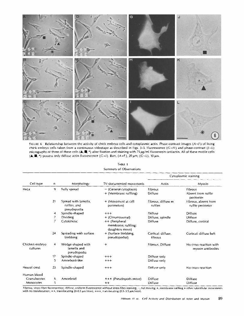
FIGURE 6
Relationship between the activity of chick embryo cells and cytoplasmic actin . Phase-contrast images (A-F) of livingchick embryo cells taken from a continuous videotape as described in Figs . 3-5 . Fluorescence (G-H) and phase-contrast (J-L)micrographs of three of these cells (", " , *) after fixation and staining with 75 pg/ml fluorescein-antiactin . All of these motile cells(", " , *) possess only diffuse actin fluorescence (G-1) . Bars, (A-F), 20 ttm ; (G-L), 10I.m .
Cell type
n
Morphology
TV-documented movements
Actin
Hela
TABLE I
Summary of Observations
Cytoplasmic staining
Myosin
Fibrous, stress fiber fluorescence ; diffuse, uniform fluorescence without stress fiber staining; -, not moving ; +, membrane ruffling + other subcellular movementswith no translocation ; ++, translocating (0-0.5 Wm/min) ; +++, translocating (0 .5-3 .5 jum/min) .
HERMAN ET AL . Cell Activity and Distribution of Actin and Myosin 89
blebbing pseudopodial) fibrous
Chicken embryo 4 Wedge-shaped with + Fibrous, Diffuse No cross-reaction withcultures lamella and myosin antibodies
pseudopodia17 Spindle-shaped +++ Diffuse only5 Amoeboid-like +++ Diffuse only
Neural crest 23 Spindle-shaped +++ Diffuse only No cross-reaction
Human bloodGranulocytes 6 Amoeboid +++ (Pseudopods move) Diffuse DiffuseMonocytes 1 ++ Diffuse Diffuse
9
21
Fully spread
Spread with lamella,ruffles, andpseudopodia
- (General cytoplasm)+ (Membrane ruffling)
+ (Movement at cellperimeters)
FibrousDiffuse
Fibrous, diffuse inruffles
FibrousAbsent from ruffle
perimeterFibrous, absent from
ruffle perimeter
4 Spindle-shaped +++ Diffuse Diffuse7 Dividing + (Chromosomal) Diffuse, spindle Diffuse7 Cytokinetic ++ (Peripheral Diffuse Diffuse, cortical
membrane ; ruffling ;daughters move)
24 Spreading with surface + (Surface blebbing, Cortical : diffuse, Cortical : diffuse belt

cedure, this seems likely from the lack of antigen extractionand the good morphological preservation .The results obtained by correlating the cellular activity with
fluorescent antibody staining were :(a) Motile cells or motile parts of cells stain diffusely with
antibodies to actin and myosin. This includes spreading HeLacells, moving chick embryo cells and blood cells, rufflingmembranes ofboth chick embryo and HeLa cells, and dividingHeLa cells .
(b) When most of the fluorescent-antiactin staining is con-fined to prominent stress fibers, cells grown on glass moveabout very slowly, if at all. However, there are active move-ments at ruffled membranes in the periphery of these cells .Such active regions stain diffusely with fluorescein-antiactin .
(c) Combinations of diffuse and fibrous antiactin (and anti-myosin) staining can be found in cells that are stationary orchange shape slowly . These slow shape changes can lead toslow locomotion .
(d) Nonlocomoting cells can have any antiactin and anti-myosin staining pattern .
These observations have led us to conclude that (a) only inthe cases of ruffled membranes, mitosis, and cytokinesis is itpossible to predict cellular activity from morphology and an-tibody staining; (b) stress fibers are not essential for motility;and (c) cellular motility is usually associated with a diffusedistribution of actin and myosin .On the basis of morphology alone, we (15, 16, 21, 22) and
others (8, 19, 25, 30-32) have argued that actin (and/or myosin)was present in parts of cells that were moving. We believe thatthis is true for ruffling membranes, the cleavage furrow, andthe mitotic apparatus . Only these events have cellular mor-phology or antibody staining patterns distinctive enough toconclude that motion was occurring at the time of fixation . Forexample, both stationary and slowly locomotiog HeLa (Fig . 4and 5) and chick embryo cells (Fig. 6) can have diffuse, or amixture of diffuse and fibrous antiactin staining .Although there is good evidence that isolated stress fibers
(26) and the stress fibers of permeable cell models (28) cancontract in Mg`-ATP, our observations on living HeLa cellsshow that stress fibers are not essential for motility . In cells orparts of cells with prominent stress fibers, little or no motionoccurs . We never observe locomotion ofwell-spread HeLa cellswhen most of the antiactin and antimyosin fluorescence isconfined to stress fibers. Although motion was inferred, notdocumented, Badley et al . (8) made similar observations oncultured chick fibroblasts. Given these observations and theevidence that many stress fibers terminate on substrate attach-ment plaques (17, 27), it seems likely that most stress fibershave a structural role in anchoring the cytoplasmic matrix tothe substrate, rather than being contractile . Their contractilepotential is expressed only when one or both of their terminalattachment plaques is released from the substrate, such as inexperimental manipulation (26, 28) . Actually, neither the func-tion of these photogenic animyosin bundles in vitro nor theirvery existence in vivo is established .
Motile regions of cells always had diffuse antiactin fluores-cence, suggesting that their motility was probably broughtabout by a diffuse actin network in the cytoplasm . Taylor andhis co-workers (40) injected fluorescent-actin into rapidly mov-ing amoebas and found that it also distributed diffusely in thestreaming cytoplasm and the stationary cortex . These lightmicroscope observations do not mean, however, that actinfilaments are not involved, because electron micrographs reveal
90
THE JOURNAL Of CELL BIOLOGY - VOLUME 90, 1981
actin filaments in the cortex of amoeba (13), as well as thecytoplasm of ruffling membranes (4, 11, 12, 37, 42) and thecleavage furrow (36) .
In general, myosin is also dispersed throughout the cyto-plasm of motile cells (Figs . 2 and 3) as well as the mitoticapparatus (15, 16) and is concentrated in the cleavage furrow(15, 16, 20) . One exception is the apparent absence of myosinfrom the margins of ruffling membranes (Fig. 2 and 3) (15, 19)and from the interior cytoplasm ofspreading HeLa cells (Figs.2 and 3) . If myosin does participate in moving ruffles, it mustdo so in the cytoplasm at the base of the ruffles where it isfound with actin (Figs . 2 and 3) . This seems similar to thesituation in the brush border, where myosin is confined in theterminal web (9, 14, 23, 34) . The existence of a morphologicallydispersed form of the cell's contractile apparatus explains whyall regions of the cytoplasm are potentially contractile (39) .The combination of time-lapse recording and fluorescent
antibody staining has provided some new information aboutcytoplasmic actin and myosin and cell activity; however, theapproach has limitations . First, the fluorescence micrographsprovide no information about the physical state of actin ormyosin except when actin is assembled into stress fibers . Indiffusely stained cells, actin could be either monomeric orpolymerized and the filaments could be arranged in either arandom network or in small bundles like the contractile ring(36) . It is entirely possible that in diffusely stained cells (Fig . 6)some of the actin filaments are also aligned in bundles bytension, like those ofthe contractile ring . These small bundlesare not detected by fluorescent antibody staining, because theactin concentration in the bundles and the surrounding cyto-plasm is the same . A second limitation is that the apparentabsence of fluorescence from part of a cell does not mean thatthe antigen is missing there . For example, by electron micros-copy we found low concentrations of ferritin-labeled anti-myosin in regions of stress fibers that appear nonfluorescentwhen stained with rhodamine-antimyosin (23) . Consequently,one can conclude from fluorescent antibody staining that theratio of actin to myosin is much higher in the margin than atthe base of the ruffles, not that myosin is absent from mem-brane ruffles .
The authors express their thanks to Dr. Dan Kiehart for his generositywith his "'I-labeled protein A and Tom Urquhart for his help with thephotographic reproductions. We also thank Drs. Keigi Fujiwara forimmunizing the goat, Alan Cohen for the neural crest cultures, andLarry Gerace for his advice on running gels.
This work was supported by National Institutes of Health grantsGM 26338-03 and GM 26132-03 and Muscular Dystrophy Associationpostdoctoral fellowships to Ira Herman and Nancy Crisona.
Received for publication 12 August 1980, and in revisedform 6 February1981 .
REFERENCES
1 . Abercrombie, M . 1961 . The bases of the locomotory behavior of fibroblasts . Exp. Cell Res.8 (Suppl .):188-198 .
2 . Abercrombie, M., J. E . M . Heaysman, and S. M . Pegrum . 1970 . The locomotion offibroblasts in culture. 1 . Movements of the leading edge. Exp. Cell Res. 59 :393-398 .
3. Abercrombie, M ., J . E. M . Heaysman, and S . M. Pegrum . 1970. The locomotion offibroblasts in culture. 11 . Ruffling. Exp. Cell Res. 69:437-444.
4 . Abercrombie, M., 1 . E . M . Heaysman, and S. M. Pegrum . 1971 . The locomotion offibroblasts in culture. IV. Electron microscopy of the leading lamellae. Exp. Cell Res. 67 :359-367 .
5, Adair, W. S ., D. Jurivich, and U . W . Goodenough . 1978 . Localization of cellular antigensin sodium dodecyl sulfate-polyacrylamide gets. J. Cell Biol. 79 :281-285 .
6 . Albrecht-Beuhler, G. 1979 . Group locomotion ofPTK cells. Exp . Cell Res. 122 :402-407 .7. Ambrose, E . J . 1961 . The movements of fibrocytes . Exp. Cell Res. 8 (Suppl .) :54-73 .8 . Badley, R . A ., 1 . R . Couchman, and D . A . Rees. 1980 . Compariso n of the cytoskeleton in
migratory and stationary chick fibroblasts . J Muscle Res. Cell. Motil. 1 :5-14.9. Bretscher, A ., and K . Weber . 1978 . Localization of actin and microfilament-associated
proteins in microvilb and the terminal web of intestinal brush borders by immunofluores-cence microscopy . J Cell Biol. 79 :839-845 .
10. Bronner, M. E., and A . M. Cohen. 1979 . Migratory patterns of cloned neural crestmelanocytes injected into host chicken embryos, Proc . Nail. Acad. Sci. U. S. A . 76 :1843-1849.
11 . Buckley, I . K . 1974. Subcellular motility: a correlated light and electron microscopic studyusing cultured cells. Tissue Cell. 6:1-20.
12. Buckley, 1 . K ., and K . R. Porter. 1967 . Cytoplasmic fibrils in living cultured cells.Protoplasma. 64 :349-380.
13 . Comly, L . T . 1973 . Microfilaments in Chaos carolinensis. Membrane association, distri-bution, and HMM binding in the glycerinated cell . J. Cell Biol. 58 :230-237.
14. Drenckhahn, D ., and U. Grüschel-Stewart. 1980 . Localization of myosin, actin andtropomyosin in rat intestinal epithelium : immunohistochemical studies at the light andelectron microscope levels . J. Cell Biol. 86 :475-483.
15 . Fujiwara, K., and T . D . Pollard . 1976. Fluorescent antibody localization of myosin in thecytoplasm, cleavage furrow and mitotic spindle of human cells . J. Cell Biol. 71 :848-875 .
16. Fujiwara, K., and T. D. Pollard . 1978. Simultaneous localization of myosin and tubulin inhuman tissue culture cells by double antibody staining. J. Cell Biol. 77 :182-195.
17 . Geiger, B . 1979. A 130 K protein from chicken gizzard . Its localization at the termini ofmicrofilament bundles in cultured chicken cells . Cell. 18 :193-205.
18 . Goldman, R ., T . Pollard, and J . Rosenbaum, editors. 1976. Cell Motility. Cold SpringHarbor Conference on Cell Proliferation. Vol 3, Book A . Cold Spring Harbor Laboratory,Cold Spring Harbor, N .Y . 1265 .pp.
19 . Gottlieb, A . L, M. H. Heggeness, J . F. Ash, and S. J . Singer . 1979 . Mechanochemica lproteins, cell motility and cell-cel] contacts : the localization of mechanochemical proteinsinside cultured cells at the edge of an in vitro "wound ." J. Cell. Physiol. 100:563-578 .
20 . Herman, I . M., P. Maupin, and T. D . Pollard. 1980 . Localization of myosin in dividingHeLa cells with ferritin-antimyosin . J. Cell Biol. 87 (2, Pt. 2):224 a (Abstr .).
21 . Herman, I. M ., and T . D. Pollard. 1978 . Actin localization in fixed dividing cells stainedwith fluorescent heavy meromyosin. Exp . Cell Res. 114 :15-25 .
22 . Herman, I. M ., andT. D . Pollard . 1979 . Comparison of purified antiactin and fluorescent-heavy meromyosin staining patterns in dividing cells . J. Cell Biol. 80 :509-520.
23 . Herman, 1. M ., and T. D. Pollard. 1981 . Electro n microscopic localization of cytoplasmicmyosin with ferritin-labeled antibodies . J. Cell Biol. 88 :346-351 .
24 . Holtfreter, 1 . 1943 . A study of the mechanics of gastrulation . I. J. Exp. Zool. 94:261-318 .25 . Hynes, R. O ., and A. T . Destree . 1978. Relationships between fibronectin and actin . Cell.
15 :875-886.
26 . Isenberg, G ., P . C . Ratlike, N . Hulsmann, W . Franke, and K . E. Wohlfarth-Botterman.1976 . Cytoplasmic actomyosin fibrils in tissue culture cells . Cell Tissue Res. 166 :427-443.
27. Izzard, C . S., and L . R . Lochner. 1976 . Cell-to-substrat e contacts in living fibroblasts. Aninterference reflexion study with an evaluation of the technique . J. Cell Sci. 21 :129-159.
28. Kreis, T . E ., and W. Birchmeier. 1980. Stress fiber sarcomeres of fibroblasts are contractile .Cell. 22 :555-561 .
29. Laemmli, U . K . 1970. Cleavage of structural proteins during the assembly of the head ofbacteriophage T4 . Nature (Land.). 227:680-685 .
30. Lazarides, E . 1975. Immunofluorescence studies on the structure of actin filaments intissue culture cells. J. Histochem . Cytochem . 23 :507-528 .
31 . Lazarides, E. 1976 . Aspects of the structural organization of actin filaments in tissueculture cells . In Cold Spring Harbor Conference on Cell Proliferation . Cell Motility . R .Goldman, T. D . Pollard, and 1 . Rosenbaum, editors. Cold Spring Harbor Laboratory,Cold Spring Harbor, N . Y . 3(Book A) :347-360 .
32. Lazarides, E ., and K. Weber . 1974 . Actin antibody: the specific visualization of actinfilaments in non-muscle cells. Proc. Nall. A cad. Sci. U. S. A . 71 :2268-2272.
33 . Mabuchi, L, and M . Okuno. 1977. The effect of myosin antibody on the division of starfishblastomeres. J. Cell Biol. 74:251-263 .
34 . Mooseker, M. S., T. D. Pollard, and K . Fujiwara. 1978 . Characterization and localizationof myosin in brush borders of intestinal epithelial cells. J. Cell Biol. 79:444-453 .
35. Pollard, T . D ., and R. R. Weiping . 1974. Actin and myosin and cell movement. CRC Crit.Rev . Biochem . 2:1-65 .
36. Schroeder, T . 1973. Actin in dividing cells . Contractile ring filaments bind heavy mero-myosin. Proc. Nall. Acad. Sci. U. S. A . 70:1688-1692 .
37 . Spooner, B ., K . Yamada, and N . Wessells. 1971 . Microfilamems and cell locomotion. J.Cell Mot 49:595-613 .
38 . Taylor, A . C. 1961 . Attachment and spreading of cells in culture . Exp . Cell Res. 8 (Suppl.) :154-173.
39. Taylor, D. L. 1976. Motile model systems of amoeboid movement. In Cold Spring HarborConference on Cell Proliferation . Cell Motility. R . Goldman, T . D. Pollard, and J .Rosenbaum, editors . Cold Spring Harbor Laboratories, Cold Spring Harbor, N . Y. 3(Book A) :797-821 .
40. Taylor, D . L ., Y. L . Wang, and 1. M . Heiple . 1980. Contractile basis of amoeboidmovement . VII. The distribution of fluorescently labeled actin in living amebas . J. CellBiol. 86:590-598 .
41 . Weilting, R . R. 1977. Effects of myosin and heavy meromyosin on actin-related gelationof HeLa cell extracts. J Cell Biol. 75:95-103 .
42. Yamada, K ., B . Spooner, and N . Wessells. 1971 . Ultrastructure and function of growthcones and axons of cultured nerve cells . J Cell Biol. 49 :614-635.
HERMAN ET AL. Cell Activity and Distribution of Actin and Myosin
91