Embed Size (px)
Citation preview
submit.radiology.or.kr J Korean Soc Radiol 2013;68(3):197-200 197
INTRODUCTION
The central nervous system (CNS) involvement in progressive systemic sclerosis (PSS) is rare, which has been considered to be either uncommon or a secondary consequence of hypertension, uremia, pulmonary dysfunction and steroid treatment (1-3). White matter hyper intensities (WMHI) on MRI were more com-mon in patients with PSS than in the control group, which might be asymptomatic and inadequate of the disease duration (2, 3).
We report a unique case of PSS where WMHI have arisen in both the middle cerebellar peduncles and left temporal lobe, and made rapid progress into tumefactive WMHI in the sub-cortices of both precentral gyri. To the best of our knowledge, tumefactive form of PSS has not been reported.
CASE REPORT
A 68-year-old man was admitted to our hospital with the on-
set of dysarthria for 1 month. Vocal cord movement was slightly impaired, but not fixed. Neurologic examination was normal, except moderate dysarthria. He was alert and normotensive. T2 weighted image (T2-WI) of MRI showed multiple small WMHI in the left pons and both cerebral white matters (Fig. 1A). On past history, the patient had presented peripheral type of the left facial palsy at age 26. He had 4-year-history of Raynaud phe-nomenon and 3-year-history of skin thickening and hyperpig-mentation. One year ago, he was diagnosed as PSS, limited cuta-neous type.
Two months later, dysarthria has been progressed, with im-pairment of vibration perception in both legs and gait distur-bance was developed. T2-WI of MRI revealed WMHI in both middle cerebellar peduncles and left temporal lobe (Fig. 1B), and increased diffusivity on diffusion weighted MR image (DWI). MR spectroscopy showed decrease in the NAA peak at 2.0 ppm. MR angiography (MRA) for evaluation of the possible cerebral macroangiopathy showed to be normal. No demonstrable en-
Case ReportpISSN 1738-2637J Korean Soc Radiol 2013;68(3):197-200
Received November 8, 2012; Accepted December 20, 2012Corresponding author: Hui Joong Lee, MDDepartment of Radiology, Kyungpook National University Hospital, 130 Dongdeok-ro, Jung-gu, Daegu 700-721, Korea.Tel. 82-53-420-5390 Fax. 82-53-422-2677E-mail: [email protected]
This research was supported by Basic Science Research Program through the National Research Foundation of Korea (NRF) funded by the Ministry of Education, Science and Technology (2012005117).
Copyrights © 2013 The Korean Society of Radiology
White matter hyper intensities (WMHI) on MRI are not rare in patients with pro-gressive systemic sclerosis (PSS). In this presentation, WMHI were developed in both middle cerebellar peduncles and temporal white matter in a patient with PSS, and regressed after medication of high dose steroid. However, new lesions were devel-oped in the subcortices of both precentral gyri, and progressed rapidly to tumefac-tive hyperintensity on MRI. We report an unusual relapsing and progressive tume-factive demyelinating form of central nervous system involvement in PSS.
Index termsProgressive Systemic SclerosisCentral Nervous System InvolvementMRI
Relapsing and Progressive Tumefactive Demyelinating Form of Central Nervous System Involvement in a Patient with Progressive Systemic Sclerosis1 진행성 전신성 경화증의 진행성 종양성 탈수초 형태의 중추신경계통 이환1
Ho Kyun Kim, MD1, Hui Joong Lee, MD2
1Department of Radiology, Catholic University of Daegu School of Medicine, Daegu, Korea2Department of Radiology, Kyungpook National University Hospital, Daegu, Korea
Relapsing and Progressive Tumefactive Demyelinating Form of CNS Involvement in a Patient with Progressive Systemic Sclerosis
submit.radiology.or.krJ Korean Soc Radiol 2013;68(3):197-200198
external lamina, with sparse media and adventitia (1, 4, 5). Ce-rebral hypoperfusion, suggestive of impaired quantitative mi-crocirculation, was observed in PSS patients (6). Reduction of regional cerebral blood flow might be related to PSS, although the vast majority of patients remained in subclinical phase. In PSS, the primary site of vascular involvement is at the level of small arteries and capillaries. However, several authors have re-ported CNS abnormalities related with carotid and intracranial vasculitis in patients with PSS (7-9).
Most common MR findings of CNS involvement of PSS is WMHI, which are thought to result from ischemic vasculopa-thy, supporting the hypothesis of early and frequent brain in-volvement in patients with PSS. In this case, WMHI of both middle cerebellar peduncles and left temporal lobe that showed at the first presentation were thought as ischemic vasculopathy as the usual complication of PSS. The lesions regressed after medication of high dose steroid. However, rapid progression
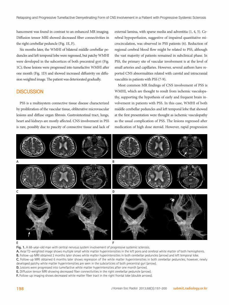
hancement was found in contrast to an enhanced MR imaging. Diffusion tensor MRI showed decreased fiber connectivities in the right cerebellar peduncle (Fig. 1E, F).
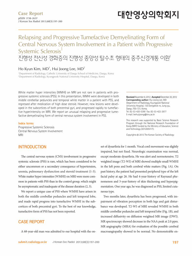
Six months later, the WMHI of bilateral middle cerebellar pe-duncles and left temporal lobe were regressed, but patchy WMHI were developed in the subcortices of both precentral gyri (Fig. 1C); those lesions were progressed into tumefactive WMHI after one month (Fig. 1D) and showed increased diffusivity on diffu-sion weighted image. The patient was deteriorated gradually.
DISCUSSION
PSS is a multisystem connective tissue disease characterized by proliferation of the vascular tissue, obliterative microvascular lesions and diffuse organ fibrosis. Gastrointestinal tract, lungs, heart and kidneys are mostly affected. CNS involvement in PSS is rare, possibly due to paucity of connective tissue and lack of
Fig. 1. A 68-year-old man with central nervous system involvement of progressive systemic sclerosis.A. Axial T2-weighted image shows multiple small white matter hyperintensities in the left pons and cerebral white matter of both hemispheres. B. Follow-up MRI obtained 2 months later shows white matter hyperintensities in both cerebellar peduncles (arrow) and left temporal lobe. C. Follow-up MRI obtained 6 months later shows regression of the white matter hyperintensities in both cerebellar peduncles; however, newly developed patchy white matter hyperintensities are seen in the subcorticies of both precentral gyri (arrow). D. Lesions were progressed into tumefactive white matter hyperintensities after one month (arrow).E. Diffusion tensor MRI showing decreased fiber connectivities in the right cerebellar peduncle (arrow). F. Follow-up imaging shows decreased white matter fiber tract in the right frontal lobe (double arrows).
A
C
E
B
D
F

Ho Kyun Kim, et al
submit.radiology.or.kr J Korean Soc Radiol 2013;68(3):197-200 199
REFERENCES
1.LuciveroV,MezzapesaDM,PetruzzellisM,CarellaA,Lam-
bertiP,FedericoF.Ischaemicstrokeinprogressivesystemic
sclerosis.NeurolSci2004;25:230-233
2.MohammedRH,SabryYY,NasefAA.BrainMRIscreening
showingevidencesofearlycentralnervoussystemin-
volvementinpatientswithsystemicsclerosis.Rheumatol
Int2011;31:667-671
3.SardanelliF, IozzelliA,CotticelliB,LosaccoC,CutoloM,
SulliA,etal.Whitematterhyperintensitiesonbrainmag-
netic resonance in systemic sclerosis.AnnRheumDis
2005;64:777-779
4.KanzatoN,MatsuzakiT,KomineY,SaitoM,SaitoA,Yoshio
T,etal.Localizedsclerodermaassociatedwithprogressing
ischemicstroke.JNeurolSci1999;163:86-89
5.SimeoniS,PuccettiA,TinazziE,TomelleriG,CorrocherR,
LunardiC.Systemicsclerosisandsuperficialsiderosisof
thecentralnervoussystem:casualityorcausality?Rheu-
matolInt2008;28:815-818
6.NobiliF,CutoloM,SulliA,CastaldiA,SardanelliF,Accardo
S,etal.Impairedquantitativecerebralbloodflowinsclero-
dermapatients.JNeurolSci1997;152:63-71
7.ChungMH,SumJ,MorrellMJ,HoroupianDS.Intracere-
bralinvolvementinsclerodermaencoupdesabre:report
ofacasewithneuropathologicfindings.AnnNeurol1995;
37:679-681
8.StoneJ,FranksAJ,GuthrieJA,JohnsonMH.Scleroderma
“encoupdesabre”:pathologicalevidenceofintracerebral
inflammation.JNeurolNeurosurgPsychiatry2001;70:
382-385
9.PathakR,GaborAJ.Sclerodermaandcentralnervoussys-
temvasculitis.Stroke1991;22:410-413
10.TorabiAM,PatelRK,WolfeGI,HughesCS,Mendelsohn
DB,TrivediJR.Transversemyelitis insystemicsclerosis.
ArchNeurol2004;61:126-128
was demonstrated later into tumefactive hyper intensities in the subcortices of both precentral gyri, which has not been described as CNS involvement of PSS. MRA for evaluation of the possible cerebral macroangiopathy showed normal. Diffusion tensor MRI showed decreased fiber connectivities in the right cerebel-lar peduncle and the subcortex of the right precentral gyrus. These lesions could not be explained as resulting from ischemic vasculopathy or as a form of microangiopathy. In our opinion, white matter change might be caused by a localized autoim-mune demyelinating disease. The evidence supporting this hy-pothesis is: A) an apparent good response to high-dose cortico-steroid treatment as suggested by the clinical improvement at first presentation; B) no steno-occlusive lesion was detected on MRA; C) no vascular risk factor was present, which was docu-mented during the hospitalisation; D) echocardiography was un-able to detect embolic sources; and E) increased diffusivity of the lesion was detected on DWI, which considered vasogenic ede-ma, as result of demyelination, rather than acute ischemic lesion.
When one identifies multifocal relapsing and remitting WMHI, the differential diagnosis may be worth the consideration: mul-tiple sclerosis, autoimmune disease, such as behcet’s disease, acute disseminated encephalomyelitis or lymphoma. PSS is a multisys-tem disease primarily affecting the skin, but may involve other or-gans including lung and kidney. Only a few cases of coexistence of PSS and multiple sclerosis have been described. Since several con-nective tissue diseases are associated with transverse myelitis, in-cluding systemic lupus erythematosus and Sjögren’s syndrome, there are discussions whether the transverse myelitis in patients with PSS were an independent overlapping event or a manifes-tation of PSS (10).
In summary, we present the first report of relapsing and pro-gressive tumefactive demyelinating form in a patient with PSS probably due to demyelination of white matter. Conventional MRI was very useful to detect the CNS lesions, and diffusion tensor imaging and MR spectroscopy could be helpful to evalu-ate the severity and the change of white matter involvement.

Relapsing and Progressive Tumefactive Demyelinating Form of CNS Involvement in a Patient with Progressive Systemic Sclerosis
submit.radiology.or.krJ Korean Soc Radiol 2013;68(3):197-200200
진행성 전신성 경화증의 진행성 종양성 탈수초 형태의 중추신경계통 이환1
김호균1 · 이희중2
진행성 전신성 경화증 환자에서 자기공명영상에서 백질부 고신호강도는 드물지 않은 소견이다. 제시된 증례에서는 중소
뇌각과 측두엽의 백질부 고신호강도가 고농도의 스테로이드 치료 후 호전을 보였으나, 새로운 병변이 양측 중심전회의 피
질하에 발생하였으며, 급속히 종양성 고신호강도로 발전하였다. 저자들은 진행성 전신성 경화증의 진행성 종양성 탈수초
형태의 중추신경계통 이환을 보고하고자 한다.
1대구가톨릭대학교 의과대학 영상의학과학교실, 2경북대학교병원 영상의학과