Embed Size (px)
Citation preview

Dynamics of Ribosomal Protein S1 on aBacterial Ribosome with Cross-Linking andMass Spectrometry*□S
Matthew A. Lauber‡, Juri Rappsilber§, and James P. Reilly‡¶
Ribosomal protein S1 has been shown to be a significanteffector of prokaryotic translation. The protein is in factcapable of efficiently initiating translation, regardless ofthe presence of a Shine-Dalgarno sequence in mRNA.Structural insights into this process have remained elu-sive, as S1 is recalcitrant to traditional techniques ofstructural analysis, such as x-ray crystallography.Through the application of protein cross-linking andhigh resolution mass spectrometry, we have detailedthe ribosomal binding site of S1 and have observedevidence of its dynamics. Our results support a previoushypothesis that S1 acts as the mRNA catching arm ofthe prokaryotic ribosome. We also demonstrate that insolution the major domains of the 30S subunit are re-markably flexible, capable of moving 30–50A with re-spect to one another. Molecular & Cellular Proteomics11: 10.1074/mcp.M112.019562, 1965–1976, 2012.
Initiation of translation is often the rate-limiting step ofprotein biosynthesis (1). In prokaryotes, this process is widelyrecognized to be directed by the Shine-Dalgarno (S.D.)1 se-quence of mRNA and its complementation with the 3� end of16S rRNA (2). However, binding of the S.D. sequence to theribosome is not obligatory for initiation. Ribosomal protein S1,widely conserved in prokaryotes, (3) has been shown to effi-ciently initiate translation, regardless of the presence of anS.D. sequence (4, 5).
S1 is a strikingly atyptical ribosomal protein, being both thelargest (61 kDa) and the most acidic (pI 4.7) (6). The protein is
composed of six homologous repeats each forming beta bar-rel domains (3) that in solution comprise a highly elongatedstructure spanning up to ca. 230 Å (7). This length is compa-rable to the diameter of the ribosome itself. In addition tothese anomalous characteristics, S1 is also one of only tworibosomal proteins that has been attributed functional signif-icance (6). Ribosomal protein S1, for instance, has no appar-ent role in the assembly of the ribosome, (2) yet is critical fortranslation in E. coli (8, 9). The functional significance of S1 isrelated to its most pronounced characteristic, the ability tosimultaneously bind mRNA and the ribosome. Analysis offragments produced by limited proteolysis and chemicalcleavage of S1 has shown that an N-terminal fragment of S1(residues 1–193) binds the ribosome (10) but not RNA (11).Likewise, a C-terminal fragment (res 172–557) binds RNA (12,13) but not the ribosome (6, 10). By nature of this bi-functionalstructure, S1 enhances the E. coli ribosome’s affinity for RNA�5000 fold (14) and can directly mediate initiation of transla-tion by binding the 5� UTR of mRNA (4, 5). These observationshave led to the hypothesis that S1 acts as a catching arm forthe prokaryotic ribosome, working to bring mRNA to the prox-imity of the ribosome and thereby facilitate initiation (6).
Unfortunately, structural analyses capturing how S1 is ableto function in this manner remain elusive. A high-resolutioncrystal structure of ribosome bound S1, or even free S1, doesnot exist, because S1 is recalcitrant to crystallography (6). Prep-aration of ribosomes for x-ray crystallography actually involvesthe deliberate removal of ribosomal protein S1 as a means toimprove the reproducibility of crystallization and the quality ofthe ribosome crystals formed (15–17). The structure and inter-actions of the protein have nevertheless intrigued structuralbiologists for decades. However, studies completed to datehave failed to convincingly demonstrate the interaction betweenS1 and the rest of the 30S subunit, because they were incapa-ble of localizing the individual S1 domains (16, 18–20).
We have studied the binding of S1 to the 30S subunit bycombining cross-linking with mass spectrometry. Chemicalcross-linking has long been appreciated as a technique toprobe protein-protein interactions (21, 22). With the advent ofmodern mass spectrometers, it can be very effectively em-ployed to confidently identify the exact residues involved inlinkages (23–28). In most cross-linking analyses, protein res-idues are targeted for covalent modification with a molecule
From the ‡Department of Chemistry, Indiana University, Blooming-ton, Indiana 47405; §Wellcome Trust Centre for Cell Biology, Instituteof Cell Biology, The University of Edinburgh, Edinburgh EH9 3JR, UKand Institut fur Biotechnologie, Technische Universitat Berlin, 13353Berlin, Germany
Author’s Choice—Final version full access.Received April 10, 2012, and in revised form, September 19, 2012Published, MCP Papers in Press, October 2, 2012, DOI
10.1074/mcp.M112.0195621 The abbreviations used are: DEST, diethyl suberthioimidate; FA,
formic acid; FDR, false discovery rate; mRNA, messenger ribonucleicacid; ppm, parts per million; rRNA, ribosomal ribonucleic acid; SCX,strong cation exchange; SD, Shine Dalgarno; SEC, size exclusionchromatography; SMTA, S-methylthioacetimidate; TCEP, tris(2-car-boxyethyl)phosphine hydrochloride; UTR, untranslated region.
Research
Author’s Choice © 2012 by The American Society for Biochemistry and Molecular Biology, Inc.This paper is available on line at http://www.mcponline.org
Molecular & Cellular Proteomics 11.12 1965
that contains two reactive groups separated by a spacer armof known length. Only protein residues closer than the lengthof the spacer arm are capable of being linked. Identification ofcross-linked residues thereby provides distance constraintsfor structural modeling. In this work, the novel amidinatingprotein cross-linker, DEST (diethyl suberthioimidate), was em-ployed (29, 30). This amine reactive reagent, unlike commer-cially available reagents, preserves the native basicity of theresidues it modifies while being effective at physiological pH.Use of the reagent is unlikely to perturb protein structure andthe modifications it imparts are compatible with ionization formass spectrometry. We have additionally shown that thecross-links it forms can be efficiently enriched from othercomponents of proteolytic digests using strong cation ex-change (SCX) chromatography, (30) and that DEST cross-linking of ribosomes yields structural information in excellentagreement with x-ray crystallography (29). Although DEST isan 11Å spacer arm cross-linker, it links alpha carbons up to24Å apart because of the length and flexibility of lysine sidechains. Nevertheless, this is sufficient resolution to approxi-mate the binding positions of the 10kDa domains of S1.Furthermore, multiple cross-linking of a single domain signif-icantly enhances the resolution with which it can be localized.
Here, through the application of protein cross-linking andhigh resolution mass spectrometry, we show that S1 binds tothe 30S subunit near the anti-S.D. motif of the 16S rRNA,demonstrate that it is highly elongated even when bound tothe ribosome, and provide evidence that its C-terminal mRNAbinding region is remarkably dynamic. Our results thus indi-cate S1 is structurally poised, as previously hypothesized, (6)to act as the mRNA catching arm of the prokaryotic ribosome.
EXPERIMENTAL PROCEDURES
Preparation of E. coli 30S Subunits—Ribosomes were preparedfrom E. coli MRE 600 (16, 17, 31) in accord with Spedding’s proce-dure, (32) except that the second sucrose/salt wash was omitted soas to minimize loss of ribosomal protein S1 (6). Cells were grown inLuria Bertani broth at 37 °C to mid-log phase and subsequentlyharvested by centrifugation. Pelleted cells were resuspended in buffercontaining protease inhibitors and lysed via four passages through aFrench press at 11,000 psi. The lysate was then clarified and ribo-somes were pelleted by centrifugation through 1.1 M sucrose. Theresulting sucrose/salt washed ribosomes were stored as �500A260/ml aliquots at �80 °C.
To dissociate and purify 30S subunits from 50S ribosomal sub-units, sucrose/salt-washed ribosomes were added to a low magne-sium (0.3 mM) buffer and subjected to sucrose density gradient frac-tionation. Fractions containing only 30S subunits were pooled, filteredand buffer exchanged into cross-linking buffer (50 mM HEPES-KOH,100 mM KCl, 20 mM MgCl2, pH 7.4 @ 20 °C) using Amicon Ultra 100Kcentrifugal filter devices (Millipore, Eschborn, Germany), and storedas 100 A260/ml aliquots at �80 °C. Details about the preparation of30S subunits can be found in the supplemental information.
Extraction of Ribosomal Proteins—Ribosomal proteins were ex-tracted from 30S subunits through acetic acid precipitation of rRNA.Glacial acetic acid and 1 M MgCl2 were added to samples such thatthe solutions contained a 3:6:1 (v/v/v) mixture of sample/glacial aceticacid/1 M MgCl2. The samples were vortexed and allowed to remain at
room temperature for 10 min before the rRNA precipitate was pelletedby centrifugation at 14,100 g for 10 min.
Analysis of Unmodified 30S Ribosomal Proteins—To assay thecontents of our 30S subunit preparation, unmodified ribosomal pro-teins were analyzed by LC-MS. Ribosomal proteins were extractedfrom �100 pmol of unmodified 30S subunits by the acetic acidprocedure described above, loaded onto a BioBasic-4 reversedphase column (5 �m, 1 � 100 mm, Thermo Fisher Scientific, Belle-fonte, PA) and eluted at 50 �l/min with a 55 min gradient between0.1% formic acid (FA) in 90:10 water/acetonitrile (ACN) and 0.1% FAin 40:60 water/ACN. Reversed-phase effluent resulting from this sep-aration was infused into the electrospray ionization (ESI) source of aquadrupole time-of-flight mass spectrometer (Q-TOF, Waters, Micro-Mass, Manchester, UK) at a flow rate of 10 �l/min. The voltagesettings for the ESI needle, sample cone, and extraction cone were�3.0 kV, 35 V, and 1.5 V, respectively. Mass spectra were acquiredover a range of 600–1800 m/z. Intact protein masses were obtainedby manually summing together the raw spectra corresponding tochromatographic peaks and deconvolution using MassLynx (Waters,Milford, MA, version 4.1) and MaxEnt 1 or by automatic deconvolutionusing Protein Trawler software (BioAnalyte, Portland, ME).
Cross-Linking of the 30S Subunit—Aliquots of 30S subunits werediluted with cross-linking buffer (50 mM HEPES-KOH, 100 mM KCl, 20mM MgCl2, pH 7.4 @ 20 °C) to 20 A260/ml (which converts to 1.4�M)(32) and mixed in equal volume with 1.9 mM DEST, such that 30Ssubunits were modified at 0.7 �M in the presence of 0.95 mM DEST(Scheme S1). These conditions yielded an �5:1 DEST to proteinprimary amine ratio, assuming that there are 288 such amines in thispreparation of 30S subunits. After proceeding at room temperaturefor 6 h, reactions were quenched by adding 1 M NH4Cl to a finalconcentration of 100 mM. These conditions are deliberately used toallow DEST to react until it is near fully hydrolyzed as well as yield onlypartial modification of proteins (30). The modified 30S subunits weresubsequently cleared of hydrolyzed reagent by exchange into a post-cross-linking buffer (20 mM HEPES-NH4OH, 100 mM NH4Cl, 20 mM
MgCl2, 5 mM DTT, pH 7.4 @ 4 °C) using Amicon Ultra 100K centrifugalfilter devices (Millipore, Eschborn, Germany) and repurified by su-crose density gradient fractionation to ensure only intra-30S subunitcross-linking was analyzed. Fractions containing DEST-modified 30Ssubunits were manually aspirated, pooled, filtered and buffer ex-changed into post-cross-linking buffer using Amicon Ultra 100K cen-trifugal filter devices (Millipore, Eschborn, Germany), and stored as100 A260/ml (6.9 �M) aliquots at �80 °C. For one replicate analysis,130 �l of this sample was prepared and analyzed according to theprocedures below. Each replicate, as a result, required 900 pmol of30S subunits and 270 �g of protein, assuming one mol of subunitcontains 300,000 g of protein. In total, the study of S1/30S cross-linking involved two replicate analyses and �540 �g of protein.
Size Exclusion Chromatography—Ribosomal proteins were ex-tracted from 450 pmol of DEST-modified 30S subunits. This sample,containing �135 �g of protein, was subsequently buffer exchangedinto a low pH urea containing buffer (6% (v/v) acetic acid, 6 M urea,100 mM NH4 acetate, pH 3.5 @ 20 °C) using an Amicon Ultra 10Kcentrifugal filter device (Millipore, Eschborn, Germany). These sam-ples were then separated by size exclusion chromatography using anAcquity UPLC BEH200 SEC column (1.7 �m, 4.6 � 150 mm, Waters,Milford, MA). It was expected that because S1 is significantly largerthan other ribosomal proteins it could be enriched particularly aftercross-linking using SEC. Proteins were eluted through the columnwith mobile phase (6% (v/v) acetic acid, 100 mM NH4 acetate, pH 3.5@ 20 °C) at 0.3 ml/min (Waters 2695, Milford, MA) and detected viaabsorbance at 280 nm (1200 series VWD SL� UV-Vis detector,Agilent, Santa Clara, CA). Fractions corresponding to 3 to 3.8 min,3.8 to 4.6 min, and 4.6 to 5.4 min were collected, dried under
The Binding Site and Dynamics of Ribosomal Protein S1
1966 Molecular & Cellular Proteomics 11.12

vacuum, and stored at �20 °C until subsequent proteolytic diges-tion. The amount of protein in each SEC fraction was estimatedbased on the amount of sample injected and the A280 trace from thechromatogram.
Unmodified ribosomal proteins were also subjected to size exclu-sion chromatography to provide a frame of reference for the elution ofthe cross-linked proteins. Ribosomal proteins were extracted from300 pmol of unmodified 30S subunits, prepared for injection, andchromatographed under the same conditions outlined for DEST-mod-ified ribosomal proteins. The amount of unmodified ribosomal pro-teins that had been analyzed was less than the amount of cross-linked ribosomal proteins that had been analyzed. The sample ofunmodified proteins was less heterogeneous and consequently pro-duced sharper, more intense chromatographic peaks. Less samplewas required to mark their elution times.
Proteolytic Digestion—Dried aliquots of SEC fractionated, DEST-modified ribosomal proteins (ca. 68 �g) and proteomics grade trypsin(3.4 �g) were reconstituted in 63 �l of 50 mM Tris/10 mM CaCl2 (pH 8)and 0.1% (w/v) Rapigest (Waters, Milford, MA). Proteins were notreduced and alkylated. Each digest reaction was allowed to proceedat 37 °C for 24 h and subsequently quenched by adding 10% TFA toa final concentration of 0.5%. Extended digestion times and Rapigest,a surfactant, were used to minimize the number of missed cleavages.To hydrolyze Rapigest, the quenched digests were incubated for 30min at 37 °C. By-products of the Rapigest regeant were cleared fromthe samples by centrifuging at 14,000 � g.
SCX Enrichment and Fractionation of DEST Cross-Links—Strongcation exchange (SCX) chromatography was employed to simplifyproteolytic digests of cross-linked proteins prior to their analysis bynanoLC-MS/MS through 1) enriching interpeptide cross-links fromnoncross-linked peptide (linear peptide) species via a separation atlow pH and 2) fractionating the enriched interpeptide cross-links via aseparation at intermediate pH. We have previously demonstrated howDEST interpeptide cross-links can be enriched using SCX chroma-tography by exploiting the fact that they contain more basic residues,and thus positive charges at low pH, than other species in a proteo-lytic digest (29, 30). Tryptic digest of the cross-linked sample wasloaded onto an SCX column (TSKgel SP-NPR, 4.6 � 35 mm, TosohBioscience, Montgomeryville, PA) equilibrated with 0.1% TFA in water(pH 2). Noncross-linked (linear peptide) species were then eluted fromthe column with a 300 mM NaCl mobile phase. The enriched inter-peptide cross-links, still adsorbed to the column, were then fraction-ated. The mobile phase was changed to a 20 mM sodium acetate pH5 buffer and a gradient of NaCl was implemented (40 min ramp from0 to 120 mM NaCl followed by a 10 min hold of 1000 mM NaCl). DESTinterpeptide cross-links eluting from the SCX column were collectedsequentially onto 5 C18 trapping columns (Javelin, 5 �m, 1.0 � 20mm, Thermo Scientific, Bellefonte, PA) using a valve system (33) toswitch the flow path from one trapping column to the next every 10min. The contents of each C18 trapping column were then desaltedwith 0.1% TFA in water at 0.3 ml/min for 3 min and eluted with a 5 minisocratic hold (flow rate: 100 �l/min) of 5% aqueous mobile phase(0.1% TFA in water) and 95% organic mobile phase (0.1% TFA inACN). The entire eluate from each trap was dried under vacuum andstored at �20 °C until being reconstituted in 0.1% TFA in water forsubsequent LC-MS/MS analysis.
Capillary LC-ESI-MS/MS—Capillary LC-ESI-MS/MS was em-ployed to analyze the digests of SEC-fractionated, DEST-modifiedribosomal proteins that had and had not been subjected to SCXchromatography. Digests that had not been subjected to SCX chro-matography were analyzed to benchmark the effect of the SCX sep-aration and because they can occasionally yield a few unique identi-fications. Two replicates of each sample were prepared and analyzed.In each run, �1 �g of sample was separated and analyzed using an
11 cm long IntegraFrit capillary trapping column packed with 1.5 cmof C18 (150 �m x 11 cm, New Objective, Woburn, MA; Magic C18, 5�m, 200Å, Michrom BioResources, Auburn, CA), a capillary emittercolumn packed with 15 cm of C18 (75 �m x 15 cm, Magic C18, 5 �m,100 Å, Michrom BioResources), an LTQ Orbitrap XL mass spectrom-eter (Thermo Fisher Scientific, San Jose, CA), and a NanoLC-Ultrachromatography system (Eksigent, Dublin, CA). In each run, samplewas injected onto the trapping column and flushed with mobile phaseA (0.1% FA in 97:3 water/ACN) for 12 min at 2.5 �l/min to removesalts and contaminants. The flow rate was then reduced to 300nL/min, the trapping column was put in line with the capillary emittercolumn, and a 150-min gradient between 4 and 35% mobile phase B(0.1% FA in ACN) was implemented. Eluting peptides were electros-prayed into an LTQ-Orbitrap XL mass spectrometer operating indata-dependent mode to acquire a full profile MS scan (300–1800m/z) and centroid MS/MS scans of the three most intense precursorions. The AGC target value was set to 7.5 � 105 for MS scans and 1 �105 for MS/MS scans. The max fill time was set to 500 ms. Precursorswere isolated with a 2 m/z window and subjected to CID in the LTQat 35% normalized collision energy. Both the MS and MS/MS scanswere acquired in the Orbitrap with resolution set to 100,000 and 7500,respectively. Dynamic exclusion was employed with the followingsettings: a 90 s exclusion duration, maximum exclusion list of 500,and one repeat count. In addition, charge state rejection was enabledfor 1� and 2� charge states. MS/MS spectra were reduced to peaklists using MaxQuant (version 1.1.1.36). Default program settingswere used, except that the value for top peaks per 100 Da waschanged to 100 and de-isotoping was disabled.
Database Searching—MS/MS data contained in the resulting fileswere searched against the sequences of the E. coli 30S ribosomalproteins for interpeptide cross-links using the in-house program, Xi(version 1.2.315), (34) an algorithm that produces identificationsbased on matching experimental precursor and fragment ion masseswith the theoretical precursor and fragment ion masses of cross-linksgenerated in silico from a sequence database. The sequence data-base contained entries for the 21 E. coli 30S ribosomal proteins(S1-S21), without entries for possible contaminating proteins, such astrypsin and keratin. More information of the sequences of the ribo-somal proteins is provided below. Searches were completed with MStolerance set to 6 ppm, MS2 tolerance set to 10 ppm, enzyme set totrypsin, cross-linker set to DEST (mass shift: 136.10005, specificity:Lys, Protein N-term), and maximum missed cleavages set to 2 (allowsfor up to 2 in an entire cross-link not considering cross-linked Lysresidues as cleavage sites). Protein modifications, other than DESTcross-linking, were not included in these searches. In addition, ionmatching was set to include b-ions, y-ions, precursor ions, water lossions, and ammonia loss ions. Matches from all searches were re-quired to have at least 50% of the ion current in a given spectrumassigned, a Xi score �5, and to correspond to peptides at least 4residues in length. We also required each match to have a score atleast 1 unit higher than the score of the assigned spectrum’s next bestmatch. For instance, a match with a Xi score of 8 was considered onlyif the assigned spectrum’s next best match was less than 7. Thesescore thresholds were established through searching both target anddecoy databases.
Moreover, many matches were manually validated to ensure thatconfident identifications were made. For example, intraprotein cross-link matches involving peptides from a contiguous sequence of pro-tein were manually inspected to determine if they indeed providedunambiguous identifications. This is imperative because in this situ-ation a potential match could also have been made to a dead-endmodified peptide derived from the same sequence, because it wouldhave the exact same mass. These matches were thus manuallyinspected and disregarded if the fragmentation did not indicate the
The Binding Site and Dynamics of Ribosomal Protein S1
Molecular & Cellular Proteomics 11.12 1967
cross-link structure. That is, it was required that one or more fragmentions unique to the cross-link be observed. In addition and perhapsmore importantly, interprotein cross-link matches were manually val-idated to ensure that a number of fragment ions had been assigned toboth peptide chains of the proposed cross-link. We have previouslydemonstrated that interprotein, but not intraprotein, cross-linkmatches require this additional scrutiny (29). Because the searchspace needed to identify interprotein cross-links is much larger thanthe minimal search space needed to identify intraprotein cross-linksfor a particular protein, there is a greater likelihood of an interproteincross-link match being a false positive. To this end, interproteincross-linking matches with Xi scores �9 were disregarded unlessthere were 4 unique fragments matched to within 10 ppm assigned oneach peptide chain. Interprotein cross-linking matches with Xi scores�5 but �9 were disregarded unless there were 4 unique fragmentsmatched to within 3 ppm assigned on each peptide chain. Fragmentsambiguously matching either peptide chain in the cross-link were notincluded in this count. Finally, spectra that corresponded to cross-links with multiple possible linkage patterns were manually inspected,and exact linkages were proposed only when the two cross-linkedresidues could be defined by the observed fragmentation. C-terminallysine residues produced by trypsin were not considered as potentiallinkage sites, because amidination is known to block tryptic cleavage(35, 36).
False Discovery Rate Analysis—The false discovery rates (FDRs) ofthis analysis were estimated by repeating searches against combinedtarget-decoy databases that contained both unaltered and random-ized sequences of the E. coli 30S ribosomal proteins. Searches werecompleted against five such target-decoy databases, each random-ized independently. Decoy searches were conducted in this mannerfor several reasons. Randomized sequences were employed ratherthan unrelated sequences to ensure that the unique amino acidcomposition of the ribosomal proteins was preserved. Furthermore,randomized sequences rather than reversed sequences were used sothat more than one decoy database could be searched and thatconsequently FDRs could be more accurately defined. Lastly, thedecoy databases were constructed with unaltered and randomizedsequences to capture the most common type of false positive, amatch in which one peptide sequence, but not both, is correctlyidentified. Combined target-decoy databases produce these falsepositives via matches in which a peptide with an unaltered sequenceis proposed to be cross-linked to a peptide with a randomizedsequence.
In these searches, there were two decoy matches conforming tothe criteria outlined in the previous section on database searching.These, so called decoy matches, were matches that involved a ran-domized sequence cross-linked to either the true or randomized S1sequences. As this was the result of searching five different sets ofrandomized sequences, the average number of false positives in ourS1 data was estimated to be 2 divided by 5, or 0.4. The FDR wascalculated by dividing this number (0.4) by the number of S1 cross-link matches, 72, produced when searching against just the unalteredribosomal protein sequences. This resulted in an FDR for spectramatches involving S1 of �0.6%. The calculation presented here is inaccord with the generally accepted definition of an FDR, (37) exceptthat the number of decoy matches corresponding to one analysis hadbeen tested numerous times.
The false discovery rate for cross-links not involving S1 was de-termined in the same manner. Searching 5 sets of randomized se-quences yielded 4 decoy matches, indicating the average number offalse positives in the data set was 0.8. Given that there were 648non-S1 cross-link matches produced when searching the unalteredribosomal protein sequences, this resulted in an FDR for spectramatches not involving S1 of �0.1%.
Crystal Structure Visualization—PyMOL v. 0.99 (DeLano Scientific,www.pymol.org) was employed for the visualization and manipulationof the crystal structures for the 30S subunit of the E. coli ribosome(PDB: 2AVY)(16, 38) and domains 4 and 6 of S1 (PDB: 2KHI and 2KHJ)(3).
Depleting S1 from 30S Subunits—Protein S1 was removed from30S subunits by taking advantage of the fact that it strongly adsorbsto polyU Sepharose and that it can be displaced from the ribosomewith 1 M NH4Cl (11). PolyU Sepharose 4B (125 mg) was placed into 1ml spin columns (Pierce #69725, Thermo Scientific, Rockford, IL) andhydrated with polyU buffer (10 mM HEPES NH4OH, 20 mM MgCl2, 1 M
NH4Cl, 5 mM DTT, pH 7.5 @ 20 °C). Purified 30S subunits (1.5 nmol)were then loaded onto and passed four times through the columns byspinning at 100g. Subsequently, four 500 �l volumes of polyU bufferwere passed through the columns. The flow through from theseseparations, containing the 30S subunits depleted of S1, was col-lected and exchanged into cross-linking buffer (50 mM HEPES-KOH,100 mM KCl, 20 mM MgCl2, pH 7.4 @ 20 °C) using Amicon Ultra 100Kcentrifugal filter devices (Millipore, Eschborn, Germany). Subunitsdepleted of S1 were analyzed by LC-MS and subjected to cross-linking as noted above.
SMTA Labeling of Proteins—Purified 30S subunits either contain-ing or not containing protein S1 were modified with SMTA (S-meth-ylthioacetimidate) under conditions similar to those used in previousribosome labeling experiments (Scheme S2) (35, 39, 40). Unlike withcross-linking, the conditions of this experiment lead to modification ofall solvent accessible primary amines. The SMTA reagent is present athigh excess. Reactions are routinely carried out for only one hydrol-ysis half-life (1 h), (41) as there is no need to consider the hydrolysisof a second intramolecular thioimidate group and its effect on sub-sequent procedures. Reaction of thioimidates with amines must befaster than reaction with OH groups, because even in aqueous solu-tion, in which water is present at very high concentrations, significantlevels of amidination can be achieved. This is also evidenced by thefact that amidinating reagents are highly selective for amines and donot readily modify other nucleophiles (42, 43). 30S subunits (4 �M) incross-linking buffer (50 mM HEPES-KOH, 100 mM KCl, 20 mM MgCl2,pH 7.4 @ 20 °C) were mixed in equal volume with 200 mM SMTA thathad been reconstituted in 160 mM KOH. The reaction pH was meas-ured to be �7.4. After 1 h, the reaction was quenched by the additionof acetic acid and precipitation of RNA. Methanethiol disulfide ad-ducts formed during the reaction (44) were thereafter reduced with 50mM TCEP (tris(2-carboxyethyl)phosphine hydrochloride). Two repli-cates of each sample type were analyzed by LC-MS as describedabove. Intensity weighted averages for the number of modificationsadded to each protein were calculated using mass spectral peak listsgenerated by Protein Trawler software (BioAnalyte, Portland, ME).
Protein Sequence Data—The protein sequences used in this anal-ysis for E. coli 30S ribosomal proteins were derived from the K12reference genome (GenBank Accession.Version U00096.2). Two re-visions, as discussed below, were made to these sequences to makethem consistent with MRE 600 sequences. The Asp residue at posi-tion 126 in S2 was changed to Glu, and the sequence of S7 wasshortened to end with Trp 155.
RESULTS AND DISCUSSION
Isolation and Characterization of E. coli 30S Subunits—Tofacilitate this analysis of S1, ribosomes were prepared withspecial consideration. Because the protein is weakly bound toand thus easily removed from the ribosome, (6, 45) ribosomeswere purified by centrifugation with only one sucrose/saltwash step, rather than the two often utilized (32, 39, 46). Inaddition, the purified sucrose/salt-washed ribosomes were
The Binding Site and Dynamics of Ribosomal Protein S1
1968 Molecular & Cellular Proteomics 11.12

dissociated and 30S subunits were isolated from 50S sub-units using sucrose density gradient fractionation.
The proteins of these 30S subunits were characterized byLC-MS, as shown in Fig. 1. In this study, ribosomes wereisolated from E. coli MRE 600, an RNase deficient strain com-monly exploited to prepare homogenous ribosomes for struc-tural analyses (16, 17, 31). The genome of this strain has notbeen sequenced. It has been assumed, particularly by x-raycrystallographers, (16) that ribosomal proteins from MRE 600and the paradigmatic K12 strain share identical sequences.Indeed, the masses we measured for 19 of the 21 smallsubunit ribosomal proteins from MRE 600 matched thosepredicted using K12 sequences and canonical post-transla-tional modifications to within the 3 Da mass accuracy of ourQTOF instrument (supplemental Table S1) (39, 46–52). Twoproteins, S2 and S7, were however suspected of having dif-ferent sequences, because their observed masses signifi-cantly differed from those previously observed for S2 and S7from K12 (39, 46). Analysis of sequences in the UniProtKnowledgebase (release 2012_03)(53) indicated that manyE. coli strains, including O8 IAI1 and O9 HS, (54, 55) have S2and S7 sequences that are both different than those from K12and consistent with our mass measurements. These E. colistrains encode S2 with a Glu rather than an Asp at residueposition 126, and they encode S7 in a truncated form with asequence terminating at Trp 155.
LC-MS analysis also revealed that in contrast with previousproteomic studies of the E. coli ribosome, (39, 46) this prep-aration yielded appreciable signal for a protein whose massmatched the theoretical mass of S1 to within 2.2 Da (Fig. 1and supplemental Table S1). Because we were particularlyinterested in confirming this identification, we internally cali-brated the ESI TOF mass spectrum that included variouscharge states of co-eluting S2, S3, S8 and S10 ribosomalproteins. The resulting 61158.2 Da mass matched the S1theoretical mass to within 0.2 Da, or 3 ppm. This mass isconsistent with the K12 reference sequence and 166 of theother 167 E. coli S1 sequences in the UniProt Knowledgebase
(release 2012_03) (53). Ribosomal subunits prepared in thismanner clearly contained protein S1 and in sufficient quanti-ties for thorough structural analysis.
Preparation of Cross-Linked Samples—Purified 30S sub-units were cross-linked at physiological pH with DEST presentat an estimated 5:1 excess over protein primary amines. Pro-teins were thereafter extracted from the modified subunits,fractionated by size exclusion chromatography (Fig. 2A), anddigested with trypsin. As in previous work, SCX chromatog-raphy was employed to enrich and fractionate the DEST in-terpeptide cross-links present in the digests by exploiting the
FIG. 1. LC-MS of 30S ribosomalproteins. A, Reversed phase chromat-ogram of proteins extracted from un-modified 30S subunits. B, Deconvo-luted ESI mass spectrum of the specieseluting between 65 and 70 min. Aster-isks mark harmonic artifacts of decon-volution corresponding to the molecu-lar weights of the proteins labeled. Thelabeled S1 mass is the result of an in-ternal calibration.
FIG. 2. Preparation of cross-linked samples for nanoLC-ESI-MS/MS. A, Size exclusion chromatogram of ribosomal proteins ex-tracted from 450 pmol of DEST cross-linked 30S subunits (solid line)and of ribosomal proteins extracted from 300 pmol of unmodified 30Ssubunits (dashed line). B, Method for strong cation exchange enrich-ment and fractionation of interpeptide DEST cross-links.
The Binding Site and Dynamics of Ribosomal Protein S1
Molecular & Cellular Proteomics 11.12 1969
fact that they contain a considerable number of basic residues(Fig. 2B) (29, 30). Not surprisingly, these chromatographicprocedures proved to be highly beneficial to this structuralanalysis of S1. Interprotein cross-links involving S1 were onlydetected in samples that had originated from the high molec-ular weight SEC fraction and were isolated by SCX.
Mass Spectrometric Analysis of Cross-Linked Peptides—Cross-links present in the prepared samples were analyzedusing nanoLC-ESI-MS/MS with an LTQ-Orbitrap mass spec-trometer. Both precursor and fragmentation mass spectrawere acquired with high resolution in the Orbitrap. The result-ing accurate precursor and fragment ion masses weresearched for matches to theoretical interpeptide cross-linksderived from sequences of the 30S ribosomal proteins. Theentire workflow for analyzing the cross-linking of the 30Sribosomal subunit is provided in Scheme S3. Using the algo-rithm, Xi, (34) along with manual validation, we were able toidentify 72 spectra matches associated with cross-linking ofS1 with a false discovery rate (FDR) of �0.6% (supplementalTables S2 and S3). This number demonstrates that these datawere obtained with very high confidence.
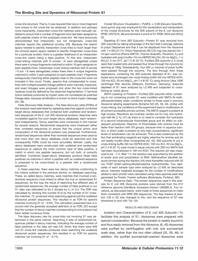
From these 72 spectra matches, 33 different linkages wereidentified, 23 corresponding to interprotein cross-linking and10 to intraprotein cross-linking (Table I). Interprotein cross-linking between K37 of S18 and K150 of S1 was, for example,identified by means of the Orbitrap MS/MS spectrum dis-played in Fig. 3. This spectrum contains ions resulting fromcleavage of two different peptide chains; peak assignmentsshown in red correspond to fragmentation of the S18 peptide,NYITESGKIVPSR, whereas those in blue correspond to frag-mentation of the S1 peptide, DTLHLEGKELEFK. In this spec-trum, 14 different fragment assignments could be attributedto the S18 peptide sequence, and 15 different fragment as-signments could be attributed to the S1 peptide sequence.The sequences of both peptides were thereby matched withvery high confidence, particularly because all fragmentmasses were required to match theoretical masses to within10 ppm (further details on spectra matching can be foundunder “Experimental Procedures”). That so many high confi-dence interprotein cross-links, such as this, were identified isnoteworthy. The density of cross-linking information obtainedhere for S1 appears to be comparable or more extensive thanthat reported in recent large-scale cross-linking analyses (34,56–59).
Cross-Linking of Protein S1—Interpretation of the identifiedprotein linkages has led to an insightful understanding of S1.S1 interprotein cross-links outnumbered S1 intraproteincross-links. Furthermore, the intraprotein cross-links thatwere detected predominately corresponded to cross-linkingwithin a domain or between domains adjacent in sequence,such as between domains 2 and 3. This is consistent with S1being an elongated protein, whose amino groups are morelikely to cross-link to amines from other proteins on the sur-face of the ribosome than to other S1 amines. Interprotein
cross-linking of S1 further demonstrates this point. S1 wasfound to cross-link to 15 different residues spanning close to240 Å across the topography of the ribosome, from S19 nearthe subunit interface to S6 on the tip of the 30S platform to S3at the mRNA entry pore (supplemental Fig. S1). These obser-vations corroborate the conclusion from small angle x-rayscattering studies that S1 adopts a highly elongated structure(6).
These experiments most importantly provide explicit infor-mation about the localization of S1’s individual domains. Amodel for the architecture of these domains and their char-acterized functions is presented in Fig. 4. Fig. 5 displays thecrystal structure of the E. coli 30S subunit (16). As mentionedearlier, the first two domains of S1 are believed to be respon-sible for binding the ribosome (10, 11). The cross-linking of
TABLE ISummary of identified linkages involving S1. (A) Intraprotein and (B)
Interprotein identifications
A
Domain Domain Linkage Matches
1 2 K88-K150 22 3 K150-K196 12 3 K150-K247 152 4 K150-K279 52 C-term K150-K555 12 3 K158-K247 14 C-term K279-K555 15 6 K363-K464 15 5 K370-K411 26 6 K450-K504 1
Total 30
B
Protein 1 Protein 2 Linkage (Prot1-Prot2) Matches
S1 S2 K14-K10 1S1 S2 K88-K36 1S1 S2 K363-K114 1S1 S2 K363-K127 1S1 S3 K279-K107 1S1 S3 K279-K149 1S1 S6 K150-K106 1S1 S7 K117-K148 2S1 S7 K150-K148 7S1 S7 K158-K148 1S1 S7 K229-K148 1S1 S7 K279-K55 1S1 S7 K279-K148 5S1 S9 K279-K99 4S1 S18 K150-K29 1S1 S18 K150-K37 1S1 S18 K272-K29 1S1 S19 K279-K86 1S1 S21 K150-K4 1S1 S21 K150-K39 1S1 S21 K247-K4 1S1 S21 K279-K4 6S1 S21 K279-K39 1
Total 42
The Binding Site and Dynamics of Ribosomal Protein S1
1970 Molecular & Cellular Proteomics 11.12

these domains can therefore be used to elucidate the ribo-somal binding site of S1. As displayed in Fig. 5A, domain 1cross-linked to the N-terminal region of S2 through two dif-ferent lysine residues. Domain 2, meanwhile, cross-linked tosix different residues of S6, S7, S18, and S21 in an adjacentregion at the platform of the 30S subunit, �30Å away. Thebinding site of S1 can therefore be defined as the cleft be-tween the platform and body/head of the 30S subunit. Amodel for the interaction between domains 1 and 2 of S1 and
the 30S subunit that is consistent with the observed cross-linking pattern is shown in Fig. 5B. More detailed structuralmodeling requires a high resolution structure for the N-termi-nal ribosome binding domains of S1. Binding of S1 to thisregion of the 30S subunit is likely to be imperative for its abilityto facilitate the initiation of translation. During initiation, mRNAbinds along a groove in the 30S subunit, such that its 5� endextends out through this cleft (60–62). This region of the 30Ssubunit contains the 3� end of 16S rRNA with a sequence
FIG. 3. Orbitrap MS/MS of an interprotein cross-link between K37 of S18 and K150 of S1. Peak assignments corresponding to the S18peptide are shown in red and those corresponding to the S1 peptide are shown in blue. Peaks marked with asterisks correspond to neutrallosses of other assignments.
FIG. 4. Domain architecture of protein S1. The NMR structures of domains 4 and 6 are shown (3).
FIG. 5. Cross-linking of the N-terminal ribosome binding region of S1 to the 30S ribosomal subunit. A, Cross-linking of domains 1 and2 involves residues localized near the 3� end of the 16S rRNA. Residues on the 30S subunit (PDB: 2AVY)(16) that cross-linked to S1 are markedwith red circles. The triangle on S6 shows the approximate position of a residue not present in the crystal structure. The S1 residues involvedin these cross-links are highlighted in red. B, A binding site for the N-terminal ribosome binding region of S1 consistent with this cross-linkinganalysis.
The Binding Site and Dynamics of Ribosomal Protein S1
Molecular & Cellular Proteomics 11.12 1971
complementary to the S.D. sequence found in many mRNA(2, 16). Our results show that S1 is also present in this loca-tion, explaining how S1, like the anti-S.D. sequence, is able tobind the 5� UTR of mRNA and facilitate initiation (4, 5).
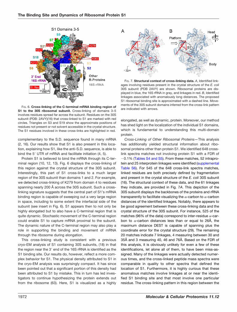
Protein S1 is believed to bind the mRNA through its C-ter-minal region (10, 12, 13). Fig. 6 displays the cross-linking ofthis region against the crystal structure of the 30S subunit.Interestingly, this part of S1 cross-links to a much largerregion of the 30S subunit than domains 1 and 2. For example,we detected cross-linking of K279 from domain 4 to residuesspanning nearly 200 Å across the 30S subunit. Such a cross-linking signature suggests that the central part of S1’s mRNAbinding region is capable of sweeping out a very large regionin space, including to some extent the interfacial side of thesubunit (see insert in Fig. 6). S1 appears then to not only behighly elongated but to also have a C-terminal region that isquite dynamic. Stochastic movement of the C-terminal regioncould enable S1 to capture mRNA proximal to the subunit.The dynamic nature of the C-terminal region may also play arole in supporting the binding and movement of mRNAthrough the ribosome during elongation.
This cross-linking study is consistent with a previouscryo-EM analysis of S1 containing 30S subunits, (18) in thatthe region near the 3� end of the 16S rRNA is identified as theS1 binding site. Our results do, however, reflect a more com-plex behavior for S1. The physical density attributed to S1 inthe cryo-EM analysis was surprisingly compact. It has sincebeen pointed out that a significant portion of this density hadbeen attributed to S1 by mistake. This in turn has led inves-tigators to continue hypothesizing the protein extends outfrom the ribosome (63). Here, S1 is visualized as a highly
elongated, as well as dynamic, protein. Moreover, our methodhas shed light on the localization of the individual S1 domains,which is fundamental to understanding this multi-domainprotein.
Cross-Linking of Other Ribosomal Proteins—This analysishas additionally yielded structural information about ribo-somal proteins other than protein S1. We identified 648 cross-link spectra matches not involving protein S1 with a FDR of�0.1% (Tables S4 and S5). From these matches, 52 intrapro-tein and 25 interprotein linkages were identified (supplementalTable S6). For 545 of the 648 cross-link spectra matches,linked residues are both precisely defined by fragmentationand present in the crystal structure of the E. coli 30S subunit(16). The structural context of these data, and the 61 linkagesthey indicate, are provided in Fig. 7A. This depiction of the30S subunit displays the backbones of the proteins and rRNAtransparently to facilitate visualizing the �-carbon to �-carbondistances of the identified linkages. Notably, there appears tobe good agreement between these cross-linking data and thecrystal structure of the 30S subunit. For instance, 525 of thematches (96% of the data) correspond to inter-residue �-car-bon to �-carbon distances less than or equal to 26Å, themaximum distance DEST is capable of spanning plus thecoordinate error for the crystal structure (29). The remaining20 matches indicate 7 linkages, 4 measuring between 30 and35Å and 3 measuring 40, 46 and 76Å. Based on the FDR ofthis analysis, it is obviously unlikely for even a few of theseidentifications, let alone all of them, to have been miss-as-signed. Many of the linkages were actually detected numer-ous times, and the cross-linked peptide mass spectra werecomparable in quality to other spectra that defined thelocation of S1. Furthermore, it is highly curious that theseanomalous matches involve linkages at or near the identi-fied S1 binding site and that most involve one particularresidue. The cross-linking pattern in this region between the
FIG. 6. Cross-linking of the C-terminal mRNA binding region ofS1 to the 30S ribosomal subunit. Cross-linking of domains 3–5involves residues spread far across the subunit. Residues on the 30Ssubunit (PDB: 2AVY)(16) that cross-linked to S1 are marked with redcircles. Triangles on S3 and S19 show the approximate positions ofresidues not present or not solvent accessible in the crystal structure.The S1 residues involved in these cross-links are highlighted in red.
FIG. 7. Structural context of cross-linking data. A, Identified link-ages involving residues present in the crystal structure of the E. coli30S subunit (PDB 2AVY) are shown. Ribosomal proteins are dis-played in blue, the 16S rRNA in gray, and linkages in red. B, Identifiedlinkages associated with anomalously long distances. The proposedS1 ribosomal binding site is approximated with a dashed line. Move-ments of the 30S subunit domains inferred from the cross-link patternare indicated with arrows.
The Binding Site and Dynamics of Ribosomal Protein S1
1972 Molecular & Cellular Proteomics 11.12
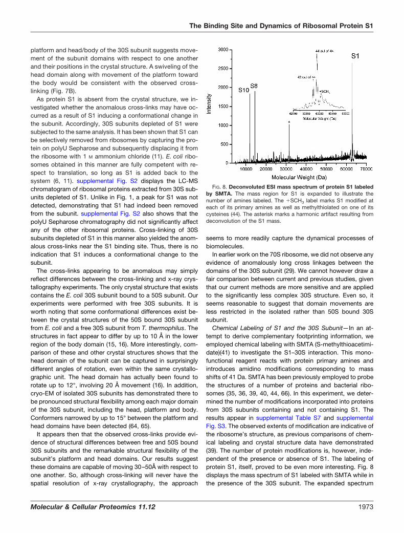
platform and head/body of the 30S subunit suggests move-ment of the subunit domains with respect to one anotherand their positions in the crystal structure. A swiveling of thehead domain along with movement of the platform towardthe body would be consistent with the observed cross-linking (Fig. 7B).
As protein S1 is absent from the crystal structure, we in-vestigated whether the anomalous cross-links may have oc-curred as a result of S1 inducing a conformational change inthe subunit. Accordingly, 30S subunits depleted of S1 weresubjected to the same analysis. It has been shown that S1 canbe selectively removed from ribosomes by capturing the pro-tein on polyU Sepharose and subsequently displacing it fromthe ribosome with 1 M ammonium chloride (11). E. coli ribo-somes obtained in this manner are fully competent with re-spect to translation, so long as S1 is added back to thesystem (6, 11). supplemental Fig. S2 displays the LC-MSchromatogram of ribosomal proteins extracted from 30S sub-units depleted of S1. Unlike in Fig. 1, a peak for S1 was notdetected, demonstrating that S1 had indeed been removedfrom the subunit. supplemental Fig. S2 also shows that thepolyU Sepharose chromatography did not significantly affectany of the other ribosomal proteins. Cross-linking of 30Ssubunits depleted of S1 in this manner also yielded the anom-alous cross-links near the S1 binding site. Thus, there is noindication that S1 induces a conformational change to thesubunit.
The cross-links appearing to be anomalous may simplyreflect differences between the cross-linking and x-ray crys-tallography experiments. The only crystal structure that existscontains the E. coli 30S subunit bound to a 50S subunit. Ourexperiments were performed with free 30S subunits. It isworth noting that some conformational differences exist be-tween the crystal structures of the 50S bound 30S subunitfrom E. coli and a free 30S subunit from T. thermophilus. Thestructures in fact appear to differ by up to 10 Å in the lowerregion of the body domain (15, 16). More interestingly, com-parison of these and other crystal structures shows that thehead domain of the subunit can be captured in surprisinglydifferent angles of rotation, even within the same crystallo-graphic unit. The head domain has actually been found torotate up to 12°, involving 20 Å movement (16). In addition,cryo-EM of isolated 30S subunits has demonstrated there tobe pronounced structural flexibility among each major domainof the 30S subunit, including the head, platform and body.Conformers narrowed by up to 15° between the platform andhead domains have been detected (64, 65).
It appears then that the observed cross-links provide evi-dence of structural differences between free and 50S bound30S subunits and the remarkable structural flexibility of thesubunit’s platform and head domains. Our results suggestthese domains are capable of moving 30–50Å with respect toone another. So, although cross-linking will never have thespatial resolution of x-ray crystallography, the approach
seems to more readily capture the dynamical processes ofbiomolecules.
In earlier work on the 70S ribosome, we did not observe anyevidence of anomalously long cross linkages between thedomains of the 30S subunit (29). We cannot however draw afair comparison between current and previous studies, giventhat our current methods are more sensitive and are appliedto the significantly less complex 30S structure. Even so, itseems reasonable to suggest that domain movements areless restricted in the isolated rather than 50S bound 30Ssubunit.
Chemical Labeling of S1 and the 30S Subunit—In an at-tempt to derive complementary footprinting information, weemployed chemical labeling with SMTA (S-methylthioacetimi-date)(41) to investigate the S1–30S interaction. This mono-functional reagent reacts with protein primary amines andintroduces amidino modifications corresponding to massshifts of 41 Da. SMTA has been previously employed to probethe structures of a number of proteins and bacterial ribo-somes (35, 36, 39, 40, 44, 66). In this experiment, we deter-mined the number of modifications incorporated into proteinsfrom 30S subunits containing and not containing S1. Theresults appear in supplemental Table S7 and supplementalFig. S3. The observed extents of modification are indicative ofthe ribosome’s structure, as previous comparisons of chem-ical labeling and crystal structure data have demonstrated(39). The number of protein modifications is, however, inde-pendent of the presence or absence of S1. The labeling ofprotein S1, itself, proved to be even more interesting. Fig. 8displays the mass spectrum of S1 labeled with SMTA while inthe presence of the 30S subunit. The expanded spectrum
FIG. 8. Deconvoluted ESI mass spectrum of protein S1 labeledby SMTA. The mass region for S1 is expanded to illustrate thenumber of amines labeled. The �SCH3 label marks S1 modified ateach of its primary amines as well as methylthiolated on one of itscysteines (44). The asterisk marks a harmonic artifact resulting fromdeconvolution of the S1 mass.
The Binding Site and Dynamics of Ribosomal Protein S1
Molecular & Cellular Proteomics 11.12 1973
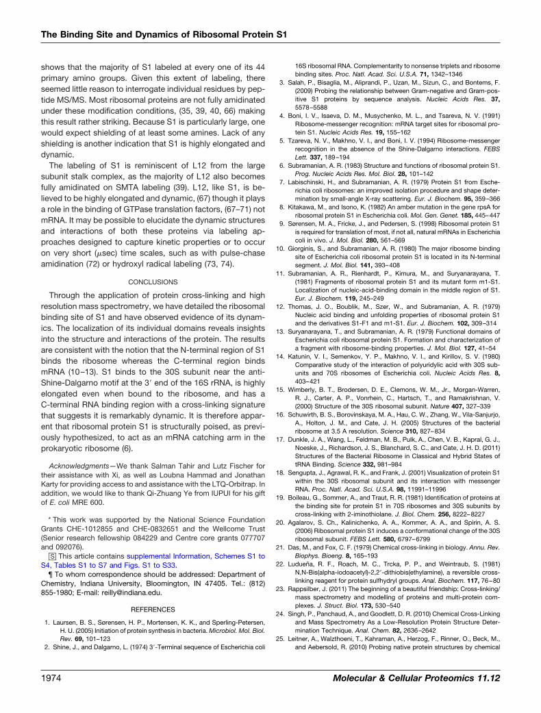
shows that the majority of S1 labeled at every one of its 44primary amino groups. Given this extent of labeling, thereseemed little reason to interrogate individual residues by pep-tide MS/MS. Most ribosomal proteins are not fully amidinatedunder these modification conditions, (35, 39, 40, 66) makingthis result rather striking. Because S1 is particularly large, onewould expect shielding of at least some amines. Lack of anyshielding is another indication that S1 is highly elongated anddynamic.
The labeling of S1 is reminiscent of L12 from the largesubunit stalk complex, as the majority of L12 also becomesfully amidinated on SMTA labeling (39). L12, like S1, is be-lieved to be highly elongated and dynamic, (67) though it playsa role in the binding of GTPase translation factors, (67–71) notmRNA. It may be possible to elucidate the dynamic structuresand interactions of both these proteins via labeling ap-proaches designed to capture kinetic properties or to occuron very short (�sec) time scales, such as with pulse-chaseamidination (72) or hydroxyl radical labeling (73, 74).
CONCLUSIONS
Through the application of protein cross-linking and highresolution mass spectrometry, we have detailed the ribosomalbinding site of S1 and have observed evidence of its dynam-ics. The localization of its individual domains reveals insightsinto the structure and interactions of the protein. The resultsare consistent with the notion that the N-terminal region of S1binds the ribosome whereas the C-terminal region bindsmRNA (10–13). S1 binds to the 30S subunit near the anti-Shine-Dalgarno motif at the 3� end of the 16S rRNA, is highlyelongated even when bound to the ribosome, and has aC-terminal RNA binding region with a cross-linking signaturethat suggests it is remarkably dynamic. It is therefore appar-ent that ribosomal protein S1 is structurally poised, as previ-ously hypothesized, to act as an mRNA catching arm in theprokaryotic ribosome (6).
Acknowledgments—We thank Salman Tahir and Lutz Fischer fortheir assistance with Xi, as well as Loubna Hammad and JonathanKarty for providing access to and assistance with the LTQ-Orbitrap. Inaddition, we would like to thank Qi-Zhuang Ye from IUPUI for his giftof E. coli MRE 600.
* This work was supported by the National Science FoundationGrants CHE-1012855 and CHE-0832651 and the Wellcome Trust(Senior research fellowship 084229 and Centre core grants 077707and 092076).
□S This article contains supplemental Information, Schemes S1 toS4, Tables S1 to S7 and Figs. S1 to S33.
¶ To whom correspondence should be addressed: Department ofChemistry, Indiana University, Bloomington, IN 47405. Tel.: (812)855-1980; E-mail: [email protected].
REFERENCES
1. Laursen, B. S., Sørensen, H. P., Mortensen, K. K., and Sperling-Petersen,H. U. (2005) Initiation of protein synthesis in bacteria. Microbiol. Mol. Biol.Rev. 69, 101–123
2. Shine, J., and Dalgarno, L. (1974) 3�-Terminal sequence of Escherichia coli
16S ribosomal RNA. Complementarity to nonsense triplets and ribosomebinding sites. Proc. Natl. Acad. Sci. U.S.A. 71, 1342–1346
3. Salah, P., Bisaglia, M., Aliprandi, P., Uzan, M., Sizun, C., and Bontems, F.(2009) Probing the relationship between Gram-negative and Gram-pos-itive S1 proteins by sequence analysis. Nucleic Acids Res. 37,5578–5588
4. Boni, I. V., Isaeva, D. M., Musychenko, M. L., and Tsareva, N. V. (1991)Ribosome-messenger recognition: mRNA target sites for ribosomal pro-tein S1. Nucleic Acids Res. 19, 155–162
5. Tzareva, N. V., Makhno, V. I., and Boni, I. V. (1994) Ribosome-messengerrecognition in the absence of the Shine-Dalgarno interactions. FEBSLett. 337, 189–194
6. Subramanian, A. R. (1983) Structure and functions of ribosomal protein S1.Prog. Nucleic Acids Res. Mol. Biol. 28, 101–142
7. Labischinski, H., and Subramanian, A. R. (1979) Protein S1 from Esche-richia coli ribosomes: an improved isolation procedure and shape deter-mination by small-angle X-ray scattering. Eur. J. Biochem. 95, 359–366
8. Kitakawa, M., and Isono, K. (1982) An amber mutation in the gene rpsA forribosomal protein S1 in Escherichia coli. Mol. Gen. Genet. 185, 445–447
9. Sørensen, M. A., Fricke, J., and Pedersen, S. (1998) Ribosomal protein S1is required for translation of most, if not all, natural mRNAs in Escherichiacoli in vivo. J. Mol. Biol. 280, 561–569
10. Giorginis, S., and Subramanian, A. R. (1980) The major ribosome bindingsite of Escherichia coli ribosomal protein S1 is located in its N-terminalsegment. J. Mol. Biol. 141, 393–408
11. Subramanian, A. R., Rienhardt, P., Kimura, M., and Suryanarayana, T.(1981) Fragments of ribosomal protein S1 and its mutant form m1-S1.Localization of nucleic-acid-binding domain in the middle region of S1.Eur. J. Biochem. 119, 245–249
12. Thomas, J. O., Boublik, M., Szer, W., and Subramanian, A. R. (1979)Nucleic acid binding and unfolding properties of ribosomal protein S1and the derivatives S1-F1 and m1-S1. Eur. J. Biochem. 102, 309–314
13. Suryanarayana, T., and Subramanian, A. R. (1979) Functional domains ofEscherichia coli ribosomal protein S1. Formation and characterization ofa fragment with ribosome-binding properties. J. Mol. Biol. 127, 41–54
14. Katunin, V. I., Semenkov, Y. P., Makhno, V. I., and Kirillov, S. V. (1980)Comparative study of the interaction of polyuridylic acid with 30S sub-units and 70S ribosomes of Escherichia coli. Nucleic Acids Res. 8,403–421
15. Wimberly, B. T., Brodersen, D. E., Clemons, W. M., Jr., Morgan-Warren,R. J., Carter, A. P., Vonrhein, C., Hartsch, T., and Ramakrishnan, V.(2000) Structure of the 30S ribosomal subunit. Nature 407, 327–339
16. Schuwirth, B. S., Borovinskaya, M. A., Hau, C. W., Zhang, W., Vila-Sanjurjo,A., Holton, J. M., and Cate, J. H. (2005) Structures of the bacterialribosome at 3.5 A resolution. Science 310, 827–834
17. Dunkle, J. A., Wang, L., Feldman, M. B., Pulk, A., Chen, V. B., Kapral, G. J.,Noeske, J., Richardson, J. S., Blanchard, S. C., and Cate, J. H. D. (2011)Structures of the Bacterial Ribosome in Classical and Hybrid States oftRNA Binding. Science 332, 981–984
18. Sengupta, J., Agrawal, R. K., and Frank, J. (2001) Visualization of protein S1within the 30S ribosomal subunit and its interaction with messengerRNA. Proc. Natl. Acad. Sci. U.S.A. 98, 11991–11996
19. Boileau, G., Sommer, A., and Traut, R. R. (1981) Identification of proteins atthe binding site for protein S1 in 70S ribosomes and 30S subunits bycross-linking with 2-iminothiolane. J. Biol. Chem. 256, 8222–8227
20. Agalarov, S. Ch., Kalinichenko, A. A., Kommer, A. A., and Spirin, A. S.(2006) Ribosomal protein S1 induces a conformational change of the 30Sribosomal subunit. FEBS Lett. 580, 6797–6799
21. Das, M., and Fox, C. F. (1979) Chemical cross-linking in biology. Annu. Rev.Biophys. Bioeng. 8, 165–193
22. Luduena, R. F., Roach, M. C., Trcka, P. P., and Weintraub, S. (1981)N,N-Bis(alpha-iodoacetyl)-2,2�-dithiobis(ethylamine), a reversible cross-linking reagent for protein sulfhydryl groups. Anal. Biochem. 117, 76–80
23. Rappsilber, J. (2011) The beginning of a beautiful friendship: Cross-linking/mass spectrometry and modelling of proteins and multi-protein com-plexes. J. Struct. Biol. 173, 530–540
24. Singh, P., Panchaud, A., and Goodlett, D. R. (2010) Chemical Cross-Linkingand Mass Spectrometry As a Low-Resolution Protein Structure Deter-mination Technique. Anal. Chem. 82, 2636–2642
25. Leitner, A., Walzthoeni, T., Kahraman, A., Herzog, F., Rinner, O., Beck, M.,and Aebersold, R. (2010) Probing native protein structures by chemical
The Binding Site and Dynamics of Ribosomal Protein S1
1974 Molecular & Cellular Proteomics 11.12

cross-linking, mass spectrometry, and bioinformatics. Mol. Cell. Pro-teomics 9, 1634–1649
26. Sinz, A. (2006) Chemical cross-linking and mass spectrometry to mapthree-dimensional protein structures and protein-protein interactions.Mass Spectrom. Rev. 25, 663–682
27. Petrotchenko, E. V., and Borchers, C. H. (2010) Crosslinking combined withmass spectrometry for structural proteomics. Mass Spectrom. Rev. 29,862–876
28. Rasmussen M. I., Refsgaard J. C., Peng, L., Houen, G., and Hojrup, P.(2011) CrossWork: software-assisted identification of cross-linked pep-tides. J Proteomics 74, 1871–1883
29. Lauber, M. A., and Reilly, J. P. (2011) Structural Analysis of a ProkaryoticRibosome Using a Novel Amidinating Crosslinker and Mass Spectrom-etry. J. Proteome Res. 10, 3604–3616
30. Lauber, M. A., and Reilly, J. P. (2010) Novel Amidinating Cross-Linker forFacilitating Analyses of Protein Structures and Interactions. Anal. Chem.82, 7736–7743
31. Cammack, K. A., and Wade, H. E. (1965) The sedimentation behaviour ofribonuclease-active and -inactive ribosomes from bacteria. Biochem. J.96, 671–680
32. Spedding, G. (1990). in Ribosomes and Protein Synthesis: A Pratical Ap-proach (Rickwood, D., and Hames, B. D. eds.), Oxford Press, New York.pp 1–29
33. Karty, J. A., Running, W. E., and Reilly, J. P. (2007) Two dimensional liquidphase separations of proteins using online fractionation and concentra-tion between chromatographic dimensions. J. Chromatogr. B. 847,103–113
34. Chen, Z. A., Jawhari, A., Fischer, L., Buchen, C., Tahir, S., Kamenski, T.,Rasmussen, M., Lariviere, L., Bukowski-Wills, J. C., Nilges, M., Cramer,P., and Rappsilber, J. (2010) Architecture of the RNA polymerase II-TFIIFcomplex revealed by cross-linking and mass spectrometry. EMBO J. 29,717–726
35. Running, W. E., and Reilly, J. P. (2009) Ribosomal Proteins of Deinococcusradiodurans: Their Solvent Accessibility and Reactivity. J. Proteome Res.8, 1228–1246
36. Liu, X., Broshears, W. C., and Reilly, J. P. (2007) Probing the structure andactivity of trypsin with amidination. Anal. Biochem. 367, 13–19
37. Choi, H., and Nesvizhskii, A. I. (2008) False Discovery Rates and RelatedStatistical Concepts in Mass Spectrometry-Based Proteomics. J. Pro-teome Res. 7, 47–50
38. DeLano, W. L. (2002) The PyMOL Molecular Graphics System. DeLanoScientific, Palo Alto, CA, U.S.A.
39. Liu, X., and Reilly, J. P. (2009) Correlating the Chemical Modification ofEscherichia coli Ribosomal Proteins with Crystal Structure Data. J. Pro-teome Res. 8, 4466–4478
40. Beardsley, R. L., Running, W. E., and Reilly, J. P. (2006) Probing theStructure of the Caulobacter crescentus Ribosome with Chemical La-beling and Mass Spectrometry. J. Proteome Res. 5, 2935–2946
41. Thumm, M., Hoenes, J., and Pfleiderer, G. (1987) S-Methylthioacetimidateis a new reagent for the amidination of proteins at low pH. Biochim.Biophys. Acta 923, 263–267
42. Hunter, M. J., and Ludwig, M. L. (1962) The reaction of imidoesters withproteins and related small molecules. J. Am. Chem. Soc. 84, 3491–3504
43. Hermanson, G. T. (2008) Bioconjugate Techniques, 2nd ed., AcademicPress, San Diego
44. Janecki, D. J., Beardsley, R. L., and Reilly, J. P. (2005) Probing ProteinTertiary Structure with Amidination. Anal. Chem. 77, 7274–7281
45. Draper, D. E., and Von Hippel, P. H. (1979) Interaction of Escherichia coliribosomal protein S1 with ribosomes. Proc. Natl. Acad. Sci. U.S.A. 76,1040–1044
46. Arnold, R. J., and Reilly, J. P. (1999) Observation of Escherichia coliribosomal proteins and their posttranslational modifications by massspectrometry. Anal. Biochem. 269, 105–112
47. Kowalak, J. A., and Walsh, K. A. (1996) �-Methylthio-aspartic acid: identi-fication of a novel posttranslational modification in ribosomal protein S12from Escherichia coli. Protein Sci. 5, 1625–1632
48. Kamp, R., and Wittmann-Liebold, B. (1980) Primary structure of protein S11from Escherichia coli ribosomes. FEBS Lett. 121, 117–122
49. Wittmann-Liebold, B., and Greuer, B. (1978) The primary structure of pro-tein S5 from the small subunit of the Escherichia coli ribosome. FEBSLett. 95, 91–98
50. Hitz, H., Schafer, D., and Wittmann-Liebold, B. (1977) Determination of thecomplete amino-acid sequence of protein S6 from the wild-type and amutant of Escherichia coli. Eur. J. Biochem. 75, 497–512
51. Yaguchi, M. (1975) Primary structure of protein S18 from the small Esch-erichia coli ribosomal subunit. FEBS Lett. 59, 217–220
52. Strader, M. B., Costantino, N., Elkins, C. A., Chen, C. Y., Patel, I., Makusky,A. J., Choy, J. S., Court, D. L., Markey, S. P., and Kowalak, J. A. (2011)A proteomic and transcriptomic approach reveals new insight into beta-methylthiolation of Escherichia coli ribosomal protein S12. Mol. Cell.Proteomics 10, M110.005199
53. Apweiler, R., Martin, M. J., O’Donovan, C., Magrane, M., Alam-Faruque, Y.,Antunes, R., Casanova, E. B., Bely, B., Bingley, M., Bower, L., Tursteinas,B., Chan, W. M., Chavali, G., Da Silva, A., Dimmer, E., Eberhardt, R.,Fazzini, F., Fedotov, A., Garavelli, J., Garcia Castro, L., Gardner, M.,Hieta, R., Huntley, R., Jacobsen, J., Legge, D., Liu, W., Luo, J., Orchard,S., Patient, S., Pichler, K., Poggioli, D., Pontikos, N., Pundir, S., Rosanoff,S., Sawford, T., Sehra, H., Turner, E., Wardell, T., Watkins, X., Corbett,M., Donnelly, M., van Rensburg, P., Goujon, M., McWilliam, H., Lopez,R., Xenarios, i., Bougueleret, L., Bridge, A., Poux, S., Redaschi, N.,Argoud-Puy, G., Auchincloss, A., Axelsen, K., Baratin, D., Blatter, M.-C.,Boeckmann, B., Bolleman, j., Bollondi, L., Boutet, E., Braconi Quitaje, S.,Breuza, L., deCastro, E., Cerutti, L., Coudert, E., Cuche, B., Cusin, I.,Doche, M., Dornevil, D., Duvaud, S., Estreicher, A., Famiglietti, L., Feuer-mann, M., Gehant, S., Ferro, S., Gasteiger, E., Gerritsen, V., Gos, A.,Gruaz-Gumowski, N., Hinz, U., Hulo, C., Hulo, N., James, J., Jimenez, S.,Jungo, F., Kappler, T., Keller, G., Lara, V., Lemercier, P., Lieberherr, D.,Martin, X., Masson, P., Mooinat, M., Morgat, A., Paesano, S., Pedruzzi,I., Pilbout, S., Pozzato, M., Pruess, M., Rivoire, C., Roechert, B., Schnei-der, M., Sigrist, C., Sonesson, K., Staehli, S., Stanley, E., Stutz, A.,Sundaram, S., Tognolli, M., Verbregue, L., Veuthey, A.-L., Wu, C. H.,Arighi, C. N., Arminski, L., Barker, W. C., Chen, C., Chen, Y., Dubey, P.,Huang, H., Kukreja, A., Laiho, K., Mazumder, R., McGarvey, P., Natale,D. A., Natarajan, T. G., Roberts, N. V., Suzek, B. E., Vinayaka, C. R.,Wang, Q., Wang, Y., Yeh, L.-S., and Zhang, J. (2012) Reorganizing theprotein space at the Universal Protein Resource (UniProt). Nucleic AcidsRes. 40, D71–D75
54. Touchon, M., Hoede, C., Tenaillon, O., Barbe, V., Baeriswyl, S., Bidet, P.,Bingen, E., Bonacorsi, S., Bouchier, C., Bouvet, O., Calteau, A., Chia-pello, H., Clermont, O., Cruveiller, S., Danchin, A., Diard, M., Dossat, C.,Karoui, M. E., Frapy, E., Garry, L., Ghigo, J. M., Gilles, A. M., Johnson, J.,Le Bouguenec, C., Lescat, M., Mangenot, S., Martinez-Jehanne, V.,Matic, I., Nassif, X., Oztas, S., Petit, M. A., Pichon, C., Rouy, Z., Ruf, C.S, Schneider, D., Tourret, J., Vacherie, B., Vallenet, D., Medigue, C.,Rocha, E. P., and Denamur, E. (2009) Organised genome dynamics in theEscherichia coli species results in highly diverse adaptive paths. PLoSGenet. 5, e1000344
55. Rasko, D. A., Rosovitz, M. J., Myers, G. S., Mongodin, E. F., Fricke, W. F.,Gajer, P., Crabtree, J., Sebaihia, M., Thomson, N. R., Chaudhuri, R.,Henderson, I. R., Sperandio, V., and Ravel, J. (2008) The pangenomestructure of Escherichia coli: comparative genomic analysis of E. colicommensal and pathogenic isolates. J. Bacteriol. 190, 6881–6893
56. Leitner, A., Reischl, R., Walzthoeni, T., Herzog, F., Bohn, S., Forster, F., andAebersold, R. (2012) Expanding the Chemical Cross-Linking Toolbox bythe Use of Multiple Proteases and Enrichment by Size Exclusion Chro-matography. Mol. Cell. Proteomics 11, M111.014126
57. Kao, A., Chiu, C.-I., Vellucci, D., Yang, Y., Patel, V. R., Guan, S., Randall, A.,Bald, P., Rychnovsky, S. D., and Huang, L. (2011) Development of anovel cross-linking strategy for fast and accurate identification of cross-linked peptides of protein complexes. Mol. Cell. Proteomics 10,M110.002212
58. Trnka, M. J., and Burlingame, A. L. (2010) Topographic studies of theGroEL-GroES chaperonin complex by chemical cross-linking using di-formyl ethynylbenzene. The power of high resolution electron transferdissociation for determination of both peptide sequences and their at-tachment sites. Mol. Cell. Proteomics 9, 2306–2317
59. Buncherd, H., Nessen, M. A., Nouse, N., Stelder, S. K., Roseboom, W.,Dekker, H. L., Arents, J. C., Smeenk, L. E., Wanner, M. J., van Maarse-veen, J. H., Yang, X., Lewis, P. J., de Koning, L. J., de Koster, C. G., andde Jong, L. (2012) Selective enrichment and identification of cross-linkedpeptides to study 3-D structures of protein complexes by mass spec-trometry. J. Proteomics 75, 2205–2215
The Binding Site and Dynamics of Ribosomal Protein S1
Molecular & Cellular Proteomics 11.12 1975
60. Selmer, M., Dunham, C. M., Murphy, F. V.,4th, Weixlbaumer, A., Petry, S.,Kelley, A. C., Weir, J. R., and Ramakrishnan, V. (2006) Structure of the70S ribosome complexed with mRNA and tRNA. Science 313,1935–1942
61. Yusupova, G. Z., Yusupov, M. M., Cate, J. H., and Noller, H. F. (2001) Thepath of messenger RNA through the ribosome. Cell 106, 233–241
62. Yusupova, G., Jenner, L., Rees, B., Moras, D., and Yusupov, M. (2006)Structural basis for messenger RNA movement on the ribosome. Nature444, 391–394
63. Dunkle, J. A., and Cate, J. H. D. (2011) The Packing of Ribosomes inCrystals and Polysomes. in Ribosomes: Structure, Function, and Dy-namics (Marina Rodnina, Wolfgang Wintermeyer, and Green, R. eds.),Springer-Verlag/Wien, New York. pp 65–73
64. Gabashvili, I. S., Agrawal, R. K., Grassucci, R., and Frank, J. (1999) Struc-ture and structural variations of the Escherichia coli 30 S ribosomalsubunit as revealed by three-dimensional cryo-electron microscopy. J.Mol. Biol. 286, 1285–1291
65. Lata, K. R., Agrawal, R. K., Penczek, P., Grassucci, R., Zhu, J., and Frank,J. (1996) Three-dimensional reconstruction of the Escherichia coli 30 Sribosomal subunit in ice. J. Mol. Biol. 262, 43–52
66. Lauber, M. A., Running, W. E., and Reilly, J. P. (2009) B. subtilis RibosomalProteins: Structural Homology and Post-Translational Modifications. J.Proteome Res. 8, 4193–4206
67. Diaconu, M., Kothe, U., Schlunzen, F., Fischer, N., Harms, J. M., Tonev-itsky, A. G., Stark, H., Rodnina, M. V., and Wahl, M. C. (2005) Structural
basis for the function of the ribosomal L7/12 stalk in factor binding andGTPase activation. Cell 121, 991–1004
68. Savelsbergh, A., Mohr, D., Kothe, U., Wintermeyer, W., and Rodnina, M. V.(2005) Control of phosphate release from elongation factor G by ribo-somal protein L7/12. EMBO J. 24, 4316–4323
69. Gao, Y. G., Selmer, M., Dunham, C. M., Weixlbaumer, A., Kelley, A. C., andRamakrishnan, V. (2009) The Structure of the Ribosome with ElongationFactor G Trapped in the Posttranslocational State. Science 326,694–699
70. Helgstrand, M., Mandava, C. S., Mulder, F. A., Liljas, A., Sanyal, S., andAkke, M. (2007) The Ribosomal Stalk Binds to Translation Factors IF2,EF-Tu, EF-G and RF3 via a Conserved Region of the L12 C-terminalDomain. J. Mol. Biol. 365, 468–479
71. Mohr, D., Wintermeyer, W., and Rodnina, M. V. (2002) GTPase Activation ofElongation Factors Tu and G on the Ribosome. Biochemistry 41,12520–12528
72. Chang, F. M., Lauber, M. A., Running, W. E., Reilly, J. P., and Giedroc, D. P.(2011) Ratiometric Pulse-Chase Amidination Mass Spectrometry as aProbe of Biomolecular Complex Formation. Anal. Chem. 83, 9092–9099
73. Stocks, B. B., and Konermann, L. (2009) Structural Characterization ofShort-Lived Protein Unfolding Intermediates by Laser-Induced OxidativeLabeling and Mass Spectrometry. Anal. Chem. 81, 20–27
74. Konermann, L., Stocks, B. B., Pan, Y., and Tong, X. (2010) Mass Spec-trometry Combined with Oxidative Labeling for Exploring Protein Struc-ture and Folding. Mass Spectrom. Rev. 29, 651–667
The Binding Site and Dynamics of Ribosomal Protein S1
1976 Molecular & Cellular Proteomics 11.12