Embed Size (px)
Citation preview
Rehabilitation of Mandibulectomy Patients is still a Challenge for Prosthodontist
International Journal of Oral Care and Research, April-June 2017;5(2):1-4 1
IJOCR
Rehabilitation of Mandibulectomy Patients is still a Challenge for Prosthodontist: A Report of Two Cases1Sapna Rani, 2Arpit Sikri, 3Amrita Pawar, 4Ricky P Singh, 5Mahinder S Chauhan, 6Mahesh Verma
IJOCR
Case RepORt10.5005/jp-journals-00000-0000
dysfunctions in such patients are observed in swallowing, speech, control of saliva, mandibular movements, masti-cation, respiration, and psychic functioning.3 This type of dysfunction radically alters the prosthetic prognosis. One of the basic objectives in rehabilitation is to retrain the muscles for mandibular denture control and repeated occlusal approximation. Cantor and Curtis3 provided a postmandibulectomy classification for edentulous patient that can also be applied in partially edentulous arches. This article describes different treatment modalities for mandibulectomy patients from prosthodontic perspective.
CASE REPORTS
Case 1
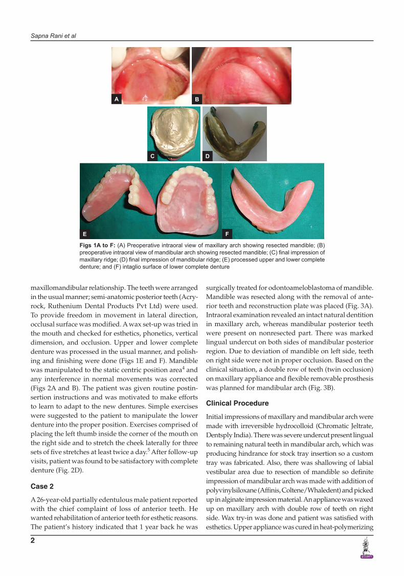
A 62-year-old completely edentulous male patient was referred to the Department of Prosthodontics for rehabilitation. His medical history revealed that he had undergone an extensive resection of mandible distal to the midline on right side due to squamous cell carcinoma 4 years back (Figs 1A and B). Deviation of mandible was there and due to loss of nerve supply to tongue, drooping of tongue was present (Fig. 2C). Based on clinical situ-ation, maxillary and mandibular complete denture was fabricated for the patient.
Clinical Procedure
Preliminary impressions of maxillary and mandibular arch were made with irreversible hydrocolloid (Chro-matic Jeltrate, Dentsply India) and primary casts were prepared. The custom trays were border-molded with low-fusing impression material (DPI Tracing stick, Mumbai) for maxillary and mandibular impression taking care to avoid overextension and also intaglio surface detail of mandibular arch on the resected area was recorded in low-fusing impression material. Final impressions of maxillary and mandibular arch were made with medium-body polyvinylsiloxane (Affinis, Coltene/Whaledent) (Figs 1C and D). This impression material was chosen to produce minimal tissue displacement. Stabilized record bases were made on definitive cast and wax rims were adjusted until a tentative occlusal vertical dimension was established.
The patient was asked to move his mandible as far as possible to the untreated side to record a functional
1Reader, 2-5Senior Lecturer, 6Head1,3,5Department of Prosthodontics, ITS Dental College, Murad Nagar, Uttar Pradesh, India2Department of Prosthodontics, Sudha Rustagi College of Dental Sciences & Research, Faridabad, Haryana, India4Department of Community Dentistry, ITS Dental College, Murad Nagar, Uttar Pradesh, India6Department of Prosthodontics, Maulana Azad Institute of Dental Sciences, New Delhi, India
Corresponding Author: Sapna Rani, Reader, Department of Prosthodontics, ITS Dental College, Murad Nagar, Uttar Pradesh, India, e-mail: [email protected]
ABSTRACT
Multidisciplinary approach is required for the management of malignant tumors in terms of the control of primary disease and rehabilitation following treatment. Role of a prosthodontist along with physical therapy is to regain the functional efficiency as well as near-normal lifestyle for the patient. This article describes the different treatment modalities for postmandibulectomy patients depending on clinical situations and challenges confronted by prosthodontist for rehabilitation of such patients. Case reports of different clinical situations with treatment modality of twin occlu-sion, modified conventional complete denture are described.
Keywords: Mandibulectomy, Prosthodontic rehabilitation, Twin occlusion.
How to cite this article: Rani S, Sikri A, Pawar A, Singh RP, Chauhan MS, Verma M. Rehabilitation of Mandibulectomy Patients is still a Challenge for Prosthodontist: A Report of Two Cases. Int J Oral Care Res 2017;5(2):1-4.
Source of support: Nil
Conflict of interest: None
INTRODUCTION
One of the most challenging and demanding maxillofacial endeavors is the construction of functional dentures for the edentulous patient who has undergone a mandibu-lar resection. Segmental resection of mandible results in physiological and esthetic problems. The most significant difficulty encountered is mandibular deviation toward the defective side.1,2 The greater the loss of tissues, greater will be the deviation of the mandible to the resected side, thus compromising the prognosis of the prosthetic reha-bilitation to a greater extent. Apart from deviation, other
Sapna Rani et al
2
maxillomandibular relationship. The teeth were arranged in the usual manner; semi-anatomic posterior teeth (Acry-rock, Ruthenium Dental Products Pvt Ltd) were used. To provide freedom in movement in lateral direction, occlusal surface was modified. A wax set-up was tried in the mouth and checked for esthetics, phonetics, vertical dimension, and occlusion. Upper and lower complete denture was processed in the usual manner, and polish-ing and finishing were done (Figs 1E and F). Mandible was manipulated to the static centric position area4 and any interference in normal movements was corrected (Figs 2A and B). The patient was given routine postin-sertion instructions and was motivated to make efforts to learn to adapt to the new dentures. Simple exercises were suggested to the patient to manipulate the lower denture into the proper position. Exercises comprised of placing the left thumb inside the corner of the mouth on the right side and to stretch the cheek laterally for three sets of five stretches at least twice a day.5 After follow-up visits, patient was found to be satisfactory with complete denture (Fig. 2D).
Case 2
A 26-year-old partially edentulous male patient reported with the chief complaint of loss of anterior teeth. He wanted rehabilitation of anterior teeth for esthetic reasons. The patient’s history indicated that 1 year back he was
surgically treated for odontoameloblastoma of mandible. Mandible was resected along with the removal of ante-rior teeth and reconstruction plate was placed (Fig. 3A). Intraoral examination revealed an intact natural dentition in maxillary arch, whereas mandibular posterior teeth were present on nonresected part. There was marked lingual undercut on both sides of mandibular posterior region. Due to deviation of mandible on left side, teeth on right side were not in proper occlusion. Based on the clinical situation, a double row of teeth (twin occlusion) on maxillary appliance and flexible removable prosthesis was planned for mandibular arch (Fig. 3B).
Clinical Procedure
Initial impressions of maxillary and mandibular arch were made with irreversible hydrocolloid (Chromatic Jeltrate, Dentsply India). There was severe undercut present lingual to remaining natural teeth in mandibular arch, which was producing hindrance for stock tray insertion so a custom tray was fabricated. Also, there was shallowing of labial vestibular area due to resection of mandible so definite impression of mandibular arch was made with addition of polyvinylsiloxane (Affinis, Coltene/Whaledent) and picked up in alginate impression material. An appliance was waxed up on maxillary arch with double row of teeth on right side. Wax try-in was done and patient was satisfied with esthetics. Upper appliance was cured in heat-polymerizing
Figs 1A to F: (A) Preoperative intraoral view of maxillary arch showing resected mandible; (B) preoperative intraoral view of mandibular arch showing resected mandible; (C) final impression of maxillary ridge; (D) final impression of mandibular ridge; (E) processed upper and lower complete denture; and (F) intaglio surface of lower complete denture
A B
C
E
D
F

Rehabilitation of Mandibulectomy Patients is still a Challenge for Prosthodontist
International Journal of Oral Care and Research, April-June 2017;5(2):1-4 3
IJOCR
resin (Fig. 3C) and lower partial denture was fabricated in flexible material (Lucitone FRS, Dentsply International) (Fig. 3D). Finishing and polishing was done after occlusal adjustments and delivered to the patient (Fig. 3E).
DISCUSSION
This article highlights the functional and esthetic reha-bilitation of partial mandibulectomy (partial edentulous and complete edentulous) patients.
Figs 2A to D: (A) Passive occlusal contacts on resected side; (B) occlusal contacts on contralateral side; (C) extraoral preoperative frontal view; and (D) extraoral postoperative frontal view showing improved esthetics
Loss of facial structures and sensory and motor innervations complicates the control factor and together with the reduced denture base contributes to a difficult complete denture situation. The maxilla–mandibular relation cannot be recorded with accuracy due to devia-tion, so a satisfactory occlusion is difficult to achieve. The occlusion given in first patient was static centric position because this position is achieved by the patient comfort-ably though it is not truly repeatable as a centric relation.
A B
C D
Figs 3A to E: (A) Intraoral view of resected mandible; (B) processed upper appliance with double row of teeth and lower partial denture. (C) intraoral occlusal view of maxillary appliance; (D) intraoral occlusal view of mandibular partial denture; and (E) intraoral view of denture showing occlusion
A
C D E
B
Sapna Rani et al
4
Tissue surface was recorded in modeling compound on the resected side so as to provide minimal pressure on this region.
Literature review advocates fabrication of guide flange or palatal ramp prosthesis for patients without reconstruction to prevent deviation of the mandible and to improve masticatory function and esthetics.1 Since a considerable period of time had elapsed after the surgical procedure, scar tissue formation has occurred and guid-ance prosthesis was not possible.6-9 Hence, a conventional removable prosthesis for the patients was advocated.
Rosenthal suggested two rows of maxillary posterior teeth on the unresected side.9 Two rows of teeth on the unresected side of the maxillary denture were arranged because the patient could not close in proper intercuspa-tion and hence, could not masticate. Double row of teeth placed on the opposing maxillary prosthesis help guide the mandible into a more desirable maxillomandibular relationship through cuspal interlocking and also pro-vides a broader occlusal table.1
Semi-anatomic teeth were used for esthetics while occlusal grinding was done to provide freedom in lateral movements. This helped in minimizing lateral stresses that would otherwise have displaced the mandibular prosthesis. The teeth slide over one another down the incline formed by the second row of teeth and into a functional occlusal position. The inner row of teeth in twin occlusion helped in restoring the function, whereas the
outer row helped in supporting the cheeks and enhancing the esthetics. With the mode of treatment provided, both patients expressed satisfaction with the mastication and esthetics (Fig. 2D).
REFERENCES
1. Mankar S, Pakhan A, Thombare R, Godbole S. Twin occlusion: a prosthetic management of hemimandibulectomy patient – a case report. Natl J Med Dent Res 2012;1:19-23.
2. Beumer J III, Curtis TA, Marunick MT. Maxillofacial rehabili-tation: prosthodontic and surgical consideration. St. Louis: Ishiyaku Euro America; 1996. p. 184-188.
3. Cantor and Curtis. Prosthetic management of edentulous mandibulectomy patients. Part I. Anatomic, physiologic, and psychologic considerations. J Prosthet Dent 1971;25:446-457
4. Desjardins RP, Laney WR. Prosthetic rehabilitation after cancer resection in the head and neck. Surg Clin North Am 1977 Aug;57(4):809-822.
5. Arora V, Singh K, Agrawal KK, Alvi AH. Management of mandibular deviation after mandibulectomy by simplified approach. BMJ Case Rep 2013 Apr 25;2013:bcr2012008492.
6. Scaaf NG. Oral construction for edentulous patients after partial mandibulectomies. J Prosthet Dent 1976 Sep;36(3):292-297.
7. Cantor R, Curtis TA. Prosthetic management of edentulous mandibulectomy patients: part II. Clinical procedures. J Pros-thet Dent 1971 May;25(5):546-555.
8. Desjardins RP. Occlusal considerations for the partial mandibulectomy patient. J Prosthet Dent 1979 Mar;41(3): 308-315.
9. Rosenthal LE. The edentulous patient with jaw defects. Dent Clin North Am 1994;8:773-779.