Embed Size (px)
DESCRIPTION
Erosion
Citation preview
Review Article
Rehabilitation of the worn dentition*
A. JOHANSSON*, A.-K. JOHANSSON†, R. OMAR‡ & G. E. CARLSSON§*Department of
Clinical Dentistry – Prosthodontics, Faculty of Medicine and Dentistry, University of Bergen, †Department of Clinical Dentistry – Cariology,
Faculty of Medicine and Dentistry, University of Bergen, Norway, ‡Department of Restorative Sciences, Faculty of Dentistry, Kuwait University,
Kuwait and §Department of Prosthetic Dentistry, Sahlgrenska Academy at University of Gothenburg, Goteborg, Sweden
SUMMARY The purpose of this review was to evaluate
the literature on the rehabilitation of tooth wear,
with some pertinent historical, epidemiological and
aetiological aspects of tooth wear provided as back-
ground information. In historical skull material,
extensive tooth wear, assumed to be the result of
coarser diets, was found even in relatively young
individuals. Such wear is seldom seen in current
populations. Although many of the factors associ-
ated with extensive tooth wear in historical mate-
rial are no longer present or prevalent, new risk
factors have emerged. In the young individual, the
literature points to a global rise in soft drink
consumption as the most significant factor in the
development of tooth wear through dental erosion.
Among older individuals, lifestyle changes and
chronic diseases that are controlled with medica-
tions that may, in turn, result in regurgitation
and ⁄ or dry mouth, are possible reasons amongst
others for the widespread clinical impression of an
increasing prevalence of tooth wear. The aetiology
of tooth wear is multifactorial and the role of
bruxism is not known. Clinical controlled trials of
restorative and prosthodontic approaches for the
range of clinical conditions that wear can give rise
to, are limited in number and quality. Equally, the
striking lack of evidence regarding the long-term
outcomes of treatment methods and materials calls
for caution in clinical decision-making. Notwith-
standing these observations, clinicians have pro-
vided and continue to provide rehabilitative
strategies for managing their patients’ worn denti-
tions that range traditionally from extensive pros-
thodontics to an increasing reliance on adhesive
techniques.
KEYWORDS: bruxism, dental care, dental materials,
prosthetic dentistry, tooth abrasion, tooth attrition,
tooth erosion
Accepted for publication 10 May 2008
Introduction
Extensive tooth wear seems to have been the norm in
all ancient societies and is mainly attributed to factors
related to the diet (1, 2). The prevalence of tooth wear
in contemporary populations has not been thoroughly
studied but it is rare to find subjects with extensive
wear comparable with that in historical skull materials.
There are indications that tooth wear is on the increase
in children and adolescents, mainly as a consequence of
dental erosion (3–6). For the elderly, the longer times
for which they remain dentate as well as their increas-
ing lifespan imply a risk of advanced tooth wear and
need for rehabilitation. Today there are many methods
and materials available for the rehabilitation of a worn
dentition, but there are also many questions about
what the most appropriate and effective approach may
be in the clinical situation.
The dental literature contains a large number of
articles related to the rehabilitation of tooth wear. Upon
scrutiny, however, many are not of sufficient scientific
quality to merit inclusion in a critical review. Specifi-
cally, only a few such reports were designed as*Based on a lecture given at the JOR Summer School 2007 sponsored
by Blackwell Munksgaard and Medotech.
ª 2008 The Authors. Journal compilation ª 2008 Blackwell Publishing Ltd doi: 10.1111/j.1365-2842.2008.01897.x
Journal of Oral Rehabilitation 2008 35; 548–566

randomized controlled trials (RCTs) or as long-term
prospective studies, which are considered to provide the
strongest scientific evidence.
For the purpose of informing this review, a literature
search was performed in November 2007, using Pub-
Med, National Library of Medicine, with the search
terms ‘tooth abrasion’ or ‘tooth attrition’ or ‘tooth
erosion’ and ‘treatment’. The search resulted in 1426
hits, which when further limited to ‘Randomized
Controlled Trial’, left 74 papers. Of these, the great
majority (50 papers) dealt with different preventive
regimens, drink modifications and toothpaste abrasivity
amongst others. Only 24 papers were concerned with
reconstructive procedures, and of these, 20 studied the
restoration of non-carious cervical lesions (NCCLs), two
evaluated the aesthetics of resin materials, one the
restoration of chipped or worn incisal edges and the last
one, the performance of posterior composite ⁄ compom-
er restorations. No paper with a RCT design that studied
fixed or removable prosthodontic treatment of the
worn dentition was found. Based on this search, it is
evident that there is a stark absence of documented
outcomes as to the rehabilitation of the worn dentition.
Bearing this constraint in mind, this review and the
recommendations are necessarily based on other types
of studies, clinical experience and opinions of respected
authorities, all these categories of evidence being
widely regarded as having lesser scientific robustness.
A similar rationalization was adopted in a presentation
of a recent consensus conference (7). The purpose of
this review was to evaluate the literature on the
rehabilitation of tooth wear, having first provided some
pertinent historical, epidemiological and aetiological
perspectives of tooth wear as background information.
Historical aspects
Common findings in material from prehistorical popu-
lations are the notable changes in dento-alveolar and
craniofacial morphology over a lifetime (1, 8, 9). The
effects of excessive function, including that of wear, on
certain dento-alveolar morphological features have
been shown to be similar in modern man and his
ancestors (Fig. 1) (10, 11). It is generally believed that
in prehistorical populations, the extensive wear of
molars was mainly the result of a coarser diet, and
more vigorous and lengthy masticatory activity is
required by such a diet. The wear of anterior teeth, in
addition to its masticatory role, may also reflect the
effects of using the teeth as tools (12). Based on single
case studies, tooth wear appears to have been a more
common cause of tooth damage and consequent cause
of tooth loss than either dental caries or periodontal
disease. Thus, in Egyptian mummies, almost all
abscesses in the jaws were ascribed to tooth wear and
only a fraction to caries (13). Similarly, high propor-
tions of tooth wear as a cause of abscesses have been
documented in much later historical materials, such as
in Medieval Britons and in Icelanders who lived in the
eleventh century (14, 15) (Fig. 2). From this, it would
seem that the natural history of the human dentition in
earlier times differed quite markedly from the one that
prevails today. Nevertheless, even though tooth wear in
modern man is usually far less extensive, its impact on
patients’ satisfaction with their dentition can be severe
and affect their quality of life (16).
In the anthropological literature, it has been assumed
that tooth wear had a linear progression, so that it could
be used for age determination in historical skull
materials (8, 9, 17, 18). While not questioning the
linearity of its progression, it has nevertheless been
emphasized that the use of the method should be
Fig. 1. Development of an ‘edge-to-edge’ bite in a contemporary
50 year-old Saudi man. (Reprinted with permission from Johans-
son AK et al., 2006)(90).
Fig. 2. Different degrees wear on the molars in a skull of a
medieval Icelander. The first molar had pulp exposure and a root
abscess. Reprinted with permission from Richter (2005)(15).
R E H A B I L I T A T I O N O F T H E W O R N D E N T I T I O N 549
ª 2008 The Authors. Journal compilation ª 2008 Blackwell Publishing Ltd

confined to a particular cultural period and diet (19),
and not universally.
In contrast to linear progression, it has been
suggested that modern man more often experiences
‘bursts’ of wear, coinciding with the presence of
certain causative factors, such as frequent acid regur-
gitation or vomiting as it occurs in those with eating
disorders or reflux disease, frequent intake of acid-
containing drinks during childhood or adolescence or
intermittent periods of intensive bruxism (20–24).
Consequently, a relatively short period of exposure
rather than on-going exposure may be responsible for
the widely differing severities and patterns of wear
found in people today, even amongst those of the
same age.
The mechanisms by which teeth wear, include
attrition, erosion and abrasion (25). These mecha-
nisms seldom operate singly, and the overlap of two
or more of them often at different times adds to the
complexity of the phenomenon of wear (26). In
ancient man, tooth wear was generally believed to be
caused by attrition and abrasion, with erosion seldom
considered or even examined. Today, dental erosion
is widely considered to be a major cause of tooth
wear (3), yet the morphological features of the wear
seen show a remarkable similarity with those seen in
our ancestors (Fig. 3). In this light, it may be
hypothesized that dental erosion has been over-
looked as a factor as regards the wear observed in
skull materials. This is supported by observations in
ancient Icelandic and British populations (15, 27).
The implications of anthropological observations and
their value when prescribing restorative therapies for
today’s patients are not insignificant, in particular with
regard to the insights into causation and progression
they may provide. These will be discussed in more
detail later in this review.
Epidemiology of tooth wear incontemporary populations
In contrast with the past, the current scientific literature
reports more frequently on the prevalence of dental
erosion than of attrition and abrasion. In addition, mostly
younger age groups are examined with strongly diverg-
ing figures reported among the different studies (4, 5). In
this regard, it is important to recall the advice from a
review performed 10 years ago that suggested that
conclusions from prevalence studies should be treated
with caution because, for example, different diagnoses
and methods of measuring tooth tissue loss are used (28).
The prevalence of tooth wear in contemporary older
individuals has not been studied much and the lack of
consensus about methods of assessment makes compar-
ison of results unreliable. In a United Kingdom study, it
was reported that the mean proportion of teeth with
some moderate wear increased from a few per cent in
adolescence to 9% in individuals over 65 years of age. In
the same older age group, 2% of the teeth exhibited
severe wear (29). In a large German epidemiological
survey, wear was scored on a scale from 0 to 3 (extensive
wear), with mean wear score increasing from 0Æ6 in
(a)
(b)
Fig. 3. Severe tooth wear with
‘cuppings’ in three contemporary
patients (a) and in an ancient skull
(b) (18). Note how closely the
patterns and distributions of wear
resemble each other.
A . J O H A N S S O N et al.550
ª 2008 The Authors. Journal compilation ª 2008 Blackwell Publishing Ltd

20–29 year-olds to 1Æ4 in 70–79 year-olds (30). A Swed-
ish epidemiological study reported the prevalence of
extensive tooth wear to be only 2%, but increases with
age (31). In another Swedish study that targeted high-
wear individuals, the prevalence of occlusal wear was
reported as modest (32). Even though the criteria for
identifying and scoring wear that are used in different
studies show wide variation, many recent reports indi-
cate that the prevalence of erosion-related tooth wear
has increased in children and young adults over the past
few decades (6). Whether this applies to older genera-
tions is not presently known.
Implications of the epidemiology of tooth wear are
important considerations in any discussion on its
management, and perhaps, especially so at the popu-
lation level. In this regard, the available literature on
the need for rehabilitation of tooth wear comprises
mainly case reports. Only one study could be found that
attempted to assess treatment need in an epidemiolog-
ical context. A population study in a northern Swedish
county found advanced wear of maxillary anterior
teeth in 14% of 35 year-olds and 36% of 65 year-olds.
The need for prosthetic treatment in these samples was
estimated as 0Æ5% and 4%, respectively (33). Yet, the
rate of treatment provided was even lower, confirming
the different ways in which dentists and patients judge
the need for treatment as well as the differences
between normative and subjective assessments of
treatment need (34).
Aetiology of tooth wear
The terms attrition, erosion and abrasion have wide-
spread acceptance as descriptors of tooth wear. It has
been suggested that the terms are not in themselves
descriptive of the wear process, nor do they imply
causation, but instead describe clinical outcomes of a
number of underlying events. In this regard, in the
science of tribology, the study of objects in relative
motion may more accurately characterize the process of
tooth wear (35).
Specific factors which have been implicated as being
aetiological and ⁄ or associated with the processes of
attrition, erosion and abrasion, include functional
activity (i.e. chewing) or parafunctional habits (e.g.
bruxism) and patterns of mandibular movement (viz.
canine guidance, anterior guidance or group function).
Similarly, diet (e.g. coarse and acidic substances),
diseases (e.g. reflux diseases, eating disorders, etc.),
salivary factors, occupational environment (e.g. air-
borne abrasives, acid, etc.), oral hygiene habits and
various aspects of the modern lifestyle have been
suggested to be associated with tooth wear. In addition,
reduced occlusal tactile sensitivity, high bite force and
increased endurance time, all of which reflect muscle
and functional proprioception, have been shown to be
correlated with extensive wear (for reviews see 4, 22,
36). There is often no strong evidence for a cause-effect
relationship, and very little is known about the impor-
tance of each in relation to another. Also, it has recently
been suggested that erosion is an aggravating factor in
tooth wear in children and certain groups of adults
(3–6). More research is needed to solve the many
remaining questions on the aetiology of tooth wear.
Bruxism
Some patients develop opposing matched wear facets
that are believed to be associated with intense tooth
grinding (Fig. 4). However, such faceting may not be
‘typical’ of bruxism alone (37) and is more likely to be
the result of a combination of different factors (38). In
addition, a diagnosis of bruxism is generally based on
the dentist’s opinion and is seldom verified by an
accurate diagnostic test (e.g. somnography or video ⁄audio recordings). Even given a reliable diagnosis of
bruxism, its frequency and intensity over time in that
patient are seldom known.
It is also the case that severe tooth wear often shows
clinical signs of erosive damage, while ‘attrition-like’,
bruxing-induced wear as the sole feature is rare. The
commonly expressed opinion among the dental
Fig. 4. Well-matching opposing facets that are considered to be
‘typical’ in patients with heavy bruxism. (Reprinted with permis-
sion from Johansson AK et al., 2006)(90).
R E H A B I L I T A T I O N O F T H E W O R N D E N T I T I O N 551
ª 2008 The Authors. Journal compilation ª 2008 Blackwell Publishing Ltd

profession that tooth wear is mainly the result of
bruxing activity may border more on the anecdotal
than scientific and is not supported by the literature
(37–43). Most of the studies reporting such an associ-
ation are population studies based on self-reported
bruxism, which is unreliable (40).
That bruxism is not the major cause of tooth wear
has considerable documented support. In a group of
subjects with extensive tooth wear, many factors apart
from bruxism were found to have contributed to the
wear (26), while elsewhere a variety of wear was
found in a consecutive series of referred patients (16).
Awareness of bruxism was not associated with wear
scores and should not be used to define bruxist groups
(41). Similar findings have been reported by others
(37, 42). In a large epidemiological study, it was
concluded that the contribution of bruxism to the
overall experience of tooth wear was only 3% (43).
Amongst 30-year-old Japanese subjects, tooth wear
status was not predictive of ongoing bruxing activity as
measured by an intra-splint bruxing detection system
(44). In another well-designed study, it was concluded
that dental erosion and not attrition was the more
likely cause of the loss of tooth tissue in patients with
bruxism (38). The foregoing observations strengthen
the theory of the multifactorial aetiology of tooth
wear. It may also be concluded that the overall
significance of bruxism as a causative factor is not
fully known, but it has probably been overestimated.
Toothbrushing and non-carious cervical lesions
There are many forms of NCCL. Definitions have not
always been precise in the studies published so that the
wide variation in prevalence reported (5% to 85%) is
not surprising. In a large population study using well-
defined criteria for wedge-shaped lesions (abfractions),
a quarter of the subjects and 5Æ3% of all teeth exhibited
such lesions (45). Using a broader definition of NCCLs,
however, the prevalence was reported as 62% in a
sample of new patients visiting a dental clinic (46).
Toothbrushing has generally been held to be responsi-
ble for the development of NCCL but as they are
sometimes subgingivally located, a designation of
‘toothbrush abrasion’ seems questionable (47).
Non-carious cervical lesions have also been observed
in individuals who seldom brush their teeth (48) and in
prehistorical populations before the toothbrush era (49).
It has been postulated that heavy stressing of the teeth
(e.g. heavy chewing or bruxism) will result in strain
microfractures along the buccal cemento-enamel junc-
tion, possibly making the area more prone to substance
loss (47, 50). On the other hand, the theory has received
criticism because of lack of robustness of the evidence
(51). A recent review concluded that toothbrushing
with or without toothpaste, only minimally contributes
to the development of wear of enamel, whereas tooth-
brushing and an acidic diet may be linked to dentine
wear and hypersensitivity (52). Other studies have
consistently found significant correlations between the
presence of erosive lesions and cervical defects (4, 53). It
is likely that NCCLs have a multifactorial aetiology (45)
and that toothbrushing is not the only important factor
but in the presence of acid, may contribute to a more
rapid development of cervical defects (46, 52). In the
context of the present review, it is clear that the
aetiology and natural history of such lesions must be
considered when prescribing preventive measures and
restorative interventions for the worn dentition.
Principles and management strategies fortooth wear
Diagnosis
Although a combination of factors is usually involved, it
is feasible in most cases to identify a perceived major
factor. An assessment of possible causative factors
should include a systematic history (Table 1) and a
methodical approach to the clinical examination
(Table 2).
To quantify the severity and progression of wear,
different techniques are available ranging from sophis-
ticated optical or laser scanning methods to relatively
simple ordinal scales (54). The latter are designed for
epidemiological studies, but can be appropriately
adapted for clinical use. Examples of such scales and
their different purposes are shown in Tables 3–5.
Besides the morphological variations of tooth wear
indicated in Tables 3 and 4, clinical symptoms may also
appear, for example sensitivity or even pain initially,
which can eventually affect eating, appearance and the
quality of life of the individual.
General management strategies
As already mentioned, our search of the literature on
tooth wear provided very little scientific evidence to
A . J O H A N S S O N et al.552
ª 2008 The Authors. Journal compilation ª 2008 Blackwell Publishing Ltd

support any unambiguous recommendations about its
management. This seems to be in line with an article
that endeavoured to present some clinical guidelines,
also cautioning: ‘There are no hard and fast rules and
the need for treatment should be established after
considering: the degree of wear relative to the age of
the patient, the aetiology, the symptoms and the
patient’s wishes’ (55). Also confirming our findings of
a paucity of evidence, a recent systematic review failed
to find sound evidence supporting the superiority of
one occlusion-based treatment over others in the
management of attrition (56). The suggestions and
advice pertaining to management that follow are,
therefore, of necessity, based on available literature of
lower scientific strength than RCTs and on our own
clinical experience.
In general terms and based on the history, clinical
examination and diagnosis, management should be
directed towards elimination of the aetiological factor(s)
and strengthening of modifying factors. The central
tenet of management is the implementation of pre-
ventive measures, followed by, where necessary,
restorative or prosthodontic corrective solutions. As
Table 1. Elements of the history to be taken for individuals with
tooth wear
General data
Age and sex
Subjective symptom(s)
Duration of wear
Treatment need
Lifestyle and behavioural factors
Occupational environment
Orofacial pain ⁄ masticatory function
Diet and Beverages
Type (e.g. citrus fruits, coarse food, cola, fruit juices, etc.)
Frequency of daily intake
Duration of consumption
Method of drinking ⁄ eating
Parafunctions
Type (e.g. bruxism, pen biting, etc.)
Frequency and duration
Oral hygiene
Type of toothbrush
Intensity, frequency and time of toothbrushing
Abrasivity of toothpaste
Other
Systemic diseases: diagnosis, duration
Medication
Mouth dryness
The questions should span a timeframe that contain both
current and past situations.
Table 2. Elements of the clinical
examination for individuals with tooth
wear
Element Procedure
Study casts Poured in vacuum-mixed diestone
Intra-oral photographs Anterior, posterior L ⁄ R, occlusal U ⁄ L views
Examination of wear
features
Wear facets: location, extension, ‘matching’ of
opposing facets, diffuse ⁄ demarcated.
Enamel ⁄ dentin texture, dentinal (secondary) exposure
Grading of the severity
of wear
Clinical, study casts and intraoral photographs
Salivary analysis Unstimulated and stimulated secretion rate,
buffering capacity
Assessment for TMD Examination of muscles, TMJ, occlusion and mandibular
movements
TMJ, temporomandibular joint; TMD, temporomandibular disorder; L ⁄ R, left ⁄ right; U ⁄ L,
upper ⁄ lower.
Table 3. Ordinal scale used for grading severity of occlusal ⁄ inci-
sal wear without reference to a presupposed cause (26)
Grade Criteria
0 No visible facets in enamel. Occlusal ⁄ incisal morphology
intact
1 Marked wear facets in enamel. Occlusal ⁄ incisal
morphology altered
2 Wear into dentin. Dentin exposed occlusally ⁄ incisally
and ⁄ or adjacent tooth surface. Occlusal ⁄ incisal
morphology changed in shape with height reduction
of tooth
3 Extensive wear into dentin. Larger dentin area (>2 mm2)
exposed occlusally ⁄ incisally and ⁄ or adjacent tooth
surface. Occlusal ⁄ incisal morphology totally lost locally
or generally. Substantial loss of crown height
4 Wear into secondary dentin (verified by photographs)
R E H A B I L I T A T I O N O F T H E W O R N D E N T I T I O N 553
ª 2008 The Authors. Journal compilation ª 2008 Blackwell Publishing Ltd

tooth wear is usually a relatively slow process, urgent
restoration, in many patients, will not be necessary.
Proceeding with caution is especially important for
adolescents as the longevity of restorations is finite and
frequently quite limited (4). However, for a patient
suffering from progressive wear, it is especially impor-
tant to hasten the investigatory phase to retard or
prevent further deterioration.
Prevention may at times involve making lifestyle
changes, which in children would not solely involve
the affected individual, but the whole family (4). In this
regard, it has been shown that children’s dietary
patterns commonly reflect their mothers’ (57).
The child with worn deciduous teeth presents both a
challenge and an opportunity to prevent later involve-
ment of the permanent dentition (58). Some advice and
information about tooth wear at the right time may in
some patients prevent further damage, while in others
the situation may be more difficult. However, even in
very severe cases such as with eating disorders, it has
been shown that information and prophylaxis is useful
for the control of further progression of the tooth wear
(59).
Preventing tooth wear is quite different from pre-
venting dental caries. Well-structured dental caries
prevention measures, especially those based on fluoride
use, oral hygiene and organized dental examinations,
regular recall and even subsidized treatments, have
long been available. The positive effect of fluoride in the
prevention of dental caries is well established. Its role in
the erosion process is considered to be far more limited
(60), although a recent experiment performed in
human volunteers reported TiF4 and SnF2 each to have
significant protective effects against erosion-like lesions
in situ (61).
Tooth wear, rather than being a community-based
problem, is still perceived as a problem of individual
patients even though it is increasing in prevalence. It is
also difficult to predict, thus limiting the achievement
of true prevention (62). Thus, a change in lifestyle is
more effective for the prevention of tooth wear (63)
than ‘oral’ measures such as topical fluoride application
and the use of special toothpastes or dentine bonding
agents.
Even once wear is identified in a given patient,
eradicating the causative factor(s) may not be a simple
task. For example, preventing bruxing activity, treating
gastrointestinal disorders that cause acid regurgitation,
stopping frequent vomiting as in cases of alcoholism or
eating disorders, are fraught with difficulties. In severe
cases, consultation with the patient’s physician, dietary
counselling, prescription of medication, salivary data,
etc., must be explored in the initial management of a
patient’s wear. If nocturnal bruxism is confirmed, a full
coverage hard acrylic resin occlusal splint should be
constructed for night-time use. However, it may be
difficult to motivate a patient in its long-term use,
which would be necessary for the full benefit of the
treatment to be realized. A long-term study of patients
with extensively worn dentitions provided with stabil-
ization splints showed that the splints were used on
average for 2 years but with varying usage frequency
(26). On the other hand, if dental erosion is the main
perceived cause of the wear, an occlusal splint may not
Table 5. Scale used for scoring the progression of occlusal ⁄ incisal
wear (104)
Grade Criteria
0 No visible change
1 Visible change, such as increase of facet areas,
without measurable reduction of tooth length;
occlusal ⁄ incisal morphology changed in shape
compared to the first examination
2 Measurable reduction of tooth length, <1 mm
3 Marked reduction of tooth length, ‡1 mm
Table 4. Ordinal scale used for grading severity of dental erosion
on buccal and lingual surfaces of maxillary anterior teeth (103)
Grade Criteria
0 No visible changes, developmental structures remain,
macro-morphology intact
1 Smoothened enamel, developmental structures have
totally or partially vanished. Enamel surface is shiny,
matt, irregular, ‘melted’, rounded or flat
macro-morphology generally intact
2 Enamel surface as described in grade 1.
Macro-morphology clearly changed, faceting or
concavity formation within the enamel, no
dentinal exposure
3 Enamel surface as described in grades 1 and 2.
Macro-morphology greatly changed (close to
dentinal exposure of large surfaces) or
dentin surface exposed by £1 ⁄ 34 Enamel surface as described in grades 1, 2
and 3. Dentin surface exposed by >1 ⁄ 3 or pulp
visible through the dentin
Note: approximal erosion, presence of ‘shoulder’ and ‘cuppings’
should be recorded.
A . J O H A N S S O N et al.554
ª 2008 The Authors. Journal compilation ª 2008 Blackwell Publishing Ltd

be protective and could even worsen the situation by
retaining acidic substances in the splint during sleep.
While it is possible to determine the major causative
element in most cases of wear, occasionally the main
cause will defy identification. Aside from such rare
exceptions, the general rule, for those cases where a
causal phase of treatment has been implemented and its
objectives accomplished, but aesthetic, functional or
other demands remain, is that intervention may be
warranted.
Observation, monitoring and palliative strategies
In addition to the identification of aetiological and
modifying ⁄ aggravating factor(s), and before any defin-
itive reconstructive procedures are carried out, the rate
of progression of wear should be assessed (26, 64). The
rationale for such a step lies in the fact that wear is
normally a slow process, with patients seldom com-
plaining of overt symptoms. In the case of erosion,
recognizing how the appearance of the affected teeth
changes with wear can be helpful in assessing the
activity. For example, a clean surface as well as
hypersensitivity suggests activity while staining
suggests inactivity.
It is recommended that serial observations be per-
formed using study casts at approximately 6–12
monthly intervals (depending on the perceived rate of
progression) and comparing the recordings. Based on
an assessment of the rate (Table 5), it would be possible
to decide whether intervention is necessary or not.
However, in cases in which a dominant and actively
ongoing erosive influence has been clearly implicated,
a very rapid deterioration of the tooth structure may be
expected; in such a case, reconstructive procedures
should be carried out without delay. Equally, if there
are severe symptoms of sensitivity, some form of
immediate, active treatment may be warranted
although other measures such as reducing soft drink
intake or changing the drinking method (65) might
manage the sensitivity sufficiently. If desensitizing
treatment is deemed necessary, potassium-containing
toothpastes are considered appropriate for at-home use,
while fluorides such as sodium fluoride and stannous
fluoride have been shown to be effective in-office
treatments (66). In a meta-analysis of seven blinded
clinical studies that compared variously 0Æ4% stannous
fluoride gel, 0Æ7% fluoride solution and placebos, the
0Æ7% fluoride solution showed a virtually immediate
and definable effect that seemed to continue for several
months. The effect of 0Æ4% stannous fluoride gel was
more gradual, and the authors concluded that an
effective strategy involving the use of stannous fluoride
gel includes the application of a 0Æ7% fluoride solution
in-office followed by at-home application of stannous
fluoride gel to achieve a long-term effect (67). Com-
posites may be placed temporarily or semi-permanently
over exposed areas, while dentine bonding agents may
be effective in reducing sensitivity and possibly pre-
venting further damage (68) although such measures
may not last too long and have to be frequently
repeated. Endodontic treatment is the last resort for
extreme sensitivity that cannot be treated more
conservatively.
If on the basis of relevant objective and subjective
criteria, the patient’s appearance, function and occlusal
stability are satisfactory, the patient is monitored
according to a customized recall schedule.
Rehabilitative strategies
As already stated in the Introduction, there is a stark
absence of documented outcomes as to the rehabilita-
tion of the worn dentition. Therefore, the recommen-
dations that follow are based largely on published
studies whose designs are conventionally regarded to be
of less scientific rigour than RCTs, clinical experience
and opinions of respected authorities. Even though
treatment recommendations based on such sources are
not without merit, their less compelling scientific value
must be noted.
Definitive restorative procedures should not be per-
formed without identification of aetiological factors, in
conjunction with adequate preventive measures and
advice. The question of restoration arises when the
patient’s needs, the severity of the wear and the
potential for progression are of concern. The evidence
that the presence of tooth wear will inevitably lead to
severe wear is scant (26, 69), and the factors that are
important in progression are not well understood
either. Therefore, the following citation should be
considered: ‘Tooth wear is a natural process that
normally does not require specific treatment. Even
patients with more extensive tooth wear do not
necessarily require oral rehabilitation if the adaptation
is good’ (70).
Costly conventional fixed and removable prostho-
dontics was, and still is, the mainstay of rehabilitation of
R E H A B I L I T A T I O N O F T H E W O R N D E N T I T I O N 555
ª 2008 The Authors. Journal compilation ª 2008 Blackwell Publishing Ltd

the extensively worn dentition when treatment is
indicated. Such treatment is also complex and generally
highly invasive. The tendency on the parts of patient
and clinician alike has therefore been to defer treat-
ment if at all possible, with the result that tooth wear
was usually well advanced by the time definitive
restorative treatment was commenced.
Dento-alveolar compensation Shortening of the clinical
crowns is an effect of wear that can have significant
restorative implications. Extensive wear may result in
changes to the occlusal vertical dimension (OVD),
possibly with increased interocclusal space. However,
it has been shown that dento-alveolar compensation
may cause the OVD to remain relatively constant or
even increased, despite the tooth wear (71, 72). This
would mean that any increase in OVD as part of the
reconstruction would be unnecessary.
If restoration is necessary, the pertinent question will
be whether the space required for restoration is avail-
able in maximum intercuspal position (MIP), and
whether retention and resistance will be adequate. If
the answer to the question is in the affirmative,
restoration in MIP is probably going to be relatively
straightforward. If, on the other hand, there is not
sufficient space, the next question will be whether the
wear is localized or generalized. For localized wear,
methods exist that can confine treatment to the worn
teeth and avoid it being disproportionately broadened;
generalized wear, on the other hand, will require a
re-organized approach with or without an increase in
OVD, and this will be discussed later.
Biomechanical factors When conventional fixed prosth-
odontic rehabilitation is necessary, single crowns
should be constructed whenever possible and fixed
dental prostheses (FDPs) should be of minimal exten-
sion. Nevertheless, many restorations fail as a result of
stress concentration from differential wear and poorly
planned or faulty occlusal contacts, a risk that is greater
if a heavy bruxing habit exists. An effective way to
increase the retention of conventionally retained
crowns on short, worn abutments is to furnish the
preparation with boxes and grooves or to include
parallel pins (73, 74). The once frequent use of surgical
crown lengthening to reposition the gingival tissues and
elective devitalization of teeth to place post and core
seems to be abating as minimal preparation or non-
preparation, adhesive techniques as well as techniques
that reverse the effect of alveolar compensation to
produce vertical space, are developed (75, 76).
Splinting should be avoided whenever possible and is
not recommended in cases of confirmed bruxism. Sim-
ilarly, splinting additional abutments to compensate for a
short, poorly retentive primary abutment is contraindi-
cated: the chances of cementation failure rather than
being reduced will probably be as great at the short
abutment, irrespective of the inclusion of secondary
abutments. These considerations apply particularly to
cases of heavy bruxism: the extremely high risk of
mechanical failure (e.g. porcelain and connector frac-
tures, cementation failure followed by secondary caries,
etc.) should therefore limit restorations to single crowns.
In this way, physiological tooth mobility will be unre-
strained: torquing forces are minimized, and in case of
cementation failure, the condition would be more easily
detected and be more easily correctable. It is often
suggested that a full coverage occlusal splint should be
constructed overlaying the restored teeth. In spite of its
frequent use in such a manner, there is no evidence
about the effectiveness of occlusal splints to prevent
future failure.
Rehabilitative techniques 1: Anterior wear. In many cases
of wear, only the anterior segments will be involved.
These are also the most commonly affected teeth partic-
ularly with erosive wear, and rarely would the complete
dentition be equally affected. The problem of restoring
worn anterior teeth when little available interocclusal
space exists is apparent. In this regard, a less radical
alternative to complete occlusal reconstruction, based on
the principles of combined forced intrusion of anterior
teeth and supra-eruption of posterior teeth was first
described by Dahl et al., (77) with subsequent adaptations
by others (78). To achieve this, an anterior cobalt–
chromium removable splint, resin-bonded cast or com-
posite palatal onlay ⁄ build-up or temporary crowns can
be utilized (Figs 5 and 6). Such an approach can greatly
simplify and curtail treatment, obviating the need for full
coverage restorations of frequently sound (albeit some-
times mildly worn) posterior teeth. Equally, relapse of
the anterior interocclusal space so gained has been
shown to be negligible in long-term follow-ups (79).
A recommendation of such a relatively conservative
treatment modality is generally appropriate when severe
wear affects the anterior segments only, and particularly
so in the younger patient. The method has successfully
withstood long-term scrutiny (80).
A . J O H A N S S O N et al.556
ª 2008 The Authors. Journal compilation ª 2008 Blackwell Publishing Ltd

A number of variations of the ‘Dahl technique’ have
been reported in the literature. In a retrospective study,
localized anterior tooth wear treated with direct compos-
ite restorations at an increased OVD of 1–4 mm posteri-
orly showed 90% servicing restorations, with good
posterior occlusion and patient satisfaction at 30 months
(81). In another such follow-up, resin-bonded type III
gold veneers cemented with Panavia Ex showed 89%
survival at a mean of 60 months in 25 patients, irrespec-
tive of the veneers having been cemented to the proper
(a) (b)
(c) (d)
(e) (f)
Fig. 6. A 20-year-old man with
severe tooth wear affecting mainly
the maxillary anterior teeth, caused
by frequent soft drink consumption
during his teens (a, b). Anterior bite-
raising temporary fixed dental
protheses was placed for a period of
5 months to create space anteriorly
and allow for increasing the crown
height of the permanent restorations
(c, d). Empress crowns were bonded
to 14–23 (e, f). The patient has
stopped drinking soft drinks, uses
home-based fluoride prophylaxis and
attends regular check-ups.
(Reprinted with permission from
Johansson AK et al., 2006)(90).
(a) (b)
(c) (d)
Fig. 5. A 36-year-old Swedish sailor with a long history of frequent citrus fruit consumption. His upper anterior teeth are extremely
worn, with reduced buccolingual dimension and little available space for full coverage restorations (a). Cobalt–chromium splint providing
anterior tooth separation of 2 mm and incorporating retentive clasps (b). After 2 months’ continuous use of the splint, adequate space had
been created to provide anterior crowns (without undue tooth tissue sacrifice), and definitive reconstruction needs to be performed
without delay (c). Final metal-ceramic crowns on 13–23 after cementation. Although full posterior intercuspation posteriorly is not yet
evident, there are posterior contacts (d). (Reprinted with permission from Johansson A et al., 1994)(64).
R E H A B I L I T A T I O N O F T H E W O R N D E N T I T I O N 557
ª 2008 The Authors. Journal compilation ª 2008 Blackwell Publishing Ltd

occlusion with all teeth contacting, but with occlusal
contacts on the restored teeth only (82).
In addition to the aforementioned ‘Dahl technique’,
space may also be gained in certain cases, if occlusal
analysis reveals a large horizontal discrepancy between
centric relation (CR) and MIP, but with little vertical
discrepancy; occlusal adjustment of such ‘centric inter-
ferences’ will produce a significantly more distal MIP
and thus adequate palatal space for full coverage
anterior restorations to be constructed (83, 84). In
cases of extensive anterior wear, such an approach can
maintain the original OVD, although in cases that are
planned for an increased OVD, the use of CR as the new
reference maxillomandibular position is in any case
implicit. The net space gained would in such cases have
been gained from a combination of the corrected
CR-MIP slide and the increase in OVD (Fig. 7).
2: Generalized wear. In cases of reduced OVD because
of wear it is generally recommended that it is so
maintained. If the patient’s adapted, worn in occlusion
has not caused any functional problems, it is not
essential to increase OVD. However, increasing the
OVD becomes necessary in those cases where interoc-
clusal space problems or aesthetic considerations are
especially critical. In such instances, there need not be
undue hesitation in increasing the OVD. Conventional
methods of determining the new OVD should be used,
and there are seldom any adaptive problems. However,
while there are hardly any difficulties involved in
increasing the OVD in healthy individuals, a cautious
approach is advocated with such procedures in patients
exhibiting signs or symptoms of temporomandibular
disorder (TMD). Such patients should first be treated
with reversible methods to reduce the signs and
symptoms of TMD and normalize function before any
prosthodontic therapy is started (85). As stated earlier,
even if extensive tooth wear is present, the OVD could
well be unaffected because of compensatory eruption,
which is an additional reason to leave it unchanged if
possible.
Removable prosthodontic strategies Fixed prostheses are
expensive and not affordable for many patients who
require treatment of tooth wear. In many countries
where removable prostheses are common because of
reasons of tradition or economics, total extraction
followed by complete dentures is the commonly sug-
gested therapy for managing such patients’ rehabilita-
tive needs. Such treatment, however, results in gradual
resorption of the residual alveolar ridges (RRR), leading
to a deteriorating situation as regards denture instability
and poor retention. The possibility for implant
(a)
(b)
(c)
Fig. 7. A 50-year-old man with extensive generalized tooth wear
resulting in dento-alveolar morphological changes, manifesting as
‘edge-to-edge’ bite and overclosure (a). Treatment was extensive,
including increasing the occlusal vertical dimension,
establishment of the new maxillomandibular relationship at a
more distal mandibular position in centric relation, crown
lengthening and some elective endodontic treatments (b). The
final illustration shows the acrylic provisional restoration phase (c)
(courtesy of Dr T. Abduljabbar, King Saud University, Riyadh,
Saudi Arabia).
A . J O H A N S S O N et al.558
ª 2008 The Authors. Journal compilation ª 2008 Blackwell Publishing Ltd

treatment also worsens. If single teeth or roots can be
retained as overdenture abutments, the risk of progres-
sive RRR is decreased. If the patient can maintain good
oral hygiene and the remaining teeth receive intensive
fluoride prophylaxis regularly, a conventional over-
denture is a relatively inexpensive option with a good
prognosis (Fig. 8). The use of gold copings on the
abutment teeth supporting overdentures may produce
surprisingly good long-term results (86). Removable
partial dentures with occlusal overlays can also be used
to re-establish OVD (87).
Materials The choice of material to be used for the
restoration could be crucial, if for example, it is opposed
by natural teeth or if the patient is a heavy bruxer.
Studies on the wear process affecting restorative mate-
rials are almost always experimental laboratory trials,
and extrapolating these results to the extremely vari-
able conditions that apply clinically is very difficult
(88). In cases of an opposing occlusion of tooth enamel,
most clinicians and researchers agree that a metal
occlusal surface and preferably one of high noble
content is preferred to minimize wear of the natural
dentition. Unpolished ceramics could be detrimental to
opposing natural teeth. Here, however, it is also very
important to consider other factors which influence the
wear resistance of natural teeth, viz. erosive influences
and salivary lubricatory factors amongst others. In cases
of heavy occlusal load such as, for example, in bruxers
where the situation becomes very complex, we need to
consider not only the risk for wear of the restorative
material itself and the opposing dentition, but also the
demand for strength in all the components of the
superstructure to be able to withstand the applied load.
Besides the risk of mechanical failures under conditions
of excessive load, biological failures are even more
likely, e.g. caries, marginal degradation, endodontic
problems and loss of retention (89). Overall, metal or
metal-ceramic restorations seem to be the safest choice
in cases of high load conditions (73), although under
extreme conditions there is no material that will last too
long (Fig. 9). Because of the risk of chipping of ceramic
veneers in metal-ceramic reconstructions many pros-
thodontists prefer gold-acrylic FDPs for heavy bruxers.
The few clinical studies published on wear of materials
in bruxers indicate only small differences in wear
resistance of gold and ceramic materials, whereas resin-
based materials showed three to four times larger
substance loss than gold or ceramics (73).
Adhesive strategies In children and especially when wear
affects permanent teeth in the mixed dentition, resin-
based restorations are the restorative option of choice.
(a)
(b)
(c)
(d)
Fig. 8. A 66-year-old woman who has been on antidepressant
medication for many years. She has pronounced xerostomia with
documented hyposalivation and is a heavy bruxer. The risks in
prescribing fixed prostheses are high, and which the patient can
neither afford (a, b). She was provided with an overdenture
supported by remaining roots which have been restored with resin
composite (c). A preventive regimen with fluoride gel inside the
prosthesis was prescribed. Follow-up after 1 year shows that the
prosthesis had functioned well and there have been no further
dental problems (d). (Reprinted with permission from Johansson
AK et al., 2006)(90).
R E H A B I L I T A T I O N O F T H E W O R N D E N T I T I O N 559
ª 2008 The Authors. Journal compilation ª 2008 Blackwell Publishing Ltd

These restorations may either be definitive or serve
intermediately for later and more permanent recon-
struction. The overwhelming majority of children with
tooth wear have an erosive background and the
restoration may serve several important functions, viz.
improve aesthetics, protect against further wear, reduce
loss of OVD and reduce dentine hypersensitivity
amongst others. The adhesive ability of resin-based
materials also makes them the material of choice for
restoring NCCLs and cuppings (90), where they seem to
perform reasonably well in the short term (91, 92), but
in the long term the success rate falls dramatically (93).
In cases of active erosion they may have poorer
prognosis (94, 95). In general, however, excellent
results both from an aesthetic and biological standpoint
can be achieved (Figs 10–12).
(a) (b)
(c) (d)
(e) (f)
(g) (h)
(i) (j)
Fig. 9. A 60-year-old man with
10-year-old upper and lower
metal-ceramic fixed dental prosthe-
ses (FDP) and with a history of heavy
chewing and bruxism which had
resulted in fracture of the veneering
porcelain (a–d). New metal-ceramic
FDPs were constructed (e) but were
again fractured after 2 years (f). New
FDPs were again constructed (g) but
fractured after a short time (h).
Acrylic-faced gold FDPs were
provided with upper palatal and
lower incisal metal surfaces (i). After
a further 1 year, the mandibular FDP
was totally dislodged with several of
the abutments and a mandibular
implant-retained overdenture was
constructed (j). The prognosis is
deemed extremely poor (courtesy of
Dr H. Gjengedal, University of
Bergen, Bergen, Germany).
A . J O H A N S S O N et al.560
ª 2008 The Authors. Journal compilation ª 2008 Blackwell Publishing Ltd
In the older patient too, the availability of increas-
ingly reliable adhesive technologies and materials
would seem to offer promise as a less invasive option
for the treatment of the worn dentition. While clinical
evidence for the efficacy of such technologies in
restoring dentitions uncomplicated by tooth wear is
appearing more and more, this is not the case for the
worn dentition. For example, labial porcelain veneers
are now established as a predictable long-term treat-
ment option with a low failure rate, at least in
controlled clinical settings (96). In the worn dentition,
direct resin-based composite restorations placed at an
increased OVD when used to manage localized anterior
tooth wear, showed a median survival of 57 months for
225 restorations in 31 patients when all types of failures
were considered; major failure requiring replacement
was uncommon within the first 5 years, with the
authors concluding the method to be conservative,
easily maintainable and with a good short to medium
term survival (97).
As regards the adhesive bonding of indirect restora-
tions in the worn dentition, evidence is similarly sparse
(82). A report of three case histories, each with one or
more teeth with complete loss of the clinical crown,
found one case to have survived 10 years, one failing at
6 years and the third treated more recently. While the
suggestion by the author that the method is a possible
prosthodontic management strategy may be viewed as
optimistic (98), it does illustrate the ongoing challenges
that the rehabilitation of the worn dentition continue
to pose.
As regards the perhaps less demanding partial cover-
age indirect restorations, the use of bonded palatal
porcelain veneers in combination with initial ortho-
dontic space creation has been advocated, although
clinical follow-ups on retention and effects on opposing
tooth surfaces are limited. Long-term follow-ups of full
coverage bonded ceramic FDPs are scant but show a
higher failure rate compared with conventional fixed
restorations (99, 100). It seems that under such
circumstances of limited information in even the
unworn state, the more demanding conditions of
restoring the worn dentition by these means suggests
that much greater caution is needed in these situations.
Adhesively retained ceramic restorations are becom-
ing almost routinely and exclusively practiced by some
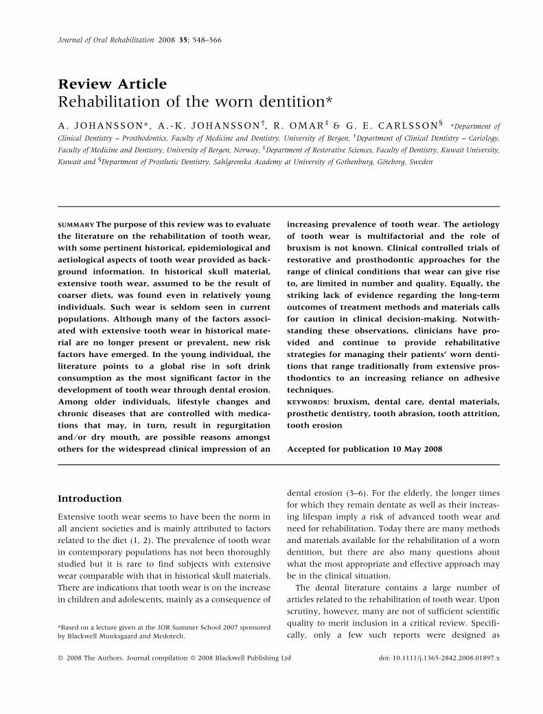
Fig. 10. A 40-year-old woman with severe buccal erosive dam-
age, restored with resin composite restorations. (Reprinted with
permission from Johansson AK et al., 2006)(90).
(a) (b)
(c) (d)
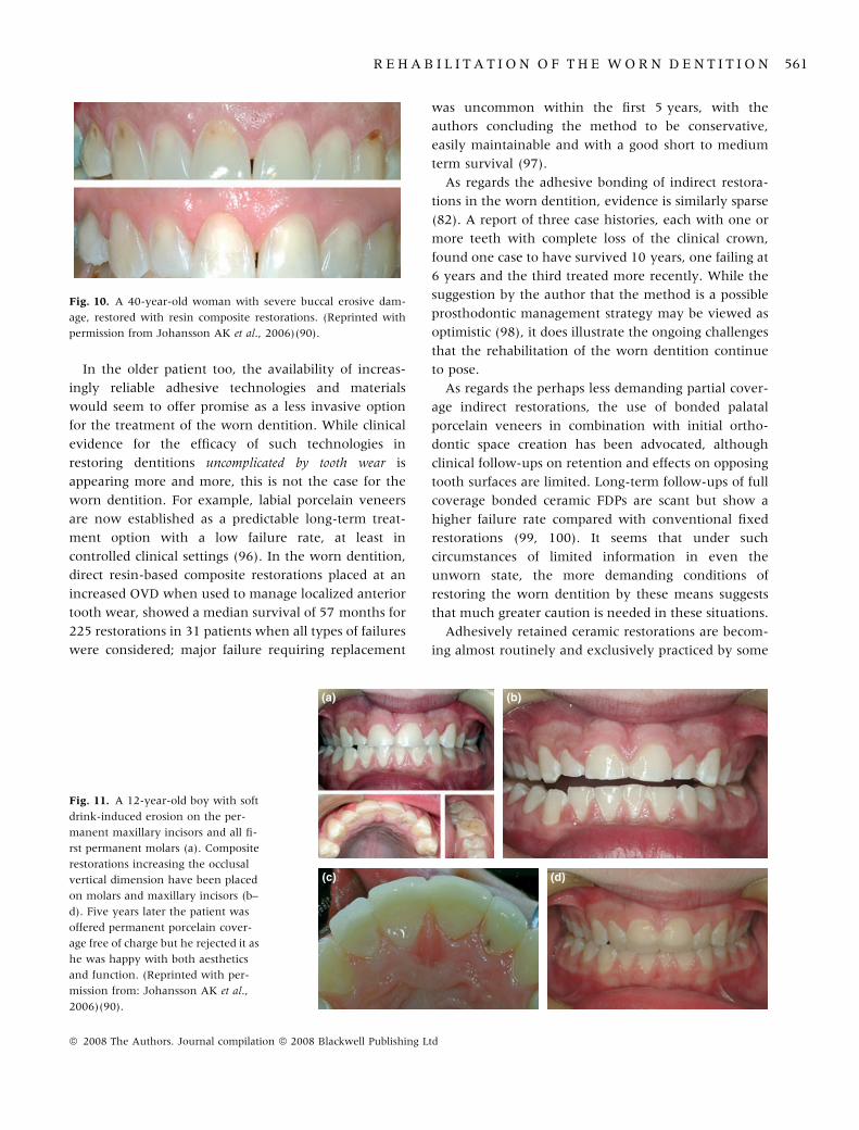
Fig. 11. A 12-year-old boy with soft
drink-induced erosion on the per-
manent maxillary incisors and all fi-
rst permanent molars (a). Composite
restorations increasing the occlusal
vertical dimension have been placed
on molars and maxillary incisors (b–
d). Five years later the patient was
offered permanent porcelain cover-
age free of charge but he rejected it as
he was happy with both aesthetics
and function. (Reprinted with per-
mission from: Johansson AK et al.,
2006)(90).
R E H A B I L I T A T I O N O F T H E W O R N D E N T I T I O N 561
ª 2008 The Authors. Journal compilation ª 2008 Blackwell Publishing Ltd

clinicians (75), but the procedures involved are tech-
nique-sensitive and the method is not yet suitable in
the hands of all dentists. For treatment of cases of tooth
wear with an erosive background, there are no system-
atic studies and only case reports have been presented.
Therefore, it is advisable to exercise some caution when
it comes to restoring worn teeth with aesthetic alter-
natives that rely solely on adhesive bonding until more
reports on its clinical longevity have appeared. Con-
ventional fixed prosthodontics, with its proven record
of long service even if only in the context of the entirely
lesser strategic demands of relatively unworn teeth
(which these data relate to), would seem in many
instances still to be the treatment of choice for exten-
sively worn teeth (Fig. 13).
An alternative rehabilitative strategy was recently
proposed based on the principle of reversibility (101).
Because the worn dentition usually produces slow
occlusal breakdown, it permits most patients to adapt to
the changing situation until a level of unacceptable
function or comfort is reached. Contrasting with this,
typical methods of reconstruction represent a sudden
change that precludes proper evaluation of the patient’s
ability to re-adapt to changed oral conditions. Just as
the pathway to the worn status may vary, so too does
the reconstructive process need to be guided, and this is
suggested by the authors to be best achievable through
staged reconstruction using adhesive techniques wher-
ever possible (101). Even if the evidence for such a
rationale is lacking, it seems possible that the all too
frequent failures seen after traditional reconstructive
efforts may be more controllable through a staged,
reversible reconstruction that relies to a large extent on
adhesive technology.
Maintenance phase
Regular follow-up of reconstructions are necessary for
several reasons. For example, a combination of short
clinical crowns, differential wear and bruxism, etc.,
increase the risks of cementation failure. Similarly,
erosion-induced wear may continue even in the pres-
ence of teeth with full coverage crowns and can
progress cervical to the crowned tooth if causal factors
(a)
(b)
(c) (d)
(e) (f) Fig. 12. A 15-year-old girl with cola-
induced palatal erosion on the max-
illary central and lateral incisors
(a, b). Palatal composite restorations
initially produce a bilateral posterior
open bite (c–e). Six weeks later the
occlusion has returned to normal
through compensatory eruption of
posterior teeth (f). (Reprinted with
permission from: Johansson AK et al.,
2006)(90).
A . J O H A N S S O N et al.562
ª 2008 The Authors. Journal compilation ª 2008 Blackwell Publishing Ltd

have not been eliminated. In addition, occlusal splint
treatment in combined attrition (bruxism) and erosion
cases may not be successful. Cases should be reviewed
at least annually when new study casts, and photo-
graphs should be taken.
A careful clinical and radiographical examination of
abutments should be performed: caries, failed reten-
tion, wear facets, porcelain integrity, etc., must be
checked, recorded, and treated as necessary. Individ-
ually designed preventive regimens should be pre-
scribed and carried out with an interval determined
on the basis of the supposed aetiology and future
progression of the tooth wear. These could comprise
topical fluoride application, dietary advice and psy-
chological motivation for lifestyle changes amongst
others. The lack of knowledge of long-term results of
restoration of tooth wear is a further reason for
regular follow-ups.
Conclusion
Tooth wear is a multifactorial process, which makes it
difficult to identify a single cause. Its progress is
usually slow which characterizes it as a physiological
condition. When it threatens tooth survival or is of
concern to the patient it may be regarded as path-
ologic. The most obvious feature is then shortened
clinical crowns, generally accompanied by dento-
alveolar compensation. This complicates definitive
rehabilitation although research, newer technologies
and materials offer possibilities of greater rationaliza-
tion of treatment modalities. Recognition of the early
signs of wear, and especially erosion, could bring
about timely prevention and improve the lifespan of
teeth.
Restoration of worn teeth will be needed in only
some patients and the measures with which need for
treatment is assessed is one of the keys to successful
outcomes. In broad terms, the decision to treat or not
should be guided by the patient’s stated and ⁄ or
perceived need, the severity of the wear as determined
by morphological changes and the potential for pro-
gression in the context of the patient’s age. The decision
will in most cases be tempered by the generally
complex and expensive nature of rehabilitation of the
worn dentition and the known risks of biomechanical
failures. There is also a striking lack of evidence
regarding long-term outcome of restorative treatment
of tooth wear using different methods and materials.
The caution in clinical decision-making that such a
complex set of circumstances demands is clear, yet the
fact that some patients will require improvements in
their condition through prosthodontics cannot be dis-
puted. Indeed, it has been suggested that while there
may be very few objective criteria for evaluating
prosthodontic treatment need, severe tooth wear may
be a notable exception (102).
(a)
(b)
(c)
Fig. 13. A 32-year-old woman with long-standing bulimia nerv-
osa which resulted in severe erosion. The maxillary teeth were
treated 1 year previously in another clinic (a). The extremely
shortened and sensitive mandibular teeth were restored with
conventional metal-ceramic single crowns (b). Radiograph after
5 years (c). In a telephone check-up 17 years after treatment the
patient says that she had not had any relapse of the eating disorder
and that her teeth had functioned well without problems
throughout the period since her dental treatment. (Reprinted
with permission from: Johansson AK et al., 2006)(90).
R E H A B I L I T A T I O N O F T H E W O R N D E N T I T I O N 563
ª 2008 The Authors. Journal compilation ª 2008 Blackwell Publishing Ltd

References
1. Molnar S. Tooth wear and culture. A survey of tooth function
among some prehistoric populations. Curr Anthropol.
1972;13:511–526.
2. Whittaker DK, Davies G, Brown M. Tooth loss, attrition and
temporomandibular joint changes in a Romano-British pop-
ulation. J Oral Rehabil. 1985;12:407–419.
3. BartlettDW.The role of erosion in tooth wear: aetiology, preven-
tion and management. Int Dent J. 2005;55 (Suppl 1):277–284.
4. Johansson AK. On dental erosion and associated factors. Swed
Dent J Suppl. 2002;(156):1–77.
5. Carlsson GE, Johansson A. Prevalens av dental erosion. In:
Johansson AK, Carlsson GE, eds. Dental erosion. Bakgrund
och kliniska aspekter. Stockholm: Forlagshuset Gothia; 2006.
6. Jaeggi T, Lussi A. Prevalence, incidence and distribution of
erosion. Monogr Oral Sci. 2006;20:44–65.
7. Gotfredsen K, Carlsson GE, Jokstad A, Arvidson Fyrberg K,
Berge M, Bergendal B et al. Implants and ⁄ or teeth: consensus
statements and recommendations. J Oral Rehabil. 2008;35
(Suppl 1):2–8.
8. Helm S, Prydso U. Assessment of age-at-death from mandib-
ular molar attrition in medieval Danes. Scand J Dent Res.
1979;87:79–90.
9. Hylander WL. Morphological changes in human teeth and
jaws in a high-attrition environment. In: Dahlberg AA, Graber
TM, eds. Orofacial growth and development. Paris, the Hauge:
Mouton Publishers; 1977:301–330.
10. Kiliaridis S, Johansson A, Haraldson T, Omar R, Carlsson GE.
Craniofacial morphology, occlusal traits, and bite force in
persons with advanced occlusal tooth wear. Am J Orthod
Dentofacial Orthop. 1995;107:286–292.
11. Johansson A, Kiliaridis S, Haraldson T, Omar R, Carlsson GE.
Covariation of some factors associated with occlusal tooth
wear in a selected high-wear sample. Scand J Dent Res.
1993;101:398–406.
12. Varrela J. Effects of attritive diet on craniofacial morphology: a
cephalometric analysis of a Finnish skull sample. Eur J
Orthod. 1990;12:219–223.
13. Marion LR. Dentistry of ancient Egypt. J Hist Dent.
1996;44:15–17.
14. Kerr NW. Dental pain and suffering prior to the advent of
modern dentistry. Br Dent J. 1998;184:397–399.
15. Richter S. Odontological investigation on archaeological
human remains from Skeljastadir in Thjorsardalur. MSc
thesis. Reykjavik: University of Iceland; 2005.
16. Al-Omiri MK, Lamey PJ, Clifford T. Impact of tooth wear on
daily living. Int J Prosthodont. 2006;19:601–605.
17. Miles AEW. Assessments of the ages of a population of Anglo-
Saxons from their dentitions. Proc R Soc Med. 1962;55:881–
886.
18. Wedel A, Borrman H, Carlsson GE. Tooth wear and tempo-
romandibular joint morphology in a skull material from the
17th century. Swed Dent J. 1998;22:85–95.
19. Maat GJ. Diet and age-at-death determinations from molar
attrition. A review related to the low countries. J Forensic
Odontostomatol. 2001;19:18–21.
20. Johansson AK, Sorvari R, Birkhed D, Meurman JH. Dental
erosion in deciduous teeth – an in vivo and in vitro study.
J Dent. 2001;29:333–340.
21. Johansson AK, Johansson A, Birkhed D, Omar R, Baghdadi S,
Khan N et al. Dental erosion associated with soft-drink
consumption in young Saudi men. Acta Odontol Scand.
1997;55:390–397.
22. Johansson A. A cross-cultural study of occlusal tooth wear.
Swed Dent J. Suppl. 1992;(86):1–59.
23. Barbosa TdeS, Miyakoda LS, Pocztaruk RdeL, Rocha CP,
Gaviao MB. Temporomandibular disorders and bruxism in
childhood and adolescence: review of the literature. Int J
Pediatr Otorhinolaryngol. 2008;72:299–314.
24. Wichelhaus A, Huffmeier S, Sander FG. Dynamic functional
force measurements on an anterior bite plane during the
night. J Orofac Orthop. 2003;64:417–425.
25. Pindborg JJ. Pathology of the dental hard tissues. Copenha-
gen: Munksgaard; 1970.
26. Carlsson GE, Johansson A, Lundqvist S. Occlusal wear.
A follow-up study of 18 subjects with extremely worn
dentitions. Acta Odontol Scand. 1985;43:83–90.
27. Robb ND, Cruwys E, Smith BGN. Regurgitation erosion as
possible cause of tooth wear in ancient British populations.
Arch Oral Biol. 1991;36:595–602.
28. Shaw L. The epidemiology of tooth wear. Eur J Prosthodont
Restor Dent. 1997;5:153–156.
29. Nunn J, Morris J, Pine C, Pitts NB, Bradnock G, Steele J. The
condition of teeth in the UK in 1998 and implications for the
future. Br Dent J. 2000;189:639–644.
30. Bernhardt O, Gesch D, Splieth C, Schwahn C, Mack F, Kocher
T et al. Risk factors for high occlusal wear scores in a
population-based sample: results of the Study of Health in
Pomerania (SHIP). Int J Prosthodont. 2004;17:333–339.
31. Hugoson A, Bergendal T, Ekfeldt A, Helkimo M. Prevalence
and severity of incisal and occlusal tooth wear in an adult
Swedish population. Acta Odontol Scand. 1988;46:255–265.
32. Johansson A, Haraldson T, Omar R, Kiliaridis S, Carlsson GE.
An investigation of some factors associated with occlusal tooth
wear in a selected high-wear sample. Scand J Dent Res.
1993;101:407–415.
33. Wanman A, Wigren L. Need and demand for dental treat-
ment. A comparison between an evaluation based on an
epidemiologic study of 35-, 50-, and 65-year olds and
performed dental treatment of matched age groups. Acta
Odontol Scand. 1995;53:318–324.
34. Elias AC, Sheiham A. The relationship between satisfaction
with mouth and number and position of teeth. J Oral Rehabil.
1998;25:649–661.
35. Mair LH. Wear in dentistry – current terminology. J Dent.
1992;20:140–144.
36. Johansson AK, Carlsson GE (eds). Dental erosion Bakgrund
och kliniska aspekter. Stockholm: Forlagshuset Gothia;
2006.
37. Pergamalian A, Rudy TE, Zaki HS, Greco CMJ. The association
between wear facets, bruxism, and severity of facial pain in
patients with temporomandibular disorders. Prosthet Dent.
2003;90:194–200.
A . J O H A N S S O N et al.564
ª 2008 The Authors. Journal compilation ª 2008 Blackwell Publishing Ltd

38. Khan F, Young WG, Daley TJ. Dental erosion and bruxism.
A tooth wear analysis from south east Queensland. Aust Dent
J. 1998;43:117–127.
39. Lavigne GJ, Khoury S, Abe S, Yamaguchi T, Raphael K.
Bruxism physiology and pathology: an overview for clinicians.
J Oral Rehabil. 2008;35:476–494.
40. Lavigne GJ, Rompre PH, Montplaisir JY. Sleep bruxism: validity
of clinical research diagnostic criteria in a controlled polysom-
nographic study. J Dent Res. 1996;75:546–552.
41. Seligman DA, Pullinger AG, Solberg WK. The prevalence of
dental attrition and its association with factors of age, gender,
occlusion, and TMJ symptomatology. J Dent Res.
1988;67:1323–1333.
42. Nystrom M, Kononen M, Alaluusua S, Evalahti M, Vartiova-
ara J. Development of horizontal tooth wear in maxillary
anterior teeth from five to 18 years of age. J Dent Res.
1990;69:1765–1770.
43. Ekfeldt A, Hugoson A, Bergendal T, Helkimo M. An individual
tooth wear index and an analysis of factors correlated to
incisal and occlusal wear in an adult Swedish population. Acta
Odontol Scand. 1990;48:343–349.
44. Baba K, Haketa T, Clark GT, Ohyama T. Does tooth wear
status predict ongoing sleep bruxism in 30-year-old Japanese
subjects? Int J Prosthodont. 2004;17:39–44.
45. Bernhardt O, Gesch D, Schwahn C, Mack F, Meyer G, John U
et al. Epidemiological evaluation of the multifactorial aetiol-
ogy of abfractions. J Oral Rehabil. 2006;33:17–25.
46. Smith WA, Marchan S, Rafeek RN. The prevalence and
severity of non-carious cervical lesions in a group of patients
attending a university hospital in Trinidad. J Oral Rehabil.
2008;35:128–134.
47. Braem M, Lambrechts P, Vanherle G. Stress-induced cervical
lesions. J Prosthet Dent. 1992;67:718–722.
48. Faye B, Kane AW, Sarr M, Lo C, Ritter AV, Grippo JO.
Noncarious cervical lesions among a non-tooth brushing
population with Hansen’s disease (leprosy): initial findings.
Quintessence Int. 2006;37:613–619.
49. McEvoy SA, Mitchell RJ, Powell ML. Wedge-shaped cervical
dental lesions in two prehistoric native American populations.
Am J Phys Anthropol. 1996;22:162.
50. Bevenius J, L’Estrange P, Karlsson S, Carlsson GE. Idiopathic
cervical lesions: in vivo investigation by oral microendoscopy
and scanning electron microscopy. A pilot study. J Oral
Rehabil. 1993;20:1–9.
51. Bartlett DW, Shah P. A critical review of non-carious cervical
(wear) lesions and the role of abfraction, erosion, and
abrasion. J Dent Res. 2006;85:306–312.
52. Addy M. Tooth brushing, tooth wear and dentine hypersen-
sitivity – are they associated? Int Dent J. 2005;55 (Suppl
1):261–267.
53. Khan F, Young WG, Shahabi S, Daley TJ. Dental cervical
lesions associated with occlusal erosion and attrition. Aust
Dent J. 1999;44:176–186.
54. Attin T. Methods for assessment of dental erosion. Monogr
Oral Sci. 2006;20:152–172.
55. Davies SJ, Gray RJ, Qualtrough AJ. Management of tooth
surface loss. Br Dent J. 2002;192:11–23.
56. van’t Spijker A, Keulen CM, Creugers NHJ. Attrition, occlu-
sion, (dys)function, and intervention: a systematic review.
Clin Oral Impl Res. 2007;18 (Suppl 3):117–126.
57. Fisher J, Mitchell D, Smiciklas-Wright H, Birch L. Maternal
milk consumption predicts the tradeoff between milk and soft
drinks in young girls’ diets. J Nutr. 2001;131:246–250.
58. Ganss C, Klimek J, Giese K. Dental erosion in children and
adolescents: a cross-sectional and longitudinal investigation
using study models. Community Dent Oral Epidemiol.
2001;29:264–271.
59. Ohrn R, Enzell K, Angmar-Mansson B. Oral status of 81
subjects with eating disorders. Eur J Oral Sci. 1999;107:157–
163.
60. Meurman JH, ten Cate JM. Pathogenesis and modifying
factors of dental erosion. Eur J Oral Sci. 1996;104:199–206.
61. Hove LH, Holme B, Young A, Tveit AB. The protective effect of
TiF4, SnF2 and NaF against erosion-like lesions in situ. Caries
Res. 2008;42:68–72.
62. Holbrook WP, Arnadottir IB, Kay EJ. Prevention. Part 3:
prevention of tooth wear. Br Dent J. 2003;195:75–81.
63. Young WG. Tooth wear: diet analysis and advice. Int Dent J.
2005;55:68–72.
64. Johansson A, Omar R. Identification and management of
tooth wear. Int J Prosthodont. 1994;7:506–516.
65. Johansson AK, Lingstrom P, Imfeld T, Birkhed D. Influence of
drinking method on tooth surface pH in relation to dental
erosion. Eur J Oral Sci. 2004;112:484–489.
66. Orchardson R, Gillam DG. Managing dentin hypersensitivity.
J Am Dent Assoc. 2006;137:990–998.
67. Thrash WJ, Dodds MW, Jones DL. The effect of stannous
fluoride on dentinal hypersensitivity. Int Dent J. 1994;1
(Suppl 1):107–118.
68. Azzopardi A, Bartlett DW, Watson TF, Sherriff M. The surface
effects of erosion and abrasion on dentine with and without a
protective layer. Br Dent J. 2004;196:351–354.
69. Bartlett DW. Retrospective long-term monitoring of tooth
wear using study models. Br Dent J. 2003;194:211–213.
70. Carlsson GE, Magnusson T. Management of temporomandib-
ular disorders in the general dental practice. Chicago: Quin-
tessence; 1999.
71. Tallgren A. Changes in adult face height due to ageing, wear
and loss of teeth and prosthetic treatment. Acta Odontol
Scand. 1957;15 (Suppl 24).
72. Berry DC, Poole DFG. Attrition: possible mechanisms of
compensation. J Oral Rehabil. 1976;3:201–206.
73. Dahl B, Øilo G. Wear of teeth and restorative materials. In:
Owall B, Kayser AF, Carlsson GE, eds. Prosthodontics.
Principles and management strategies. London: Mosby-Wolfe;
1996:187–200.
74. Setchell DJ. Conventional crown and bridgework. Br Dent J.
1999;187:68–74.
75. Toreskog S, Myrin C. A minimally invasive and esthetic
bonded porcelain technique – the concept and the vision. I:
Schou L (red). Nordic Dentistry 2003 Yearbook. Copenhagen:
Quintessence; 2003:1–25.
76. Bartlett DW. Erosion and tooth surface loss. Int J Prosthodont.
2003;16(Suppl):87–88.
R E H A B I L I T A T I O N O F T H E W O R N D E N T I T I O N 565
ª 2008 The Authors. Journal compilation ª 2008 Blackwell Publishing Ltd

77. Dahl BL, Krogstad O, Karlsen K. An alternative treatment in
cases with advanced localized attrition. J Oral Rehabil.
1975;2:209–214.
78. Gough MB, Setchell DJ. A retrospective study of 50 treatments
using an appliance to produce localised occlusal space by
relative axial tooth movement. Br Dent J. 1999;187:134–139.
79. Dahl BL, Krogstad O. Long-term observations of an increased
occlusal face height obtained by a combined orthodon-
tic ⁄ prosthetic approach. J Oral Rehabil. 1985;12:173–176.
80. Poyser NJ, Porter RW, Briggs PF, Chana HS, Kelleher MG. The
Dahl Concept: past, present and future. Br Dent J.
2005;198:669–676.
81. Hemmings KW, Darbar UR, Vaughan S. Tooth wear treated
with direct composite restorations at an increased vertical
dimension: results at 30 months. J Prosthet Dent.
2000;83:287–293.
82. Chana H, Kelleher M, Briggs P, Hooper R. Clinical evaluation
of resin-bonded gold alloy veneers. J Prosthet Dent.
2000;83:294–300.
83. Wise MD. Occlusion and restorative dentistry. Br Dent J.
1977;143:45–52.
84. Dawson PE. Vertical dimension. In: Dawson PE, ed. Evalua-
tion, diagnosis, and treatment of occlusal treatment. St Louis,
MO: CV Mosby Company; 1974:275–285.
85. De Boever JA, Carlsson GE, Klineberg IJ. Need for occlusal
therapy and prosthodontic treatment in the management of
temporomandibular disorders. Part II. Tooth loss and prostho-
dontic treatment. J Oral Rehabil. 2000;27:647–659.
86. Ettinger RL, Qian F. Abutment tooth loss in patients with
overdentures. J Am Dent Assoc. 2004;135:739–746.
87. Almog DM, Ganddini MR. Maxillary and mandibular overlay
removable partial dentures for restoration of worn teeth.
A three-year follow-up. N Y State Dent J. 2006;72:32–35.
88. Bayne SC. Dental restorations for oral rehabilitation – testing
of laboratory properties versus clinical performance for clinical
decision making. J Oral Rehabil. 2007;34:921–932.
89. Yip KH, Smales RJ, Kaidonis JA. Differential wear of teeth and
restorative materials: clinical implications. Int J Prosthodont.
2004;17:350–356.
90. Carlsson GE, Johansson A, Milleding P. Rekonstruktiv
behandling av erosionsskador. In: Johansson AK, Carlsson
GE, eds. Dental erosion. Bakgrund och kliniska aspekter.
Stockholm: Forlagshuset Gothia AB; 2006:163–180.
91. Jaeggi T, Gruninger A, Lussi A. Restorative therapy of erosion.
Monogr Oral Sci. 2006;20:200–214.
92. van Dijken JW. Retention of a resin-modified glass ionomer
adhesive in non-carious cervical lesions. A 6-year follow-up.
J Dent. 2005;33:541–547.
93. van Dijken JW, Sunnegardh-Gronberg K, Lindberg A. Clinical
long-term retention of etch-and-rinse and self-etch adhesive
systems in non-carious cervical lesions. A 13 years evaluation.
Dent Mater. 2007;23:1101–1107.
94. Bartlett D, Sundaram G. An up to 3-year randomized clinical
study comparing indirect and direct resin composites used to
restore worn posterior teeth. Int J Prosthodont. 2006;19:613–
617.
95. Oncag G, Tuncer AV, Tosun YS. Acidic soft drinks effects on
the shear bond strength of orthodontic brackets and a
scanning electron microscopy evaluation of the enamel.
Angle Orthod. 2005;75:247–253.
96. Layton D, Walton T. An up to 16-year prospective study of
304 porcelain veneers. Int J Prosthodont. 2007;20:389–396.
97. Redman CD, Hemmings KW, Good JA. The survival and
clinical performance of resin-based composite restorations
used to treat localised anterior tooth wear. Br Dent J.
2003;194:566–572.
98. Bartlett DW. Three patient reports illustrating the use of
dentin adhesives to cement crowns to severely worn teeth. Int
J Prosthodont. 2005;18:214–218.
99. Marquardt P, Strub JR. Survival rates of IPS empress 2 all-
ceramic crowns and fixed partial dentures: results of a 5-year
prospective clinical study. Quintessence Int. 2006;37:253–
259.
100. Sailer I, Feher A, Filser F, Gauckler LJ, Luthy H, Hammerle
CH. Five-year clinical results of zirconia frameworks for
posterior fixed partial dentures. Int J Prosthodont.
2007;20:338–383.
101. Creugers NH, van’t Spijker A. Tooth wear and occlusion:
friends or foes? Int J Prosthodont. 2007;20:348–350.
102. Jokstad A. Oral prosthetics from a Nordic perspective. Int J
Prosthodont. 2002;15:145–153.
103. Johansson AK, Johansson A, Birkhed D, Omar R, Baghdadi S,
Carlsson GE. Dental erosion, soft-drink intake, and oral health
in young Saudi men, and the development of a system for
assessing erosive anterior tooth wear. Acta Odontol Scand.
1996;54:369–378.
104. Johansson A, Haraldson T, Omar R, Kiliaridis S, Carls-
son GE. A system for assessing the severity and pro-
gression of occlusal tooth wear. J Oral Rehabil. 1993;20:
125–131.
Correspondence: Professor Anders Johansson, Department of Clinical
Dentistry – Prosthodontics, Faculty of Medicine and Dentistry,
University of Bergen, Arstadveien 17, 5009 Bergen, Norway.
E-mail: [email protected]
A . J O H A N S S O N et al.566
ª 2008 The Authors. Journal compilation ª 2008 Blackwell Publishing Ltd