Embed Size (px)
Citation preview

188 R.A. JUNGMANN ET AL.THE JOURNAL OF EXPERIMENTAL ZOOLOGY 282:188–195 (1998)
© 1998 WILEY-LISS, INC.
Regulation of LDH-A Gene Expression byTranscriptional and Posttranscriptional SignalTransduction Mechanisms
RICHARD A. JUNGMANN,* DELAI HUANG, AND DI TIANCancer Center and Department of CM Biology, Northwestern UniversityMedical School, Chicago, Illinois 60611
ABSTRACT The lactate dehydrogenase-A (LDH-A) gene, whose product plays a pivotal role innormal anaerobic glycolysis and is frequently increased in human cancers, is highly regulated atthe transcriptional and posttranscriptional levels. Our laboratory has carried out extensive stud-ies concerning the regulation of LDH-A subunit expression. We have elucidated complex regula-tory mechanisms by identifying multiple cis-acting promoter elements including functional sitesfor Sp1 and c-Myc interactions as well as sites that interact with the protein kinase A and proteinkinase C substrates, CREB and AP1, respectively. Furthermore, we have reported the existence ofa CRE-dependent silencer element in the LDH-A promoter. LDH-A expression is additionally regu-lated through the protein kinase A and C signal pathways at the posttranscriptional level, specifi-cally mRNA stability. J. Exp. Zool. 282:188�195, 1998. © 1998 Wiley-Liss, Inc.
BIOLOGY OF THE LDH ISOZYME SYSTEMLactate dehydrogenase (LDH) plays a pivotal
role in normal anaerobic glycolysis. It catalyzes theinterconversion of lactate and pyruvate with NAD+
as cofactor. The LDH enzymes are tetrameric pro-teins consisting of two kinds of 35-kDa subunits:the A type (or M) predominates in skeletal muscleand liver, and the B (or H) type in heart. Variouscombinations of these two subunits produce fivepossible isozymes in somatic cell types: LDH-1(B4), LDH-2 (AB3), LDH-3, (A2B2), LDH-4 (A3B),and LDH-5 (A4). A third type of LDH isozyme(LDH C4) was shown to be present only in mam-malian testes and spermatozoa (Goldberg, ’63).
In mammals, the isozymes follow a binomial dis-tribution pattern, suggesting a random combina-tion of two subunits to the overall isozyme patternresulting from the relative abundance of A and Bsubunits. Genetic evidence shows that the two sub-units are separate products encoded by genes lo-cated on different chromosomes. For example, thehuman LDH-A gene is located on chromosome 11and the LDH-B gene on chromosome 12 (Li et al.,’88; Seawright et al., ’88). Various pathological con-ditions are associated with LDH-A deficiency(exertional myoglobinuria) and LDH-B deficiency(squamous cell lung cancer, colorectal adenoma).
An unusual property of LDH-A was identifiedseveral years ago, suggesting that it may play aregulatory role during gene transcription and DNA
replication. For instance, a rat liver single-stranded DNA-binding protein was identified asLDH-5 (Williams et al., ’85), and human and bo-vine LDH-5 were shown to be capable of bindingsingle-strand DNA (Sharief et al., ’86).
DIFFERENTIAL EXPRESSIONOF LDH ISOZYMES
The LDH-A and B subunit genes offer an inter-esting and biologically important system forstudying the developmental, tissue-specific, andhormonal regulation of the metabolically impor-tant LDH isozymes. Not only is there tissue-specific expression of LDH subunits, but the sametissue in the same organism may possess vary-ing proportions of the A and B subunits duringdifferent phases of prenatal and postnatal devel-opment or at different hormonal stages. Theexpression of the LDH genes appears tightly co-ordinated in a tissue and developmental stage-specific fashion. Furthermore, any one tissue maygreatly change its LDH isozyme pattern duringontogeny. For example, the heart muscle of mouseand rat changes from a B subunit pattern during
Grant sponsor: NIH; Grant number: GM53115.*Correspondence to: Richard A. Jungmann, Ph.D., Department CM
Biology, Northwestern University Medical School, 303 E. Chicago Ave.,Chicago, IL 60611.
Received 27 March 1998; Accepted 27 March 1998
LDH-A SUBUNIT GENE EXPRESSION 189
oogenesis to a predominantly A subunit one inembryonic life and again back to a predominantlyB subunit pattern in adult life (Markert andUrsprung, ’62; Fritz et al., ’73). During pregnancyand lactation, the LDH isozyme pattern in ratmammary gland acquires primarily the LDH-Asubunit (Richards and Hilf, ’72a). Involution ofthe mammary gland resulting from cessation oflactation is associated with a return of the LDHisozyme pattern to the original “B state.” Richardsand Hilf (’72b) have shown that estrogen elevatesLDH activity in both normal rat mammary glandsand mammary tumors by selectively increasingLDH-4 and 5 isozymes. There are profoundalterations of LDH isozyme patterns during neo-plastic development (Hilf et al., ’76). The abnor-malities of LDH isozyme expression in tumortissue are often two-sided: Not only are fetalisozyme forms activated or reexpressed, but insome instances isozymes that characterize adulttissue are lost. During the tumorigenesis of themammary gland, a shift from the A/B isozymepattern to the LDH A pattern has been found(Rosado et al., ’69; Farron et al., ’72; Lee et al.,’79). On the other hand, ovariectomy of tumor-bearing rats, resulting in tumor regression, leadsto a concomitant decrease of the LDH-A and cor-responding increase of the LDH-B subunit in themammary gland. Recently, it has been shown thatc-Myc is able to transactivate the LDH-A gene infibroblasts; this may have significant implicationsfor tumor metabolism and growth (Shim et al.,’97). In rat C6 glioma cells (which express boththe LDH-A and -B subunit mRNAs), the relativeLDH isozyme composition is shifted in favor ofthe LDH-5 (A4) isozyme after catecholamine (viacyclic AMP) stimulation because of an increasedde novo synthesis of the LDH-A but not the Bsubunit (Derda et al., ’80). From all these obser-vations, the nature of the controls that allow se-lective LDH-A and B gene expression duringdevelopment and differentiation represent a vi-tal area for future research.
REGULATION OF LDH GENEEXPRESSION
Transcriptional regulationIn the case of mammalian LDH isozyme sys-
tems, transcription of the LDH-A and B genes iscontrolled in a tissue-specific manner, with theliver the major site of LDH-A and the heart themajor site of LDH-B subunit synthesis. In themajority of tissues, the relative expression of
LDH-A/B subunits is specific for that tissue, al-though a number of physiologic factors may alterthat ratio. The differential regulation is mani-fested, for instance, by the fact that LDH-A ex-pression is regulated at the transcriptional levelby the protein kinase A and C signal pathways,whereas LDH-B expression is not (Miles et al.,’81; Jungmann et al., ’83; Short et al., ’94; Huangand Jungmann, ’95). Examples of the effects ofvarious effector agents on LDH-A transcription areshown in Table 1. Activation of protein kinase sig-nal pathways by the appropriate second messen-gers, Sp-cAMPS or the phorbol ester TPA, resultsin a marked increase of the rate of LDH-A sub-unit transcription. In accordance with the tran-scriptional mechanism of activation, actinomycinD inhibits transcriptional activation. Addition ofspecific inhibitors, such as Rp-cAMPS, a proteinkinase A antogonist, and BIM, an inhibitor of pro-tein kinase C, also blocks transcriptional activa-tion. In all of these cases, the experimentalevidence supports a transcriptional mechanism ofLDH-A subunit gene regulation through appropri-ate promoter regulatory elements.
The regulatory (promoter) region of the LDH-Agene identified so far includes several distal ele-ments located within the 1.2-kb region upstreamfrom the transcription initiation site (see Fig. 1).These elements include two Sp1 sites, necessaryfor efficient expression of basal transcriptional ac-
TABLE 1. Effect of activators of the protein kinases A and Csignal transduction pathways on transcription of LDH-A
and -B subunit genes1
Rate of LDH mRNAsynthesis (ppm)
Treatment A subunit B subunit
None 1150 ± 88 310 ± 24Sp-cAMPS (0.5 mM) 4230 ± 155 330 ± 29TPA (100 nM) 3780 ± 110 258 ± 31Actinomycin D (5 µg/ml) 277 ± 12 198 ± 18Sp-cAMPS + Actinomycin D 189 ± 25 212 ± 49TPA + actinomycin D 265 ± 44 199 ± 43Rp-cAMPS (0.5 mM) 299 ± 57 202 ± 58BIM (100 nM) 66 ± 35 178 ± 34BIM + TPA 267 ± 37 207 ± 271Nuclear run-off experiments were carried out as described previ-ously (Jungmann et al., ’83). Nuclei were isolated from rat C6 gliomacells that had been treated for 4 hours with or without the indicatedconcentration of agents. Isolated nuclei were allowed to incorporate[α-32P]UTP and mRNA was determined by hybridization analysis.RNA synthesis results are given in parts per million (ppm) and arethe average and S.E. determined from five separate experiments. Sp-cAMPS is a protein kinase A agonist and Rp-cAMPS is a proteinkinase A antagonist. BIM, bisindolylmaleimide is a specific proteinkinase C inhibitor (Huang and Jungmann, ’95).

190 R.A. JUNGMANN ET AL.
tivity (Short et al., ’94); two functional E2 sites(Shim et al. ’97); a negative regulatory element(NRE) (Chung et al., ’95); a phorbol ester-induc-ible AP1 site regulated by protein kinase C (Huangand Jungmann, ’95); as well as a cAMP- induc-ible CRE site (Short et al., ’94). The CRE site bindsan isoform of CREB (∆CREB) (M.L. Short et al.,’91) and activates transcription in response toCREB phosphorylation by the protein kinase Asignal pathway (S. Short et al., ’91). An analysisof the promoter sequence of the human LDH-Bgene indicated the absence of a CRE motif (Takanoand Li, ’89). This is consistent with our observa-tion that the expression of LDH-A but not of LDH-B is induced by cAMP (Derda et al., ’80).
Detailed mutational analysis of a 1.2-kb pro-moter region revealed the presence of both posi-tive as well as negative transcription controlmodules. The negatively acting sequence, NRE,is located in a distal segment between positions–873 and –850 bp and is characterized by the factthat its silencing activity depends on the pres-ence of the CRE module and has no repressingeffect on promoters lacking CRE (Chung et al.,’95). The silencer element is of dyad symmetryand consists of a palindromic sequence with twohalf-sites, 5´-TCTTG-3´. Gel mobility shift, foot-printing, Southwestern blotting, and UV-cross-linking studies demonstrated the presence of a69-kDa protein with specific binding activity forthe NRE. Gel supershift analysis with anti-phospho-CREB antibody and anti-Fos antibodyindicated the presence of CREB and Fos or anti-genically closely related proteins within the NRE-protein complex. How the silencer module acts invivo and the physiological role of the silencer ele-ment in regulation of the LDH isozyme systemsstill remain to be elucidated.
The promoter for the LDH-A gene also containsa conserved consensus E box sequence, -CACGTG,a potential binding site for the cMyc/Max hetero-dimer transcription factor. The E box elementhas the consensus sequence that interacts witha family of transcription factors known as thebasic helix-loop-helix proteins. These proteinsare characterized by a basic region that is re-quired for binding of the protein to the E boxand a helix-loop-helix motif required for proteindimerization. Overexpression of c-Myc in rat fi-broblasts causes a marked activation of theLDH-A promoter and increased transcription ofLDH-A mRNA (Shim et al., ’97).
Modulation of LDH-A expression occurs througheffector agents that activate the protein kinase Aand C signal transduction pathways. In the caseof protein kinase C activation, stimulation of ratC6 glioma cells with either tetradecanoylphorbolacetate (TPA) or dioctanoylglycerol (DG) causes amarked accumulation of LDH-A subunit mRNA(Huang and Jungmann, ’95). Use of the specificprotein kinase C inhibitor bisindolylmaleimide(BIM) (GF 109203X) prevented the TPA-inducedincrease of LDH-A subunit mRNA, indicating thatprotein kinase activation is necessary for LDH-Ainduction. Transient transfection analysis of LDH-A promoter deletion/CAT constructs, DNA/proteinbinding assays, including footprint and gel shiftanalyses, identified a TRE-AP-1 enhancer mod-ule at position –294 bp, which is the target forthe protein kinase C signal pathway and prob-ably requires phosphorylation of proteins of theAP-1 complex.
Cyclic AMP-regulated transcriptional activity isdependent upon the 8-bp cAMP-responsive element(CRE), -TGACGTCA-, located at the –48/–41-bpupstream region (Short et al., ’94). Mutations inthe CRE abolish cAMP-mediated induction and re-duce basal activity by about 65%. The CRE bindsa 47-kDa protein, which has been identified asCRE-binding protein (CREB-327 or ∆CREB) (M.L.Short et al., ’91), an isoform of the CREB tran-scription factor family. Addition of the dephos-phoform of CREB-327 to in vitro transcriptionassays using the LDH-A promoter as template hadno effect, whereas the phosphoform of CREB-327markedly stimulated transcription from the pro-moter (S. Short et al., ’91).
In summary, the overall regulatory mechanismof LDH-A subunit expression by effector agentsthat activate protein kinase A may briefly be de-scribed as follows (Fig. 2). Stimulation of β-adr-energic receptor activates adenylyl cyclase, leading
Fig. 1. Organization of the rat LDH-A subunit promoter.Shown above is the promoter fragment –1173/+1 bp (+1 isthe transcription start site) including transcription regula-tory elements identified so far. See text for details.

LDH-A SUBUNIT GENE EXPRESSION 191
to the intracellular synthesis of cAMP, which, inturn, causes the dissociation and concomitant ac-tivation of cytoplasmic cAMP-dependent proteinkinase (protein kinase A) into its catalytic (C) andregulatory subunits (Fig. 2, step a). Because thenucleus is devoid of protein kinase A, the cata-lytic subunit translocates to nuclear sites (Fig. 2,step b) (Jungmann et al., ’80; Kuettel et al., ’85;Kwast-Welfeld and Jungmann, ’88; Squinto andJungmann, ’89), where it achieves the phos-phorylative modification of a serine residue inCREB-327 (Fig. 2, step c). This, in turn, allowsphospho-CREB to associate with the CRE lead-ing to an increased rate of LDH-A mRNA tran-scription. It should be noted that protein kinaseA and C activators do not act synergistically atthe transcriptional level and that LDH-B subunitgene transcription is not affected by these agents.
Posttranscriptional regulationModulation of mRNA stability is an important
mechanism for controlling gene expression. Sev-eral lines of evidence indicate that the processof mRNA degradation is of biological importance.First, cytoplasmic mRNA turnover is universaland appears in both prokaryotic and eukaryoticcells. Second, turnover rates can vary widelywithin a single cell and are often influenced byextracellular factors. Some mRNAs have half-lives of several hours or even days, whereas oth-ers like inducible growth regulators—such asoncogenes (c-Myc and c-Fos), cytokines, and tran-scriptional activators (Shaw and Kamen, ’86)—tend to have extremely unstable messages withhalf-lives on the order of 10 to 30 min. Third,the time required for any specific molecule to
reach a new steady-state level depends solely onthe rate constant of degradation. Fourth, theturnover rates of a number of mRNA species canbe modulated by differential biological signals.A prime example of this latter effect is repre-sented by the LDH-A mRNA. Our past studieshave shown that in rat C6 glioma cells the half-life of the relatively unstable LDH-A mRNA (t½≈ 55 min) is markedly increased after treatmentof cells with cAMP analogs and phorbol ester in-dicating that the stability of LDH-A mRNA isregulated via the protein kinase A and C signalpathways. Moreover, we have shown that theprotein kinase A and C signal pathways inter-act synergistically to regulated LDH-A subunitstability (Jungmann et al., ’83; Huang et al., ’95).
Based on these studies, we postulated thatmRNA stability could be an inherent property ofmRNA primary or secondary structure and thattrans-regulatory factors could be involved actingas modulators of the actions of protein kinases Aor C. The question arose: What is the nature ofthe mRNA structure that determines stability andregulation by protein kinases A and C?
Determinants of mRNA stability/instabilityhave been identified in several regions of mRNAmolecules including the 3´-untranslated region(3´UTR) of the message (Shaw and Kamen, ’86;Jones and Cole, ’87; Brewer and Ross, ’88; Brewer,’91). The major determinants of stability locatedin the 3´UTR of several mRNAs are sequencesrich in A and U bases. Multiple copies of the se-quence AUUUA are frequently found in the3´UTR of many short-lived mRNAs.
Characterization of LDH-A 3´UTRAlthough the translated regions of LDH-A and
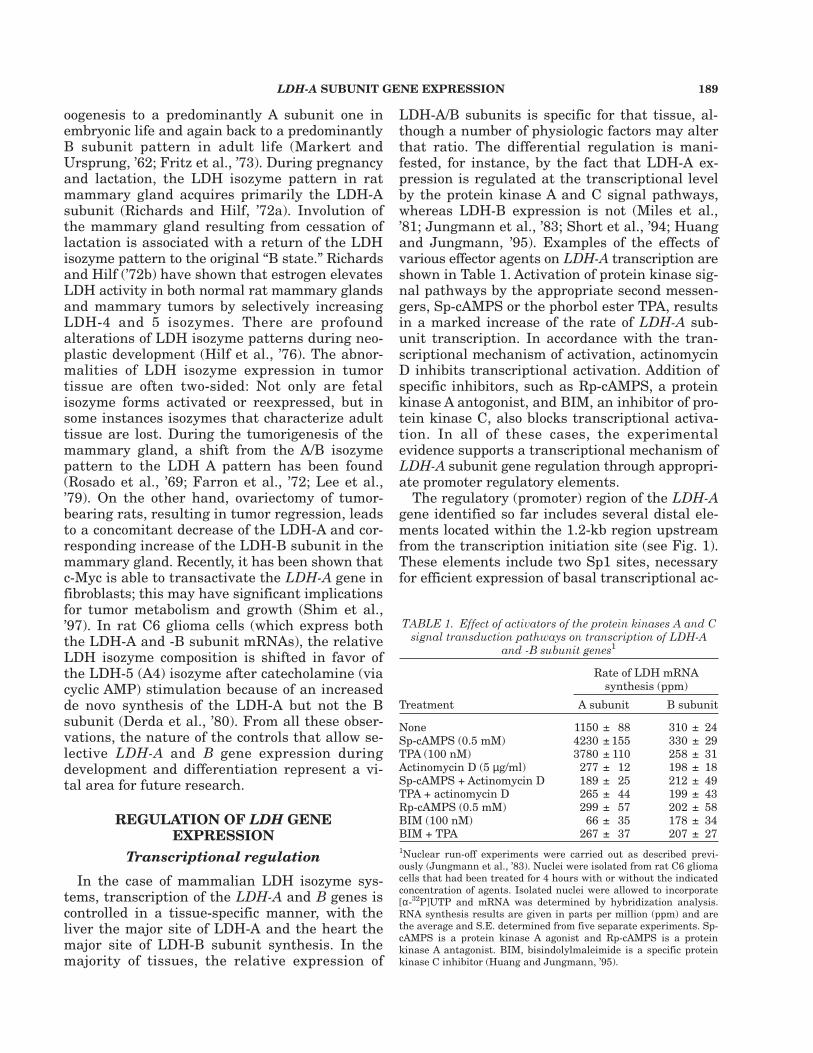
B mRNAs show a high percentage of sequencesimilarity, the size (in number of bases) of tsheir3´UTRs is markedly different (507 bases in LDH-A versus 194 bases in LDH-B). Additionally, the3´UTRs exhibit little, if any, sequence similarity.However, untranslated sequences that are A- andU-rich are found in the 3´UTRs of the LDH-A andLDH-B mRNAs, but both 3´UTRs lack the previ-ously identified consensus AU-rich element,AUUUA, which has been identified as one of therecognition signals involved in mRNA stability/instability and as a binding site for regulatoryRNA-binding proteins. The sequence of LDH-A3´UTR is shown in Figure 3. LDH-A 3´UTR con-tains about 24% adenine, 28% uridine, 23% guani-dine, and 25% cytosine. Thus, as a whole, it isnot AU rich. However, a region of about 120 nucle-
Fig. 2. Scheme summarizing the molecular events lead-ing to protein kinase A-mediated induction of LDH-A sub-unit mRNA. See text for details.
192 R.A. JUNGMANN ET AL.
otides (underlined in Fig. 3) has a high AU con-tent of about 70%. It does not seem to share se-quence similarity with some of the well-studied3´UTRs, such as c-fos, c-myc, GM-CSF, or histone.No UUAUUU(A/U)(A/U) or AUUUA motif isfound, implying that its degradation may not beregulated in the same manner as mRNAs con-taining AUUA or UUAUUU(A/U)(A/U).
LDH-A 3´UTR but not LDH-B 3´UTRdestabilizes b-globin mRNA
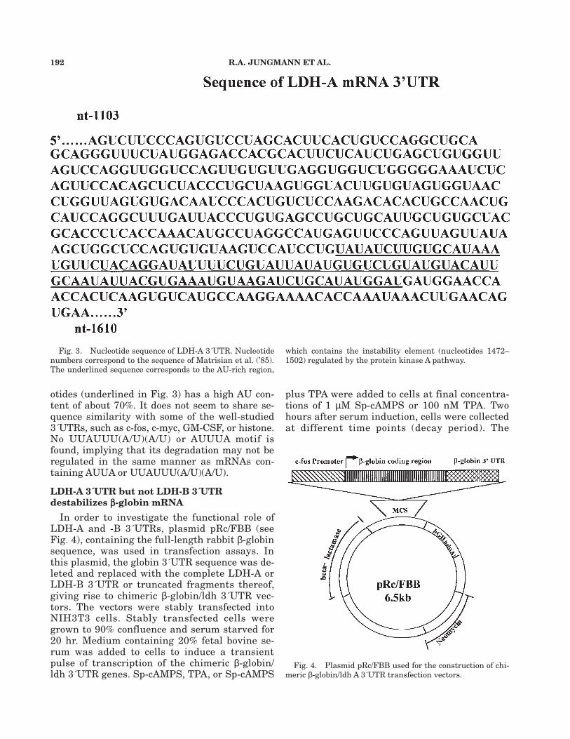
In order to investigate the functional role ofLDH-A and -B 3´UTRs, plasmid pRc/FBB (seeFig. 4), containing the full-length rabbit β-globinsequence, was used in transfection assays. Inthis plasmid, the globin 3´UTR sequence was de-leted and replaced with the complete LDH-A orLDH-B 3´UTR or truncated fragments thereof,giving rise to chimeric β-globin/ldh 3´UTR vec-tors. The vectors were stably transfected intoNIH3T3 cells. Stably transfected cells weregrown to 90% confluence and serum starved for20 hr. Medium containing 20% fetal bovine se-rum was added to cells to induce a transientpulse of transcription of the chimeric β-globin/ldh 3´UTR genes. Sp-cAMPS, TPA, or Sp-cAMPS
plus TPA were added to cells at final concentra-tions of 1 µM Sp-cAMPS or 100 nM TPA. Twohours after serum induction, cells were collectedat different time points (decay period). The
Fig. 3. Nucleotide sequence of LDH-A 3´UTR. Nucleotidenumbers correspond to the sequence of Matrisian et al. (’85).The underlined sequence corresponds to the AU-rich region,
which contains the instability element (nucleotides 1472–1502) regulated by the protein kinase A pathway.
Fig. 4. Plasmid pRc/FBB used for the construction of chi-meric β-globin/ldh A 3´UTR transfection vectors.

LDH-A SUBUNIT GENE EXPRESSION 193
amounts of chimeric β-globin/ldh 3´UTR mRNAwere measured by ribonuclease protection assay,and decay curves were plotted. Under the ex-perimental conditions, wild-type β-globin mRNAexhibits a very slow rate of decay (Fig. 5). In con-trast, the chimeric β-globin/ldh 3´UTR mRNAsare markedly destabilized, with a half-life of 55min for β-globin/ldh A mRNA (Fig. 5) and about45 min for β-globin/ldh B mRNA (Fig. 6). Thehalf-lives of the two chimera mRNAs are simi-lar to the wild-type LDH-A and LDH-B mRNAhalf-lives observed in rat C6 glioma cells. Thesefindings indicate that the 3´UTRs of both sub-unit mRNAs contain a destabilizing element(s)which is responsible for the accelerated decay ofchimeric β-globin/ldh 3´UTR.
Treatment of cells with the protein kinase Aagonist and cAMP analogue Sp-cAMPS or thephorbol ester tetradecanoylphorbol acetate (TPA)increased the half-life of chimeric β-globin/ldh AmRNA to 8 hr and 5 hr, respectively. Moreover,treatment with a combination of Sp-cAMPS andTPA caused a synergistic stabilizing effect and in-creased the half-life t½ to about 30 hr. It is im-portant to note that stimulation of cells with theSp-cAMPS (Fig. 6) or TPA (not shown) had no ef-fect on the half-life of β-globin/ldh B mRNA. Takentogether, the results indicate that the 3´UTR se-quences of both LDH-A and -B mRNA contain adestabilizing sequence(s) which needs to be fur-ther identified. However, only the LDH-A 3´UTRresponds to agents that activate the protein ki-nase A and C signal pathways and very likely con-tains the regulatory sequences responsible for thestabilizing effect.
The protein kinase A-regulatory sequence islocated to a 30-nucleotide LDH-A 3´UTRfragment
To identify the 3´UTR sequence responsible forthe protein kinase A regulation of LDH-A mRNAstability, truncated fragments systematically cov-ering the entire LDH-A 3´UTR sequence (Fig. 7)were synthesized and used to replace the β-globin3´UTR in plasmid pRc/FBB. After stable transfec-tion of these vectors, the half-lives of the chimericβ-globin/ldh mRNAs were determined. The resultsare shown in Figure 8. In agreement with the dataof Figure 5, chimeric β-globin/ldh mRNA with thecomplete LDH-A 3´UTR (nt 1103–1610) was sta-bilized synergistically by the combined stimulatoryeffect of Sp-cAMPS and the protein kinase C ago-
Fig. 5. Decay of chimeric β-globin/ldh A 3´UTR mRNA andeffect of protein kinase A and C agonists.
Fig. 6. Decay of chimeric β-globin/ldh B 3´UTR mRNA inunstimulated and Sp-cAMPS-stimulated rat C6 glioma cells.
Fig. 7. Diagram of LDH-A 3´UTR fragments used for theconstruction of chimeric β-globin/ldh A 3´UTR plasmids. Frag-ment consisting of nucleotides 1472–1502 was mutated bydeleting two nucleotides (C and A) and changing nucleotides-AUAU- to -CAGA- (underlined).

194 R.A. JUNGMANN ET AL.
nist dioctanoylglycerol (DG). After truncation, frag-ments consisting of nucleotides 1103–1177, 1173–1247, 1243–1317, 1313–1387, 1383–1457 (data notshown), 1453–1471, and 1503–1527 (see Fig. 8) losttheir responsiveness to Sp-cAMPS and DG. How-ever, some of them (nucleotides 1243–1317, 1453–1471, and 1503–1527) retained their ability todestabilize the corresponding chimeric mRNA, al-though to a lesser degree than the complete 3´UTR.It is clear from Figure 8 that the 30-nucleotidesequence comprising nucleotides 1472 through1502 is the regulatory site targeted by the proteinkinase A pathway and appears to be responsiblefor protein kinase A-mediated stabilization of LDH-A mRNA. A partial modification of this sequencethrough mutation (for detailed base sequence, seeFig. 7) abolished the effect of cAMP and proteinkinase A.
CONCLUSIONIntracellular LDH-A mRNA subunit levels are
regulated through alteration of the mRNA stabil-ity in addition to transcriptional modulation. A30-nucleotide instability element has been iden-tified, which is the target of the stabilizing effectby protein kinase A. This instability element prob-ably interacts with phosphoproteins that are thetarget of protein kinase A and, together with theelement, form a regulatory module determiningLDH-A mRNA half-life under physiologic condi-tions that cause activity changes of protein kinaseA. Further studies need to be carried out to iden-tify the target of protein kinase C.
LITERATURE CITEDBrewer, G. (1991) An A+U-rich element RNA-binding factor
regulates c-myc mRNA stability in vitro. Mol. Cell. Biol.,11:2460–2466.
Brewer, G., and J. Ross (1988) Poly(A) shortening and degra-dation of the 3´ AU-rich sequences of human c-myc mRNAin a cell-free system. Mol. Cell. Biol., 8:1697–1708.
Chung, K.C., D. Huang, Y. Chen, S. Short, M.L. Short, Z.Zhang, and R.A. Jungmann (1995) Identification of a si-lencer module which selectively represses cyclic AMP-re-sponsive element-dependent gene expression. Mol. Cell.Biol., 15:6139–6149.
Derda, D.F., M.F. Miles, J.S. Schweppe, and R.A. Jungmann(1980) Cyclic AMP regulation of lactate dehydrogenase. Iso-proterenol and N6, O2´-dibutyryl cyclic AMP increase thelevels of lactate dehydrogenase-5 isozyme and its messen-ger RNA in rat C6 glioma cells. J. Biol. Chem., 255:11112–11121.
Farron, F., H.H.T. Hsu, and E.W. Knox (1972) Fetal-typeisoenzymes in hepatic and nonhepatic rat tumors. CancerRes., 32:302–308.
Fritz, P.J., E.L. White, K.M. Pruitt, and E.S. Vesell (1973)Lactate dehydrogenase isozymes. Turnover in rat heart,skeletal muscle, and liver. Biochemistry, 12:4034–4039.
Goldberg, E. (1963) Lactic and malic dehydrogenases in hu-man spermatozoa. Science, 139:602–603.
Hilf, R., W.D. Rector, and R.A. Orlando (1976) Multiple mo-lecular forms of lactate dehydrogenase and glucose 6-phos-phate dehydrogenase in normal and abnormal human breasttissues. Cancer, 37:1825–1830.
Huang, D., and R.A. Jungmann (1995) Transcriptional regu-lation of the lactate dehydrogenase A subunit gene by thephorbol ester 12-O-tetradecanoylphorbol-13-acetate. Mol.Cell. Endocrinol., 108:87–94.
Huang, D., C.J. Hubbard, and R.A. Jungmann (1995) Lac-tate dehydrogenase A subunit messenger RNA stability issynergistically regulated via the protein kinase A and Csignal transduction pathways. Mol. Endocrinol., 9:994–1004.
Jones, T.R., and M.D. Cole. (1987) Rapid cytoplasmic turn-over of c-myc mRNA: requirement of the 3´ untranslatedsequences. Mol. Cell. Biol., 7:4513–4521.
Jungmann, R.A., M.I. Mednieks, and J.S. Schweppe (1980)Nuclear compartmentation of protein kinase and the regu-lation of gene expression. In: Cell Compartmentation andMetabolic Channeling. L. Noyer, F. Lynen, and K. Mothes,eds. Elsevier/North-Holland Biomedical Press, Amsterdam,pp. 415–426.
Jungmann, R.A., D.C. Kelley, M.F. Miles, and D.M. Milkowski(1983) Cyclic AMP regulation of lactate dehydrogenase. Iso-proterenol and N6, O2´-dibutyryl cyclic AMP increase therate of transcription and change the stability of lactate de-hydrogenase A subunit messenger RNA in rat C6 gliomacells. J. Biol. Chem., 258:5312–5318.
Kuettel, M.R., S.P. Squinto, J. Kwast-Welfeld, G. Schwoch,J.S. Schweppe, and R.A. Jungmann (1985) Localizationof nuclear subunits of cyclic AMP-dependent protein ki-nase by the immunocolloidal gold method. J. Cell Biol.,101:965–975.
Kwast-Welfeld, J., and R.A. Jungmann (1988) Hormonalregulation of nuclear cyclic AMP-dependent protein kinasesubunit levels in rat ovaries. J. Biol. Chem., 263:14343–14350.
Lee, C., L. Oliver, E.L. Coe, and R. Oyasu (1979) Lactate de-
Fig. 8. Half-lives of various chimeric mRNAs arising fromthe expression of β-globin/ldh A 3´UTR deletion plasmids. TheLDH-A 3´UTR fragments (in nucleotides) tested are indicated.Fragment ‘mut’ is a mutation of fragment 1472–1502 (for se-quence, see Fig. 7).

LDH-A SUBUNIT GENE EXPRESSION 195
hydrogenase in normal mammary glands and in 7,12-dime-thyl[a]anthracene-induced mammary tumors in Sprague-Dawley rats. J. Natl. Cancer Inst., 62:193–199.
Li, S.S.-L., Y.-C.E. Pan, F.S. Sharief, M.J. Evans, M.-F. Lin,G.M. Clinton, and J.J. Holbrook (1988) Cancer-associatedlactate dehydrogenase is a tyrosylphosphorylated form ofhuman LDH-A skeletal muscle isozyme. Cancer Invest.,6:93–101.
Markert, C.L., and H. Ursprung (1962) The ontogeny ofisozyme patterns of lactate dehydrogenase in the mouse.Dev. Biol., 5:363–381.
Matrisian, L.M., G. Rautmann, B.E. Magun, and R. Breath-nach (1985) Epidermal growth factor or serum stimulationof rat fibroblasts induces an elevation in mRNA levels forlactate dehydrogenase and other glycolytic enzymes. NucleicAcids Res., 13:711–726.
Miles, M.F., P. Hung, and R.A. Jungmann (1981) Cyclic AMPregulation of lactate dehydrogenase. Quantitation of lac-tate dehydrogenase M-subunit messenger RNA in isoprot-erenol- and N6, O2´-dibutyryl cyclic AMP-stimulated rat C6glioma cells by hybridization analysis using a cloned cDNAprobe. J. Biol. Chem., 256:12545–12552
Richards, A.H., and R. Hilf (1972a) Influence of pregnancy,lactation and involution on glucose-6-phosphate dehydro-genase and lactate dehydrogenase isoenzymes in the ratmammary gland. Endocrinology, 91:287–295.
Richards, A.H., and R. Hilf (1972b) Effect of estrogen admin-istration on glucose 6-phosphate dehydrogenase and lactatedehydrogenase isoenzymes in rodent mammary tumors andnormal mammary glands. Cancer Res., 32:611–616.
Rosado, A., H.P. Morris, and S. Weinhouse (1969) Lactate de-hydrogenase subunits in normal and neoplastic tissues ofthe rat. Cancer Res., 29:1673–1680.
Seawright, A., J.M. Fletcher, J.A. Fantes, H. Morrison, D.J.Porteous, S.S.-L. Li, N.D. Hastie, and V. van Heyningen(1988) Analysis of WAGR deletions and related transloca-
tions with gene-specific probes, using FACS-selected cellhybrids. Somat. Cell. Mol. Genet., 14:21–30
Sharief, F.S., S.H. Wilson, and S.S.-L. Li (1986) Identifica-tion of the mouse low-salt eluting single-stranded DNA-bind-ing protein as a mammalian lactate dehydrogenase-Aisoenzyme. Biochem. J., 233:913–916.
Shaw, G., and R. Kamen (1986) A conserved AU sequencefrom the 3´ untranslated region of GM-CSF mRNA medi-ates selective mRNA degradation. Cell, 45:659–667.
Shim, H., C. Dolde, B.C. Lewis, C-S. Wu, G. Dang, R.A.Jungmann, R. Dalla-Favera, and C.V. Dang (1997) c-Myctransactivation of LDH-AImplications for tumor metabolismand growth. Proc. Natl. Acad. Sci. USA, 94:6658–6663.
Short, M.L., C.F. Manohar, M.R. Furtado, G.D. Ghadge, S.M.Wolinsky, B. Thimmapaya, and R.A. Jungmann (1991a)Nucleotide and derived amino-acid sequences of the CRE-binding protein from rat C6 glioma and HeLa cells. NucleicAcids Res., 19:4290.
Short, S., M.L. Short, D.M. Milkowski, and R.A. Jungmann(1991b) Functional analysis of cis- and trans-regulatory el-ements of the lactate dehydrogenase A subunit promoterby in vitro transcription. J. Biol. Chem., 266:22164–22172.
Short, M.L., D. Huang, D.M. Milkowski, S. Short, K. Kunst-man, C-J. Soong, K.C. Chung, and R.A. Jungmann (1994)Analysis of the rat lactate dehydrogenase A subunit genepromoter/regulatory region. Biochem. J., 304:391–398.
Squinto, S.P., and R.A. Jungmann (1989) Modulation ofnuclear cyclic AMP-dependent protein kinase in dibutyrylcyclic AMP-treated rat H4IIE hepatoma cells. Biochem. J.,260:673–682.
Takano, T., and S.S.-L. Li (1989) Structure of human lactatedehydrogenase-B gene. Biochem. J., 257:921–924.
Williams, K.R., S. Reddigari, and G.L. Patel (1985) Identifi-cation of a nucleic acid helix-stabilizing protein from ratliver as lactate dehydrogenase-5. Proc. Natl. Acad. Sci. USA,82:5260–5264.