Embed Size (px)
Citation preview

Vol. 169, No. 2JOURNAL OF BACTERIOLOGY, Feb. 1987, P. 646-6530021-9193/87/020646-08$02.00/0Copyright © 1987, American Society for Microbiology
Regulation, Initiation, and Termination of the cenA and cexTranscripts of Cellulomonas fimi
N. M. GREENBERG, R. A. J. WARREN, D. G. KILBURN, AND R. C. MILLER, JR.*Department of Microbiology, University of British Columbia, Vancouver, British Columbia, Canada V6T IWS
Received 13 June 1986/Accepted 28 October 1986
We characterized the in vivo transcripts of two Celulomonas fimi genes, the cenA gene, which encodes anextracellular endo-0-1,4-glucanase (EC 3.2.1.4) and the cex gene, which encodes an extracellular exo-P-1,4-glucanase (EC 3.2.1.91). By Northern blot analysis, cenA mRNA was detected in C. fimi RNA preparationsfrom glycerol- and carboxymethyl cellulose-grown cells but not from glucose-grown cells. In contrast, cexmRNA was detected only in the preparations from carboxymethyl cellulose-grown cells. Therefore, thetranscription of these genes is subject to regulation by the carbon source provided to C. fimi. By nuclease Siprotection studies with unique 5'-labeled DNA probes and C. fimi RNA isolated in vivo, 5' termini were found51 and 62 bases before the cenA translational initiation codon and 28 bases before the cex translationalinitiation codon. S1 mapping with unlabeled DNA probes and C. fimi RNA which had been isolated in vivo butwhich had been 5' labeled in vitro with guanylyltransferase and [a-32P]GTP confirmed that true transcriptioninitiation sites for cenA and cex mRNA hod been identified. Comparative analysis of the DNA sequencesimmediately upstream of the initiation sites of the cenAi and cex mRNAs revealed a 30-base-pair region wherethese two sequences display at least 66% homology. Si mapping was also used to locate the 3' termini of thecenA and cex transcripts. Three 3' termini were found for cenA messages, whereas only one 3' terminus wasidentified for cex mRNA. The transcripts of both genes terminate in regions where their corresponding DNAsequences contain inverted repeats.
Cellulomonas fimi is a gram-positive, nonsporeformingfacultative anaerobe which grows best at 30°C (3, 25, 42). Aninteresting feature of C. fimi DNA is its G+C content of 72mol% (42). At least three classes of P-1,4-glucanases areproduced by C. fimi under appropriate physiological condi-tions: P-1,4-endoglucanases (EC 3.2.1.4) (1, 15, 28, 44, 52)and at least one --1,4-exoglucanase (EC 3.2.1.91) (17, 36, 50)which can act synergistically to hydrolyze carboxymethylcellulose (CMC) (17) and a P-glucosidase (EC 3.2.1.21) (48)which hydrolyzes cellobiose to glucose.We previously reported the molecular cloning of two C.
fimi genes in Escherichia coli: the cenA gene, which encodesan extracellular endo-P-1,4,-glucanase (Eng) (15, 52), andthe cex gene, which encodes an extracellular exo-P-1,4-glucanase (Exg) (15, 36, 50). Although these cloned geneshave been well characterized, very little information isavailable on the molecular mechanisms which govern theirexpression in C. fimi. In this study we used Northernblotting to investigate the in vivo regulation of cenA and cextranscription and nuclease S1 protection analysis to map theinitiation and termination sites of the cenA and cex tran-scripts. DNA probes derived from the cloned genes in E. coliwere used in the analysis. To our knowledge, this is the firstcharacterization of transcription in C. fimi, and cenA and cexare the first cellulase-encoding genes that have been showndirectly to be regulated at the transcriptional level.
MATERIALS AND METHODS
Bacterial strains and plasmids. The bacterial strains usedwere C. fimi ATCC 484 and E. coli JM101 (31) and JM83(46). Plasmids pBR322 (9), pUC12, pUC13 (46), and pUC18(46, 53) and their derivatives (as described below) werepropagated in E. coli JM83 or JM101.
* Corresponding author.
Plasmid pcEC2 is a derivative of pBR322 that contains a2.2-kilobase-pair BamHI-SmaI fragment (Fig. 1A) carryingthe cenA gene of C. fimi (52). Plasmid pUC12A25 is aderivative of pUC12 which contains a 2.6-kilobase-pairBamHI-SalI fragment (Fig. 1B) carrying the cex gene of C.fimi (36).
Plasmids pNG101 and pNG102 are derivatives of pUC18which contain the 636-base-pair (bp) BamHI-SalI and 939-bpSmaI fragments, respectively, of pcEC2. Plasmid pNG201 isa derivative of pUC18, from which the 216-bp SmaI-PvuIIpUC18 fragment was deleted, and carries the 138-bp PstI-BanI fragment of pUC12A25. Plasmid pNG202 is a deriva-tive of pUC18 carrying the 253-bp Sau3Al-SalI fragment ofpUC12A25. Plasmids pNG101, -102, -201, and -202 wereconstructed to facilitate the preparation of high-specific-activity hybridization probes by minimizing the number ofunwanted fragments which could compete for label in end-labeling reactions. Plasmid pUC13Bam31 carries the 397-bpSau3A1 fragment of pUC12A25 in the BamHI site of pUC13and was kindly provided by G. P. O'Neill.Enzymes and reagents. Restriction endonucleases BamHI,
BglII, Sau3A1, and SmaI, Si nuclease, T4 polynucleotidekinase, guanylyltransferase, yeast tRNA, and redistilledphenol were from Bethesda Research Laboratories, Inc.Enzymes BanI, HindIII, PstI, and SalI, dextran sulfate, andDNA-grade Sephadex G-50 were from Pharmacia P.-L. NarIand StyI were from New England BioLabs, Inc.Diethylpyrocarbonate, MOPS (morpholinepropanesulfonicacid), HEPES (N-2-hydroxyethylpiperazine-N'-2-ethane-sulfonic acid), and Trizma base were from Sigma ChemicalCo. Formamide was from BDH. Radionuclides were fromNew England Nuclear Corp. All other chemicals were ofreagent grade or higher and were purchased from commer-cial suppliers.Media and growth conditions. C. fimi was grown in basal
medium (43) supplemented with either 0.2% (wt/vol) glyc-
646
on April 1, 2018 by guest
http://jb.asm.org/
Dow
nloaded from
CELLULASE GENE TRANSCRIPTION IN C. FIMI 647
erol, 0.2% (wt/vol) glucose, or 1% (wt/vol) CMC (Sigma; lowviscosity) as a carbon source. E. coli strains were grown in2 x YT medium (31). All strains were grown at 30°C. Whensolid medium was required, agar (Difco Laboratories) wasadded to 1.5% (wt/vol) except for basal medium containingCMC, in which 1.0% agar was used. When appropriate,ampicillin (Sigma) was added to 100,ug ml-' in liquid or solidmedium.
RNase-free work. Chemicals and reagents used for RNAwork were purchased solely for this purpose and were keptseparate from regular laboratory supplies. All glasswareused for RNA work was either baked at 300°C for 3 h or wasbought as disposable labware. When appropriate, solutionswere treated with 0.2% (vol/vol) diethylpyrocarbonate asdescribed previously (12, 29). All plastics (pipette tips andmicrofuge tubes) were sterilized by autoclaving withoutfurther pretreatment.RNA extraction. C. fimi RNA was prepared by a modifi-
cation of the procedures of Miller et al. (32) and Kennell andBicknell (26). Cultures (up to 100 ml) in the late log phasewere rapidly chilled on ice and centrifuged for 5 min at 6,000x g. Cells were washed with 1/10 volume of ice-cold 10 mMTris hydrochloride (pH 7.5)-i mM EDTA (TE), transferredto polycarbonate tubes (Oak Ridge type), and centrifuged for5 min at 6,000 x g. The cells were then suspended in 1/25volume of 50 mM Tris hydrochloride (pH 6.8)-2 mMEDTA-1% sodium dodecyl sulfate (SDS), and the tubeswere placed immediately into a boiling water bath for 90 s.The tubes were chilled on ice for 5 min, and an equal volumeof ice-cold 5 M NaCl was added and mixed briefly on avortex mixer. After 5 min on ice, the resultant slurry wascentrifuged for 10 min at 30,000 x g, and the clearedsupernatant fluid was carefully decanted into a 30-ml Corex(Corning Glass Works) glass tube. The nucleic acids wereprecipitated with 2.5 volumes of 95% ethanol at -20°C for 12to 16 h and recovered by centrifugation for 20 min at 10,000x g. The pellets were washed with 70% ethanol at -20°Cand redissolved in 2 ml of 10 mM Tris hydrochloride (pH7.5)-S5 mM MgCl2. Samples were treated with 5 U of RQ1DNase I (Promega) for 15 min at 37°C, EDTA was added to5 mM, and the mixture was extracted twice with phenol-chloroform (1:1) and once with chloroform. The organicphases were combined and back extracted with 1 ml of TE(pH 7.5). The aqueous phases were pooled, and RNA wasrecovered by precipitation with 3 volumes of 95% ethanoland centrifugation for 10 min at 10,000 x g. The pellets werewashed with 70% ethanol and redissolved in 20 mM NaPO4(pH 6.5)-i mM EDTA (RNA storage buffer). The RNApreparations were compared for similar banding patternsafter analytical electrophoresis on agarose gels and subse-quent staining with ethidium bromide. RNA concentrationswere determined by A26, and samples were divided intoaliquots and stored at -70°C.DNA extraction and purification. Plasmid DNA was iso-
lated by a modification of the alkaline lysis procedure ofBirnboim and Doly (8). When required for the preparation ofhigh-specific-activity probes, DNA was further purified bycentrifugation to equilibrium in CsCl density gradients con-taining ethidium bromide (29).
Preparation of 32P end-labeled DNA. To end label DNAfragments, plasmid DNA was digested with restriction en-zyme for 1 h at 37°C, extracted twice with phenol-chloroform (1:1) and once with chloroform, and precipitatedwith ethanol. For 5' labeling, fragments were treated withcalf intestinal alkaline phosphatase and labeled with [-y-32P]ATP (3,000 Ci mmol-1) and T4 polynucleotide kinase as
described previously (29). The 3' ends were labeled with[a-32P]dGTP (3,000 Ci mmol-') and the Klenow fragment ofDNA polymerase I as described previously (29). Incorpora-tion of label was monitored by liquid scintillation spectro-photometry in an ISOCAP-300.
Preparation of hybridization probes. 32P-end-labeled DNAwas digested wih an appropriate restriction endonuclease toliberate fragments uniquely labeled at one end. The diges-tions were routinely performed under conditions recom-mended by the suppliers. The hybridization probes werepurified by electrophoresis in 5% polyacrylamide gels,electroeluted, and precipitated from ethanol, as describedpreviously (29). In some instances, yeast tRNA (20 jig ml-')was added as a carrier in the final precipitation. Pellets werewashed with 70% ethanol, dried briefly in air, and redis-solved in TE (pH 7.5). Samples were removed for quantita-tion by liquid scintillation counting. Typical specific activi-ties were 1 x 10⁴ to 4 x 10⁴ dpm ng of DNA probe-'.
Single-stranded M13 molecular weight standards. To pre-pare single-stranded DNA molecular weight standards,M13mpll single-stranded DNA was digested with HaeIII(Bethesda Research) by the procedure of von Gabain et al.(47) and then 5' end labeled as described above. LabeledHaeIII fragments were recovered by centrifugation throughsmall columns of Sephadex G-50 (29). Fragments wereethanol precipitated with yeast tRNA carrier, redissolved inTE (pH 7.5), and stored at -20°C. Fragment sizes weredetermined from the complete nucleotide sequence ofM13mpll (45, 53). The 525-base fragment arises from partialdigestion (N. M. Greenberg, unpublished observations).Northern blot analysis of C. fimi RNA. For Northern
blotting, 20 ,ug of C. fimi RNA was precipitated with ethanol,redissolved in 10 ,ul of 20 mM MOPS-1 mM EDTA-5 mMsodium acetate (running buffer [pH 7]) with 50% formamideand 2.2 M formaldehyde, heated for 5 min at 68°C, andcooled briefly on ice. Loading dye was added (to give 3%[wt/vol] Ficoll [Pharmacia] and 0.02% [wt/vol] bromphenolblue and xylene cyanol), and samples were electrophoresedalongside single-stranded M13 molecular weight markers on1.0% agarose-6.6% formaldehyde gels at 20 to 40 mA withrecirculation of running buffer. Nucleic acids were blotted toBiotrans membranes (Pall, Inc.) in 20x SSC (ix SSC is 0.15M NaCl plus 0.015 M sodium citrate) for 12 to 16 h (41), andthe membranes were baked at 80°C for 1 h. Prehybridiza-tions and hybridizations were performed essentially by theprotocols supplied with the membranes. Briefly,prehybridizations were done in 5x SSC-50% formamide-4mM PP1-5 x Denhardt buffer (29) 10% dextran sulfate-250,ug of heat-denatured (100°C for 5 min) salmon sperm DNAml-'. Incubations were for 1 to 2 h at 42°C with constantagitation. For hybridizations, the probe DNA (8 x 10⁵ to 10X 105 dpm ml-1) and carrier were denatured together byheating. The probes were allowed to hybridize to filters for12 to 16 h at 42°C with constant agitation. Blots were washedat 20°C with three changes of 2x SSC-0. 1% SDS and then at60°C with three changes of 0.1x SSC-0.1% SDS and ex-posed to XAR-2 film (Eastman Kodak Co.) at -70°C withintensifying screens.
In vitro cap labeling of RNA. To characterize primarytranscripts, total C. fimi RNA from CMC-grown cultureswas labeled in vitro at the 5' end by using the vaccinia viruscapping enzyme, guanylyltransferase, as described previ-ously (34, 51). Briefly, up to 60 ,ug of total C. fimi RNA waslabeled in 0.1-ml mixtures containing 25 mM Tris hydrochlo-ride (pH 7.5)-2 mM MgCl2-1 mM dithiothreitol-250 ,uCi of[a-32P]GTP (3,000 Ci mmol-1)-10 to 25 U of guanylyltrans-
VOL. 169, 1987
on April 1, 2018 by guest
http://jb.asm.org/
Dow
nloaded from

648 GREENBERG ET AL.
ferase. After 30 min at 37°C, the reaction was stopped by theaddition of EDTA to 4 mM and SDS to 0.2%. The RNA wasextracted twice with phenol-chloroform (1:1) and precipi-tated twice from ethanol in the presence of 2 M ammoniumacetate. The RNA was finally recovered by ethanol precip-itation from 0.3 M sodium acetate. Typical specific activitieswere 1 x 10³ to 3 x 10³ dpm ng-1.
Si nuclease transcript mapping. Analysis for 5' and 3' endsof C. fimi transcripts with labeled DNA probes was doneessentially as described by Favaloro et al. (13) and Berk andSharp (4, 5, 49). Up to 30 ,ug of C. fimi RNA was precipitatedwith 0.01 to 0.03 pmol of end-labeled DNA probe (1 x 10⁴ to4 x 104 dpm), redissolved in 30 ,ul of hybridization buffer (0.4M NaCl, 0.04 M sodium phosphate [pH 6.5], 0.4 mM EDTA,80% formamide), heated for 15 min at 85°C, and held at 60°Cfor 3 h. Samples were diluted with 300 ,ul of ice-cold Sibuffer (30 mM sodium acetate [pH 4.5], 28 mM NaCl, 4.5mM ZnSO4) and treated with 1,400 U of S1 nuclease for 30min at 37°C. The reaction was terminated by the addition of75 ,ul of stop buffer (2.5 mM ammonium acetate, 50 mMEDTA), and yeast tRNA (20 ,ug) was added. The mixturewas precipitated with 400 ,ul of isopropanol and centrifuged.When capped RNA and unlabeled DNA probes were used
in the S1 mapping experiments, the procedure was modifiedas follows (51). Up to 50 pug of capped RNA was precipitatedwith up to 500 ng of unlabeled DNA probe, redissolved inhybridization buffer, heated to 85°C, held at 60°C for 3 h, andthen treated with S1, as described above. After the S1treatment, the 0.33-ml samples were treated with 25 ng ofRNase A for 15 min at 22°C to reduce the background ofunhybridized RNA. This reaction was terminated by theaddition of SDS to 0.25% and two extractions with phenol-chloroform (1:1). Trimmed hybrids were recovered by pre-cipitation from ethanol.
After either of these procedures, pellets were dissolved insequencing dye buffer (80% formamide, 0.5x TBE [29],0.02% [wt/vol] bromphenol blue and xylene cyanol), heatedto 90°C for 2 min, and electrophoresedurea gels with appropriate size markers (
A.
B.
Bm Sm Ss Sa
cen A
Al1- A2
Bm S3 Ps St Bn S3I lIr f
cex
0 OObp I-'B Ba
FIG. 1. Representation of cloned segmejcontaining the cenA (52) and cex (36) genes. TIshown as boxed regions. Both genes are transl(A) The 2.2-kilobase BamHI-SmaI fragment othe cenA gene. A-1, SstI-SalI Northern blot p5' Si probe; A-3, BgiII-SmaI 3' Si probe.BamHI-SaiI fragment of pUC12A25 that contzStyI-SalI Northern blot probe; B-2, PstI-BaiSau3A1 fragment used in 5' mapping experimby guanylyltransferase and GTP; B-3, Sau3A1restriction endonucleases are abbreviated as fBgiII; Bm, BamHI; S3, Sau3Ai; Sa, Sall; SrStyI.
A. B.
>- (-)M oc3u
25277
849525 52--
1 23
2527-l
1623 !
849525
1 2 3
FIG. 2. Northern blot analysis of cenA- and cex-specific tran-scripts. RNA was extracted from C. fimi cultures grown on basalmedium supplemented with glycerol, glucose, or CMC, denaturedwith formaldehyde, fractionated on formaldehyde gels containing1.0% agarose, and transferred to Biotrans membranes. (A) Hybrid-ization with cenA intragenic SstI-SaIl (site of 5' end labeled with 32P)fragment (Fig. 1, A-1). Lanes: M, HaeIII restriction fragments ofsingle-stranded M13mpll (sizes in nucleotides are indicated on theleft); 1, RNA from glycerol-grown cells; 2, RNA from glucose-grown cells; 3, RNA from CMC-grown cells. (B) Hybridization withcex intragenic StyI-Sall (site of 5' end labeled with 32P) fragment(Fig. 1, B-1). Lanes are as indicated in panel A. Arrows indicatemajor hybrids.
The gels were dried to Whatmann 3MM filter paper andexposed to XRP-1 film (Eastman Kodak) at -70°C withintensifying screens.
RESULTS
in polyacrylamide- Regulation by carbon source and approximate lengths of(see figure legends). cenA and cex mRNA transcripts. The lengths of the specific
cenA and cex transcripts and the effects of the carbonsources provided during growth in culture on the relative
Bg Sm mRNA levels were characterized by Northern blot analysis.Bg lThe intragenic cenA probe (Fig. 1, A-1) hybridized strongly
to a species of C. fimi RNA approximately 1,400 bases longisolated from CMC-grown cells (Fig. 2A, lane 3). A lessabundant hybrid of about the same size was detectable in
A3 RNA from glycerol-grown cells (Fig. 2A, lane 1). HybridsSa S3 Sa Sa between this probe and RNA isolated from glucose-grown
r e' cultures were not detected (Fig. 2A, lane 2). The intrageniccex probe (Fig. 1, B-1) hybridized strongly to an RNA
B-1 approximately 1,500 bases long which was present only inB-3 preparations from CMC-grown cells (Fig. 2B, lane 3). These
results indicate that the carbon source provided duringgrowth can regulate the levels of both the cenA and cex gene
nts of C. fimi DNA transcripts and that the cex gene appears to be more strin-he structural genes are gently regulated than the cenA gene.lated from left to right. Mapping cenA and cex transcriptional start sites with S1if pcEC2 that contains nuclease. To identify the 5' ends of the cenA and cex mRNA,probe; A-2, SmaI-Sall transcripts synthesized in vivo were analyzed by Si nuclease(B) The 2.6-kilobase mapping with the 5'-end-labeled probes A-2 (labeled at theains the cex gene.B2, SalI site) and B-2 (labeled at the BanI site) (Fig. 1). Fourent with RNA labeled distinct cenA-specific hybrids were protected from Si nucle-L-SaltI3'Si probe.The ase degradation (Fig. 3A, lane 6), three of which wereollows: Bn, BanI; Bg, closely spaced, with the fourth running a little higher in them, SmaI; Ss, SstI; St, gel. The two predominant hybrids (Fig. 3A, +1 and +2) were
considered to map the transcriptional start sites for the cenA
J. BACTERIOL.
on April 1, 2018 by guest
http://jb.asm.org/
Dow
nloaded from

CELLULASE GENE TRANSCRIPTION IN C. FIMI 649
A. B.
-11-1
-.1\2
.1
;<2*',:
FIG. 3. Mapping 5' end of cenA and cex mRNA. After hybrid-ization with RNA and treatment with S1 nuclease, cenA- or cex-specific 32P-labeled DNA probes were analyzed on 8% polyacryl-amide-7 M urea sequencing gels alongside probe sequenced by thebase-specific chemical cleavage method of Maxam and Gilbert (30).(A) Si protection of cenA SmaI-Sall (site of 5' end labeled with 32P)fragment (Fig. 1, A-2) by RNA from glucose-grown C. fimi (lane 5),RNA from CMC-grown C. fimi (lane 6), and yeast tRNA (lane 7).Lanes 1 through 4 contained chemical sequencing ladders G > A, G+ A, T + C, and C > T, respectively. (B) S1 protection of cexPstI-BanI (site of 5' end labeled with 32p) fragment (Fig. 1, B-2).Lanes are as indicated in panel A. Numbers on the right identifyingspecies of protected hybrids.
gene. These corresponded to two adjacent G residues 51 and50 bases upstream of the translational initiation codon (ATG)for the unprocessed cenA gene product (see Fig. 5) (52). Theother two protected hybrids mapped to a C and a G, 52 and62 bases upstream, respectively, from the ATG.Four adjacent cex-specific hybrids were resolved by Si
mapping (Fig. 3B, lane 6). These were not observed in thecontrol lanes (lanes 5 and 7). The two predominant bandsmapped to adjacent C residues, located 28 and 27 basesupstream of the translational initiation codon for the unproc-essed cex gene product (36). These were considered startsites for cex gene transcription. The assignment of Si bandsto a position in a DNA sequence reflects a correction of 1.5bases to compensate for the slower mobility of the Si-cleaved hybrids relative to the chemical degradation ladder(33, 49).Si mapping using capped RNA to confirm location of
transcription initiation. Since the locations of the 5' terminifor the cenA and cex transcripts were detected by S1nuclease mapping with DNA probes that were each labeledat the 5' terminus, the transcript map obtained in this fashioncould represent the 5' ends of processed transcripts and notprimary transcript initiation sites. Therefore, an S1 mappingexperiment was performed with total RNA from CMC-
grown C. fimi which had been labeled in vitro with thevaccinia virus capping enzyme. In this system, only RNAspecies with 5' di- or triphosphates (i.e., the primary tran-scripts) are suitable substrates for guanylyltransferase (34).Hence, only RNA transcripts having intact, labeled 5' endscan be resolved in the final analysis and only if they can forma hybrid with a DNA probe and be protected from Sidigestion.To map the cenA mRNA 5' end in this fashion, the A-2
probe was tested for its ability to protect an RNA species of162 bases after Si digestion. This size was predicted basedon the calculated distance between the +1 site identified inFig. 3A and the Sall end of the probe. A protected hybrid ofabout this size was indeed observed after Si mapping (Fig.4A, lane 2), which was conspicuously absent in the controllane (lane 1, RNA without probe). That the protected cappedtranscript migrated as a species which was a few nucleotideslonger than expected may be explained by the presence ofthe 5' cap (G5'ppp5'Np. . .), which is also known to stericallyhinder Si digestion (34, 51). In this experiment we wereunable to detect a larger protected species which wouldcorrespond to the weak -11 band observed in Fig. 3A (seeDiscussion).To map the cex mRNA 5' end in a similar fashion, the B-2
probe was used to determine whether it could protect anRNA species about 69 bases long. However, the analysiscould not resolve the specific signal from a background ofnuclease-treated, unhybridized RNA in this size range (re-sults not shown). Therefore the B-2a probe was used toprotect an RNA species which would be slightly longer andresolve higher in the gel. This B-2a probe was tested for itsability to protect an RNA species of 246 bases after Sidigestion. This size was based on the calculated distancebetween the + 1 site identified in Fig. 3B and the Sau3A1 siteof the probe within cex. A protected hybrid of this size wasobserved (Fig. 4B, lane 2) which was not present in thecontrol lane (lane 1, RNA without probe). These resultsconfirmed the previous mapping of start sites for cenA andcex transcription.The DNA sequences immediately upstream of the mapped
mRNA start sites of cenA and cex are compared in Fig. 5.
A.
169158
117.
B.
251
219
FIG. 4. S1 mapping with C. fimi RNA labeled withguanylyltransferase and [a-32P]GTP. After hybridization betweenDNA probes and C. fimi RNA and Si and RNase A treatment,cenA- and cex-specific hybrids were analyzed on 5% polyacryl-amide-7 M urea gels. The numbers on the left indicate size andmigration of M13mpll HaeIII fragments. The arrows on the rightindicate the specific probe-protected RNA species (lane 2). Resultsof the parallel negative control experiments without probe added tothe labeled RNA are also shown (lane 1). (A) Si protection oflabeled RNA by cenA SmaI-Sall fragment (Fig. 1, A-2). (B) S1protection of labeled RNA by cex Sau3A1 fragment (Fig. 1, B-2a).
VOL. 169, 1987
on April 1, 2018 by guest
http://jb.asm.org/
Dow
nloaded from
650 GREENBERG ET AL.
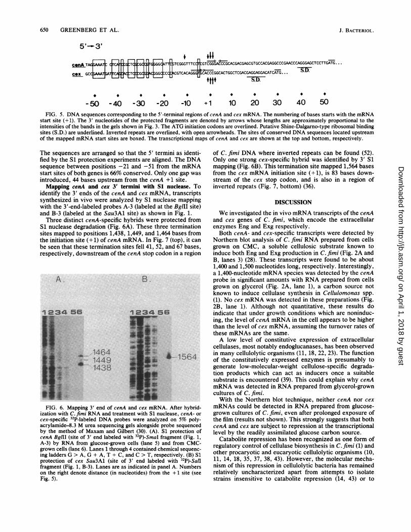
cenA TAGFr TCA TGTGCGGCATT1'TCGGGTTT
Cex GCC;WT;A7TCAG:kBTC:JGCG;GGCCCJ CGTCACAG
-50 -40 -30 -20 -10
.11jCC GTCGGGACCCGCACGACGAGCGTGCCACGAGGCCCGAACCCAGGGAGCTCCTTGATG...
GGGffCACCCGGCACTGGCTCGAC#I.LLL('iL#SGAGGAGGACATCATG. . .
S.D.
+1 10 20 30 40 50FIG. 5. DNA sequences corresponding to the 5'-terminal regions of cenA and cex mRNA. The numbering of bases starts with the mRNA
start site (+1). The 3' nucleotides of the protected fragments are denoted by arrows whose lengths are approximately proportional to theintensities of the bands in the gels shown in Fig. 3. The ATG initiation codons are overlined. Putative Shine-Dalgarno-type ribosomal bindingsites (S.D.) are underlined. Inverted repeats are overlined, with open arrowheads. The sites of conserved DNA sequences located upstreamof the mapped mRNA start sites are boxed. The transcriptional maps of cenA and cex are shown at the top and bottom, respectively.
The sequences are arranged so that the 5' ternfied by the S1 protection experiments are alignsequence between positions -21 and -51 frostart sites of both genes is 66% conserved. Onlintroduced, 44 bases upstream from the cenAMapping cenA and cex 3' termini with S1
identify the 3' ends of the cenA and cex mRNsynthesized in vivo were analyzed by Si nuclwith the 3'-end-labeled probes A-3 (labeled atand B-3 (labeled at the Sau3A1 site) as showrThree distinct cenA-specific hybrids were p
Si nuclease degradation (Fig. 6A). These thresites mapped to positions 1,438, 1,449, and 1,4the initiation site (+ 1) of cenA mRNA. In Fig.be seen that these termination sites fell 41, 52,respectively, downstream of the cenA stop cod
A.
s.:]_i
_J:
1_=:'s__s
Q
w.''ll1
B.
5
1464+- 1449
1438
FIG. 6. Mapping 3' end of cenA and cex mRN,ization with C. fimi RNA and treatment with Si nuccex-specific 32P-labeled DNA probes were analyzeacrylamide-8.3 M urea sequencing gels alongside pby the method of Maxam and Gilbert (30). (A). ScenA BglII (site of 3' end labeled with 32P)-SmaI frA-3) by RNA from glucose-grown cells (lane 5) a
grown cells (lane 6). Lanes 1 through 4 contained ching ladders G > A, G + A, T + C, and C > T, resp
protection of cex Sau3Al (site of 3' end labeledfragment (Fig. 1, B-3). Lanes are as indicated in paion the right denote distance (in nucleotides) from tFig. 5).
mini as identi-ied. The DNAom the mRNA[y one gap was+1 site.nuclease. ToA, transcriptslease mappingthe BgIII site)
of C. fimi DNA where inverted repeats can be found (52).Only one strong cex-specific hybrid was identified by 3' S1mapping (Fig. 6B). This termination site mapped 1,564 basesfrom the cex mRNA initiation site (+1), is 83 bases down-stream of the cex stop codon, and is also in a region ofinverted repeats (Fig. 7, bottom) (36).
DISCUSSION
a in Fig. 1. We investigated the in vivo mRNA transcripts of the cenA)rotected from and cex genes of C. fimi, which encode the extracellular-e termination enzymes Eng and Exg respectively..64 bases from Both cenA- and cex-specific transcripts were detected by7 (top), it can Northern blot analysis of C. fimi RNA prepared from cellsand 67 bases, grown on CMC, a soluble cellulosic substrate known toIon in a region induce both Eng and Exg production in C. fimi (Fig. 2A and
B, lanes 3) (28). These transcripts were found to be about1,400 and 1,500 nucleotides long, respectively. Interestingly,a 1,400-nucleotide mRNA species was detected by the cenAprobe in significant amounts with RNA prepared from cellsgrown on glycerol (Fig. 2A, lane 1), a carbon source notknown to induce cellulase synthesis in Cellulomonas spp.(1). No cex mRNA was detected in these preparations (Fig.2B, lane 1). Although not quantitative, these results do
6 indicate that under growth conditions which are noninduc-ing, the level of cenA mRNA in the cell appears to be higherthan the level of cex mRNA, assuming the turnover rates ofthese mRNAs are the same.A low level of constitutive expression of extracellular
cellulases, most notably endoglucanases, has been observed^-1564 in many cellulolytic organisms (11, 18, 22, 23). The function4i1564 of the constitutively expressed enzymes is presumably to
generate low-molecular-weight cellulose-specific degrada-tion products which can act as inducers once a suitablesubstrate is encountered (39). This could explain why cenAmRNA was detected in RNA prepared from glycerol-growncultures of C. fimi.With the Northern blot technique, neither cenA nor cex
k. After hybrid- mRNAs could be detected in RNA prepared from glucose-clease, cenA- or grown cultures of C. fimi, even after prolonged exposure ofad on 5% poly- the film (results not shown). This strongly suggests that bothirobe sequenced cenA and cex are subject to repression at the transcriptional,1 protection of level by the readily assimilated glucose carbon source.ragment (Fig. 1, Catabolite repression has been recognized as one form of
mid from CMC- regulatory control of cellulase biosynthesis in C. fimi (1) andemical sequenc- other procaryotic and eucaryotic cellulolytic organisms (10,I with 32p)_SalI 11, 14, 18, 35, 37, 38, 43). However, the molecular mecha-nel A. Numbers nism of this repression in cellulolytic bacteria has remainedhe +1 site (see relatively uncharacterized apart from attempts to isolate
strains insensitive to catabolite repression (14, 43) or to
J. BACTERIOL.
1234 56
on April 1, 2018 by guest
http://jb.asm.org/
Dow
nloaded from

CELLULASE GENE TRANSCRIPTION IN C. FIMI 651
1438 1449I
1464I
cenA - -TGAGCTGAGACCTCGCCCACGACGAG;CCCGCGGACGGCGCACGTGCGTCCGCGGGCTCGTCCGTCCGGCCGCCGCGGGCGCCCGGACGTCGGGGCGGCGGACAATGGG
ce x --TGACGGGCCGTCGGTCGTCGGGTCCCGACGGGCCCGGGCACCGGGCCGGTGGTCGCGCACGCCGCGCGGTCACCGGCCCGGCGCCC.TCTGCGTCGATACGCTGGGCCGAT
1564FIG. 7. DNA sequences corresponding to the 3'-terminal regions of cenA and cex mRNA. Shown are the DNA sequences downstream
of the translational stop codons of cenA (top) and cex (bottom). Only the noncoding strands are shown (5'--3', left to right). The numberingcorresponds to the number of bases from the +1 sites (Fig. 5). The stop codons are overlined. The arrows, whose lengths are approximatelyproportional to the intensities of the bands on the Si gels (Fig. 6), denote the corresponding 5' nucleotides of the protected fragments. Theinverted repeats are overlined, with open arrowheads.
reconstruct cellulolytic systems in more suitable hosts bymolecular cloning (2, 11, 16, 27). While the effect ofcatabolite repression on gene expression has been wellstudied in E. coli (24, 38), we believe that this is the firstreport of repression of cellulase biosynthesis at the level oftranscription in C. fimi as a consequence of growth onglucose.To map the C. fimi promoters for cenA and cex, the 5'
ends of cenA and cex mRNAs were mapped by nuclease Slprotection studies with DNA probes that had been 5' labeledto high specific activities. With the 5'-end-labeled probe A-2,four cenA mRNA 5' termini were found between 62 and 50bases upstream from the ATG codon, three closely spacedand one about 11 bases further upstream. This suggested thatcenA transcription was being directed from two promoters:promoter cenAp1 directed transcription from position +1,which appeared to represent the strongest signal on the Sigels, and the promoter cenAp2 directed transcription fromposition -11, which was a weak signal relative to the +1signal.With the 5'-end-labeled probe B-2, four cex mRNA 5'
termini were found between 26 and 29 bases from the ATGcodon. We could not detect any bands higher in the Sl gels,even after prolonged exposure (data not shown). Transcrip-tion of the cex gene appears to be directed from only onepromoter in this region.
Since the transcript maps obtained for cenA and cex withhigh-specific-activity 5'-labeled DNA probes could representthe location of 5' termini of processed message, we tookadvantage of the vaccinia virus capping enzyme to label, invitro, the C. fimi RNA from CMC-grown cells and map it bynuclease Sl studies with unlabeled DNA restriction frag-ments. Guanylyltransferase can only cap the 5' di- andtriphosphate ends of RNA primary transcripts with GMP(34). Using an [a-32P]GTP donor, we were able to label theRNA to a specific activity sufficient to detect mRNAspresent in the 0.003 to 0.01% range. The rationale was thatsince only primary transcripts could be labeled, only labeledprimary transcripts would be protected by cenA- or cex-specific, unlabeled, DNA probes in an Sl mapping study.As a result of this procedure, we were able to confirm that
we had mapped transcriptional start sites for both cenA andcex. However, we could not confirm that there are twopromoters for the cenA gene, although this had been indi-cated in the previous experiments. One possibility is thatthere are indeed two tandem promoters for cenA. ThecenA-specific 5'-labeled DNA probe displayed a weakersignal at -11 than at + 1 (Fig. 3A). In the RNA populationlabeled in vitro with guanylyltransferase, the specific activityof the RNA labeled in this fashion was only about 1/10 that
of the 5' labeled probe. Therefore, the signal from themRNAs originating at -11 would probably go undetected inour system. Of course, further biochemical studies would beneeded to confirm this observation.The finding that only G and C corresponded to the start
sites mapped by nuclease Sl protection studies was notunexpected considering the unusually high G+C content ofC. fimi DNA. However, this observation is of interest sincemost procaryotic gene transcripts initiate with G or A inA+T rich stretches of DNA (20, 33, 40), although excep-tions are known (6, 7, 20, 21, 33, 40).When the regions of DNA upstream of the sites where
both genes initiate transcription were cQmpared they dis-played considerable sequence homology (Fig. 5). The highlyconserved sequences 20 bases upstream of the start sites ofboth genes do not resemble known consensus promoters asrecognized by E. coli, Bacillus subtilis, or StreptomycesRNA polymerase holoenzymes (6, 7, 19, 20). However, asshown in Fig. 8, there are a number of similarities betweenpositions only 4 bases upstream of the identified start sites ofboth cenA and cex and -10 regions of other characterizedpromoters. Of particular interest are the -10 region homol-ogies between the putative cenAp1 and cex promoters andthe tet prornoter of pBR322. In addition, the putative cenAp2promoter, which probably does not direct efficient transcrip-tion in E. coli (52), shows considerable -10 region homologywith the tsrp1 promoter of Streptomyces azureus, which is
PROMOTER
E. coli /B. subtilis
pBR322 tet
cenA P1
cex
cenA P2
tsr P1
Streptomyces
-35 REGION
TTGACa
GTTTGAfATCGCGCC
T CCLGG
IIITCCTCATC
TCAGGGCA
11TTGaca
-10 REGION
-17bp- TAtAaT1111
-17bp- TTTMT111
-16bp- TTTCCT
- 16bp- ATTCGT
-16bp- CAGG T-l9bp- TALGGT
-17bp- tigaT
uRNASTART
-6bp- G
-4bp- G
-4bp-
-4bp-
-6bp-
C
G
A
FIG. 8. Homology of putative C. fimi promoters for "-10" and"-35" regions with other procaryotic consensus promoter se-quences. Shown are comparisons of the putative cenApl, cenAp2,and cex promoters with the E. colilB. subtilis consensus promoter(20), pBR322 tet promoter (20), Streptomyces consensus promoter(21), and tsrp1 promoter (21). Matches are denoted by vertical lines.
VOL. 169, 1987
on April 1, 2018 by guest
http://jb.asm.org/
Dow
nloaded from
652 GREENBERG ET AL.
not known to direct efficient transcription in E. coli or B.subtilis (21).The high degree of sequence conservation in the region 20
to 50 bases upstream from the +1 sites of cenA and cex,which overlaps their putative -35 regions, suggests thatthese sequences have a common regulatory function. Possi-bly these sequences are binding sites for regulatory elementsand, together with the promoter sequences, could play a rolein the regulation of transcription of cenA and cex.The inverted repeats immediately downstream of the
translational stop codons resemble rho-independent termi-nation signals as found in E. coli (40, 54). The mapping oftranscript 3' ends in these regions suggested that thesesequences function as termination signals for transcription inC. fimi.The complete nucleotide sequences of cenA (52) and cex
(36) have been determined. The minimum length of geneticmaterial needed to encode their characterized Eng and Exgproteins, from the first base of the initiation codon to the lastbase of the stop codon, is 1,350 bases for cenA and 1,455bases for cex. We mapped the 5' and 3' termini of the cenAand cex transcripts and found them to be about 1,464 and1,564 bases long, respectively. ENG and EXG, then, appearto be translated from monocistronic mRNAs.
ACKNOWLEDGMENTS
We thank P. Bdguin, J. T. Beatty, and P. P. Dennis for helpfulsuggestions.
This research was supported by the Natural Sciences and Engi-neering Research Council of Canada through a Strategic Grant(67-0941) to R.A.J.W., D.G.K., and R.C.M., an Operating Grant(67-6608) to R.C.M., and a Postgraduate Scholarship to N.M.G.
LITERATURE CITED1. Beguin, P., H. Eisen, and A. Roupas. 1977. Free and cellulase-
bound cellulases in a Cellulomonas species. J. Gen. Microbiol.101:191-196.
2. Beguin, P., M. Rocancourt, M.-C. Chebrou, and J.-P. Aubert.1986. Mapping of mRNA encoding endoglucanase A fromClostridium thermocellum. Mol. Gen. Genet. 202:251-254.
3. Bergey, D. H., R. S. Breed, R. W. Hammer, F.-C. Harrison, andF. M. Huntoon. 1923. Bergey's manual of determinative bacte-riology, 1st ed. p. 165. The Williams & Wilkins Co., Baltimore.
4. Berk, A. J., and P. A. Sharp. 1977. Sizing and mapping of earlyadenovirus mRNA's by gel electrophoresis of S1 endonucleasedigested hybrids. Cell 12:721-732.
5. Berk, A. J., and P. A. Sharp. 1978. Structure of the adenovirus2 early mRNA's. Cell 14:695-711.
6. Bibb, M. J., M. J. Bibb, J. M. Ward, and S. N. Cohen. 1985.Nucleotide sequences encoding and promoting expression ofthree antibiotic resistance genes indigenous to Streptomyces.Mol. Gen. Genet. 199:26-36.
7. Bibb, M. J., G. R. Janssen, and J. M. Ward. 1985. Cloning andanalysis of the promoter region of the erythromycin resistancegene (ermE) of Streptomyces erythraeus. Gene 38:215-226.
8. Birnboim, H. C., and J. Doly. 1979. A rapid alkaline extractionprocedure for screening recombinant plasmid DNA. NucleicAcids Res. 7:1513-1523.
9. Bolivar, F., R. L. Rodriguez, P. J. Greene, M. C. Betlach, H. L.Heyneker, A. W. Boyer, J. H. Crosa, and S. Falkow. 1977.Construction and characterization of new cloning vehicles. II. Amultipurpose cloning system. Gene 2:95-113.
10. Canevascini, G., M. R. Coudray, J. P. Rey, J. G. Southgate, andH. Meier. 1979. Induction and catabolite repression of cellulasesynthesis in the thermophilic fungus Sporotrichium thermo-phile. J. Gen. Microbiol. 110:291-303.
11. Coughlan, M. P. 1985. The properties of fungal and bacterialcellulases with comment on their production and application.Biotechnol. Genet. Eng. Rev. 3:39-109.
12. Ehrenberg, L., I. Fedorcsak, and F. Solymosy. 1976. Diethyl-pyrocarbonate in nucleic acid research. Prog. Nucleic Acid Res.Mol. Biol. 16:189-262.
13. Favaloro, J., R. Treisman, and R. Kamen. 1980. Transcriptionmaps of polyoma virus-specific RNA: analysis by two-dimensional nuclease S1 gel mapping. Methods Enzymol.65:718-749.
14. Fennington, G., D. Neubauer, and F. Stutzenberger. 1984.Cellulase biosynthesis in a catabolite repression-resistant mu-tant of Thermomonospora curvata. Appl. Environ. Microbiol.47:201-204.
15. Gilkes, N. R., D. G. Kilburn, M. L. Langsford, R. C. Miller, Jr.,W. W. Wakarchuk, R. A. J. Warren, D. J. Whittle, andW. K. R. Wong. 1984. Isolation and characterization of Esche-richia coli clones expressing cellulase genes from Cellulomonasfimi. J. Gen. Microbiol. 130:1377-1384.
16. Gilkes, N. R., D. G. Kilburn, R. C. Miller, Jr., and R. A. J.Warren. 1984. A mutant of Escherichia coli that leaks cellulaseactivity encoded by cloned cellulase genes from Cellulomonasfimi. Bio/Technology 2:259-263.
17. Gilkes, N. R., M. L. Langsford, D. G. Kilburn, R. C. Miler, Jr.,and R. A. J. Warren. 1984. Mode of action and substratespecificities of cellulases from cloned bacterial genes. J. Biol.Chem. 259:10455-10459.
18. Gong, C. S., and G. T. Tsao. 1979. Cellulase and biosynthesisregulation. Annu. Rep. Ferment. Processes 3:111-140.
19. Grossman, A. D., and R. Losick. 1986. RNA polymerase heter-ogeneity in bacteria. Symp. Soc. Gen. Microbiol. 39:127-138.
20. Hawley, D. K., and W. R. McClure. 1983. Compilation andanalysis of Escherichia coli promoter DNA sequences. NucleicAcids Res. 11:2237-2255.
21. Hopwood, D. A., M. J. Bibb, K. F. Chater, G. R. Janssen, F.Ma!partida, and C. P. Smith. 1986. Regulation of gene expres-sion in antibiotic producing Streptomyces. Symp. Gen. Micro-biol. 39:251-276.
22. Hulme, M. A., and D. W. Stranks. 1970. Induction and theregulation of production of cellulases by fungi. Nature (London)226:469-470.
23. Hulme, M. A., and D. W. Stranks. 1971. Regulation of cellulaseproduction by Myrothecium verrucaria grown on non-cellulosicsubstrates. J. Gen. Microbiol. 69:145-155.
24. Jacob, F., and J. Monod. 1961. Genetic regulatory mechanismsin the synthesis of proteins. J. Mol. Biol. 3:318-356.
25. Keddie, R. M. 1974. Genps III. Cellulomonas Bergey et al. 1923,154, emend. mut. char. Clark 1952, 50, p. 629-631. In R. E.Buchanan and N. E. Gibbons (ed.), Bergey's manual of deter-minative bacteriology, 8th ed. The Williams & Wilkins Co.,Baltimore.
26. Kennell, D., and I. Bicknell. 1973. Decay of messengerribonucleic acid from the lactose operon of Escherichia coli as afunction of growth temperature. J. Mol. Biol. 74:21-31.
27. Koide, Y., A. Nakamura, T. Uozumi, and T. Beppu. 1986.Molecular cloning of a cellulase gene from Bacillus subtilis andits expression in Escherichia coli. Agric. Biol. Chem. 50:233-237.
28. Langsford, M. L., N. R. Gilkes, W. W. Wakarchuk, D. G.Kilburn, R. C. MiUler, Jr., and R. A. J. Warren. 1984. Thecellulase system of Cellulomonas fimi. J. Gen. Microbiol.130:1367-1376.
29. Maniatis, T., E. F. Fritsch, and J. Sambrook. 1982. Molecularcloning: a laboratory manual, Cold Spring Harbor Laboratory,Cold Spring Harbor, N.Y.
30. Maxam, A. M., and W. Gilbert. 1980. Sequencing end-labeledDNA with base-specific chemical cleavages. Methods Enzymol.65:499-560.
31. Messing, J. 1983. New M13 vectors for cloning. MethodsEnzymol. 101:20-79.
32. Miller, R. C., Jr., E. T. Young H, R. H. Epstein, H. M. Krisch,T. Mattson, and A. BoUe. 1981. Regulation of the synthesis ofthe T4 DNA polymerase (gene 43). Virology 110:98-112.
33. Moran, C, P., N. Lang, S. F. C. LeGrice, G. Lee, M. Stephens,A. L. Sonenshein, J. Pero, and R. Losick. 1982, Nucleotidesequences that signal the initiation of transcription and transla-
J. BACTERIOL.
on April 1, 2018 by guest
http://jb.asm.org/
Dow
nloaded from
CELLULASE GENE TRANSCRIPTION IN C. FIMI 653
tion in Bacillus subtilis. Mol. Gen. Genet. 186:339-346.34. Moss, B. 1981. 5' end labelling of RNA with capping and
methylating enzymes, p. 253-266. In J. G. Chirikjian and T. S.Papas (ed.), Gene amplification and analysis, vol. 2.Elsevier/North-Holland Publishing Co., Amsterdam.
35. Nisizawa, T., H. Suzuki, and K. Nisizawa. 1972. Cataboliterepression of cellulase formation in Trichoderma viride. J.Biochem. 71:999-1007.
36. O'Neill, G., S. H. Goh, R. A. J. Warren, D. G. Kilburn, andR. C. Miller, Jr. 1986. Structure of the gene encoding theexoglucanase of Cellulomonasfimi. Gene 44:325-330.
37. Paigen, K., and B. Williams. 1970. Catabolite repression andother control mechanisms in carbohydrate utilization, p.
251-324. In A. H. Rose and J. F. Wilkinson (ed.), Advances inmicrobial physiology, vol. 4. Academic Press, Inc. (London),Ltd., London.
38. Postma, P. W. 1986. Catabolite repression and related pro-
cesses. Symp. Soc. Gen. Microbiol. 39:21-49.39. Priest, F. G. 1977. Extracellular enzyme synthesis in the genus
Bacillus. Bacteriol. Rev. 41:711-753.40. Rosenberg, M., and D. Court. 1979. Regulatory sequences
involved in the promotion and termination of RNA transcrip-tion. Annu. Rev. Genet. 13:319-353.
41. Southern, E. M. 1975. Detection of specific sequences amongDNA fragments separated by gel electrophoresis. J. Mol. Biol.98:503-517.
42. Stackebrandt, E., and 0. Kandler. 1979. Taxonomy of the genusCellulomonas, based on phenotypic characters and deoxyribo-nucleic acid-deoxyribonucleic acid homology, and proposal ofseven neotype strains. Int. J. Syst. Bacteriol. 29:273-282.
43. Stewart, B. J., and J. M. Leatherwood. 1976. Derepressedsynthesis of cellulase by Cellulomonas. J. Bacteriol. 128:609-615.
44. Thayer, D. W., S. V. Lowther, and J. G. Phillips. 1984.Cellulolytic activities of strains of the genus Cellulomonas. Int.
J. Syst. Bacteriol. 34:432-438.45. van Wezenbeek, P. M. G. F., T. J. M. Hulsebos, and J. G. G.
Schoenmakers. 1980. Nucleotide sequence of the filamentousbacteriophage M13 DNA genome: comparison with phage fd.Gene 11:129-148.
46. Vieira, J., and J. Messing. 1982. The pUC plasmids andM13mp7-derived system for insertion mutagenesis and sequenc-ing with synthetic universal primers. Gene 19:259-268.
47. von Gabain, A., J. G. Belasco, J. L. Schottel, C. Y. Chang, andS. N. Cohen. 1983. Decay of mRNA in Escherichia coli: inves-tigation of the fate of specific segments of transcripts. Proc.Natl. Acad. Sci. USA 80:653-657.
48. Wakarchuk, W. W., D. G. Kilburn, R. C. Miller, Jr., andR. A. J. Warren. 1984. The preliminary characterization of theP-glucosidases of Cellulomonas fimi. J. Gen. Microbiol.130:1385-1389.
49. Weaver, R. F., and C. Weissman. 1979. Mapping of RNA by amodification of the Berk and Sharp procedure: the 5' termini of15S 0-globin mRNA precurssor and mature 10S P-globin mRNAhave identical map units. Nucleic Acids Res. 7:1175-1193.
50. Whittle, D. J., D. G. Kilburn, R. A. J. Warren, and R. C. Miller,Jr. 1982. Molecular cloning of a Cellulomonas firni cellulasegene in Escherichia coli. Gene 17:139-145.
51. Wich, G., H. Hummel, M. Jarsch, U. Bar, and A. Bock. 1986.Transcription signals for stable RNA genes in Methanococcus.Nucleic Acids Res. 14:2459-2479.
52. Wong, W. K. R., B. Gerhard, Z. M. Guo, D. G. Kilburn,R. A. J. Warren, and R. C. Miller, Jr. 1986. Characterizationand structure of an endoglucanase gene of Cellulomonas fimi.Gene 44:315-324.
53. Yanisch-Perron, C., J. Vieira, and J. Messing. 1985. ImprovedM13 phage cloning vectors and host strains: nucleotide se-quences of the M13mpl8 and pUC19 vectors. Gene 33:103-119.
54. Yanofsky, C. 1981. Attenuation in the control of expression ofbacterial operons. Nature (London) 289:751-758.
VOL. 169, 1987
on April 1, 2018 by guest
http://jb.asm.org/
Dow
nloaded from