Embed Size (px)
Citation preview
Regional Variation in Morphology of Vertebral Centraand Intervertebral Joints in Striped Bass, Moronesaxatilis
B.N. Nowroozi,1* C.J. Harper,1 B. De Kegel,2 D. Adriaens,2 and E.L. Brainerd1
1Department of Ecology and Evolutionary Biology, Brown University, Providence, Rhode Island 029122Evolutionary Morphology of Vertebrates, Ghent University, Gent 9000, Belgium
ABSTRACT The vertebral column of fishes has tradi-tionally been divided into just two distinct regions, ab-dominal and caudal. Recently, however, developmental,morphological, and mechanical investigations havebrought this traditional regionalization scheme intoquestion. Alternative regionalization schema advocatethe division of the abdominal vertebrae into cervical, ab-dominal, and in some cases, transitional regions. Here,we investigate regional variation at the level of the ver-tebrae and intervertebral joint (IVJ) tissues in thestriped bass, Morone saxatilis. We use gross dissection,histology, and polarized light imaging to quantify verte-bral height, width, length, IVJ length, IVJ tissue volumeand cross-sectional area, and vertical septum fiber popu-lations, and angles of insertion. Our results reveal re-gional differences between the first four (most rostral)abdominal vertebrae and IVJs and the next six abdomi-nal vertebrae and IVJs, supporting the recognition of adistinct cervical region. We found significant variation invertebral length, width, and height from cranial to cau-dal. In addition, we see a significant decline in the vol-ume of notochordal cells and the cross-sectional area ofthe fibrous sheath from cranial to caudal. Further, polar-ized light imaging revealed four distinct fiber popula-tions within the vertical septum in the cervical and ab-dominal regions in contrast with just one fiber popula-tion found in the caudal region. Measurement of theinsertion angles of these fiber populations revealed sig-nificant differences between the cervical and abdominalregions. Differences in vertebral, IVJ, and vertical sep-tum morphology all predict greater range of motion anddecreased stiffness in the caudal region of the fish com-pared with the cervical and abdominal regions. J. Mor-phol. 273:441–452, 2012. � 2011 Wiley Periodicals, Inc.
KEY WORDS: vertebral column; intervertebral joints;Morone saxatilis; vertebrae; regionalization
INTRODUCTION
Regionalization of the vertebral column has beenidentified across a variety of vertebrate taxa(Heilman, 1927; Romer, 1956; Wake and Lawson,1973; Wake, 1980). Typically, this regionalization isdefined based on variation in morphology andmechanics along the length of the spine. For exam-ple, in humans, we see regionalization of the verte-bral column based on the size and shape of vertebral
centra as well as the presence or absence of ribs.Mechanically, the lumbar region of the human ver-tebral column has a smaller range of motion, andgreater mechanical stiffness compared with themore flexible cervical region (Panjabi et al., 1976;White and Panjabi, 1978; Panjabi et al., 1994;Panjabi et al., 2001; Kulig et al., 2007; McDonaldet al., 2010). Similarly, the regionalization of the fishaxial skeleton has been defined based on vertebralmorphology.
The vertebral column of actinopterygian fisheshas traditionally been divided into two regions, com-monly termed abdominal and caudal (e.g., Bond,1996; Grande and Bemis, 1998; Kacem et al., 1998;Ward and Brainerd, 2007), but also called trunk andtail (e.g., Gadow and Abbott, 1895), trunk and cau-dal (e.g., Kardong, 1998; Liem et al., 2001), and pre-caudal and caudal (e.g., Rockwell et al., 1938; Birdand Mabee, 2003). Although the number of verte-brae in these two regions can be highly variable,even within a species (Ward and Brainerd, 2007),the regions are defined primarily by longitudinalposition relative to the anus and the presence or ab-sence of ribs and hemal spines (Rockwell et al.,1938; Helfman et al., 1997). The abdominal verte-brae are located anterior to the anus and they gener-ally bear ribs, with the exception of the most rostralvertebrae in some species. The caudal vertebrae aredefined as the hemal spine-bearing vertebraelocated posterior to the anus.
Recently, however, these traditional abdominaland caudal divisions have been called into question.
Contract grant sponsor: US National Science Foundation; Con-tract grant number: 0629372; Contract grant sponsors: BushnellGraduate Education and Research Fund, Sigma Xi Grant-in-Aid ofResearch.
*Correspondence to: Bryan N. Nowroozi, 80 Waterman St., BoxG-W, Providence, RI 02912. E-mail: [email protected]
Received 22 July 2011; Revised 10 September 2011;Accepted 1 October 2011
Published online 23 November 2011 inWiley Online Library (wileyonlinelibrary.com)DOI: 10.1002/jmor.11034
JOURNAL OF MORPHOLOGY 273:441–452 (2012)
� 2011 WILEY PERIODICALS, INC.
Studies of zebrafish development have suggestedthat the pattern of ossification, as well as Hox geneexpression, result in distinct cervical and transi-tional regions of the vertebral column in addition tothe abdominal and caudal regions (Morin-Kensickiet al., 2002, Bird and Mabee, 2003). Of particularimportance, the expression of hoxc6 has been shownto align with the border between the cervical(nonrib bearing) and abdominal (rib-bearing) verte-brae in zebrafish (Morin-Kensicki et al., 2002). Thesame Hoxc6 expression correlates with the borderbetween cervical to thoracic vertebrae in both thechick and mouse, suggesting similarities in thisregionalization across taxa (Burke et al., 1995). Inaddition, evidence suggests the anterior expressionborder of hoxd12 may correspond to a transitionalregion intermediate in morphology between the ab-dominal and caudal regions (Bird and Mabee,2003). Thus, Hox gene expression in zebrafish sug-gests regionalization from anterior to posterior ofcervical, abdominal, transitional, and caudal.
In addition, the results of mechanical tests onvarious species suggest different species-specificregionalization schema. In the blue marlin,Makaira nigricans, mechanical tests show anincrease in angular stiffness in the caudal region,suggesting a mechanical division between the tra-ditional abdominal and caudal joints (Hebranket al., 1990; Long, 1992). However, a comparativestudy on skipjack tuna (Katsuwonus pelamis) andNorfolk spot (Leisostomus xanthurus) found me-chanical regionalization in angular stiffness ofintervertebral joints (IVJs) along the vertebral col-umn divided into abdominal, caudal peduncle, andcaudal fin associated joints in the skipjack tuna,whereas uniform mechanics along the length ofthe vertebral column were reported in the Norfolkspot (Hebrank, 1982).
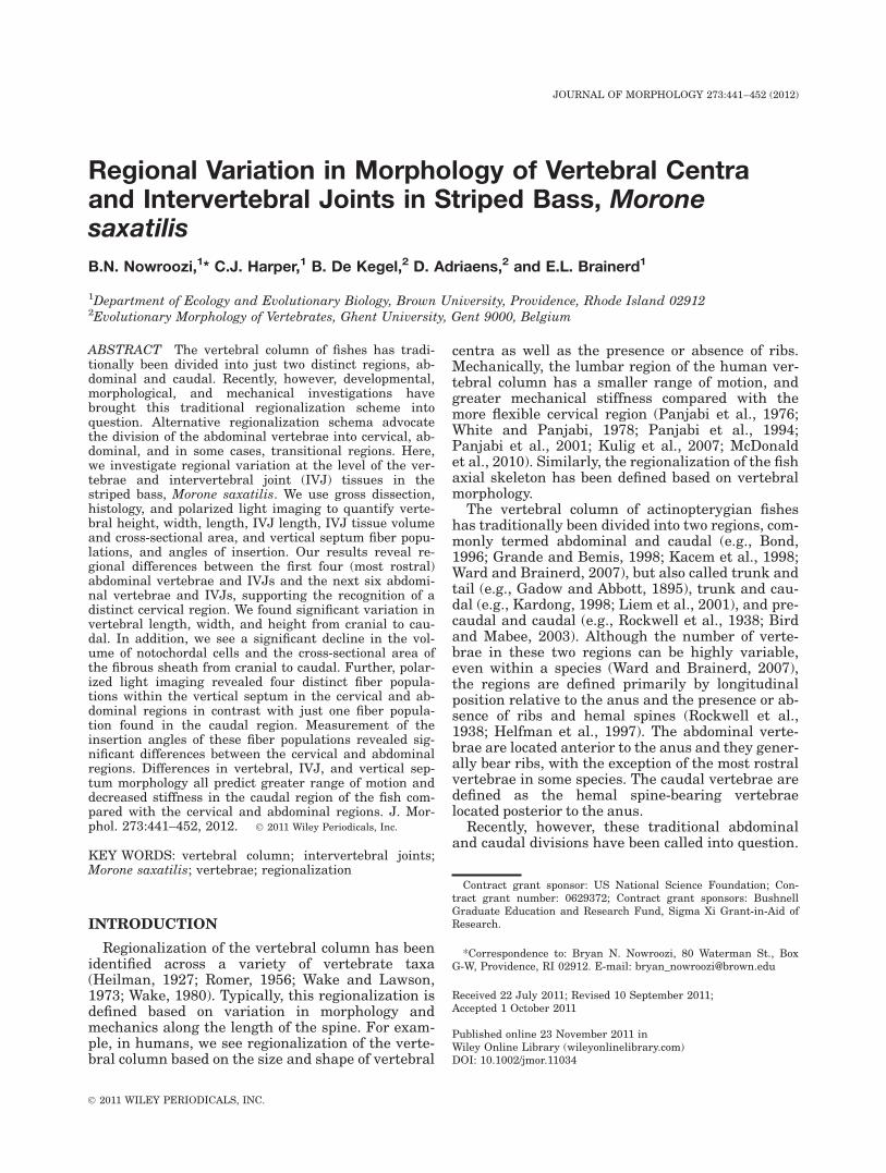
Most previous morphological analyses of region-alization in fishes have been based on gross mor-phology of vertebrae, and very little is knownabout the regional variation in soft tissue anatomyof the IVJs. The IVJ tissues likely play an impor-tant role in the mechanical properties of each jointalong the length of the vertebral column, andshould be included in a regionalization analysis. Inamphicoelous fish vertebrae, the IVJ structuresare housed within two adjacent vertebrae (Fig. 1).IVJ soft tissue structures can be divided into twoclasses: the tissues that encapsulate the notochor-dal remnants (encapulating complex), and thenotochordal remnants themselves [the notochordalcell mass (NCM), the notochordal epithelium (NE),and the notochordal strand, (Nst)].
The tissues that form the encapsulating complex(i.e., the joint capsule) consist of the fibroussheath, the elastica externa, and the externalintervertebral ligament (Schmitz, 1995; Nordviket al., 2005). Lying between the two vertebrae andextending just deep to the bone of the vertebra is
the fibrous sheath (Fig. 1). The fibrous sheath is ahelically wound structure in both the shortnosesturgeon (Acipenser brevirostratus) and Africanlungfish (Protopterus annectens), and can be acel-lular, as seen in the sturgeon, or contain fibro-blasts, as seen in the lungfish (Schmitz, 1998).Because of the putative homology of the fibroussheath with that of other craniates, it has beensuggested that this tissue is composed of Type IIcollagen and associated proteoglycans in fishes(Linsenmayer et al., 1973; Kimura and Kamimura,1982; Eikenberry et al., 1984; Schmitz, 1998; Grotmolet al., 2006). Between the rims of the two vertebrae,two additional encapsulating tissues can be found.The first is the elastica externa, an elastic layer posi-tioned superficially to the fibrous sheath. Superficialto that elastic layer is a layer composed of scleroto-mally derived collagen called the external interverte-bral ligament (Fig. 1). Each of the three tissueswithin the encapsulating complex is thought to resisttensile loading during the lateral bending of locomo-tion (Symmons, 1979, Schmitz, 1995, 1998).
Within the IVJ capsule, the cellular remnants ofthe notochordal tissue form the notochordal cellmass, notochordal strand, and notochordal epithe-lium (Fig. 1). The notochordal cell mass consists ofnotochordal cells of varying degrees of vacuoliza-tion with each vacuole surrounded by networks ofintermediate filaments. These dense filamentousnetworks of adjacent cells are connected to oneanother by desmosomes that promote cell-to-celladhesion, allowing the vacuoles to resist compres-sive loads as the filaments distribute this load(Schmitz, 1995; Junqueira and Carneiro, 2005).The notochordal strand, thought to be present onlyin basal ray-finned fishes, runs longitudinallyalong the length of the vertebral column passingthrough the center of each amphicoel via a noto-chordal foramen (Fig. 1). Previous studies suggestthat this axial strand is under a tensile load that
Fig. 1. Sagittal section schematic of three amphicoelous ver-tebral centra and two intervertebral joints (IVJs). Adapted fromSymmons, 1979. Abbreviations: C, centrum; DLL, dorsal longi-tudinal ligament; EC, encapsulating complex; EE, elasticaexterna; EIL, external intervertebral ligament; FS, fibroussheath; L, lacunae; NC, nerve cord; NCM, notochordal cellmass; NE, notochordal epithelium; NF, notochordal foramen;Nst, notochordal strand.
442 B.N. NOWROOZI ET AL.
Journal of Morphology
might play a role in elastic resilience during loco-motion (Symmons, 1979). In the persistant noto-chord of sturgeon and lungfishes, this strand iscomposed of the intermediate filaments of col-lapsed vacuolated notochordal cells (Schmitz,1998). The last of the remnants of the notochordaltissues is a lining composed of a thin layer of noto-chordal cells called the notochordal epithelium.This tissue separates the two extracellular lacunaefrom the notochordal cell mass. The lacunae arefluid filled spaces that occupy a substantial portionof the IVJ cavity and are thought to resist com-pressive loads related to locomotion (Schmitz,1995), and possibly supply nutrients to the noto-chordal cell mass (Symmons, 1979) (Fig. 1).
Finally, there are two tissues that lie external tothe IVJ capsule that we have included in ourinvestigation: the dorsal longitudinal ligament andthe vertical septum. The dorsal longitudinal liga-ment passes through the neural arch and lies dor-sal to the nerve chord (Fig. 1). The vertical septumis a sheet of connective tissue that spans the areabetween adjacent neural and hemal spines. Bothof these tissues have been implicated in resistinglateral bending and contributing to elastic energystorage during locomotion (Symmons, 1979; Vid-eler, 1993). In particular, the vertical septum isthought to store energy elastically not only in thefibers of the connective tissue but also by transfer-ring load to the associated neural and hemalspines (Videler, 1993). Given these hypotheses, anyvariation in these tissues will impact the mechan-ics of IVJ function during locomotion.
The primary goals of the present study are: (1)to describe and quantify regional variation in themorphology of the vertebral centra and IVJ tissuesalong the length of the vertebral column of an acti-nopterygian fish; (2) to determine whether thisvariation supports any of the proposed regionaliza-tion schema in fishes; and (3) to hypothesize howregional variation might affect the mechanical per-formance of the IVJs. We use gross dissection, se-rial sectioning of IVJs, and polarized light imagingof the vertical septum to investigate this variation.
We focused our investigation on the striped bass,Morone saxatilis (Walbaum), a perciform fish withrelatively small number of vertebrae (24) com-pared with other fishes of similar size. The lowvertebral number results in larger vertebrae andIVJs, which should facilitate our morphologicalmeasurements as well as future mechanical and invivo studies. In addition, the striped bass swimswith a nonspecialized, subcarangiform swimmingstyle that we hope will be a general case studyfrom which we can later compare the more special-ized swimming species.
MATERIALS AND METHODSSpecimens
We studied a total of 11 striped bass, M. saxatilis, by grossdissection, histology, and polarized light imaging. Individualsvaried in size from 0.17 m to 0.95 m in total length (TL) and0.20 kg to 11.1 kg in body mass (Table 1). The two largest speci-mens, 0.95 m in length and 0.66 m in length, were obtainedfrom local fish markets and freshly dissected for morphologicalmeasurements on the same day. The remaining nine individualswere obtained live from the Susquehanna Aquaculture, fishhatchery (York Haven, PA) and housed in a 300 gallon circularaquarium at 19–228C. Individuals of relatively similar TL andweight were chosen for both the histology and polarized lightimaging of the vertical septum. Conversely, individuals ofdiverse lengths and weights were used for gross morphologicalmeasurements (Table 1).
Morphological Measurements
Five individuals of M. saxatilis (Table 1) were studied bygross dissection and measurements were made using Mitotoyodigital calipers. Measurement precision was determined to be60.02 mm based on the standard deviation of 50 repeatedmeasurements of the length of a single vertebra. To preservethe natural hydration of the IVJs, all individuals were freshlydissected (i.e., specimens were never frozen and thawed).
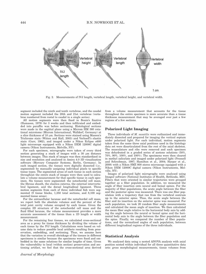
The axial musculature was removed from the axial skeletonand measurements were made of the length of the IVJs. Thislength was taken as the distance between the rims of adjacentvertebrae at the level of the lateral ridge (Fig. 2). We repeatedeach measurement three times and report the mean for eachIVJ. Once this measurement had been made for each IVJ alongthe length of the vertebral column, the vertebrae were disar-ticulated from one another and the remaining tissue removed tofacilitate centrum measurements.
For each individual centrum, we measured vertebral length,vertebral width, and vertebral height. Vertebral length wasmeasured along the lateral ridge of each centrum. Vertebralheight and width were measured from the anterior amphicoelfor each vertebra (Fig. 2). For the urostyle of each individual,the length measurement was taken as the length of the rostralhalf amphicoel. We repeated each measurement three timesand report the mean for each centrum. In addition, aspect ratio(vertebral centrum length/vertebral centrum width, see Wardand Brainerd, 2007) was calculated as a proxy for vertebralshape.
Histology
Three individuals of M. saxatilis were euthanized and imme-diately dissected to prepare for sectioning. For each individual,three vertebra-joint-vertebra specimens, called motion seg-ments, were dissected: the most rostral motion segmentincluded the third and fourth vertebrae, the abdominal motion
TABLE 1. Summary of individual Morone saxatilis
Total length (m) Body mass (kg)
Gross morphologyIndividual 01 0.36 0.52Individual 02 0.95 11.11Individual 03 0.66 3.35Individual 04 0.32 0.43Individual 05 0.34 0.45
HistologyIndividual 06 0.26 0.33Individual 07 0.28 0.39Individual 08 0.28 0.36
Polarized lightIndividual 09 0.33 0.73Individual 10 0.35 0.41Individual 11 0.28 0.42
REGIONAL VARIATION IN VERTEBRAL CENTRA AND IVJ 443
Journal of Morphology
segment included the ninth and tenth vertebrae, and the caudalmotion segment included the 20th and 21st vertebrae (verte-brae numbered from rostal to caudal in a single series).All motion segments were then fixed in Bouin’s fixative
(Humason, 1979) for 3 weeks and then infiltrated and embed-ded into paraffin wax before sectioning. Histological sectionswere made in the sagittal plane using a Microm HM 360 rota-tional microtome (Microm International, Walldorf, Germany) ata slice thickness of 10 lm. Sections were stained using Masson’sTrichrome stain (Witten and Hall, 2003) and Verhoeff ’s elastinstain (Gurr, 1962), and imaged under a Nikon Eclipse e600light microscope equipped with a Nikon DXM 12000C digitalcamera (Nikon Instruments, Melville, NY).For each specimen, micrographs were taken of every third
section generating a stack of images with a 30 lm distancebetween images. This stack of images was then standardized insize and resolution and analyzed in Amira 4.0 3D visualizationsoftware (Mercury Computer Systems, Berlin, Germany). Ineach imaged section, the tissues were digitally dissected (i.e.,segmented) by manually assigning individual pixels to specifictissue types. The segmented areas of each tissue in each sectionthroughout the entire stack of images were then used to calcu-late a volume measurement for that specific tissue in each spec-imen. Six tissues were segmented: the notochordal cell mass,the fibrous sheath, the elastica externa, the external interverte-bral ligament, and the dorsal longitudinal ligament. Threemotion segments from each of three individual fish were seg-mented (9 tissue blocks, 1,350 total sections, and 8,100 seg-mented tissue areas).For the extracellular lacunae and the notochordal cell mass,
we report both the absolute volumes and the percent of thetotal joint cavity volume occupied by each tissue. Given thecomplex 3D structure of the notochordal cell mass and thefluid filled lacunae, the volume measurement provides a moreaccurate assessment of the tissue than a 1D length or widthmeasurement.For the remaining four tissues, we calculated cross-sectional
area as a proxy for tissue thickness by dividing the volume ofeach tissue by the IVJ length. Calculations were based on vol-ume data to reduce possible local artifacts resulting from pres-ervation, embedding, and sectioning. Thus, we assume herethat the variation in overall shrinkage of the tissues in differentspecimens is similar because the specimens were fixed and em-bedded in the same solutions for similar lengths of time. Giventhe vulnerability to local (within section) preservation and sec-tioning artifact, we feel that the cross-sectional area derived
from a volume measurement that accounts for the tissuethroughout the entire specimen is more accurate than a tissuethickness measurement that may be averaged over just a fewregions of a few sections.
Polarized Light Imaging
Three individuals of M. saxatilis were euthanized and imme-diately dissected and prepared for imaging the vertical septumunder polarized light. For each individual, motion segmentstaken from the same three axial positions used in the histologydata set were disarticulated from the rest of the axial skeleton.The musculature and ribs were removed and each specimenwas dehydrated in a graded series of acetone solutions (50%,70%, 90%, 100%, and 100%). The specimens were then clearedin methyl salicylate and imaged under polarized light (Presnelland Schreibman, 1997; Hamilton et al., 2004; Harper et al.,2008) with a Nikon SMZ 800 stereo microscope equipped with aNikon DXM 12000C digital camera (Nikon Instruments, Mel-ville, NY).
Images of polarized light micrographs were analyzed usingImageJ software (National Institutes of Health, Bethesda, MD).Fibers that were oriented in similar trajectories were groupedtogether as a fiber population. In addition, we measured theangle of fiber insertion onto neural and hemal spines. For themajority of fiber populations, the acute angle between the fiberand the posterior spine was measured. There was one fiber pop-ulation with a trajectory that ran from the vertebral centrumup to the anterior spine. Thus, the acute angle between thefiber and its insertion on the anterior spine was measured. Foreach population, we took 20 random fiber angle measurementsand calculated the mean angle of insertion. We then calculatedthe mean fiber angle relative to the horizontal body axis by add-ing the angle between the neural or hemal spine and the hori-zontal body axis to the angle between the fiber population andthe spine. Finally, we compared the number of fiber popula-tions, and the normalized angles of each population, betweendifferent longitudinal regions of the three individuals.
Statistical Analysis
We analyzed data using a nested ANOVA analysis with axialposition nested within individual for all three quantitative datasets: gross morphological measurements, tissue volumes from
Fig. 2. Measurements of IVJ length, vertebral length, vertebral height, and vertebral width.
444 B.N. NOWROOZI ET AL.
Journal of Morphology
histology, and vertical septum fiber angles. The various meas-urements were incorporated as the response variables. For thehistological and polarized light data, differences between eachpair of regions were tested post hoc using Tukey HSD multiplecomparisons based on least-squared means. All statistical anal-yses were performed using JMP software (version 8.0.1, SASInstitute, Cary, NC).
RESULTSGross Morphology
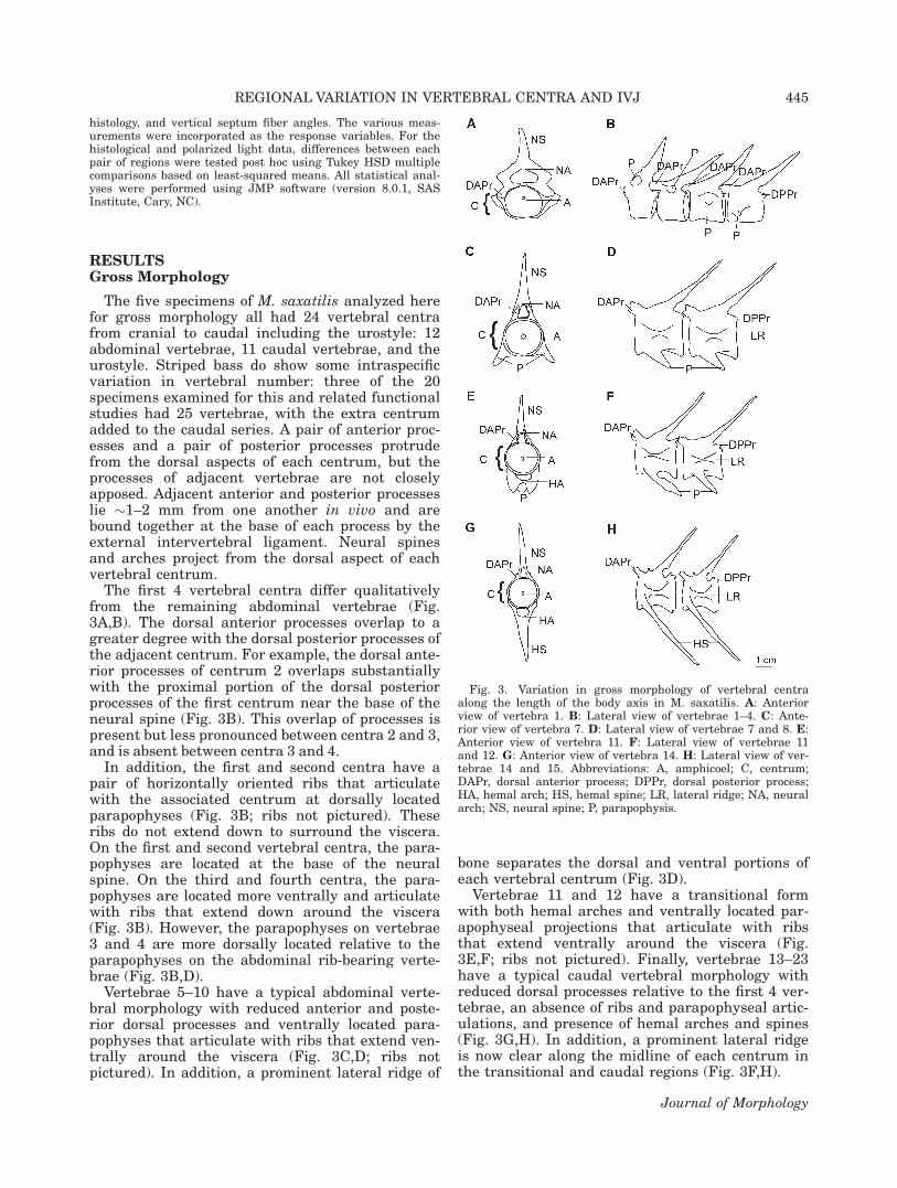
The five specimens of M. saxatilis analyzed herefor gross morphology all had 24 vertebral centrafrom cranial to caudal including the urostyle: 12abdominal vertebrae, 11 caudal vertebrae, and theurostyle. Striped bass do show some intraspecificvariation in vertebral number: three of the 20specimens examined for this and related functionalstudies had 25 vertebrae, with the extra centrumadded to the caudal series. A pair of anterior proc-esses and a pair of posterior processes protrudefrom the dorsal aspects of each centrum, but theprocesses of adjacent vertebrae are not closelyapposed. Adjacent anterior and posterior processeslie �1–2 mm from one another in vivo and arebound together at the base of each process by theexternal intervertebral ligament. Neural spinesand arches project from the dorsal aspect of eachvertebral centrum.
The first 4 vertebral centra differ qualitativelyfrom the remaining abdominal vertebrae (Fig.3A,B). The dorsal anterior processes overlap to agreater degree with the dorsal posterior processes ofthe adjacent centrum. For example, the dorsal ante-rior processes of centrum 2 overlaps substantiallywith the proximal portion of the dorsal posteriorprocesses of the first centrum near the base of theneural spine (Fig. 3B). This overlap of processes ispresent but less pronounced between centra 2 and 3,and is absent between centra 3 and 4.
In addition, the first and second centra have apair of horizontally oriented ribs that articulatewith the associated centrum at dorsally locatedparapophyses (Fig. 3B; ribs not pictured). Theseribs do not extend down to surround the viscera.On the first and second vertebral centra, the para-pophyses are located at the base of the neuralspine. On the third and fourth centra, the para-pophyses are located more ventrally and articulatewith ribs that extend down around the viscera(Fig. 3B). However, the parapophyses on vertebrae3 and 4 are more dorsally located relative to theparapophyses on the abdominal rib-bearing verte-brae (Fig. 3B,D).
Vertebrae 5–10 have a typical abdominal verte-bral morphology with reduced anterior and poste-rior dorsal processes and ventrally located para-pophyses that articulate with ribs that extend ven-trally around the viscera (Fig. 3C,D; ribs notpictured). In addition, a prominent lateral ridge of
bone separates the dorsal and ventral portions ofeach vertebral centrum (Fig. 3D).
Vertebrae 11 and 12 have a transitional formwith both hemal arches and ventrally located par-apophyseal projections that articulate with ribsthat extend ventrally around the viscera (Fig.3E,F; ribs not pictured). Finally, vertebrae 13–23have a typical caudal vertebral morphology withreduced dorsal processes relative to the first 4 ver-tebrae, an absence of ribs and parapophyseal artic-ulations, and presence of hemal arches and spines(Fig. 3G,H). In addition, a prominent lateral ridgeis now clear along the midline of each centrum inthe transitional and caudal regions (Fig. 3F,H).
Fig. 3. Variation in gross morphology of vertebral centraalong the length of the body axis in M. saxatilis. A: Anteriorview of vertebra 1. B: Lateral view of vertebrae 1–4. C: Ante-rior view of vertebra 7. D: Lateral view of vertebrae 7 and 8. E:Anterior view of vertebra 11. F: Lateral view of vertebrae 11and 12. G: Anterior view of vertebra 14. H: Lateral view of ver-tebrae 14 and 15. Abbreviations: A, amphicoel; C, centrum;DAPr, dorsal anterior process; DPPr, dorsal posterior process;HA, hemal arch; HS, hemal spine; LR, lateral ridge; NA, neuralarch; NS, neural spine; P, parapophysis.
REGIONAL VARIATION IN VERTEBRAL CENTRA AND IVJ 445
Journal of Morphology
Gross Morphological Measurements
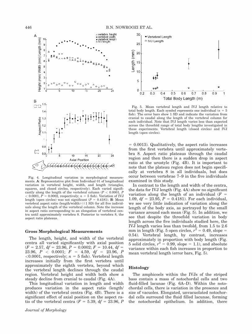
The length, height, and width of the vertebralcentra all varied significantly with axial position(F 5 2.57, df 5 23.96, P 5 0.0002; F 5 10.44, df 523.96, P < 0.0001; F 5 4.59, df 5 23.96, P<0.0001, respectively; n 5 5 fish). Vertebral lengthincreases initially from the first vertebra untilapproximately the eighth vertebra, beyond whichthe vertebral length declines through the caudalregion. Vertebral height and width both show asteady decline from cranial to caudal (Fig. 4A).
This longitudinal variation in length and widthproduces variation in the aspect ratio (length/width) of the vertebral centra (Fig. 4B). There is asignificant effect of axial position on the aspect ra-tio of the vertebral centra (F 5 5.39, df 5 23.96, P
5 0.0013). Qualitatively, the aspect ratio increasesfrom the first vertebra until approximately verte-bra 8. Aspect ratio plateaus through the caudalregion and then there is a sudden drop in aspectratio at the urostyle (Fig. 4B). It is important tonote that the plateau region does not begin specifi-cally at vertebra 8 in all individuals, but doesoccur between vertebrae 7–9 in the five individualsexamined in this study.
In contrast to the length and width of the centra,the data for IVJ length (Fig. 4A) show no significantvariation along the length of an individual (F 51.09, df 5 23.95, P 5 0.4181). For each individual,we see very little indication of variation along thelength of the body axis, as portrayed by the smallvariance around each mean (Fig. 5). In addition, wesee that despite the threefold variation in bodylength across the five individuals studied here, theIVJ length varies less than twofold, from 1.5 to 2.6mm in length (Fig. 5 open circles, r2 5 0.49, slope 50.54). Vertebral length, by contrast, increasesapproximately in proportion with body length (Fig.5 solid circles, r2 5 0.99, slope 5 1.1), and absolutevariance within each fish increases in proportion tomean vertebral length (error bars, Fig. 5).
Histology
The amphicoels within the IVJs of the stripedbass contain a mass of notochordal cells and twofluid-filled lacunae (Fig. 6A–D). Within the noto-chordal cells, there is variation in the presence andsize of vacuoles. Elongated, unvacuolated notochor-dal cells surround the fluid filled lacunae, formingthe notochordal epithelium. In addition, there
Fig. 4. Longitudinal variation in morphological measure-ments. A: Representative plot from Individual 01 of longitudinalvariation in vertebral height, width, and length (triangles,squares, and closed circles, respectively). Each varied signifi-cantly along the length of the vertebral column (P < 0.0001, P< 0.0001, P 5 0.0002, respectively; n 5 5 fish). Variation of IVJlength (open circles) was not significant (P 5 0.4181). B: Meanvertebral aspect ratio (length/width) (61 SD) for all five individ-uals along the length of the vertebral column. Note the increasein aspect ratio corresponding to an elongation of vertebral cen-tra until approximately vertebra 8. Posterior to vertebra 8, theaspect ratio plateaus.
Fig. 5. Mean vertebral length and IVJ length relative tototal body length. Each symbol represents one individual (n 5 5fish). The error bars show 1 SD and indicate the variation fromcranial to caudal along the length of the vertebral column foreach individual. Note that IVJ length varies less than expectedacross the threefold range of total body lengths investigated inthese experiments. Vertebral length (closed circles) and IVJlength (open circles).
446 B.N. NOWROOZI ET AL.
Journal of Morphology
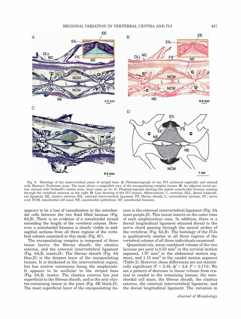
appears to be a loss of vacuolization in the notochor-dal cells between the two fluid filled lacunae (Fig.6A,B). There is no evidence of a notochordal strandextending the length of the vertebral column. How-ever, a notochordal foramen is clearly visible in mid-sagittal sections from all three regions of the verte-bral column examined in this study (Fig. 6C).
The encapsulating complex is composed of threetissue layers: the fibrous sheath, the elasticaexterna, and the external intervertebral ligament(Fig. 6A,B, insets,D). The fibrous sheath (Fig. 6Ablue,D) is the deepest layer of the encapsulatingtissues. It is thickened in the intervertebral region,but has narrow extensions lining the amphicoels.It appears to be acellular in the striped bass(Fig. 6A,B, insets). The elastica externa lies justsuperficial to the fibrous sheath, and is the only elas-tin-containing tissue in the joint (Fig. 6B black,D).The most superficial layer of the encapsulating tis-
sues is the external intervertebral ligament (Fig. 6Ainset purple,D). This tissue inserts on the outer rimsof each amphicoelous cone. In addition, there is adorsal longitudinal ligament situated dorsal to thenerve chord passing through the neural arches ofthe vertebrae (Fig. 6A,B). The histology of the IVJsis qualitatively similar in all three regions of thevertebral column of all three individuals examined.
Quantitatively, mean combined volume of the twolacunae per joint is 0.55 mm3 in the cervical motionsegment, 1.97 mm3 in the abdominal motion seg-ment, and 1.15 mm3 in the caudal motion segment(Table 2). However, these differences are not statisti-cally significant (F 5 2.38, df 5 2.6, P 5 0.174). Wesee a pattern of decrease in tissue volume from cra-nial to caudal in the remaining tissues: the noto-chordal cell mass, the fibrous sheath, the elasticaexterna, the external intervertebral ligament, andthe dorsal longitudinal ligament. The variation in
Fig. 6. Histology of the intervertebral joints of striped bass. A: Photomicrograph of one IVJ sectioned sagittally and stainedwith Masson’s Trichrome stain. The inset shows a magnified view of the encapsulating complex tissues. B: An adjacent serial sec-tion stained with Verhoeff ’s elastin stain. Inset same as (A). C: Photomicrograph showing the patent notochordal foramen passingthrough the vertebral centrum on the right. D: Line drawing of the IVJ tissues. Abbreviations: C, centrum; DLL, dorsal longitudi-nal ligament; EE, elastica externa; EIL, external intervertebral ligament; FS, fibrous sheath; L, extracellular lacunae; NC, nervecord; NCM, notochordal cell mass; NE, notochordal epithelium; NF, notochordal foramen.
REGIONAL VARIATION IN VERTEBRAL CENTRA AND IVJ 447
Journal of Morphology
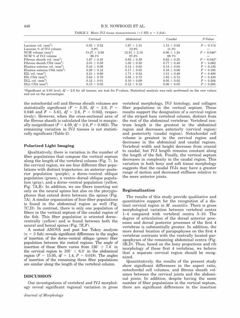
the notochordal cell and fibrous sheath volumes arestatistically significant (F 5 5.35, df 5 2.6, P 50.046 and F 5 5.61, df 5 2.6, P 5 0.042, respec-tively). However, when the cross-sectional area ofthe fibrous sheath is calculated the trend is margin-ally insignificant (F5 4.59, df5 2.6, P5 0.062). Theremaining variation in IVJ tissues is not statisti-cally significant (Table 2).
Polarized Light Imaging
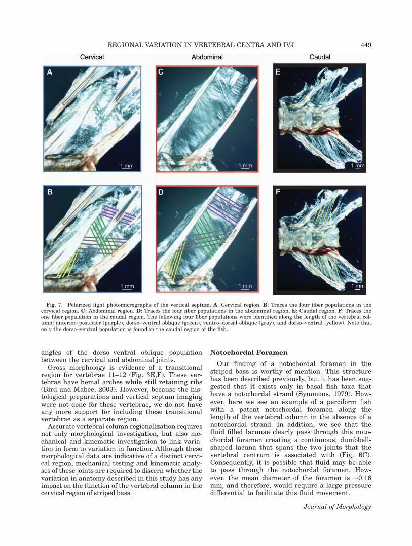
Qualitatively, there is variation in the number offiber populations that compose the vertical septumalong the length of the vertebral column (Fig. 7). Inthe cervical region, we see four different fiber popu-lations with distinct trajectories: an anterior–poste-rior population (purple), a dorso–ventral obliquepopulation (green), a ventro–dorsal oblique popula-tion (gray), and a dorso–ventral population (yellow,Fig. 7A,B). In addition, we see fibers inserting notonly on the neural spines but also on the pterygio-phores that extend down between the spines (Fig.7A). A similar organization of four fiber populationsis found in the abdominal region as well (Fig.7C,D). In contrast, there is only one population offibers in the vertical septum of the caudal region ofthe fish. This fiber population is oriented dorso–ventrally (yellow) and is found between both theneural and hemal spines (Fig. 7E,F).
A nested ANOVA and post hoc Tukey analysis(n 5 3 fish) reveals significant differences in the anglesof insertion of the dorso–ventral oblique (green) fiberpopulation between the rostral regions. The angle ofinsertion of these fibers varies from 1268 6 7.48 inthe cervical region to 1058 6 6.38 in the abdominalregion (F 5 15.05, df 5 1.4, P 5 0.018). The anglesof insertion of the remaining three fiber populationsare similar along the length of the vertebral column.
DISCUSSION
Our investigations of vertebral and IVJ morphol-ogy reveal significant regional variation in gross
vertebral morphology, IVJ histology, and collagenfiber populations in the vertical septum. Theseresults support the designation of a cervical regionof the striped bass vertebral column, distinct fromthe rest of the abdominal vertebrae. Vertebral cen-trum length is the greatest in the abdominalregion and decreases anteriorly (cervical region)and posteriorly (caudal region). Notochordal cellvolume is greatest in the cervical region anddecreases in the abdominal and caudal regions.Vertebral width and height decrease from cranialto caudal, but IVJ length remains constant alongthe length of the fish. Finally, the vertical septumdecreases in complexity in the caudal region. Thisvariation in both bony and soft tissue morphologysuggests that the caudal IVJs may have a greaterrange of motion and decreased stiffness relative tothe more anterior joints.
Regionalization
The results of this study provide qualitative andquantitative support for the recognition of a dis-tinct cervical region in M. saxatilis. There is grossmorphological variation between vertebral centra1–4 compared with vertebral centra 5–10. Thedegree of articulation of the dorsal anterior proc-esses and dorsal posterior processes of the first 4vertebrae is substantially greater. In addition, themore dorsal location of parapophyses on the first 4vertebrae contrasts with the ventrally located par-apophyses of the remaining abdominal centra (Fig.3B,D). Thus, based on the bony projections and ribmorphology of these first 4 vertebrae, we believethat a separate cervical region should be recog-nized.
Quantitatively, the results of the present studyshow significant differences in the aspect ratio,notochordal cell volumes, and fibrous sheath vol-umes between the cervical joints and the abdomi-nal joints. In addition, despite having the samenumber of fiber populations in the vertical septum,there are significant differences in the insertion
TABLE 2. Mean IVJ tissue measurements (61 SD; n 5 3 fish)
Cervical Abdominal Caudal P-Value
Lacunae vol. (mm3) 0.55 6 0.22 1.97 6 1.31 1.15 6 0.02 P 5 0.174Lacunae % of IVJ volume 3.9% 12.8% 11.3% —NCM volume (mm3) 13.27 6 2.00 12.91 6 2.10 8.90 6 1.24 P 5 0.046*NCM % of IVJ volume 96.1% 87.2% 88.7% —Fibrous sheath vol. (mm3) 1.67 6 0.45 0.85 6 0.39 0.63 6 0.35 P 5 0.042*Fibrous sheath CSA (mm2) 2.01 6 0.59 1.02 6 0.50 0.77 6 0.48 P 5 0.062Elastica externa vol. (mm3) 0.24 6 0.09 0.14 6 0.02 0.13 6 0.04 P 5 0.138Elastica externa CSA (mm2) 0.29 6 0.12 0.17 6 0.03 0.16 6 0.06 P 5 0.165EIL vol. (mm3) 2.21 6 0.69 1.71 6 0.54 1.51 6 0.49 P 5 0.400EIL CSA (mm2) 2.64 6 0.79 2.05 6 0.72 1.82 6 0.74 P 5 0.438DLL vol. (mm3) 0.12 6 0.01 0.10 6 0.09 0.05 6 0.02 P 5 0.284DLL CSA (mm2) 0.15 6 0.02 0.12 6 0.10 0.06 6 0.03 P 5 0.265
*Significant at 0.05 level, df 5 2.6 for all tissues; see text for F-values. Statistical analysis was only performed on the raw valuesand not on the percentages.
448 B.N. NOWROOZI ET AL.
Journal of Morphology
angles of the dorso–ventral oblique populationbetween the cervical and abdominal joints.
Gross morphology is evidence of a transitionalregion for vertebrae 11–12 (Fig. 3E,F). These ver-tebrae have hemal arches while still retaining ribs(Bird and Mabee, 2003). However, because the his-tological preparations and vertical septum imagingwere not done for these vertebrae, we do not haveany more support for including these transitionalvertebrae as a separate region.
Accurate vertebral column regionalization requiresnot only morphological investigation, but also me-chanical and kinematic investigation to link varia-tion in form to variation in function. Although thesemorphological data are indicative of a distinct cervi-cal region, mechanical testing and kinematic analy-ses of these joints are required to discern whether thevariation in anatomy described in this study has anyimpact on the function of the vertebral column in thecervical region of striped bass.
Notochordal Foramen
Our finding of a notochordal foramen in thestriped bass is worthy of mention. This structurehas been described previously, but it has been sug-gested that it exists only in basal fish taxa thathave a notochordal strand (Symmons, 1979). How-ever, here we see an example of a perciform fishwith a patent notochordal foramen along thelength of the vertebral column in the absence of anotochordal strand. In addition, we see that thefluid filled lacunae clearly pass through this noto-chordal foramen creating a continuous, dumbbell-shaped lacuna that spans the two joints that thevertebral centrum is associated with (Fig. 6C).Consequently, it is possible that fluid may be ableto pass through the notochordal foramen. How-ever, the mean diameter of the foramen is �0.16mm, and therefore, would require a large pressuredifferential to facilitate this fluid movement.
Fig. 7. Polarized light photomicrographs of the vertical septum. A: Cervical region. B: Traces the four fiber populations in thecervical region. C: Abdominal region. D: Traces the four fiber populations in the abdominal region. E: Caudal region. F: Traces theone fiber population in the caudal region. The following four fiber populations were identified along the length of the vertebral col-umn: anterior–posterior (purple), dorso–ventral oblique (green), ventro–dorsal oblique (gray), and dorso–ventral (yellow). Note thatonly the dorso–ventral population is found in the caudal region of the fish.
REGIONAL VARIATION IN VERTEBRAL CENTRA AND IVJ 449
Journal of Morphology
Functional Implications
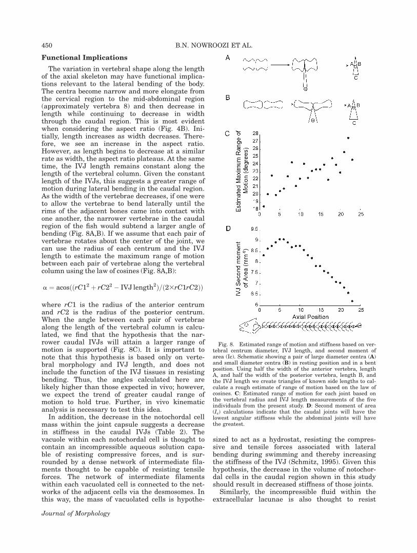
The variation in vertebral shape along the lengthof the axial skeleton may have functional implica-tions relevant to the lateral bending of the body.The centra become narrow and more elongate fromthe cervical region to the mid-abdominal region(approximately vertebra 8) and then decrease inlength while continuing to decrease in widththrough the caudal region. This is most evidentwhen considering the aspect ratio (Fig. 4B). Ini-tially, length increases as width decreases. There-fore, we see an increase in the aspect ratio.However, as length begins to decrease at a similarrate as width, the aspect ratio plateaus. At the sametime, the IVJ length remains constant along thelength of the vertebral column. Given the constantlength of the IVJs, this suggests a greater range ofmotion during lateral bending in the caudal region.As the width of the vertebrae decreases, if one wereto allow the vertebrae to bend laterally until therims of the adjacent bones came into contact withone another, the narrower vertebrae in the caudalregion of the fish would subtend a larger angle ofbending (Fig. 8A,B). If we assume that each pair ofvertebrae rotates about the center of the joint, wecan use the radius of each centrum and the IVJlength to estimate the maximum range of motionbetween each pair of vertebrae along the vertebralcolumn using the law of cosines (Fig. 8A,B):
a ¼ acosððrC12 þ rC22 � IVJ length2Þ=ð23rC1rC2ÞÞ
where rC1 is the radius of the anterior centrumand rC2 is the radius of the posterior centrum.When the angle between each pair of vertebraealong the length of the vertebral column is calcu-lated, we find that the hypothesis that the nar-rower caudal IVJs will attain a larger range ofmotion is supported (Fig. 8C). It is important tonote that this hypothesis is based only on verte-bral morphology and IVJ length, and does notinclude the function of the IVJ tissues in resistingbending. Thus, the angles calculated here arelikely higher than those expected in vivo; however,we expect the trend of greater caudal range ofmotion to hold true. Further, in vivo kinematicanalysis is necessary to test this idea.
In addition, the decrease in the notochordal cellmass within the joint capsule suggests a decreasein stiffness in the caudal IVJs (Table 2). Thevacuole within each notochordal cell is thought tocontain an incompressible aqueous solution capa-ble of resisting compressive forces, and is sur-rounded by a dense network of intermediate fila-ments thought to be capable of resisting tensileforces. The network of intermediate filamentswithin each vacuolated cell is connected to the net-works of the adjacent cells via the desmosomes. Inthis way, the mass of vacuolated cells is hypothe-
sized to act as a hydrostat, resisting the compres-sive and tensile forces associated with lateralbending during swimming and thereby increasingthe stiffness of the IVJ (Schmitz, 1995). Given thishypothesis, the decrease in the volume of notochor-dal cells in the caudal region shown in this studyshould result in decreased stiffness of those joints.
Similarly, the incompressible fluid within theextracellular lacunae is also thought to resist
Fig. 8. Estimated range of motion and stiffness based on ver-tebral centrum diameter, IVJ length, and second moment ofarea (Ic). Schematic showing a pair of large diameter centra (A)and small diameter centra (B) in resting position and in a bentposition. Using half the width of the anterior vertebra, lengthA, and half the width of the posterior vertebra, length B, andthe IVJ length we create triangles of known side lengths to cal-culate a rough estimate of range of motion based on the law ofcosines. C: Estimated range of motion for each joint based onthe vertebral radius and IVJ length measurements of the fiveindividuals from the present study. D: Second moment of area(Ic) calculations indicate that the caudal joints will have thelowest angular stiffness while the abdominal joints will havethe greatest.
450 B.N. NOWROOZI ET AL.
Journal of Morphology
compressive forces associated with locomotion. Thesquamous, nonvacuolated cells lining the lacunaeand separating the two lacunae in the interverte-bral septum contain dense intermediate filaments.The adjacent cells adhere to one another via des-mosomal connections that are thought to resistshearing forces (Schmitz, 1995). Thus, the extrac-ellular lacunae are thought to increase joint stiff-ness hydrostatically as well (Schmitz, 1995). Theresults of this study do not yield a clear conclusionabout longitudinal variation in the volumes of theextracellular lacunae. Mean lacuna size was larg-est in the abdominal region and smaller in thecervical region and the caudal region (Table 2).Although this variation was not significant, itagain points towards decreased stiffness in thecaudal joints, and possibly the cervical joints,compared with the abdominal joints.
The volume of the fibrous sheath decreases signif-icantly along the length of the vertebral column(Table 2). This decreasing trend holds when thecross-sectional area of the tissue is calculated, how-ever, the result is no longer significant. This dis-crepancy is likely related to the small sample sizeincluded in the analysis, thus the results should beviewed with caution. The fibrous sheath is thoughtto be made up of helically wound, type II collagen,similar to the fibrous sheath of cyclostomes and tet-rapods (Linsenmayer et al., 1973; Kimura andKamimura, 1982; Eikenberry et al., 1984; Schmitz,1998). Additionally, the fibrous sheath of the stripedbass lines the bony cone of the amphicoel and thick-ens in the intervertebral region (Fig. 6A,B). In thismanner, the fibrous sheath connects the two adja-cent vertebrae, and thus, likely plays a role inresisting loading during bending. The decrease inthe volume of this fibrous sheath from cranial tocaudal suggests a potential decrease in IVJ stiffnessalong the length of the fish.
These differences in soft tissue anatomy all sug-gest a decrease in stiffness along the length of thevertebral column. These structural tissues shouldinfluence the second moment of area, a structuralpredictor of flexural stiffness, of each IVJ. Specifi-cally, the encapsulating tissues should have thegreatest impact on the second moment of areabecause they are located furthest from the neutralaxis of bending. Thus, we expect a decline in thesecond moment of area from cranial to caudalassociated with the decline of the fibrous sheathand possibly the notochordal cell mass.
If we model the encapsulating tissues of eachIVJ as circular in cross section (see Porter et al.,2009), we can calculate the second moment of areawith the following equation:
Ic ¼ pr4=4
where r is the estimated radius of each IVJ takenas the average of the height and transverse width
of the associated anterior centrum. The mean Icacross the five individuals results in an initialincrease in stiffness from the cervical to the ab-dominal region, and then a decline in stiffnessthrough the caudal region (Fig. 8D). This initialincrease indicates that the flexural stiffness of thecervical joints may actually be less than the ab-dominal joints. This again supports the inclusionof a distinct cervical region, although it is in con-trast to our predictions of greater stiffness in themost rostral joints. All the rostral joints, however,appear to have greater flexural stiffness than thecaudal region, again indicating decreased stiffnesscaudally.
It is important to note that engineering theoryassumes a homogenous material when calculatingthe second moment of area. The IVJs violate thisassumption and are not exactly circular in crosssection. However, this simplified model does indi-cate that the variation in anatomy revealed in thisstudy should result in decreased structural stiff-ness in the caudal region of the vertebral column.Further, mechanical testing is necessary to sup-port this hypothesis.
Finally, this study has shown a substantial dif-ference in the number of fiber populations found inthe vertical septum along the length of an individ-ual striped bass (Fig. 7). In the cervical and ab-dominal regions of the fish, we identify four fiberpopulations with distinct trajectories. However, inthe caudal region of the fish, only one fiber popula-tion is present (Fig. 7C,F). If the vertical septumplays a role in resisting bending during swimming,this variation in fiber population again points to-ward lower stiffness in the caudal region of thefish, relative to the cervical and abominal regions.
These morphological results consistently predicta decrease in stiffness and an increase in therange of motion in the caudal region of the stripedbass. However, previous studies in the blue marlinshow increased joint stiffness caudally (Hebranket al., 1990; Long, 1992). In addition, investigationof the mechanics of Norfolk spot found uniformstiffness along the length of the spine (Hebrank,1982). These previous mechanical results do notagree with our morphological findings. Thus, fur-ther mechanical analysis is necessary to determinewhether the morphological regionalizationdescribed here corresponds to mechanical regional-ization of the striped bass vertebral column.
ACKNOWLEDGMENTS
The authors thank Susquehanna Aquaculture,Inc. for providing the live animals for this study.They also thank Eric LoPresti and Nicholas Gid-mark for help with fish housing and husbandry. Aspecial thanks to Steve Gatesy, Thomas Roberts,Sharon Swartz, John Long, Manny Azizi, Erika
REGIONAL VARIATION IN VERTEBRAL CENTRA AND IVJ 451
Journal of Morphology
Giblin, and the Morphology Group at Brown forhelpful discussion and suggestions.
LITERATURE CITED
Bird NC, Mabee PM. 2003. Developmental morphology of theaxial skeleton of the Zebrafish, Danio rerio (Ostariophysi:Cyprinidae). Dev Dyn 228:337–357.
Bond CE. 1996. Biology of Fishes, 2nd ed. Philadelphia: Saun-ders College Publishing.
Burke AC, Nelson CE, Morgan BA, Tabin C. 1995. Hox genesand the evolution of vertebrate axial morphology. Develop-ment 121:333–346.
Eikenberry EF, Childs B, Sheren SB, Parry DAD, Craig AS,Brodsky B. 1984. Crystalline fibril structure of type II colla-gen in lamprey notochord sheath. J Mol Biol 176:261–277.
Gadow H, Abbott EC. 1895. On the evolution of the vertebralcolumn of fishes. Philos Trans R Soc London B 186:163–221.
Grande L, Bemis WE. 1998. A comprehensive phylogeneticstudy of amiid fishes (Amiidae) based on comparative skeletalanatomy. An empirical search for interconnected patterns ofnatural history. J Vertebr Paleontol 18:1–690.
Grotmol S, Kryvi H, Keynes R, Krossoy C, Nordvik K, TotlandGK. 2006. Stepwise enforcement of the notochord and itsintersection with the myoseptum: An evolutionary path lead-ing to development of the vertebra? J Anat 209:339–357.
Gurr E. 1962. Staining Animal Tissues: Practical and Theoreti-cal. London: L. Hill.
Hamilton JL, Dillaman RM, McLellan WA, Pabst DA. 2004.Structural fiber reinforcement of keel blubber in harbor por-poise (Phocoena phocoena). J Morph (2004) 261:105–117.
Harper CJ, McLellan WA, Rommel SA, Gay DM, Dillaman RM,Pabst DA. 2008. Morphology of the melon and its tendinousconnections to the facial muscles in bottlenose dolphins (Tur-siops truncatus). J Morph 269:820–839.
Hebrank MR. 1982. Mechanical properties of fish backbones inlateral bending and tension. J Biomech 15:85–89.
Hebrank JH, Hebrank MR, Long JH, Block BA, WainwrightSA. 1990. Backbone mechanics of the blue marlin Makairanigricans (Pisces, Istiophoridae). J Exp Biol 148:449–459.
Heilman G. 1927. The Origin of Birds. New York: D. Appeltonand Company.
Helfman GS, Collette BB, Facey DE. 1997. The Diversity ofFishes. Malden: Blackwell Science.
Humason GL. 1979. Animal Tissue Techniques, 4th ed. SanFrancisco: WH Freeman and Company.
Junqueira L, Carneiro,J. 2005. Basic Histology, 11th ed. Colum-bus: McGraw-Hill Medical.
Kacem A, Meunier FJ, Bagliniere JL. 1998. A quantitativestudy of morphological and histological changes in the skele-ton of Salmo salar during its anadromous migration. J FishBiol 53:1096–1109.
Kardong KV. 1998. Vertebrates: Comparative Anatomy, Func-tion, Evolution. Boston: WCB/McGraw-Hill.
Kimura S, Kimimura,T. 1982. Characterization of lamprey noto-chord collagen with special reference to its skin collagen.Comp Biochem Physiol 73B:335–339.
Kulig K, Powers CM, Landel RF, Chen H, Fredericson M, Guil-let M, Butts K. 2007. Segmental lumbar mobility in individu-als with low back pain: In vivo assessment during manualand self-imposed motion using dynamic MRI. BMC Musculos-kelet Disord 8:8–18.
Liem KF, Bemis WE, Walker WF, Grande L. 2001. FunctionalAnatomy of the Vertebrates, 3rd ed. Belmont: Brooks Cole.
Linsenmayer TF, Trelstad RL, Gross J. 1973. The collagen ofchick embryonic notochord. Biochem Biophys Res Commun53:39–45.
Long JH. 1992. Stiffness and damping forces in the interverte-bral joints of blue marlin (Makaira nigricans). J Exp Biol162:131–155.
McDonald CP, Bachison CC, Chang V, Bartol SW, Bey MJ.2010. Three-dimensional dynamic in vivo motion of the cervi-cal spine: Assessment of measurement accuracy and prelimi-nary findings. Spine J 10:497–504.
Morin-Kensicki EM, Melancon E, Eisen JS. 2002. Segmentalrelationship between somites and vertebral column in zebra-fish. Development 129:3851–3860.
Nordvik K, Kryvi H, Totland GK, Grotmol S. 2005. The salmonvertebral body develops through mineralization of two pre-formed tissues that are encompassed by two layers of bone. JAnat 206:103–114.
Panjabi MM, Brand RA, White AA. 1976. Mechanical propertiesof the human thoracic spine as shown by three-dimensionalload-displacement curves. J Bone Joint Surg Am 58:642–652.
Panjabi MM, Oxland TR, Yamamoto I, Crisco JJ. 1994. Mechan-ical behavior of the human lumbar and lumbosacral spine asshown by three-dimensional load-displacement curves. J BoneJoint Surg Am 76:413–424.
Panjabi MM, Crisco JJ, Vasavada A, Oda T, Cholewicki J, NibuK, Shin E. 2001. Mechanical properties of the human cervicalspine as shown by three-dimensional load-displacementcurves. Spine 26:2692–2700.
Porter ME, Roque CM, Long JH. 2009. Turning maneuvers insharks: Predicting body curvature from axial morphology. JMorph 270:954–965.
Presnell J, Schreibman M. 1997. Humanson’s Animal TissueTechniques. Baltimore: Johns Hopkins University Press.
Rockwell H, Evans FG, Homer,CP. 1938. The comparative mor-phology of the vertebrate spinal column. Its form as relatedto function. J Morph 63:87–117.
Romer AS. 1956. Osteology of Reptiles. Chicago: University ofChicago Press.
Schmitz RJ. 1995. Ultrastructure and function of cellular com-ponents of the intercentral joint in the percoid vertebral col-umn. J Morph 226:1–24.
Schmitz RJ. 1998. Comparative ultrastructure of the cellularcomponents of the unconstricted notochord in the sturgeonand the lungfish. J Morphol 236:75–104.
Symmons S. 1979. Notochordal and elastic components of theaxial skeleton of fishes and their functions in locomotion. JZool 189:157–206.
Videler JJ. 1993. Fish Swimming. London: Chapman and Hall.Wake MH. 1980. Morphometrics of the skeleton of Dermophis
mexicanus (Amphibia: Gymnophiona). I. The vertebrae, withcomparisons to other species. J Morph 165:117–130.
Wake DB, Lawson RL. 1973. Developmental and adult morphol-ogy of the vertebral column in the plethodontid salamanderEurycea bislineata, with comments on vertebral evolution inthe amphibia. J Morph 139:251–300.
Ward AB, Brainerd EL. 2007. Evolution of axial patterning inelongate fishes. Biol J Linn Soc 90:97–116.
White A, Panjabi MM. 1978. Clinical Biomechanics of theSpine, 2nd ed. Philadelphia: Lippincott Williams and Wilkins.
Witten PE, Hall BK. 2003. Seasonal changes in the lower jawskeleton in male Atlantic salmon (Salmo salar L): Remodel-ing and regression of the kype after spawning. J Anat203:435–450.
452 B.N. NOWROOZI ET AL.
Journal of Morphology


![[Accessory] [9361] Rock of Bral](https://img.pdfslide.us/doc/110x75/577cd5761a28ab9e789ad89a/accessory-9361-rock-of-bral.jpg)