Embed Size (px)
Citation preview
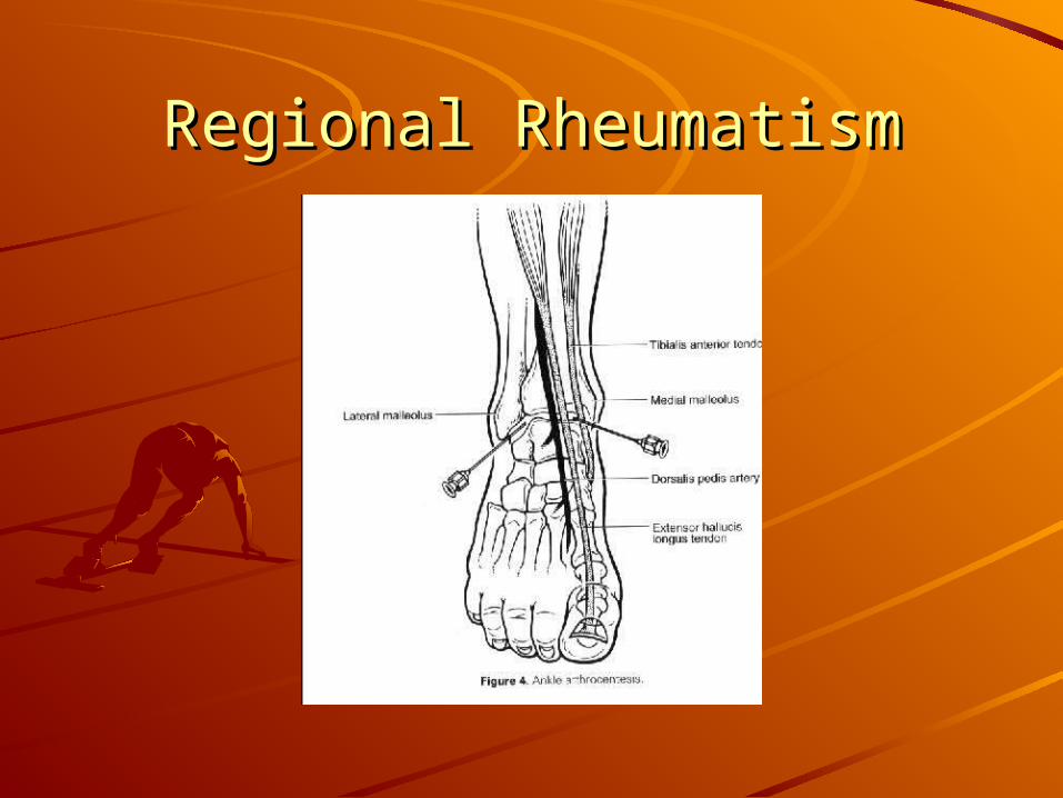
Regional RheumatismRegional Rheumatism
Andres Quiceno, MDAndres Quiceno, MDRheumatology Division PHDRheumatology Division PHD
Clinical Assistant Professor of Clinical Assistant Professor of Medicine UTSWMedicine UTSW
Regional RheumatismRegional RheumatismThese conditions are among the most These conditions are among the most poorly taught subjects in medical school.poorly taught subjects in medical school.
Even in the orthopedic and rheumatology Even in the orthopedic and rheumatology programs.programs.
These ailments are extremely common in These ailments are extremely common in medical practice.medical practice.
The medical conditions included here are The medical conditions included here are tenosynovitis, bursitis, fasciitis, tenosynovitis, bursitis, fasciitis, enthesopathy and compression enthesopathy and compression neuropathy.neuropathy.
Regional RheumatismRegional RheumatismApproximately 33% of United States adults Approximately 33% of United States adults have a musculoskeletal complaint.have a musculoskeletal complaint.
In patients over 65, musculoskeletal In patients over 65, musculoskeletal symptoms are the most common symptoms are the most common complaint reported and the most common complaint reported and the most common cause of functional limitation.cause of functional limitation.
Musculoskeletal and rheumatological Musculoskeletal and rheumatological conditions are frequently chronic and have conditions are frequently chronic and have a significant social and economical cost.a significant social and economical cost.
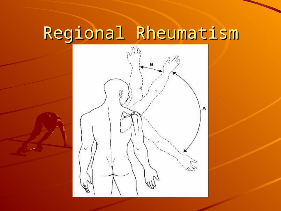
Regional RheumatismRegional RheumatismIMPINGEMENT SYNDROMEIMPINGEMENT SYNDROMEChronic shoulder pain is the most common upper Chronic shoulder pain is the most common upper extremity problem in recreational, competitive extremity problem in recreational, competitive and elite athletes.and elite athletes.This problem is more common in throwing This problem is more common in throwing athletes, racquet sports, volleyball, gymnasts and athletes, racquet sports, volleyball, gymnasts and swimmers.swimmers.This kind of athletes need full, unrestricted upper This kind of athletes need full, unrestricted upper extremity function to perform in their sport.extremity function to perform in their sport.Even mild degree of pain and dysfunction can Even mild degree of pain and dysfunction can result in complete disability for their respective result in complete disability for their respective sports.sports.
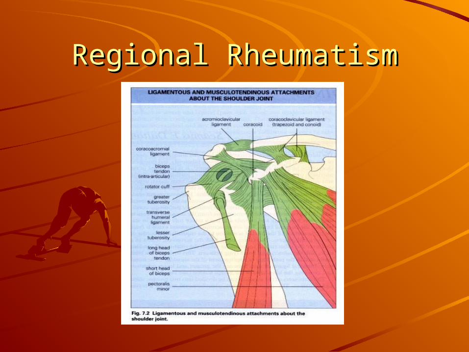
Regional RheumatismRegional RheumatismThe glenohumeral joint represents the The glenohumeral joint represents the articulation of the humerus and glenoid fossa.articulation of the humerus and glenoid fossa.
It is the most mobile joint in the body.It is the most mobile joint in the body.
The joint is stabilized by multiple ligaments and The joint is stabilized by multiple ligaments and muscles including the rotator cuff.muscles including the rotator cuff.
The rotator cuff comprises four muscles and their The rotator cuff comprises four muscles and their tendons: the subscapularis, the supraspinatus, tendons: the subscapularis, the supraspinatus, the infraspinatus and the teres minor.the infraspinatus and the teres minor.
The most commonly affected tendon is the The most commonly affected tendon is the supraspinatus.supraspinatus.
Regional RheumatismRegional Rheumatism
Regional RheumatismRegional Rheumatism
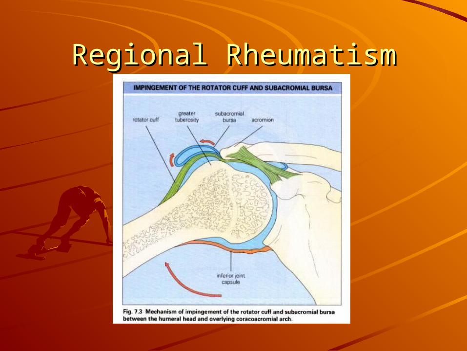
Regional RheumatismRegional RheumatismProblems of the rotator cuff involve many Problems of the rotator cuff involve many tendon abnormalities.tendon abnormalities.The most common cause full-thickness The most common cause full-thickness rotator cuff tears are chronic and most rotator cuff tears are chronic and most likely represent the final pathway of likely represent the final pathway of chronic subacromial pathology.chronic subacromial pathology.Other conditions in the spectrum of this Other conditions in the spectrum of this syndrome includes: rotator cuff tendinitis, syndrome includes: rotator cuff tendinitis, subacromial bursitis and partial rotator subacromial bursitis and partial rotator cuff tears.cuff tears.
Regional RheumatismRegional RheumatismThe earliest stage of rotator cuff pathology is The earliest stage of rotator cuff pathology is rotator cuff tendinitis, this is a condition of rotator cuff tendinitis, this is a condition of athletes in their 20s and 30s.athletes in their 20s and 30s.
There are many hypothesis for this tendinopathy.There are many hypothesis for this tendinopathy.
These includes mechanical impingement of the These includes mechanical impingement of the coracoacromial arch onto the supraspinatus coracoacromial arch onto the supraspinatus tendon with the arm abducted or forward-flexed tendon with the arm abducted or forward-flexed position.position.
This position is part of the throwing motion in This position is part of the throwing motion in overhead throwers such us baseball pitchers and overhead throwers such us baseball pitchers and quarterbacks.quarterbacks.
Regional RheumatismRegional RheumatismImpingement also affects the subacromial bursa.Impingement also affects the subacromial bursa.
Weakness or imbalance in the rotator cuff is Weakness or imbalance in the rotator cuff is associated with increase risk of subacromial associated with increase risk of subacromial pathology.pathology.
Clinical ManifestationsClinical Manifestations
A relative gradual onset of symptoms associated A relative gradual onset of symptoms associated with activity and that increase with overhead with activity and that increase with overhead activities.activities.
Pain can be diffuse and difficult to localize.Pain can be diffuse and difficult to localize.
Often they refer the pain to the deltoid muscle Often they refer the pain to the deltoid muscle area.area.
Regional RheumatismRegional RheumatismPatients with acromioclavicular pathology usually Patients with acromioclavicular pathology usually are able to point directly to this joint.are able to point directly to this joint.Limitation in the passive range of motion suggest Limitation in the passive range of motion suggest adhesive capsulitis.adhesive capsulitis.Patients with rotator cuff impingement avoid Patients with rotator cuff impingement avoid abductionabductionAbduction is more painful between 70 and 120 Abduction is more painful between 70 and 120 degrees.degrees.ImagingImagingPlain radiographs are usually no needed.Plain radiographs are usually no needed.MRI can reveal many details of this pathology.MRI can reveal many details of this pathology.
Regional RheumatismRegional Rheumatism
Regional RheumatismRegional RheumatismTreatment Treatment
Activity modification or even completely avoiding the Activity modification or even completely avoiding the impingement position.impingement position.
A physical therapy program that focuses in flexibility and A physical therapy program that focuses in flexibility and strength of the rotator cuff is recommended.strength of the rotator cuff is recommended.
NSAID are often used but is not clear if they are effective.NSAID are often used but is not clear if they are effective.
Conservative approach is keep for 2 to 3 months.Conservative approach is keep for 2 to 3 months.
Other options include subacromial corticosteroid injection.Other options include subacromial corticosteroid injection.
If no improvement in 4 to 6 months of conservative therapy If no improvement in 4 to 6 months of conservative therapy consider surgery.consider surgery.
Arthroscopic treatment has similar results to open surgery Arthroscopic treatment has similar results to open surgery with less complications.with less complications.
Success rate is between 70% to 80%.Success rate is between 70% to 80%.
Regional RheumatismRegional Rheumatism
Regional RheumatismRegional RheumatismElbow RegionElbow Region
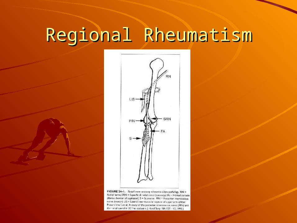
The elbow is formed by three articulations: the humerus The elbow is formed by three articulations: the humerus with the radius, the humerus with the ulna and the radius with the radius, the humerus with the ulna and the radius with the ulna.with the ulna.
The ulnar nerve passes medial to the olecranon process The ulnar nerve passes medial to the olecranon process and behind the medial epicondyle in the cubital tunnel.and behind the medial epicondyle in the cubital tunnel.
Lateral epicondyle is the site of origin of the wrist extensor-Lateral epicondyle is the site of origin of the wrist extensor-supinator muscle group.supinator muscle group.
The medial epicondyle is the site of origin of the wrist The medial epicondyle is the site of origin of the wrist flexor-pronator.flexor-pronator.
Pathology includes chronic degenerative changes of the Pathology includes chronic degenerative changes of the tendons.tendons.
Regional RheumatismRegional RheumatismLateral epicondylitis or tennis elbow, is a Lateral epicondylitis or tennis elbow, is a syndrome of pain in the wrist extensor muscles.syndrome of pain in the wrist extensor muscles.
Clinically the patient presents with discomfort if Clinically the patient presents with discomfort if the lateral elbow.the lateral elbow.
Point of tenderness is at the epicondyle or slightly Point of tenderness is at the epicondyle or slightly distal, pain at resisted wrist extension is distal, pain at resisted wrist extension is suggestive of the diagnosis.suggestive of the diagnosis.
Risk factors include high hand force with Risk factors include high hand force with repetitive use, repetitive rotation of the forearm repetitive use, repetitive rotation of the forearm and forceful gripping with wrist extension.and forceful gripping with wrist extension.
Regional RheumatismRegional Rheumatism
Regional RheumatismRegional RheumatismTreatmentTreatmentThis disorder may be slow to improve.This disorder may be slow to improve.Initial therapy includes rest, splinting, ice and Initial therapy includes rest, splinting, ice and heat application.heat application.Anti-inflammatories or pain medications could be Anti-inflammatories or pain medications could be helpful.helpful.Steroid injection is consider when conservative Steroid injection is consider when conservative treatment fails.treatment fails.Injections are relatively safe and give relief for Injections are relatively safe and give relief for two to six weeks.two to six weeks.Steroid injection is not recommended in medial Steroid injection is not recommended in medial epicondylitis.epicondylitis.
Regional RheumatismRegional RheumatismOlecranon BursitisOlecranon Bursitis
Commonly occurs after repetitive trauma to the Commonly occurs after repetitive trauma to the elbow.elbow.
Other etiologies include: rheumatoid arthritis and Other etiologies include: rheumatoid arthritis and crystalloid arthritis.crystalloid arthritis.
Aspiration of the bursa can be performed to relief Aspiration of the bursa can be performed to relief discomfort.discomfort.
If symptoms recur Steroid injection can be done.If symptoms recur Steroid injection can be done.
This bursa is a common site of infection This bursa is a common site of infection frequently caused byfrequently caused by Staphylococcus aureus. Staphylococcus aureus.
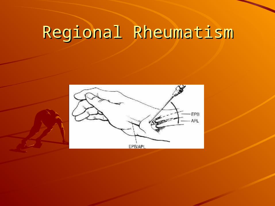
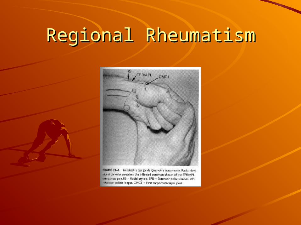
Regional RheumatismRegional RheumatismDE Quervain's DiseaseDE Quervain's Disease
This is the name given to the tenosynovitis to the This is the name given to the tenosynovitis to the extensor tendons of the thumb.extensor tendons of the thumb.
The most clinical manifestation is pain over the The most clinical manifestation is pain over the styloid process.styloid process.
Swelling and warmth over the radial wrist is Swelling and warmth over the radial wrist is common.common.
A positive Finkelstein test is the classic diagnostic A positive Finkelstein test is the classic diagnostic maneuver.maneuver.
Differential diagnosis include osteoarthritis and Differential diagnosis include osteoarthritis and Ulnar nerve compression at the wrist.Ulnar nerve compression at the wrist.
Regional RheumatismRegional RheumatismRisk factors include: assembly line work, small Risk factors include: assembly line work, small goods manufacturing, meat and poultry goods manufacturing, meat and poultry processing, textile production and computer use.processing, textile production and computer use.
Treatment includes rest with a thumb in a spica-Treatment includes rest with a thumb in a spica-splint, NSDAIDS and physical therapy.splint, NSDAIDS and physical therapy.
Steroid injection is an option after conservative Steroid injection is an option after conservative treatment.treatment.
If symptoms persist changes in the work place If symptoms persist changes in the work place could be necessary.could be necessary.
Regional RheumatismRegional Rheumatism
Regional RheumatismRegional Rheumatism
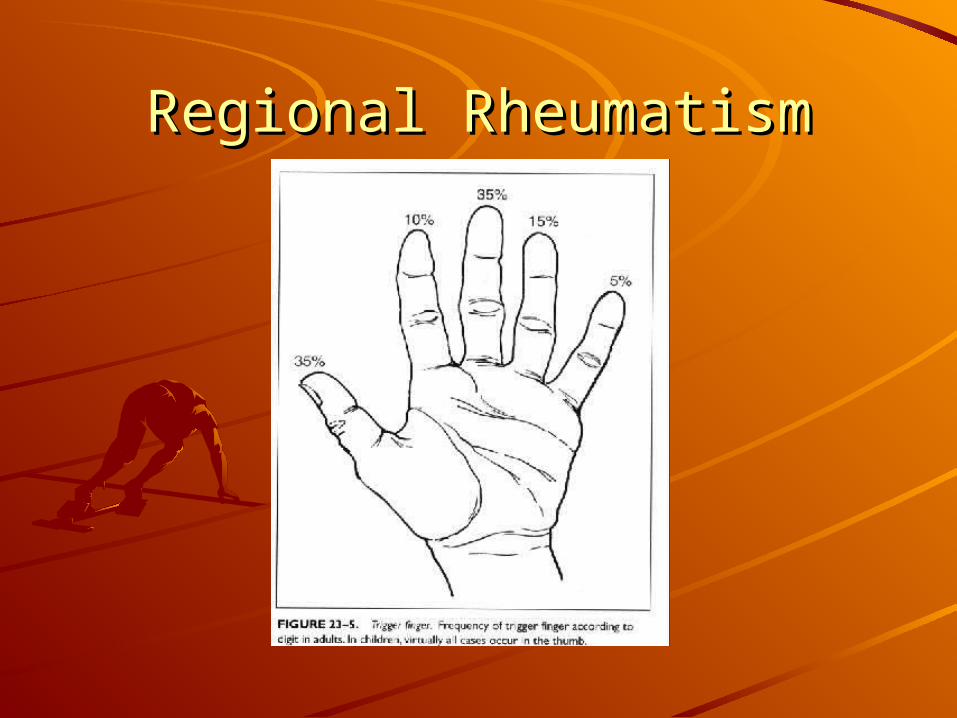
Regional RheumatismRegional RheumatismTrigger FingerTrigger Finger
Trigger finger is caused by swelling of the flexor tendon or Trigger finger is caused by swelling of the flexor tendon or narrowing of the tendon pulley superficial to the MCP joint.narrowing of the tendon pulley superficial to the MCP joint.
Trigger finger manifests with pain or crepitus in the flexor Trigger finger manifests with pain or crepitus in the flexor sheath and impaired finger flexion with triggering or sheath and impaired finger flexion with triggering or locking.locking.
Pain over the MCP joint is a classic feature.Pain over the MCP joint is a classic feature.
Risk factors include pressure over hard objects, such us tool Risk factors include pressure over hard objects, such us tool handles and repeated movements.handles and repeated movements.
Often is seen middle age women and can be associated Often is seen middle age women and can be associated with endocrinologic or rheumatoid diseases.with endocrinologic or rheumatoid diseases.
Regional RheumatismRegional Rheumatism
Regional RheumatismRegional RheumatismHip pain involves a wide differential diagnosis.Hip pain involves a wide differential diagnosis.The anatomy of this region is complex.The anatomy of this region is complex.The hip is ball-and-socket joint.The hip is ball-and-socket joint.The bone structures that conform this area The bone structures that conform this area include: acetabulum, femoral head, ischium, ilium include: acetabulum, femoral head, ischium, ilium and pubis.and pubis.A large number of muscles enable the hip to A large number of muscles enable the hip to move in a wide range of motion.move in a wide range of motion.Flexion is performed by the iliopsoas and Flexion is performed by the iliopsoas and quadriceps, extension by the hamstring.quadriceps, extension by the hamstring.The nerves that more commonly cause pain are The nerves that more commonly cause pain are the Sciatic and the femoral cutaneus.the Sciatic and the femoral cutaneus.
Regional RheumatismRegional RheumatismThe age of the patient suggest different The age of the patient suggest different diagnostic possibilities.diagnostic possibilities.
Younger patients are more prone to apophyseal Younger patients are more prone to apophyseal injuries.injuries.
Avulsion fractures are more common in skeletally Avulsion fractures are more common in skeletally immature patients.immature patients.
Bursitis and muscle strains are more common in Bursitis and muscle strains are more common in skeletally mature patients and DJD is more skeletally mature patients and DJD is more common in older adults.common in older adults.
Regional RheumatismRegional RheumatismPhysical examination is similar for all groups of age.Physical examination is similar for all groups of age.
Observation includes determining whether the Observation includes determining whether the affected leg can bear weight. Observe the patient affected leg can bear weight. Observe the patient posture and evaluate height symmetry of the iliac posture and evaluate height symmetry of the iliac crests.crests.
Palpation can help localize vague complains to an Palpation can help localize vague complains to an specific structure.specific structure.
Range of motion is dependent of patient’s age, with Range of motion is dependent of patient’s age, with range decreasing with age.range decreasing with age.
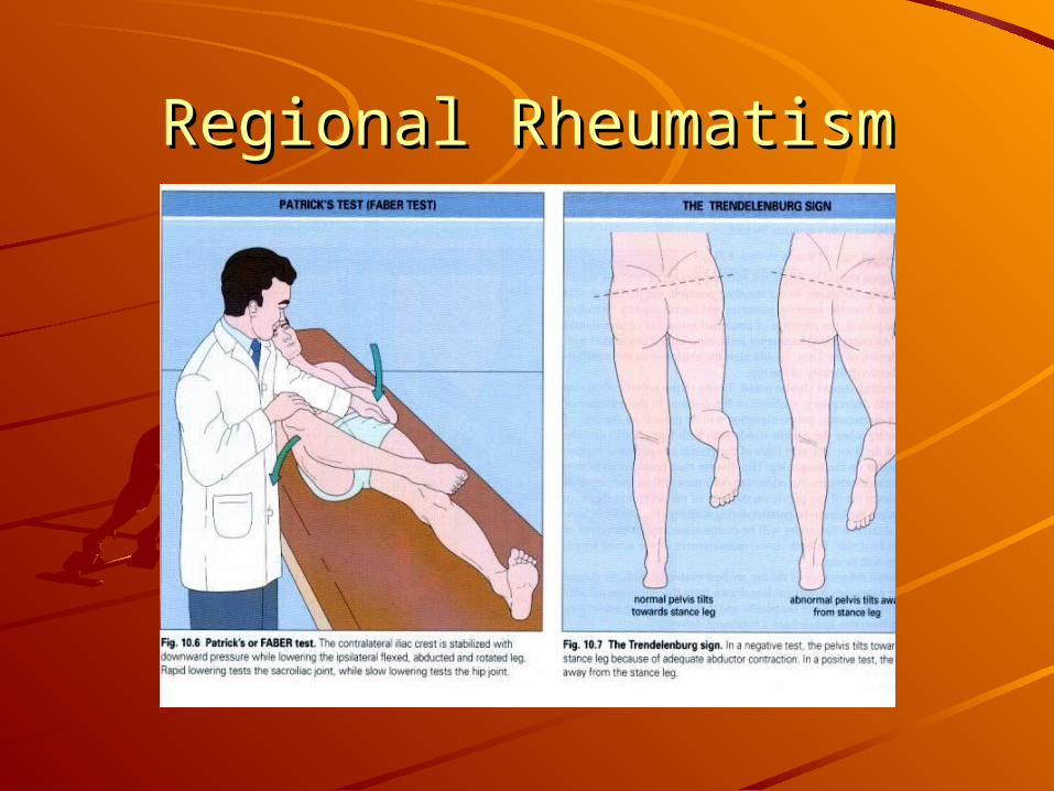
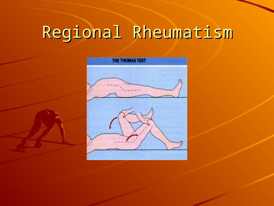
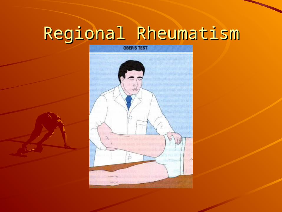
Some specific tests such us the Trendelenburg’s and Some specific tests such us the Trendelenburg’s and Ober’s are helpful to diagnose specific pathologies.Ober’s are helpful to diagnose specific pathologies.
Regional RheumatismRegional Rheumatism
Regional RheumatismRegional Rheumatism
Regional RheumatismRegional RheumatismRadiology is not as helpful as is in ankle or knee Radiology is not as helpful as is in ankle or knee pain.pain.
Radiographs anteroposterior and frog leg lateral Radiographs anteroposterior and frog leg lateral hip are recommended in all acutely injured hip are recommended in all acutely injured patients, patients with marked reduced range of patients, patients with marked reduced range of motion, point tenderness at the site of muscular motion, point tenderness at the site of muscular insertion and inability to bear weight.insertion and inability to bear weight.
Plain films are helpful in the diagnosis of slipped Plain films are helpful in the diagnosis of slipped capital femoral epiphysis, Legg-Calve-Perthes, capital femoral epiphysis, Legg-Calve-Perthes, dysplasia and apophyseal injuries.dysplasia and apophyseal injuries.
Regional RheumatismRegional RheumatismUltrasound is limited in the evaluation of the adult hip, but Ultrasound is limited in the evaluation of the adult hip, but can be helpful in the evaluation of the intraarticular can be helpful in the evaluation of the intraarticular effusions and soft tissue swelling.effusions and soft tissue swelling.
In pediatric patients could be helpful in the diagnosis of hip In pediatric patients could be helpful in the diagnosis of hip subluxation.subluxation.
CT scan provides an excellent detail of the osseus CT scan provides an excellent detail of the osseus structures, can define fractures and intraarticular loose structures, can define fractures and intraarticular loose bodies.bodies.
Bone scan is sensitive for stress fractures but lacks Bone scan is sensitive for stress fractures but lacks specificity.specificity.
MRI is helpful defining soft tissue inflammation, synovitis, MRI is helpful defining soft tissue inflammation, synovitis, neoplasm, infection and stress fractures.neoplasm, infection and stress fractures.
Regional RheumatismRegional RheumatismAge-Specific Hip ProblemsAge-Specific Hip Problems
Prepubescent: Prepubescent:
Transient synovitis is the most common cause of Transient synovitis is the most common cause of hip pain in children.hip pain in children.
Legg-Calve-Perthes is an inflammatory disease of Legg-Calve-Perthes is an inflammatory disease of the femoral head, with a male-female ratio of 5 to the femoral head, with a male-female ratio of 5 to 1, peak incidence is between four to eight year 1, peak incidence is between four to eight year old.old.

Regional RheumatismRegional RheumatismAdolescence:Adolescence:
Slipped femoral epiphysis is another age specific Slipped femoral epiphysis is another age specific entity. It is most common in kids 11 to 14 year entity. It is most common in kids 11 to 14 year old. Obesity and male sex increase the risk.old. Obesity and male sex increase the risk.
This disease increase the risk of avascular This disease increase the risk of avascular necrosis of the femoral head or ostearthritis in necrosis of the femoral head or ostearthritis in the adults.the adults.
This entity requires early referral to and This entity requires early referral to and orthopedic surgeon because this disease benefits orthopedic surgeon because this disease benefits from surgical pinning of the slipped bone.from surgical pinning of the slipped bone.
Regional RheumatismRegional RheumatismYoung AdultYoung AdultYoung adults have the longest list of possible Young adults have the longest list of possible diagnoses. Because the practice of high intensity diagnoses. Because the practice of high intensity sports, avulsion fractures, femoral neck stress sports, avulsion fractures, femoral neck stress fractures, iliotibial band syndrome are more fractures, iliotibial band syndrome are more common in this group of age.common in this group of age.The most critical diagnosis to make early is stress The most critical diagnosis to make early is stress fracture.fracture.Females are in higher risk such us endurance Females are in higher risk such us endurance athletes.athletes.This fractures can progress to unstable fractures This fractures can progress to unstable fractures and increase the risk for avascular necrosis.and increase the risk for avascular necrosis.
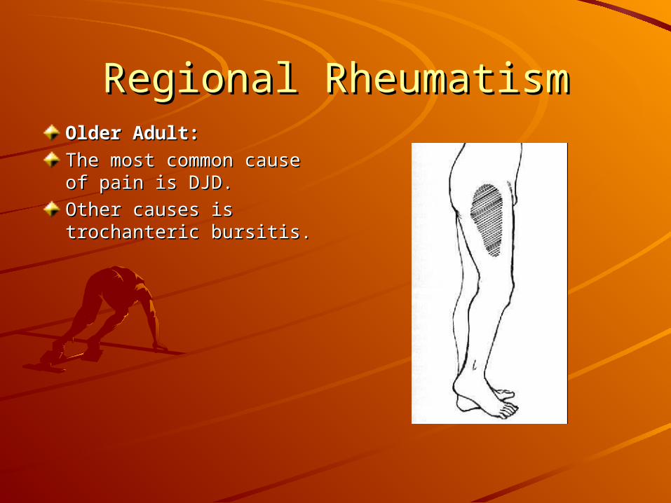
Regional RheumatismRegional RheumatismOlder Adult:Older Adult:
The most common cause The most common cause of pain is DJD.of pain is DJD.
Other causes is Other causes is trochanteric bursitis.trochanteric bursitis.
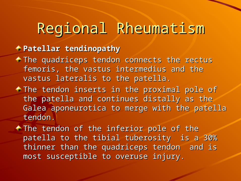
Regional RheumatismRegional RheumatismPatellar tendinopathyPatellar tendinopathy
The quadriceps tendon connects the rectus The quadriceps tendon connects the rectus femoris, the vastus intermedius and the vastus femoris, the vastus intermedius and the vastus lateralis to the patella.lateralis to the patella.
The tendon inserts in the proximal pole of the The tendon inserts in the proximal pole of the patella and continues distally as the Galea patella and continues distally as the Galea aponeurotica to merge with the patella tendon.aponeurotica to merge with the patella tendon.
The tendon of the inferior pole of the patella to The tendon of the inferior pole of the patella to the tibial tuberosity is a 30% thinner than the the tibial tuberosity is a 30% thinner than the quadriceps tendon and is most susceptible to quadriceps tendon and is most susceptible to overuse injury.overuse injury.
Regional RheumatismRegional Rheumatism
Regional RheumatismRegional RheumatismThe pathophysiology of patellar tendinopathy The pathophysiology of patellar tendinopathy shows mucoid degeneration of the tendon.shows mucoid degeneration of the tendon.
At light microscopy the tendon show abnormal At light microscopy the tendon show abnormal collagen, tenocytes and abnormal blood vessels collagen, tenocytes and abnormal blood vessels ingrowth.ingrowth.
A major feature is the absence of inflammation, A major feature is the absence of inflammation, for this reason some authors call this finding as for this reason some authors call this finding as tendinosis instead of tendinitis.tendinosis instead of tendinitis.
This suggest that this condition is more a This suggest that this condition is more a degenerative condition.degenerative condition.
Regional RheumatismRegional RheumatismPatellar tendinoapthy is more often located in the Patellar tendinoapthy is more often located in the lower pole of the patella. lower pole of the patella.The cause is repeated overloads on the extensor The cause is repeated overloads on the extensor mechanism.mechanism.It is more common in that requires maximal It is more common in that requires maximal muscle-tendon unit exertion such us jumping.muscle-tendon unit exertion such us jumping.Pain is elicited by activity, pain when sitting for Pain is elicited by activity, pain when sitting for long periods and going up and down stairs.long periods and going up and down stairs.The most common physical finding is tenderness The most common physical finding is tenderness and in chronic cases swelling.and in chronic cases swelling.MRI and US are the modalities of choice to MRI and US are the modalities of choice to evaluate patellar disorders.evaluate patellar disorders.
Regional RheumatismRegional RheumatismConservative management includes correction of Conservative management includes correction of the predisposing factors, stretching and the predisposing factors, stretching and strengthening, physical therapy, NSDAID and strengthening, physical therapy, NSDAID and steroid injection.steroid injection.
Surgery is indicated in patients that not improve Surgery is indicated in patients that not improve after three to six months of conservative therapy.after three to six months of conservative therapy.
Iliotibial bandIliotibial band
Iliotibial band friction syndrome results of Iliotibial band friction syndrome results of excessive friction between the band and lateral excessive friction between the band and lateral femoral condyle.femoral condyle.
Regional RheumatismRegional RheumatismThe iliotibial band originates proximally from the The iliotibial band originates proximally from the confluence of the fascia from the tensor fascia confluence of the fascia from the tensor fascia lata, the gluteus maximus and gluteus medius.lata, the gluteus maximus and gluteus medius.
At the knee the iliotibial band attaches to the At the knee the iliotibial band attaches to the patella, crosses the knee and attach in the patella, crosses the knee and attach in the Gerdy’s tubercle and lateral to the tibial tubercle.Gerdy’s tubercle and lateral to the tibial tubercle.
The pathogenesis of this condition is attributed to The pathogenesis of this condition is attributed to the friction of the deep layer of the band and the the friction of the deep layer of the band and the lateral femoral epicondyle.lateral femoral epicondyle.
Clinically presents with pain or burning over the Clinically presents with pain or burning over the lateral aspect of the knee.lateral aspect of the knee.
Regional RheumatismRegional RheumatismActivities such as distance running or running downhill Activities such as distance running or running downhill aggravate the symptoms.aggravate the symptoms.
Physical examination reveals tenderness over the lateral Physical examination reveals tenderness over the lateral femoral epicondyle, greater with knee at 30 degrees of femoral epicondyle, greater with knee at 30 degrees of flexion.flexion.
Ober’s test indicates tightness of iliotibial band.Ober’s test indicates tightness of iliotibial band.
In ITB syndrome, there should be no knee effusion, In ITB syndrome, there should be no knee effusion, instability or positive McMurray test.instability or positive McMurray test.
MRI confirms the diagnosis in patients considered for MRI confirms the diagnosis in patients considered for surgery.surgery.
Majority of the patients improve with conservative Majority of the patients improve with conservative management, if symptoms persist for more than six management, if symptoms persist for more than six months, surgery should be considered.months, surgery should be considered.
Regional RheumatismRegional Rheumatism
Regional RheumatismRegional RheumatismConditions of the Achilles tendonConditions of the Achilles tendon
The Achilles tendon is the largest tendon in the The Achilles tendon is the largest tendon in the body.body.
Its limited blood supply and the combination of Its limited blood supply and the combination of forces which is subjected increase the risk of forces which is subjected increase the risk of injury.injury.
Achilles tendinosis occurs in 10% of the runners, Achilles tendinosis occurs in 10% of the runners, but is also common in dancers, gymnasts and but is also common in dancers, gymnasts and tennis players.tennis players.
Regional RheumatismRegional RheumatismInjury typically occurs in active persons.Injury typically occurs in active persons.
The typical symptoms is pain or tenderness The typical symptoms is pain or tenderness proximal or at the insertion of the calcaneus.proximal or at the insertion of the calcaneus.
Peritendinitis, inflammation of the tendon sheath, Peritendinitis, inflammation of the tendon sheath, causes localized tenderness and burning about 2 causes localized tenderness and burning about 2 to 6 cm above the tendon insertion.to 6 cm above the tendon insertion.
At exam the patient should lying prone, feet At exam the patient should lying prone, feet hanging out of the examination table.hanging out of the examination table.
Palpation often elicits pain.Palpation often elicits pain.
Thompson test the physician squeezed the calf Thompson test the physician squeezed the calf and watches for plantar flexion.and watches for plantar flexion.
Regional RheumatismRegional RheumatismIn patient with tendinosis the treatment should be In patient with tendinosis the treatment should be conservative using ice, rest and NSAIDS.conservative using ice, rest and NSAIDS.Control of the biomechanical factors and a slow Control of the biomechanical factors and a slow gentle warm-up before exercise and icing after gentle warm-up before exercise and icing after exercise help patients that want to continue exercise help patients that want to continue athletic training.athletic training.In patients with Achilles tendon rupture, the In patients with Achilles tendon rupture, the treatment is controversial.treatment is controversial.The main treatment is surgery plus The main treatment is surgery plus immobilization or immobilization alone.immobilization or immobilization alone.The trend in younger patients is surgery and The trend in younger patients is surgery and immobilization in the elderly patient.immobilization in the elderly patient.
Regional RheumatismRegional Rheumatism
ReferencesReferencesTallia, Alfred and Dennis Cardone. Diagnostic and Tallia, Alfred and Dennis Cardone. Diagnostic and Therapeutic Injection of the Shoulder Region. Therapeutic Injection of the Shoulder Region. American Family Physician. Volume 67, Number American Family Physician. Volume 67, Number 6, March 15, 20036, March 15, 2003
Almekinders, Louis. Impingement Syndrome. Almekinders, Louis. Impingement Syndrome. Clinics is Sport Medicine. Volume 20, Number 3, Clinics is Sport Medicine. Volume 20, Number 3, July 2001.July 2001.
Cardone, Dennis and Alfred Tallia. Diagnostic and Cardone, Dennis and Alfred Tallia. Diagnostic and Therapeutic Injection of the Elbow Region. Therapeutic Injection of the Elbow Region. American Family Physician. Volume 66, Number American Family Physician. Volume 66, Number 11, December 1, 2002.11, December 1, 2002.
ReferencesReferencesMani, Lisa and Fredric Gerr. Work Related Upper Mani, Lisa and Fredric Gerr. Work Related Upper Extremity Musculoskeletal Disorders. Primary Extremity Musculoskeletal Disorders. Primary Care: Clinics in Office Practice. Volume 27, Care: Clinics in Office Practice. Volume 27, Number 4, December 2000.Number 4, December 2000.
Adkins, Samuel and Richard Figler. American Adkins, Samuel and Richard Figler. American Family Physician. Volume 61, Number 7, April 1, Family Physician. Volume 61, Number 7, April 1, 2000.2000.
Scopp, Jason and Claude Moorman. The Scopp, Jason and Claude Moorman. The Assessment of Athletic Hip Injury. Clinics in Assessment of Athletic Hip Injury. Clinics in Sports Medicine. Volume 20, Number 4, October Sports Medicine. Volume 20, Number 4, October 2001.2001.
ReferencesReferencesCardone, Dennis and Alfred Tallia. Diagnostic and Cardone, Dennis and Alfred Tallia. Diagnostic and Therapeutic Injection of the Hip and Knee.Therapeutic Injection of the Hip and Knee.
American Family Physician. Volume 67, Number American Family Physician. Volume 67, Number 10, May 15, 2000.10, May 15, 2000.
Mazzone, Michael and Timothy MC Cue. Common Mazzone, Michael and Timothy MC Cue. Common Conditions of the Achilles Tendon. American Conditions of the Achilles Tendon. American Family Physician. Volume 65, Number 9, May 1, Family Physician. Volume 65, Number 9, May 1, 2002.2002.
Canoso, Juan. Regional Rheumatic Diseases. Canoso, Juan. Regional Rheumatic Diseases. Rheumatology in Primary Care. W.B Saunders Rheumatology in Primary Care. W.B Saunders Company, 1997.Company, 1997.
ReferencesReferencesCush, John and Arthur Kavanaugh. Rheumatology Cush, John and Arthur Kavanaugh. Rheumatology Diagnosis and Therapeutics. Lippincott Williams & Diagnosis and Therapeutics. Lippincott Williams & Wilkins, 2000.Wilkins, 2000.
Canoso, Juan and Simon Carette. Rheumatology Canoso, Juan and Simon Carette. Rheumatology Second Edition. Mosby, 1998.Second Edition. Mosby, 1998.