Embed Size (px)
Citation preview

CADASILCADASIL
Mary Quiceno, M.D.Mary Quiceno, M.D.Clinical Assistant Professor Clinical Assistant Professor
Department of NeurologyDepartment of NeurologyUT Southwestern Medical UT Southwestern Medical
CenterCenter
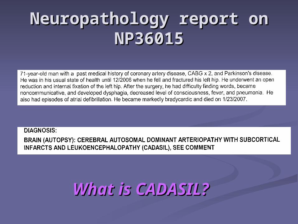
Neuropathology report on Neuropathology report on NP36015NP36015
What is CADASIL?What is CADASIL?
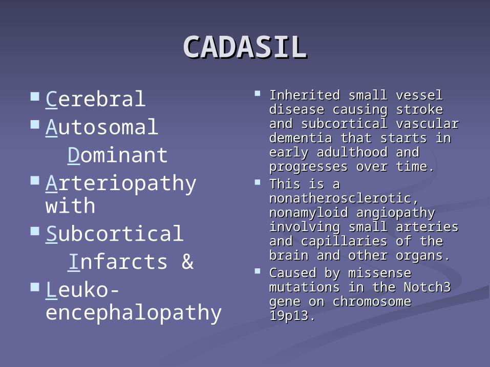
CADASILCADASIL
Cerebral Autosomal Dominant Arteriopathy
with Subcortical Infarcts & Leuko-
encephalopathy
Inherited small vessel Inherited small vessel disease causing stroke and disease causing stroke and subcortical vascular subcortical vascular dementia that starts in dementia that starts in early adulthood and early adulthood and progresses over time. progresses over time.
This is a This is a nonatherosclerotic, nonatherosclerotic, nonamyloid angiopathy nonamyloid angiopathy involving small arteries involving small arteries and capillaries of the brain and capillaries of the brain and other organs.and other organs.
Caused by missense Caused by missense mutations in the Notch3 mutations in the Notch3 gene on chromosome gene on chromosome 19p13.19p13.
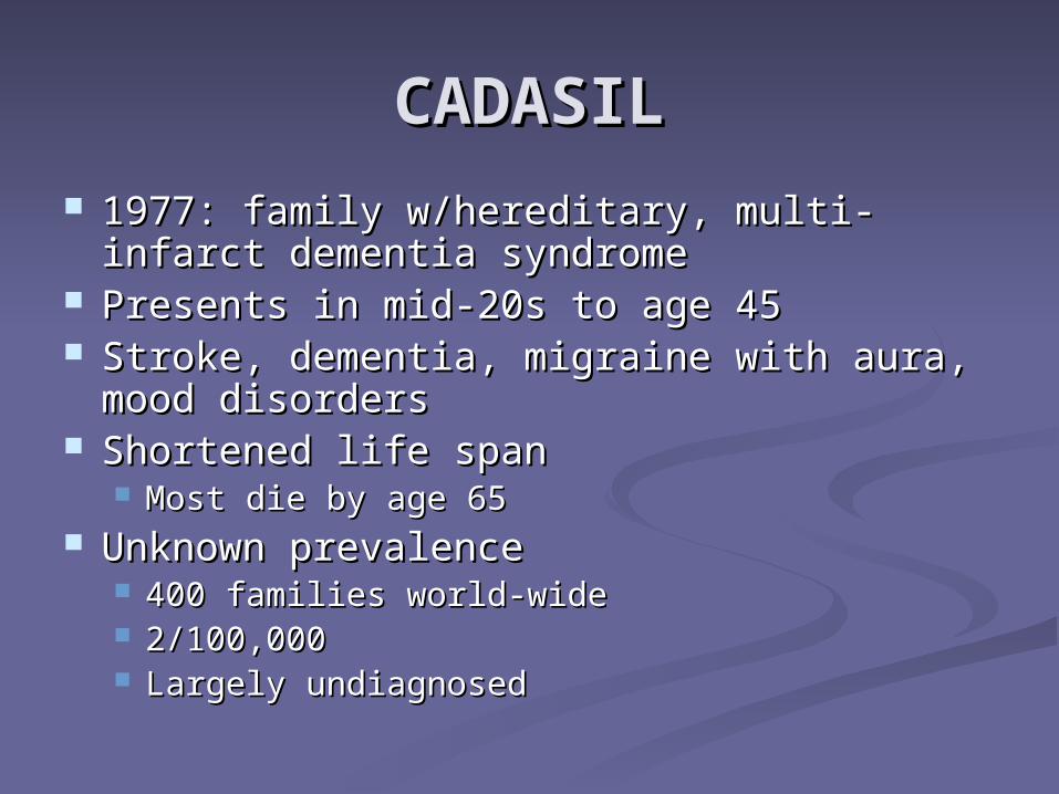
CADASILCADASIL 1977: family w/hereditary, multi-infarct 1977: family w/hereditary, multi-infarct
dementia syndromedementia syndrome Presents in mid-20s to age 45Presents in mid-20s to age 45 Stroke, dementia, migraine with aura, Stroke, dementia, migraine with aura,
mood disordersmood disorders Shortened life spanShortened life span
Most die by age 65Most die by age 65 Unknown prevalenceUnknown prevalence
400 families world-wide400 families world-wide 2/100,0002/100,000 Largely undiagnosedLargely undiagnosed
Case StudiesCase Studies
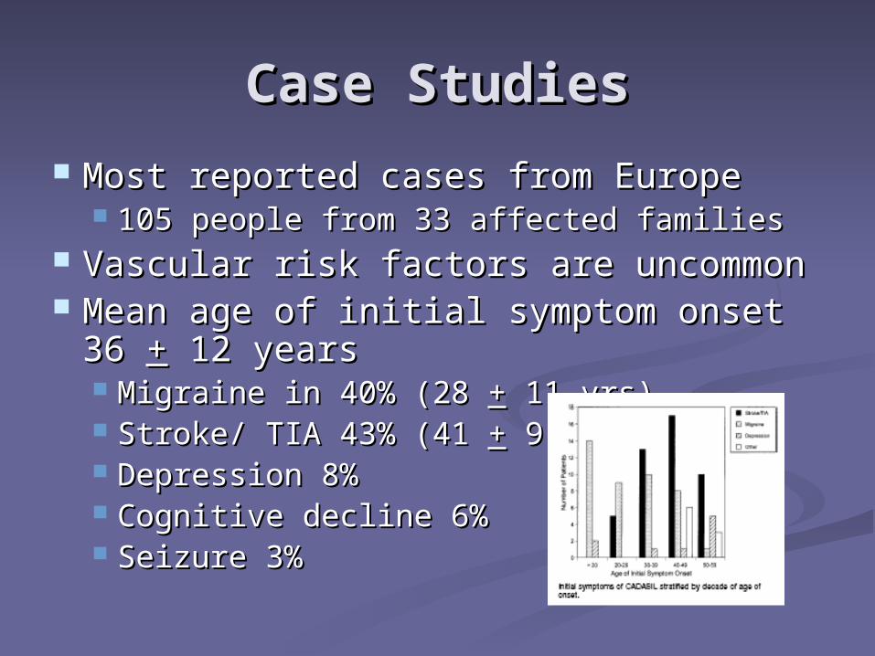
Most reported cases from EuropeMost reported cases from Europe 105 people from 33 affected families105 people from 33 affected families
Vascular risk factors are uncommonVascular risk factors are uncommon Mean age of initial symptom onset 36 Mean age of initial symptom onset 36
++ 12 years 12 years Migraine in 40% (28 Migraine in 40% (28 ++ 11 yrs) 11 yrs) Stroke/ TIA 43% (41 Stroke/ TIA 43% (41 ++ 9 yrs) 9 yrs) Depression 8%Depression 8% Cognitive decline 6%Cognitive decline 6% Seizure 3%Seizure 3%

overall, 67% had a TIA or strokeoverall, 67% had a TIA or stroke overall, 42% had dementiaoverall, 42% had dementia >30% with migraine w/aura and 15% w/mood >30% with migraine w/aura and 15% w/mood
d/od/o overall, age of death, in the 20% of the cohort overall, age of death, in the 20% of the cohort
that was deceased, was 54.8 that was deceased, was 54.8 ++ 10 years 10 years
Course is heterogeneous even in the same Course is heterogeneous even in the same family: some remain asymptomatic until their family: some remain asymptomatic until their 70s whereas others are severely affected by the 70s whereas others are severely affected by the age of 50.age of 50.
MIGRAINE with auraMIGRAINE with aura
Often initial featureOften initial feature 1/3 of families1/3 of families Occurs earlier as compared to stroke Occurs earlier as compared to stroke Consider CADASIL in migraineur Consider CADASIL in migraineur
with diffuse white matter lesions on with diffuse white matter lesions on MRIMRI Not small, scattered hyperintensities, Not small, scattered hyperintensities,
which can be seen in migraineurs (16%) which can be seen in migraineurs (16%) who don’t have CADASILwho don’t have CADASIL
STROKESTROKE TIAs and subcortical ischemic strokesTIAs and subcortical ischemic strokes Accumulating sensory, motor, and Accumulating sensory, motor, and
cognitive deficitscognitive deficits Most common featureMost common feature
Typical stroke risk factors NOT presentTypical stroke risk factors NOT present Cerebral non-atherosclerotic, Cerebral non-atherosclerotic,
nonamyloid angiopathynonamyloid angiopathy Primarily affecting small vessels that Primarily affecting small vessels that
penetrate white matter and basal penetrate white matter and basal gangliaganglia
MOOD DISORDERSMOOD DISORDERS
DepressionDepression Bipolar disorderBipolar disorder Like migraine, CADASIL should only Like migraine, CADASIL should only
be considered when MRI changes be considered when MRI changes are presentare present
Tend to predate cognitive declineTend to predate cognitive decline
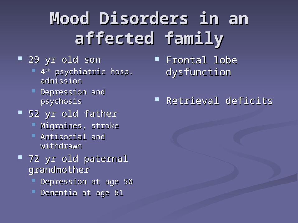
Mood Disorders in an Mood Disorders in an affected familyaffected family
29 yr old son29 yr old son 44thth psychiatric hosp. psychiatric hosp.
admissionadmission Depression and Depression and
psychosispsychosis 52 yr old father52 yr old father
Migraines, strokeMigraines, stroke Antisocial and Antisocial and
withdrawnwithdrawn 72 yr old paternal 72 yr old paternal
grandmothergrandmother Depression at age 50Depression at age 50 Dementia at age 61Dementia at age 61
Frontal lobe Frontal lobe dysfunctiondysfunction
Retrieval deficitsRetrieval deficits
COGNITIVE DEFICITSCOGNITIVE DEFICITS
Slowly progressive in addition to stepwise Slowly progressive in addition to stepwise deteriorationdeterioration Typically appears after stroke symptoms appearTypically appears after stroke symptoms appear Can be presenting featureCan be presenting feature
Frontal lobe dysfunctionFrontal lobe dysfunction Memory impairmentMemory impairment Pseudobulbar palsy, gait disturbances, Pseudobulbar palsy, gait disturbances,
pyramidal signs, sphincter incontinencepyramidal signs, sphincter incontinence Subcortical dementia Vascular dementia
Cognitive profileCognitive profile
CADASIL compared to normalsCADASIL compared to normals Impaired on executive function and speed Impaired on executive function and speed
measuresmeasures Delis-Kaplan Executive Function System (D-KEFS)Delis-Kaplan Executive Function System (D-KEFS) Trails motor speed subtest from the D-KEFSTrails motor speed subtest from the D-KEFS
CADASIL w/stroke and cerebral small vessel CADASIL w/stroke and cerebral small vessel disease (SVD)disease (SVD)
SVD typically olderSVD typically older Both impaired similarly on executive fx and speedBoth impaired similarly on executive fx and speed CADASIL worse on verbal fluency (letter)CADASIL worse on verbal fluency (letter)
Executive FunctionExecutive Function refers to a wide range of central control processes in the brain that refers to a wide range of central control processes in the brain that
connect, prioritize, and integrate operation of subordinate brain connect, prioritize, and integrate operation of subordinate brain functionsfunctions
this central management system, often attributed to operations in the this central management system, often attributed to operations in the prefrontal cortex, is crucial to organizing and integrating cognitive prefrontal cortex, is crucial to organizing and integrating cognitive processes over time and plays an increasingly important role as we processes over time and plays an increasingly important role as we mature mature
organizes, activates, focuses, integrates, and directsorganizes, activates, focuses, integrates, and directs
Executive functions require several higher-level cognitive abilities for Executive functions require several higher-level cognitive abilities for successful performance.successful performance.
These can be assessed with tasks that require:These can be assessed with tasks that require: – – initiation of effortful and novel thinkinginitiation of effortful and novel thinking – – isolation of a common feature or attribute from among the array of target isolation of a common feature or attribute from among the array of target
stimulistimuli – – formation of a higher-level concept that captures the defining properties formation of a higher-level concept that captures the defining properties
of those common featuresof those common features – – flexibility of thinking in order to abandon one conceptual relationship in flexibility of thinking in order to abandon one conceptual relationship in
order to apprehend new onesorder to apprehend new ones
Other organ diseaseOther organ disease In some patients w/CADASILIn some patients w/CADASIL
silent retinal microvascular circulatory changes silent retinal microvascular circulatory changes 18 pts: No visual symptoms. VA was normal in all. 18 pts: No visual symptoms. VA was normal in all.
Ophthalmologic abnormalities were found in 8 patients. Ophthalmologic abnormalities were found in 8 patients. FE and FA revealed silent retinal abnormalities in CADASIL FE and FA revealed silent retinal abnormalities in CADASIL
patients with nerve fiber loss in 22% and cotton wool spots in patients with nerve fiber loss in 22% and cotton wool spots in 17%. 17%.
may be considered as peripheral markers of this genetic may be considered as peripheral markers of this genetic disease. disease.
high frequency of myocardial infarction in a single high frequency of myocardial infarction in a single series of Dutch patientsseries of Dutch patients
Distinct from CADASIL, hereditary Distinct from CADASIL, hereditary endotheliopathy with retinopathy, nephropathy, endotheliopathy with retinopathy, nephropathy, and stroke (HERNS) is an autosomal dominant and stroke (HERNS) is an autosomal dominant multi-infarct syndrome with systemic involvement. multi-infarct syndrome with systemic involvement.
Brain Imaging in Brain Imaging in CADASILCADASIL
Diffuse white matter hyperintensities on T2 Diffuse white matter hyperintensities on T2 and FLAIR weighted imagesand FLAIR weighted images Subcortical white matterSubcortical white matter Basal gangliaBasal ganglia
Changes on MRI may be evident in persons Changes on MRI may be evident in persons who are in their 20swho are in their 20s Penetrance complete by age 35 and all will have Penetrance complete by age 35 and all will have
MRI findingsMRI findings The syndrome may not be suspected until The syndrome may not be suspected until
affected individuals are in their 50s or olderaffected individuals are in their 50s or older Lesion volume is inversely correlated with Lesion volume is inversely correlated with
cognitive functioncognitive function
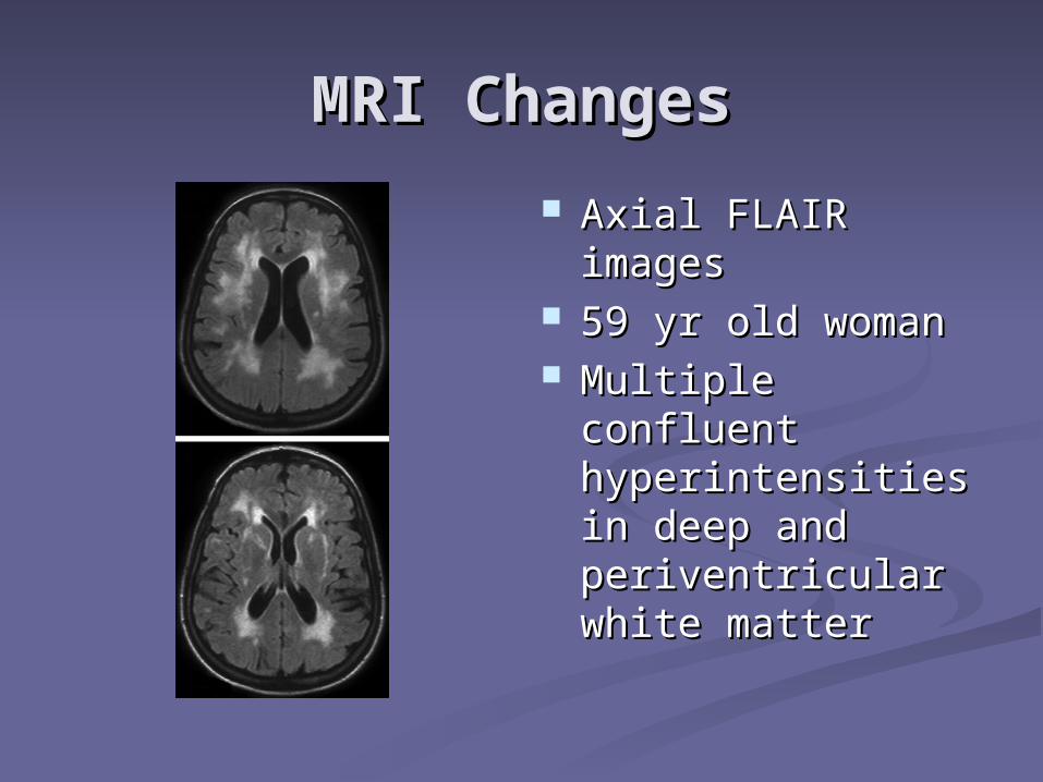
MRI ChangesMRI Changes
Axial FLAIR Axial FLAIR imagesimages
59 yr old woman59 yr old woman Multiple confluent Multiple confluent
hyperintensities in hyperintensities in deep and deep and periventricular periventricular white matterwhite matter
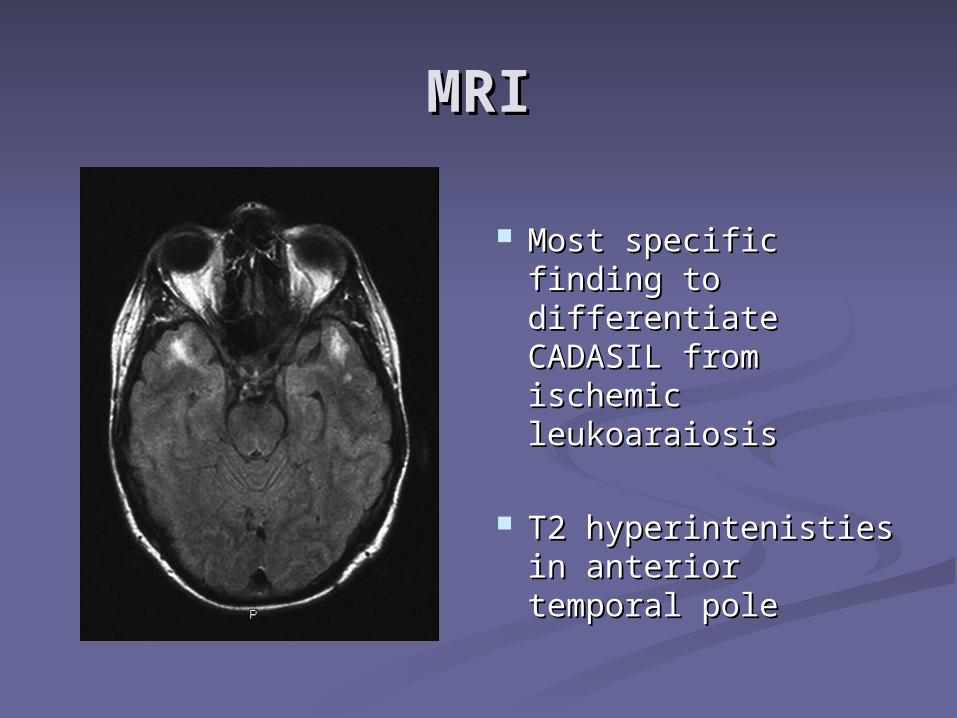
MRIMRI
Most specific finding Most specific finding to differentiate to differentiate CADASIL from CADASIL from ischemic ischemic leukoaraiosisleukoaraiosis
T2 hyperintenisties T2 hyperintenisties in anterior temporal in anterior temporal polepole
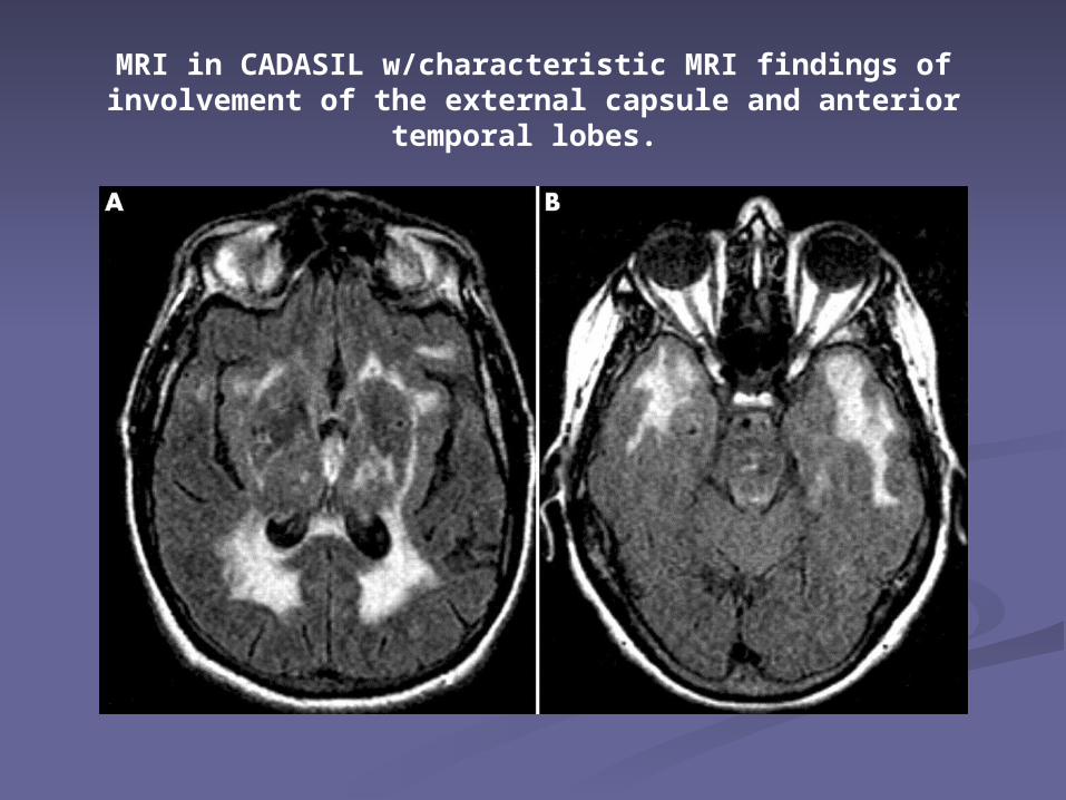
MRI in CADASIL w/characteristic MRI findings of involvement of the external capsule and anterior
temporal lobes.
Differentiating CADASIL Differentiating CADASIL from other diseases from other diseases
affecting the white matteraffecting the white matter Ischemic small-vessel disease Ischemic small-vessel disease
Usually occurs after fifth decadeUsually occurs after fifth decade Vascular risk factors presentVascular risk factors present
Multiple SclerosisMultiple Sclerosis More likely to see spinal cord and More likely to see spinal cord and
corpus callosum lesionscorpus callosum lesions Periventricular lesions are ovoid and/or Periventricular lesions are ovoid and/or
oriented perpendicular to lateral oriented perpendicular to lateral ventriclesventricles
When to consider MRI in When to consider MRI in migraineurmigraineur
Consider MRI if Migraine attacks with aura begin in
mid-adulthood Atypical aura
Hemiplegic, basilar, prolonged Family history of stroke, dementia,
depression Focal neurological signs
When to Suspect When to Suspect CADASILCADASIL
Recurrent subcortical ischemic strokesRecurrent subcortical ischemic strokes Esp. <60 yrs oldEsp. <60 yrs old Esp. in absence of vascular risk factorsEsp. in absence of vascular risk factors
Early cognitive declineEarly cognitive decline Migraine with auraMigraine with aura Comorbid psychiatric symptomsComorbid psychiatric symptoms
DepressionDepression BipolarBipolar

When to Suspect When to Suspect CADASILCADASIL
Abnormal MRIAbnormal MRI Significant white matter lesions before age Significant white matter lesions before age
3535 Multiple T2 hyperintensities w/o vascular Multiple T2 hyperintensities w/o vascular
risk factorsrisk factors Bilateral T2 hyperintensities in white matter, Bilateral T2 hyperintensities in white matter,
esp. w/lesions in ant. Temporal polesesp. w/lesions in ant. Temporal poles Family historyFamily history
Stroke, dementia, depression, migraine w/aura, Stroke, dementia, depression, migraine w/aura, other white matter diseases (which may be other white matter diseases (which may be misdiagnosed)misdiagnosed)
Premature CADPremature CAD
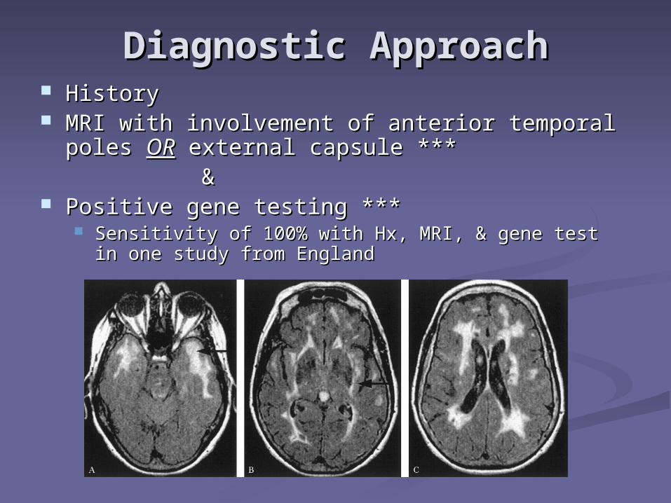
Diagnostic ApproachDiagnostic Approach HistoryHistory MRI with involvement of anterior temporal poles MRI with involvement of anterior temporal poles OROR external capsule *** external capsule ***
&& Positive gene testing ***Positive gene testing ***
Sensitivity of 100% with Hx, MRI, & gene test in one Sensitivity of 100% with Hx, MRI, & gene test in one study from Englandstudy from England

BiopsyBiopsy
Skin biopsy was positive in approximately Skin biopsy was positive in approximately half of the 18 patients testedhalf of the 18 patients tested Skin biopsy was negative in all of the gene Skin biopsy was negative in all of the gene
negative patientsnegative patients Sensitivity of 100%Sensitivity of 100% Granular osmiophilic material seen on EMGranular osmiophilic material seen on EM
Sensitivity 50%, specificity 100%Sensitivity 50%, specificity 100% Tissue samples stained with monoclonal Ab top Tissue samples stained with monoclonal Ab top
Notch3 proteinNotch3 protein Sensitivity 96%, specificity 100%Sensitivity 96%, specificity 100%
The hallmark of the disease is the presence of The hallmark of the disease is the presence of granular osmiophilic material which is seen granular osmiophilic material which is seen adjacent to the basement membrane of the adjacent to the basement membrane of the smooth muscle cells of arterioles on electron smooth muscle cells of arterioles on electron microscopy. microscopy.
This is pathognomic for CADASIL. This is pathognomic for CADASIL. The deposition of GOM in skin arterioles may The deposition of GOM in skin arterioles may
vary depending on the exact mutation involved.vary depending on the exact mutation involved. The vascular defects are present in every tissue The vascular defects are present in every tissue
and may be detected histologically by examining and may be detected histologically by examining arterioles in skin biopsy, where accumulation of arterioles in skin biopsy, where accumulation of granular and osmiophilic material within the granular and osmiophilic material within the smooth muscle cell basement membrane and smooth muscle cell basement membrane and the surrounding extracellular matrix.the surrounding extracellular matrix.
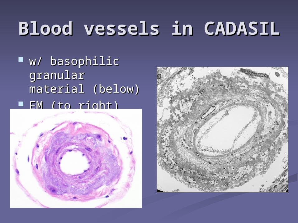
Blood vessels in Blood vessels in CADASILCADASIL
w/ basophilic w/ basophilic granular material granular material (below)(below)
EM (to right)EM (to right)
Blood vessels in Blood vessels in CADASILCADASIL
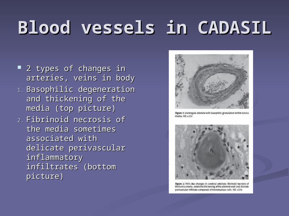
2 types of changes in 2 types of changes in arteries, veins in bodyarteries, veins in body
1.1. Basophilic degeneration Basophilic degeneration and thickening of the and thickening of the media (top picture)media (top picture)
2.2. Fibrinoid necrosis of Fibrinoid necrosis of the media sometimes the media sometimes associated with delicate associated with delicate perivascular perivascular inflammatory infiltrates inflammatory infiltrates (bottom picture)(bottom picture)
Notch3 ab in brain blood Notch3 ab in brain blood vesselsvessels
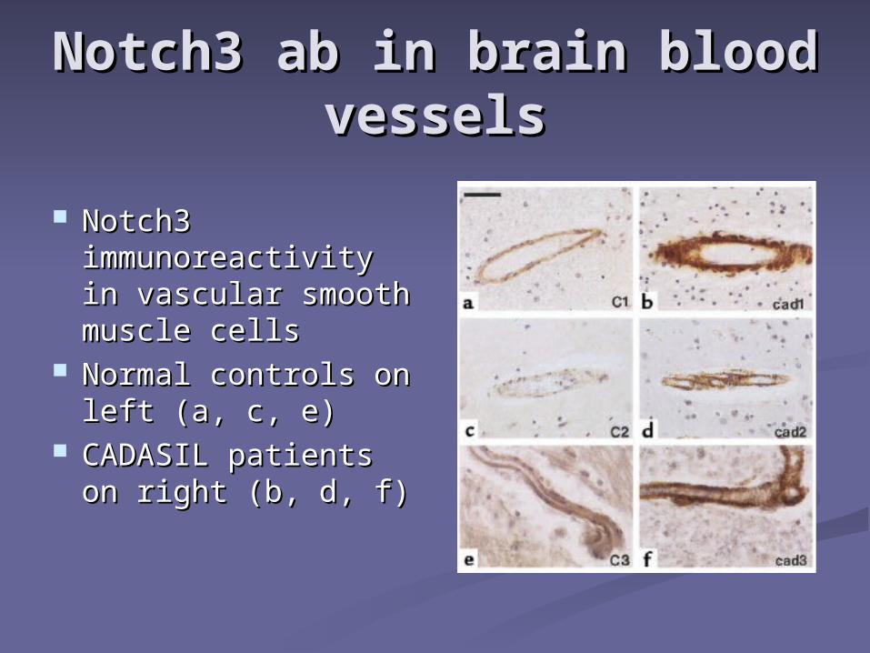
Notch3 Notch3 immunoreactivity immunoreactivity in vascular smooth in vascular smooth muscle cells muscle cells
Normal controls on Normal controls on left (a, c, e)left (a, c, e)
CADASIL patients CADASIL patients on right (b, d, f)on right (b, d, f)

What leads to CADASIL?What leads to CADASIL? Mutations in Mutations in notch3notch3
genegene Odd number of Odd number of
cysteine residues in cysteine residues in Notch3 receptor Notch3 receptor extracellular domainextracellular domain
Impaired clearance of Impaired clearance of cleavage productcleavage product
Alterations of Alterations of vascular smooth vascular smooth musclemuscle
Presence of granular Presence of granular osmiophilic depositsosmiophilic deposits
Notch3 gene mutationNotch3 gene mutation
Usually missense mutationUsually missense mutation More than 50 have been foundMore than 50 have been found Spontaneous mutations have been Spontaneous mutations have been
describeddescribed The protein folds incorrectlyThe protein folds incorrectly Leads to accumulation of protein in Leads to accumulation of protein in
membranes of smooth muscles and, membranes of smooth muscles and, ultimately, fibrosis and luminal ultimately, fibrosis and luminal narrowing of themnarrowing of them
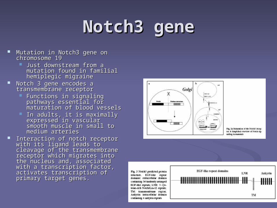
Notch3 geneNotch3 gene Mutation in Notch3 gene on Mutation in Notch3 gene on
chromosome 19chromosome 19 Just downstream from a Just downstream from a
mutation found in familial mutation found in familial hemiplegic migrainehemiplegic migraine
Notch 3 gene encodes a Notch 3 gene encodes a transmembrane receptortransmembrane receptor Functions in signaling Functions in signaling
pathways essential for pathways essential for maturation of blood vesselsmaturation of blood vessels
In adults, it is maximally In adults, it is maximally expressed in vascular smooth expressed in vascular smooth muscle in small to medium muscle in small to medium arteries arteries
Interaction of notch receptor Interaction of notch receptor with its ligand leads to cleavage with its ligand leads to cleavage of the transmembrane receptor of the transmembrane receptor which migrates into the nucleus which migrates into the nucleus and, associated with a and, associated with a transcription factor, activates transcription factor, activates transcription of primary target transcription of primary target genes. genes.
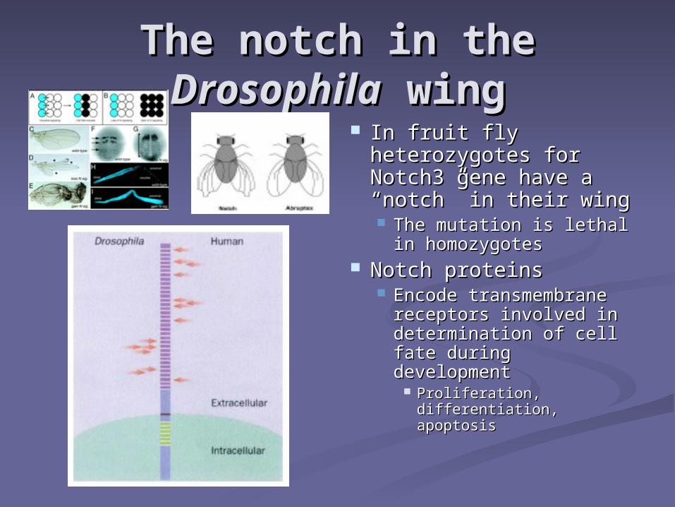
The notch in the The notch in the DrosophilaDrosophila wing wing
In fruit fly In fruit fly heterozygotes for heterozygotes for Notch3 gene have a Notch3 gene have a “notch” in their wing“notch” in their wing The mutation is lethal in The mutation is lethal in
homozygoteshomozygotes Notch proteinsNotch proteins
Encode transmembrane Encode transmembrane receptors involved in receptors involved in determination of cell determination of cell fate during developmentfate during development
Proliferation, Proliferation, differentiation, apoptosisdifferentiation, apoptosis
Pathogenic HypothesisPathogenic Hypothesis Notch 3 expression is limited to vascular Notch 3 expression is limited to vascular
smooth muscle cellssmooth muscle cells Mature vascular smooth muscle cells Mature vascular smooth muscle cells
require continued function of the Notch 3 require continued function of the Notch 3 pathwaypathway Continued survivalContinued survival
Blood vessels are narrowed and weak and Blood vessels are narrowed and weak and do not react to fluctuations of COdo not react to fluctuations of CO22 and BP and BP
Capillaries, veins are involvedCapillaries, veins are involved Generalized vasculopathyGeneralized vasculopathy
Brain PredilectionBrain Predilection
Cerebral vessels have fewer smooth Cerebral vessels have fewer smooth muscle cells than vessels of other muscle cells than vessels of other organsorgans Increased susceptibilityIncreased susceptibility
Limited ability for regeneration of Limited ability for regeneration of CNS tissueCNS tissue
White matter predilectionWhite matter predilection Insufficient collateral circulationInsufficient collateral circulation Density less than in grey matterDensity less than in grey matter

What can be done for these What can be done for these patients?patients?
TreatmentTreatment Control vascular disease risk factorsControl vascular disease risk factors
BPBP Increased SBP independent risk factor for Increased SBP independent risk factor for
progression of CADASILprogression of CADASIL CholesterolCholesterol DMDM SmokingSmoking ObesityObesity Avoid OCP, HRTAvoid OCP, HRT
TreatmentTreatment
Antiplatelet therapyAntiplatelet therapy Investigate for other causes of stroke Investigate for other causes of stroke
(cardiac, afib, hypercoag state, etc.)(cardiac, afib, hypercoag state, etc.) Cholinesterase inhibitorsCholinesterase inhibitors
Work in vascular dementiaWork in vascular dementia Screen for mood disorders, cognitive Screen for mood disorders, cognitive
decline, seizuredecline, seizure Life expectancy may be shortened by Life expectancy may be shortened by
6 years6 years
NP36015NP36015
The key findingThe key finding Abundant basophilic (blue on H&E), PAS Abundant basophilic (blue on H&E), PAS
positive, osmiophilic (black on EM) positive, osmiophilic (black on EM) granular material seen in the markedly granular material seen in the markedly thickened blood vessel wallsthickened blood vessel walls
Differential diagnosisDifferential diagnosis Atheroscerotic diseaseAtheroscerotic disease
Blood vessel walls are also thickenedBlood vessel walls are also thickened Granular material is not usually present (if Granular material is not usually present (if
present, it differs from that seen in CADASIL)present, it differs from that seen in CADASIL)
No treatmentNo treatment Screening not indicated, unless family Screening not indicated, unless family
member is affectedmember is affected Family may wish to seek genetic Family may wish to seek genetic
counselingcounseling Control vascular risk factorsControl vascular risk factors Do not smokeDo not smoke Screen for mood disorders, cognitive Screen for mood disorders, cognitive
decline, focal neurologic signs, seizuredecline, focal neurologic signs, seizure

Questions?Questions?