Embed Size (px)
Citation preview

Regenerative medicine using Cardiac
Stem Cells or Exosomes
Lawrence S.C. Czer, M.D.Med. Dir., Heart Transplant Program
Cedars-Sinai Heart Institute
Eduardo Marbán, M.D., Ph.D.Director,Cedars-Sinai Heart Institute
Los Angeles, CA
Disclosure: St. Jude Medical (research study).
Prevailing dogma before 2000• Heart cells have little or no proliferative capacity
• Myocardial scar is irreversible
• Lost heart muscle cannot regrow
• The best hope for treating cardiac injury is to limit the injury or to block secondary maladaptive pathways

• Self-renewing.
• Able to form clones (clonogenicity).
• Potential for multi-lineage differentiation
(multi-potentiality).
Becker AJ, McCulloch EA, Till JE. Cytologic demonstration of the clonal nature of spleen colonies derived
from transplanted mouse marrow cells. Nature 1963: 197: 454 – 454.
Stem CellFunctional Definition

Types of stem cells• Embryonic stem cells* or iPS (genetically
induced pluripotent stem) cells#
– Can evolve into all tissues
– Tumors
– Immune reactions
– Ethically problematic*
– Genetic modification#
• Adult stem cells
– Skeletal myoblasts
– Bone marrow stem cells
– Cardiac-derived cells
Skeletal myoblasts
• First to be translated into human studies (Jan. 2001)
• Advantages: autologous, contractile
• Disadvantage: do not couple with surrounding
myocardium, forming islands of conduction block
• VT, SCD observed in 10/22 phase 1 pts
• Clinically failed phase 2 trial as adjunct to surgery in
patients with LV dysfunction (MAGIC)
• Experience with myoblasts raised generalized fears
about stem cells and arrhythmias
Lessons learned from human
bone marrow-derived cell trials
• >1000 subacute MI patients treated worldwide since
Aug. 2001
• Modest functional benefit*, possibly transient#
• Sicker patients have greater functional improvement*
• Excellent safety profile with coronary catheter
delivery, including arrhythmias and SCD
• MRI: small reductions in scar (~3 g), but no increase in
viable myocardium
• Encouraging, but much room for improvement
#BOOST 2004, 2007 *REPAIR-MI 2006

Safety meta-analysis of five BMC trials
From : Clin Cardiol. 2009 Aug;32(8):458-66. Five trials (BOOST, REPAIR-AMI, ASTAMI,
Janssens, and Yao) were included in the analysis

• Cardiac stem cells (CSCs)– First recognized in 2000 (Deisher)
– Antigenically-selected in rats and mice (Beltrami et al., Oh et al., 2003)
• Human cardiospheres (CSps)– Outgrowth of human surgical biopsies in primary culture
(Messina et al., 2004)
– Self-organize in suspension, increase post-ischemic function
• Cardiosphere-derived cells (CDCs)– Millions of cardiac stem cells from percutaneous
endomyocardial biopsies (Smith et al., 2007)
– Paradigm for autologous therapeutics
Major landmarks 2000-present

100 mm
3
How we harvest and grow CDC’s
R. Smith et al., Circulation 115: 896 – 908, 2007
200 mm
1
Explants (1)Biopsy
specimen
200 mm
5
Cardiospheres
(CSp’s, 5)
4
% o
f cell
to
tal
c-Kit+ CD90+CD105+CD133+
0
20
40
60
80
100
Cardiac outgrowth
flat & round cells (4)
50 mm
6
Cardiosphere-
derived cells
(CDC’s, 6)Harvest (2,3)
@ 1-2 wks
2
200 mm
Minced
Digested
collagenaseTrypsin
Cultured
Poly-D-lysine Fibronectin
Seeded
Fibronectin

100 mm
3
How we harvest and grow CDCs
R. Smith et al., Circulation 115: 896 – 908, 2007
200 mm
1
Explants (1)Biopsy
200 mm
5
Cardiospheres (5)
4
% o
f cell
to
tal
c-Kit+ CD90+CD105+CD133+
0
20
40
60
80
100
Cardiosphere
-forming cells (4)
50 mm
6
Cardiosphere-
derived cells (CDCs, 6)
2,3
2
200 mm
Yield: 30M
CDCs in
3-5 weeks

CADUCEUS Trial Design AnimationBiopsy
30 minutesdigestion
in 0.2 mg/mlcollagenase
Cell harvest by
0.05% trypsin
Cardiospheres(CSps)
Poly-D-lysine coated Fibronectin coated
Cardiosphere-derived cells
(CDCs) Infusion into the same patient
Biopsy

The idea
Proof of concept
Protocol optimization
Pivotal protocol
Clinical trial
Vital Step Along The Translational Path

CADUCEUS:CArdiosphere-Derived aUtologous stem
CElls to reverse ventricUlar dySfunction
Clinicaltrials.gov identifier: NCT00893360
• Recent MI & ischemic LV dysfunction (EF 25-45%)
• NIH-funded, Phase I/II randomized, controlled, dose-escalation
safety and preliminary efficacy study (MRI for scar size, volumes,
& function)
• Two centers (Cedars-Sinai Heart Institute; Johns Hopkins)
• Endomyocardial biopsies; CDCs manufactured at Cedars-Sinai
Heart Institute
• Intracoronary infusions of autologous CDCs

0005-CDC01-002
Control
0005-CDC01-014
CDC-treated
baseline 6m
Representative CADUCEUS MR images

Controls
n=10
CDCs
n=15
p=0.000
Scar mass goes down in CDC-treated
patients but not controls….
(g, 6 months minus baseline; independent samples t-test,
pooled CDC group vs controls, means + SD)

Meanwhile, viable myocardium increases
(g,12 months minus baseline; independent samples t-test,
pooled CDC group vs controls, means + SD)
Controls
n=10
CDCs
n=15
p=0.002

Effects maintained at 12 months
(g,12 months minus baseline; independent samples t-test,
pooled CDC group vs controls, means + SD)
Decrease in scar mass Increase in viable mass

CADUCEUS Trial

Mechanism of benefit involves
direct regeneration#….
#Smith et al, Circulation, 2007;
Davis et al., PLoS One, 2009;
Davis et al., Stem Cells, 2010
Mechanism of benefit involves
direct regeneration as well as
paracrine effects*
*Chimenti et al., Circ. Res., 2010;
Cheng et al., Circ. Res., 2010

Presumptive mechanism (2004)
Transplanted stem cells
Cell proliferation
Differentiation
New myocardium of
donor origin

LV function in post-MI rats: syn = alloK. Malliaras et al., Circ 2012
Fractional Area Change Ejection Fraction
D0 3wks 3mos 6mos D0 3wks 3mos 6mos
% %
Fractional Shortening
%
Treatment effect
ΔFA
C (
%)
3 weeks post injectionD0 3wks 3mos 6mos
Syngeneic Allogeneic Control WKYControl BNXenogeneic
* *
**
**
**
**
**
** *
***
**
No surviving
transplanted
cells after 4
weeks
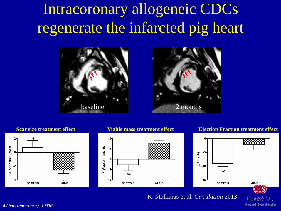
Intracoronary allogeneic CDCs
regenerate the infarcted pig heart
Scar size treatment effect Viable mass treatment effect Ejection Fraction treatment effect
*
* *
All bars represent +/- 1 SEM.
baseline 2 months
K. Malliaras et al. Circulation 2013

Mechanistic rationale for allogeneic therapy
Transplanted CDCs
Cell proliferation
Differentiation
New healthy tissue of
donor origin
Short-term engraftment
Secreted factors
New healthy tissue of
host origin
CDCs anti-inflammatory, immunomodulatory and evanescent1-6
1. I. Chimenti et al, Circ Res 2010; 2. K. Malliaras et al, Circ 2012, 2013; 3. M.
Aminzadeh et al, Eur Heart J 2014; 4. E. Tseliou et al., Basic Res Cardiol 2014;
5. L. Lauden et al, Circ Res 2013; 6. E. Marbán, Mayo Clin Proc 2014

• Regenerative √ 1,2
• Antifibrotic √ 1-4
• Anti-apoptotic √ 3-5
• Angiogenic √ 1,6
• Anti-inflammatory √ 9
• Immunomodulatory √ 9,10
1. RR Smith et al, Circ 2007; 2. Makkar et al., Lancet 2012; 3. E. Tseliou et al., PLoSOne
2014; 4. E. Tseliou et al., BRIC 2014; 5. T-S Li et al, JACC 2012; 6. I. Chimenti et al, Circ
Res 2010; 7. K. Malliaras et al, Circ 2012; 8. K. Malliaras et al., EMBO Mol Med 2013; 9.
M. Aminzadeh et al., EHJ 2014; 10. L. Lauden et al, Circ Res 2013
Therapeutic bioactivity of CDCs:
beyond regeneration

Follow the data
1. Autologous therapy (2004)
2. Recognition of durable benefits despite cell
transience (2010)
Allogeneic paradigm (2012)

Summary of criteria for the perfect stem cell
to treat heart disease• Easily harvested using routine clinical methods
• Readily grown in large numbers
• Unmodified: no genes, limited processing
• “Off the shelf” availability for acute disease
• No ethical or moral quandaries
• Safe
– No immune reaction or “rejection”
– No tumors
– No arrhythmias
• Regrows healthy heart after cardiac injury (heart attack or chronic heart failure)

Cell Type Human heart progenitor cell
Characteristics CD105+, CD45-; secreted SDF-1
Clinical Trials CADUCEUS-completed-autologous phase 1. Twenty-five patient study
showed regeneration in CDC-treated post-MI subjects with mild HFrEF
ALLSTAR-phase 1&2b study of allogeneic CDCs post-MI with mild HFrEF
DYNAMIC- phase 2a study of allogeneic CDCs in patients with advanced
HFrEF
Mechanism of
action
Paracrine effects
Promote cardiomyomyogenesis
Prevent cardiomyocyte apoptosis
Anti-fibrotic
Anti-inflammatory
Cardiosphere-derived cells (CDCs)
First described by RR Smith et al., Circulation 2007

• CDCs in HFpEF (Regress-HFpEF)
• CDCs in pulmonary hypertension (ALPHA)
• Duchenne muscular dystrophy (HOPE)
Additional allogeneic CDC trials
in progress

The graveyard of failed clinical trials
30
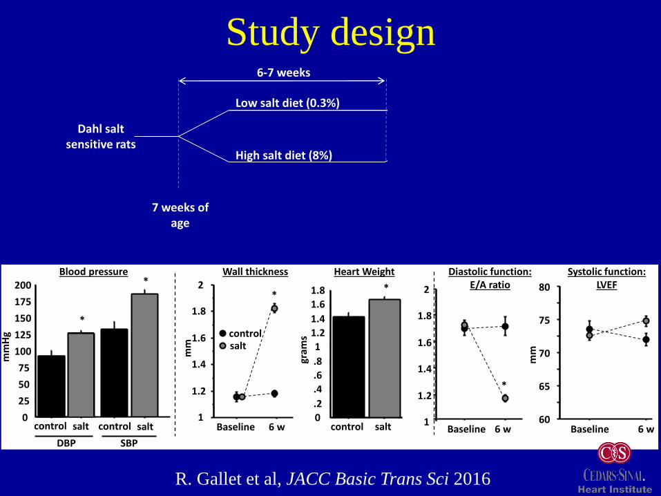
Study design
High salt diet (8%)
Low salt diet (0.3%)
Treatment
CDCs
Placebo(PBS)
6-7 weeks 4 weeks
7 weeks of age
Dahl salt sensitive rats
Endpoint
Heart Weight
0
.2
.4
.6
.8
1
1.2
1.4
1.6
1.8
control salt
*
gram
s
Blood pressure
0
25
50
75
100
125
150
175
200
control salt control salt
*
*
SBPDBP
mm
Hg
1
1.2
1.4
1.6
1.8
2
Baseline 6 w
*
Wall thickness
mm
1
1.2
1.4
1.6
1.8
2
*
Diastolic function:E/A ratio
Baseline 6 w60
65
70
75
80
Systolic function:LVEF
mm
Baseline 6 w
controlsalt
R. Gallet et al, JACC Basic Trans Sci 2016

Inflammation: attenuated by CDCs
0
.2
.4
.6
.8
†
ControlPlaceboCDC
†
†
†
†
†
†
†
Serum
inflammatory
cytokinesPlacebo
CDCCD68 DAPI
CD68 DAPI
CD45 DAPI
CD45 DAPI
Control
CDC
Control
Placebo
CD68 DAPI CD68 DAPI
CD68+ cells CD45+ cells
CD68+ cells
0
10
20
30
40
50
60
CDCPlacebo
#/fi
eld
P<0.0001
*
†
Control
CD45+ cells
0
10
20
30
40
50
60
70
80
CDCPlacebo
#/fi
eld
P<0.0001
†
Control
Histology for
cardiac
macrophages
and
leukocytes

Fibrosis:attenuated by CDCs
CDCPlaceboControl
0
1
2
3
4
5
6
CDCPlaceboControl
mR
NA
exp
ress
ion
rat
io
to c
on
tro
l
P=0.001
Collagen 3
0
1
2
3
4
5
CDCPlaceboControl
mR
NA
exp
ress
ion
rat
io
to c
on
tro
l
P<0.001
Collagen 1A1
0
2
4
6
8
10
12
CDCPlaceboControl
P<0.0001
Tota
l fib
rosi
s(%
LV
)
Total fibrosis (% LV)
††
†

“Clinical” impact
150
200
250
300
350
400
Baseline Pre-ttt 1 week 4 weeks
PlaceboControl
CDC
*
*
†gr
ams
Body weight
0
1
2
3
4
5
6
mg/
g
P=0.006
Lung/body weight
Control Placebo CDC
†
gram
s
0
.5
1
1.5
2P=0.02
Lung weight
Control Placebo CDC
‡
0
.2
.4
.6
.8
1
0 5 10 15 20 25 30 35 40 45
LogrankP=0.03
Survival
*P<0.05 vs. placebo and CDC; †P<0.05 vs. control and CDC

Regress-HFpEF Trial
Regression of fibrosis & reversal of diastolic dysfunction in
HFPEF patients treated with allogeneic CDCs
M. Zile, PI
NCT02941705
recruiting

• CDCs in HFpEF (Regress-HFpEF)
• CDCs in pulmonary hypertension (ALPHA)
• Duchenne muscular dystrophy (HOPE)
Additional allogeneic CDC trials
in progress

CDCs reduce RV systolic pressure and
hypertrophy in monocrotaline rat model of PAH
Experimental Protocol
Tracking CDCs in the LungsRV Hypertrophy
RV Systolic Pressure
R. Middleton, M. Lewis et al., PLoS One 2018

CDCs reduce vessel wall thickness and
macrophage migration in the lungs
Arteriolar Wall Thickness Macrophage Infiltration in the Lungs

ALPHA Trial
ALlogeneic CDCs for Pulmonary arterial Hypertension
therApy
M. Lewis, PI
NCT03145298
recruiting

• CDCs in HFpEF (Regress-HFpEF)
• CDCs in pulmonary hypertension (ALPHA)
• Duchenne muscular dystrophy (HOPE)
Additional allogeneic CDC trials
in progress

• Duchenne muscular
dystrophy
• X-linked recessive
disorder
• Skeletal myopathy
• Cardiomyopathy Cellular
[Ca2+]
Dystrophin
deficiency
Inflammation
Oxidative
stress
Myocyte loss
Fibrosis
Cell membrane
damage
Mitochondrial
inefficiency/loss

Duchenne cardiomyopathy:
Progressive increase in cardiac scar with patient age
Animesh Tandon et al. J Am Heart Assoc 2015;4:e001338

Mdx+VehicleCTL(WT) Mdx+CDC
0.3
0.33
0.36
0.39
0.42
0.45
0.48
0.51
CTL Mdx+Vehicle Mdx+CDC0.4
0.45
0.5
0.55
0.6
0.65
0.7
CTL Mdx+Vehicle Mdx+CDC
Collagen III A1
GAPDH
†
CTL(WT) Mdx+CDC
Mdx+
Vehicle
Collagen I A1
GAPDH
CTL(WT) Mdx+CDC
Mdx+
Vehicle
†
Coll
agen
III
A1
/GA
PD
H
Coll
agen
I A
1/G
AP
DH
CDC treatment reduced cardiac collagen
content and fibrosis in mdx mice
Aminzadeh et al., Stem Cell Reports 2018
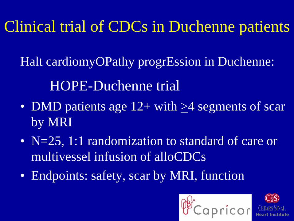
Clinical trial of CDCs in Duchenne patients
Halt cardiomyOPathy progrEssion in Duchenne:
HOPE-Duchenne trial
• DMD patients age 12+ with >4 segments of scar
by MRI
• N=25, 1:1 randomization to standard of care or
multivessel infusion of alloCDCs
• Endpoints: safety, scar by MRI, function

HOPE 6-month data
Full data set at Capricor.com

Cell-free therapeutics
• Is there a single entity that can mimic all the
salient benefits?
• Does the cell-free entity have features
superior to those of the parent cells?
Disadvantages of cells for therapeutics
• Cells work, but fragile living material
• QA/QC, release & identity criteria complex
• Suboptimal in closed compartments
• Immune memory?

20
25
30
35
40
45
50
1 15 30
LV
EF
(%
)
Days (after MI)
CDC-DMSO
CDC-GW4869 *
CDC exosomes ↑ EF
Ibrahim et al., Stem Cell Reports 2014
Exosomes are bioactive nanoparticles
• 30-150 nm particles
• Present in all body fluids
• Released by nearly all cell
types
• Loaded with miRs and
other bioactive contents
• Payload very cell-specific
Blocking exosome biosynthesis
abrogates CDC benefit
20
25
30
35
40
45
50
1 15 30
LV
EF
(%
)
Days (after MI)
Vehicle
CDC-exo
Fibroblast-exo*
**

CDCs CDC-XO
• Regenerative √ 1,2 √ 11,12
• Antifibrotic √ 1-4 √ 11,12
• Anti-apoptotic √ 3-5 √ 11,13
• Angiogenic √ 1,6 √ 11
• Anti-inflammatory √ 9 √ 12,13
• Immunomodulatory √ 9,10 √ 12
1. RR Smith et al, Circ 2007; 2. Makkar et al., Lancet 2012; 3. E. Tseliou et al., PLoSOne 2014; 4. E.
Tseliou et al., BRIC 2014; 5. T-S Li et al, JACC 2012; 6. I. Chimenti et al, Circ Res 2010; 7. K. Malliaras
et al, Circ 2012; 8. K. Malliaras et al., EMBO Mol Med 2013; 9. M. Aminzadeh et al., EHJ
2014; 10. L. Lauden et al, Circ Res 2013; 11. A. Ibrahim et al. Stem Cell Reports 2014;
12. M. Aminzadeh et al., submitted 2017; 13. G. DeCouto et al., Circulation 2017
Exosome payloads mimic CDC effects
on multiple biological processes

Cell Type Human heart progenitor cell
Characteristics CD105+, CD45-; secreted SDF-1, exosomes
Clinical Trials CADUCEUS-completed-autologous phase 1. Twenty-five patient study
showed regeneration in CDC-treated post-MI subjects with mild HFrEF
ALLSTAR-phase 1&2b study of allogeneic CDCs post-MI with mild HFrEF
DYNAMIC- phase 2a study of allogeneic CDCs in patients with advanced
HFrEF
HOPE-Duchenne-allo CDCs for DMD cardiomyopathy
Mechanism of
action
Paracrine effects mediated by exosomes
Promote cardiomyomyogenesis
Prevent cardiomyocyte apoptosis
Anti-fibrotic
Anti-inflammatory
Cardiosphere-derived cells (CDCs)
First described by RR Smith et al., Circulation 2007; methods
and bioactivity reproduced by >40 labs worldwide

When will therapeutic exosomes reach
the clinic?

51
CDC-exosome clinical manufacturing
DONOR HEART
MCB
EDCs
CARDIOSPHERES CDCs CAP-1002
CAP-2003
Confluent CDCs Conditioned medium
Ultrafiltration
EDC Expansion
CDC ExpansionMCB
w

Collagen
content
Cardiomyogenesis
Repeat xenogeneic dosing of human exosomes
recaps effects of syngeneic CDCs in mdx mice
Functional
benefit on
heart
Aminzadeh et al.,
Stem Cell Reports
2018

Follow the data
1. Autologous therapy (2004)
2. Recognition of durable benefits despite cell
transience (2010)
Allogeneic paradigm (2012)
3. Identification of exosomes as mediators (2014)
Cell-free therapeutics (2018)
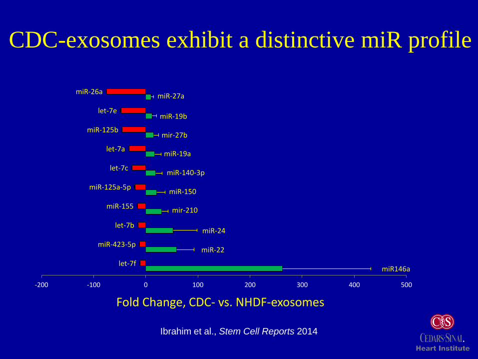
miR146a
miR-22
miR-24
mir-210
miR-150
miR-140-3p
miR-19a
mir-27b
miR-19b
miR-27a
let-7f
miR-423-5p
let-7b
miR-155
miR-125a-5p
let-7c
let-7a
miR-125b
let-7e
miR-26a
-200 -100 0 100 200 300 400 500
Fold Change, CDC- vs. NHDF-exosomes
CDC-exosomes exhibit a distinctive miR profile
Ibrahim et al., Stem Cell Reports 2014

miRs are minority of exosomal RNA
33.71
18.467.60
7.072.13
0.04
21.22
3.995.78
CDC-exo
85.70
0.92 2.240.67
1.65
0.29
4.98
1.661.89
NHDF-exo
L. Cambier et al., EMBO Mol Med 2017

0
5
10
15
20
25
Ys Yb
Re
lati
ve m
RN
A IL
10
e
xpre
sssi
on
0
10
20
30
40
50
60
70
80
90
Ys Yb
IL1
0 c
on
cen
trat
ion
(p
g/m
l)
24h
48h
72h
Plentiful Y RNA fragment regulates
IL-10 expression
L. Cambier et al., EMBO Mol Med 2017
050000
100000150000200000250000300000350000400000
Yb Yc Yd
Nb
of
cou
nts
Y RNA abundance Y RNA alignment reveals Yb homology to Y4
dG=-14.00 kcal/mol
Predicted structure
**
****
****
IL-10 transcript (left) and secreted protein (right) in macrophages

Exosomes: defined contents as next-gen TCs
miR-181b
miR-146a
Y RNA fragment
Cardioprotection
↑ PKCδ↑ IL-10
↓ TRAF-6
G. DeCouto et al.,
Circ 2017
L. Cambier et al.,
EMBO Mol Med 2017
A. Ibrahim et al., Stem
Cell Reports 2014

Working hypotheses
• No single RNA species can account for all the
benefits of exosomes
• Individual miRs or other RNA species may
prevail in any given setting
• The totality of exosomal contents required for
full manifestation of bioactivity

Deconstruction: follow the data
1. Autologous therapy (2004)
2. Recognition of durable benefits despite cell
transience (2010)
Allogeneic paradigm (2012)
3. Identification of exosomes as mediators (2014)
Cell-free therapeutics (2018)
4. Mining of exosome contents identifies defined
factors (2014-present)
Next-gen therapeutics (?)

Our initial goal with cell therapy (2004)
To repair the “permanently” injured heart
Our updated goals after 14 years of
discovery work (2018)
To use cells, exosomes or defined factors as
novel therapeutic candidates for a broad
range of inflammatory/fibrotic diseases

AHSP
in the Advanced
Health Sciences
Pavilion


Deconstruction: follow the data
1. Autologous therapy (2004)
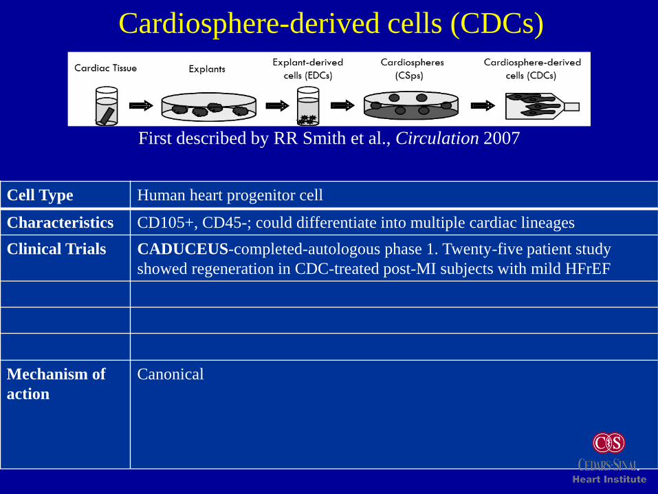
Cell Type Human heart progenitor cell
Characteristics CD105+, CD45-; could differentiate into multiple cardiac lineages
Clinical Trials CADUCEUS-completed-autologous phase 1. Twenty-five patient study
showed regeneration in CDC-treated post-MI subjects with mild HFrEF
Mechanism of
action
Canonical
Cardiosphere-derived cells (CDCs)
First described by RR Smith et al., Circulation 2007

Dis
tan
ce(m
)
0
200
400
600
800
1000
3 4 5 6 7 8 9 10 11 12
CTL(WT)
Mdx+CDC
Mdx+Vehicle
Mdx+VehicleMdx+CDC
Weeks
Functional improvement in mdx mice with
CDC treatment and repeat dosing
30
40
50
60
70
80
Baseline Wk3 M2 M3 Wk3 M2 M3
Mdx+CDC
Mdx+Vehicle
*** **
* *
1st injection 2nd injection
EF
(%)
Global LV function
Exercise capacity
* *********
• Graft versus host disease
• Hypoplastic left heart syndrome
• Recurrent ventricular tachycardia
Candidate clinical indications for
CDC exosomes
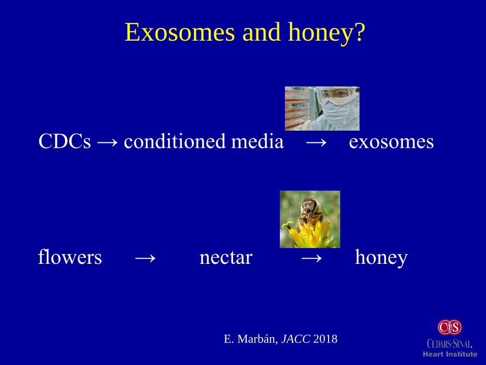
CDCs → conditioned media → exosomes
Exosomes and honey?
flowers → nectar → honey
E. Marbán, JACC 2018
Acknowledgments
Cedars-Sinai Heart Institute
Kostas Malliaras
Ryan Middleton
Mark Aminzadeh
Ahmed Ibrahim
Geoffrey DeCouto
Linda Cambier
Russell Rogers
Ronald Victor
Michael Lewis
Raj Makkar
Capricor
Linda MarbánRachel R. Smith Luis Rodriguez BorladoDeborah Ascheim
Funding
NIH
California Institute for Regenerative Medicine
Department of Defense
Coalition Duchenne


![Targeting regenerative exosomes to myocardial infarction using … · 2018. 2. 12. · C-Cure [3], CADUCEUS [4], and SCIPIO [5]—have demonstrated the cardiac regenerative potential](https://img.pdfslide.us/doc/110x75/60d6f390ad1cc314a94a4423/targeting-regenerative-exosomes-to-myocardial-infarction-using-2018-2-12-c-cure.jpg)