Embed Size (px)
Citation preview
1 / 14
Reduction and Functional Exhaustion of T Cells in Patients with Coronavirus
Disease 2019 (COVID-19)
Bo Diao1#
, Chenhui Wang2#
, Yingjun Tan1#
, Xiewan Chen3, Ying Liu
4, Lifen Ning
5, Li Chen
1, Min
Li1, Yueping Liu
1, Gang Wang
1, Zilin Yuan
1, Zeqing Feng
2, Yuzhang Wu
2#, Yongwen Chen
2#
1. Department of Medical Laboratory Center, General Hospital of Central Theater Command,
Wuhan, Hubei province, 430015, People’s Republic of China
2. Institute of Immunology, PLA, Third Military Medical University, Chongqing, 400038,
People’s Republic of China
3. Medical English Department, College of Basic Medical Sciences, Army Medical University,
Chongqing, 400038, People’s Republic of China
4. Department of Medical Laboratory Medicine, General Hospital of Central Theater
Command, Wuhan, Hubei province, 430015, People’s Republic of China
5. Hanyang Hospital Affiliated to Wuhan University of science and technology, Wuhan, Hubei
province, 430015, People’s Republic of China
# Equally to this work
*Corresponding author: Yongwen Chen (Ph. D.), Institute of Immunology, PLA, Third Military
Medical University, Chongqing, 400038, People’s Republic of China. Fax: +8602368752228; Phone:
+8602368752228. E-mail: [email protected] Or Yuzhang Wu (Prof. & Chair). Email:
. CC-BY-NC-ND 4.0 International licenseIt is made available under a perpetuity.
is the author/funder, who has granted medRxiv a license to display the preprint in(which was not certified by peer review)preprint The copyright holder for thisthis version posted February 20, 2020. .https://doi.org/10.1101/2020.02.18.20024364doi: medRxiv preprint
2 / 14
Summary
BACKGROUND The outbreak of coronavirus disease 2019 (COVID-19) caused by severe acute
respiratory syndrome coronavirus 2 (SARS-CoV-2) has posed great threat to human health, which
has been declared a public health emergency of international concern (PHEIC) by the WHO. T cells
play a critical role in antiviral immunity but their numbers and functional state in COVID-19
patients remain largely unclear.
METHODS We retrospectively reviewed the counts of total T cells, CD4+, CD8
+ T cell subsets,
and serum cytokine concentration from inpatient data of 522 patients with laboratory-confirmed
COVID-19, admitted into two hospitals in Wuhan from December 2019 to January 2020, and 40
healthy controls, who came to the hospitals for routine physical examination. In addition, the
expression of T cell exhaustion markers PD-1 and Tim-3 were measured by flow cytometry in the
peripheral blood of 14 COVID-19 cases.
RESULTS The number of total T cells, CD4+ and CD8
+ T cells were dramatically reduced in
COVID-19 patients, especially among elderly patients (≥60 years of age) and in patients requiring
Intensive Care Unit (ICU) care. Counts of total T cells, CD8+T cells or CD4
+T cells lower than
800/μL, 300/μL, or 400/μL, respectively, are negatively correlated with patient survival. Statistical
analysis demonstrated that T cell numbers are negatively correlated to serum IL-6, IL-10 and TNF-α
concentration, with patients in decline period showing reduced IL-6, IL-10 and TNF-α
concentrations and restored T cell counts. Finally, T cells from COVID-19 patients have
significantly higher levels of the exhausted marker PD-1 as compared to health controls. Moreover,
increasing PD-1 and Tim-3 expression on T cells could be seen as patients progressed from
prodromal to overtly symptomatic stages, further indicative of T cell exhaustion.
CONCLUSIONS T cell counts are reduced significantly in COVID-19 patients, and the surviving
T cells appear functionally exhausted. Non-ICU patients, with total T cells, CD8+T cells CD4
+T
cells counts lower than 800/μL, 300/μL, and 400/μL, respectively, may still require aggressive
intervention even in the immediate absence of more severe symptoms due to a high risk for further
deterioration in condition.
. CC-BY-NC-ND 4.0 International licenseIt is made available under a perpetuity.
is the author/funder, who has granted medRxiv a license to display the preprint in(which was not certified by peer review)preprint The copyright holder for thisthis version posted February 20, 2020. .https://doi.org/10.1101/2020.02.18.20024364doi: medRxiv preprint
3 / 14
Introduction
In December 2019, a series of acute respiratory illness were reported in Wuhan, Hubei
Province, China.1,2
A novel coronavirus, initially named severe acute respiratory syndrome
coronavirus 2 (SARS-CoV-2), was identified as the cause of this disease by the Chinese Center for
Disease Control and Prevention (CDC).3 This disease, now designated as coronavirus disease 2019
(COVID-19) by the WHO, rapidly spread to other cities of China, and has become a public health
emergency of international concern (PHEIC) following its global spread. COVID-19 is clinically
manifests as fever, cough, muscle pain, fatigue, diarrhea and pneumonia, and can cause death in
severe cases.4-6
Up through February 18, 2020, China has reported 72436 cases of confirmed
COVID-19 and 1868 fatalities.7
Since an effective immune response against viral infections depends on the activation of
cytotoxic T cells that can clear infection by killing virus-infected cells,8 boosting the numbers and
function of T cells in COVID-19 patients is critical for successful recovery. A recent study reported
that the 82.1% of COVID-19 cases displayed low circulating lymphocyte counts.4-6
However, the
factors which might cause the reduction in count, and the activation status of T cells in COVID-19
patients, remain uninvestigated. We retrospectively analyze here the clinical data from 522 cases of
COVID-19 who were admitted into the General Hospital of Central Theatre Command and
Hanyang Hospital in Wuhan from December 2019 to January 2020. We also compared the
expression of exhaustion markers PD-1 and Tim-3 on the surface of CD4+ and CD8
+ T cells from
COVID-19 and healthy controls. Our results thus provide a preliminary demonstration of T cell
exhaustion during COVID-19 infection and suggest that more aggressive early intervention may be
required in patients with low T lymphocyte counts.
Methods
Patients Medical records from 522 patients (aged from 5 days to 97 years) with confirmed
COVID-19 and admitted into the General Hospital of Central Theatre Command or Hanyang
Hospital in Wuhan from December 2019 to January 2020, and 40 healthy people (aged from 2 to 62
years), who came to the hospitals for routine physical examination, were collected and
retrospectively analyzed. Diagnosis of COVID-19 was based on the New Coronavirus Pneumonia
Prevention and Control Program (5th edition) published by the National Health Commission of
. CC-BY-NC-ND 4.0 International licenseIt is made available under a perpetuity.
is the author/funder, who has granted medRxiv a license to display the preprint in(which was not certified by peer review)preprint The copyright holder for thisthis version posted February 20, 2020. .https://doi.org/10.1101/2020.02.18.20024364doi: medRxiv preprint
4 / 14
China.9 All the patients were laboratory-confirmed positive for SARS-CoV-2 by use of quantitative
RT-PCR (qRT-PCR) of throat swab samples. This study was approved by the National Health
Commission of China and Ethics Commission of General Hospital of Central Theatre Command
([2020]-004-1) and Hanyang Hospital (20200217). Written informed consent was waived by the
Ethics Commission of the designated hospital for emerging infectious diseases.
Definitions The classification of clinical types, which consist of mild/moderate/severe/critical, was
based on the New Coronavirus Pneumonia Prevention and Control Program (5th edition) published
by the National Health Commission of China.9 Within the cohort analyzed 43 were admitted to the
intensive care unit (ICU), because they required high-flow nasal cannula or higher-level oxygen
support measures to correct hypoxaemia. Hypoxaemia was defined as arterial oxygen tension (PaO2)
over inspiratory oxygen fraction (FIO2) of less than 300 mm Hg or arterial oxygen saturation of 93%
or lower. According to the staging of infectious disease,10
the prodromal period is a phase in which
the host begins to experience general signs and symptoms. The illness period (overtly symptomatic
period) is a phase in which the signs or symptoms of disease are most obvious and severe, with
positive laboratory findings and chest images. For ICU patients, ICU period is a phase in which the
symptoms are most obvious and severe The decline period is a phase in which the clinical
symptoms begin to decline, laboratory findings and chest images improve, and arterial oxygen
saturation to return to the normal.
Data collection We reviewed clinical records, nursing records, laboratory findings, and chest x-rays
or CT scans for all the patients and physical examination records of the 40 healthy people. All
information was obtained and curated with a customised data collection form. Three investigators
(C Wang, Z Fen and Y Chen) independently reviewed the data collection forms to verify data
accuracy.
Sample collection and flow cytometric analysis Peripheral blood samples from 14 patients and 3
healthy volunteers were simultaneously processed in the Central Lab of General Hospital of Central
Theatre Command to isolate peripheral blood mononuclear cells (PBMCs) for further testing. The
peripheral blood was supplemented with anticoagulants and PBMCs were harvested by density
gradient centrifugation. Isolated PBMCs were stained with a BD multitest IMK Kit (Cat340503,
BD Biosciences) for analyzing the frequency and cell number of total T, CD4+ T,CD8+ T, B and
NK in healthy controls and patients. The exhaustion of T cells was detected using human
CD4-percp (RPA-T4, Biolegend), CD8-APC (SK1, BD Biosciences), CD8-PE (SK1, Biolegend),
. CC-BY-NC-ND 4.0 International licenseIt is made available under a perpetuity.
is the author/funder, who has granted medRxiv a license to display the preprint in(which was not certified by peer review)preprint The copyright holder for thisthis version posted February 20, 2020. .https://doi.org/10.1101/2020.02.18.20024364doi: medRxiv preprint
5 / 14
PD-1-PE (EH12.2H7, Biolegend) and TIM-3-FITC (F38-2E2, Biolegend) antibodies. After being
stained, the cells were measured by flow cytometry on an LSR Fortessa Cell Analyzer (BD
Biosciences) and data analyzed using the FolwJo software (TreeStar). All experimental procedures
were completed under biosafety level II plus condition.
Statistical analysis Statistical analyses were performed using GraphPad Prism version 8.0
(GraphPad Software, Inc., San Diego, CA, USA). Continuous variables were directly expressed as a
range. Categorical variables were expressed as numbers/NUMBERS (%). p values are from χ2,
non-paired t test or paired t test.
Role of the funding source The funding agencies did not participate in study design, data
collection, data analysis, or manuscript writing. The corresponding authors were responsible for all
aspects of the study to ensure that issues related to the accuracy or integrity of any part of the work
were properly investigated and resolved. The final version was approved by all authors.
Results
1. Decreasing the numbers of total T cells, CD4+ and CD8
+ subsets in COVID-19 patients
From our retrospective analysis of 522 patients, 499 cases had lymphocyte count recorded. 75.75%
(359/499), 75.95% (379/499) and 71.54% (357/499) patients had remarkably low total T cell counts,
CD4+ and CD8
+ T cell counts, respectively. Among milder patients in the Non-ICU group, the
median value of total T cells, CD4+ and CD8
+ T cell counts are 652, 342 and 208, respectively, the
median value decreased to 261, 198 and 64.3, respectively, in the ICU group. (Figure 1A). The
counts of total T cells, CD4+ and CD8
+ T cells were significantly lower in ICU patients than
Non-ICU cases (Figure 1B). These patients were further categorized into three groups based on age
(<20 years old, 20~60 years and ≥60 years), and an age-dependent reduction of T cell numbers
was observed in COVID-19 patients, with the lowest T cells numbers found in patients ≥60 years
old (Figure 1C), suggesting a potential cause for increased susceptibility in elderly patients.
We next retrospectively reviewed T cell numbers in 212 cases from Non-ICU patients within one
center (the General Hospital of Central Theatre Command). The Non-ICU patients were further
divided into four groups based on clinical outcomes. Among these patients, 151 cases are
mild/moderate, 40 cases are severe, while 13 cases in critical condition, and 8 perished occurred.
Statistical analysis showed that T cell numbers including total T cells, CD4+ and CD8
+ T cells in
. CC-BY-NC-ND 4.0 International licenseIt is made available under a perpetuity.
is the author/funder, who has granted medRxiv a license to display the preprint in(which was not certified by peer review)preprint The copyright holder for thisthis version posted February 20, 2020. .https://doi.org/10.1101/2020.02.18.20024364doi: medRxiv preprint
6 / 14
severe, critical and perished groups are significantly lower than in the mild/moderate group. Most
importantly, the numbers of total T cell, CD8+T cells and CD4
+ T cells in severe and perished
groups are lower than 800/μL, 300/μL, or 400/μL, respectively (Figure 1D). This result suggests
that aggressive interventions may be required for non-ICU patients even in the absence of more
severe symptoms should their T cell counts fall below the critical threshold.
2. Negatively correlated between T cell numbers and cytokines in COVID-19 patients
The expression of angiotensin converting enzyme 2(ACE2), the predicted receptor of
SARS-CoV-2 viruses, is absent on T cells,11
suggesting that the depressed T counts in COVID-19
patients mentioned above (Figure 1) was likely not caused by direct infection of T cells. We
therefore examined the concentrations of serum cytokines, including TNF-α, IFN-γ, IL-2, IL-4, IL-6
and IL-10, from these COVID-19 patients to explore the influence of cytokine signaling. We only
found the levels of TNF-α, IL-6 and IL-10 were significantly increased in infected patients, and
statistical analysis illustrated that their levels in ICU patients are significantly higher than in
Non-ICU patients (Figure 2A).
We next investigated the relationships between IL-10, IL-6, TNF-α and T cell count within
Non-ICU patients. Interestingly, the concentration of these three cytokines was negatively
correlated with total T cell counts, CD4+
counts, and CD8+ counts, respectively (Figure 2B). We
subsequently summarized the follow up data of cytokine concentrations and T cell numbers in ten
patients that were follower over the course of inpatient care. Interestingly, serum levels of IFN-γ,
IL-10, IL-6 and TNF-α were significantly decreased in these patients in the decline period as
compared with illness period, while counts of total T cells, CD4+, and CD8
+ T cell subsets
recovered during the decline period (Figure 2C). The phenomena suggests that the decrease of T
cells seen in COVID-19 patients is likely the result of high serum concentration of TNF-α, IL-6 and
IL-10 negatively regulating T cell survival or proliferation.
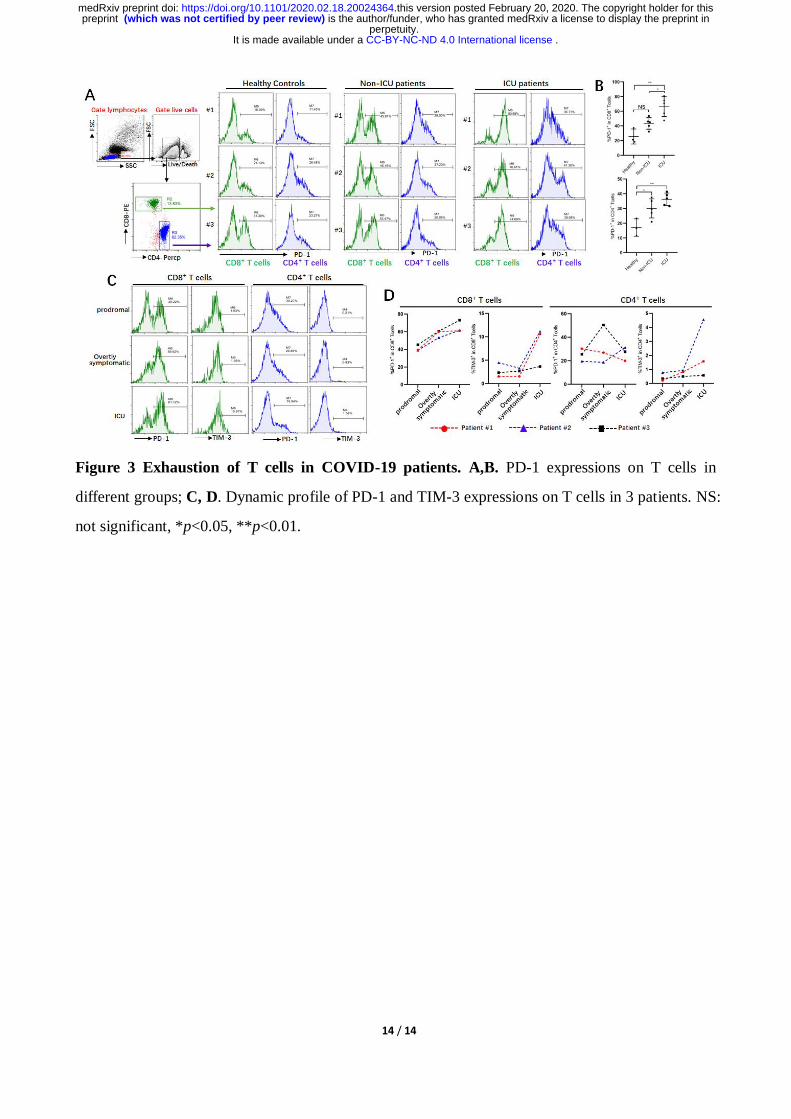
3. Enhancing T cell exhaustion in COVID-19 patients
Beyond changing in numbers during the course of infection, T cells may display limited function
during prolonged infection as a result of exhaustion, which has been associated with the expression
of some immune-inhibitory factors including PD-1, Tim-3 on cell surface.12
We therefore examined
whether T cells in COVID-19 patients have exhaustion phenotypes. FACs analysis illustrated that, T
. CC-BY-NC-ND 4.0 International licenseIt is made available under a perpetuity.
is the author/funder, who has granted medRxiv a license to display the preprint in(which was not certified by peer review)preprint The copyright holder for thisthis version posted February 20, 2020. .https://doi.org/10.1101/2020.02.18.20024364doi: medRxiv preprint
7 / 14
cells from COVID-19 patients have markedly higher levels of PD-1 compared to healthy controls
(Figure 3A). Furthermore, statistical analysis showed that the percentage of PD-1+CD8
+ T cells
from ICU patients was significantly higher than from both Non-ICU cases and healthy controls
(Figure 3B), indicating that SARS-CoV-2 viruses induce T cell exhaustion in COVID-19 patients,
particularly in those requiring ICU care.
Three patients were follow-up during inpatient care, and the expression of the exhausted markers
including PD-1 and Tim-3 on surface of T cells during disease progress was detected. FACs showed
that these patients have very low level of PD-1 and Tim-3 on CD8+ and CD4
+ T cells in the
prodromal stage, however, their levels on CD8+ T cells was increased in overtly symptomatic stages,
and highest levels were seen in ICU period (Figure 3C, D). Similarly, higher levels of Tim-3 was
observed on CD4+ T cells from patients who are in ICU stage, although enhancing the expression of
PD-1 on CD4+
T cells was not obviously during disease progress (Figure 3C, D). These results
demonstrated that T cells are exhaustion in COVID-19 patients during SARS-CoC-2 infection.
Discussion
T cells play a vital role in viral clearance, with CD8+ cytotoxic T cells (CTLs) capable of
secreting an array of molecules such as perforin, granzymes, and IFN-γ to eradicate viruses from
the host.13
At the same time, CD4+
helper T cells (Th) can assist cytotoxic T cells and B cells and
enhance their ability to clear pathogen.14
However, persistent stimulation by the virus may induce T
cell exhaustion, leading to loss of cytokine production capability and reduced functions. 15,16
Earlier
studies have been unclear regarding the numbers and function of T cells in COVID-19 patients,
albeit with suggestions of depressed lymphocyte counts.4,6
In this report, we retrospectively
reviewed the numbers of total T cells, CD4+, CD8
+ T cell subsets in a total of 499 COVID-19
patients. In Non-ICU patients, we found that over 70.56% cases underwent decreased in the total T
cells, CD4+ T cells and CD8
+ T cells. However, in the ICU group, a total of 95% (19/20) patients
showed a decrease in both total T cells and CD4+ T cells, and most importantly, all of the patients
displayed decreases in CD8+ T cells. We also analyzed Non-ICU patients in greater detail, and
found that aggressive intervention may be necessary to preempt the development of severe
symptoms in patients with low T cell counts.
Cytokine storm is a phenomenon of excessive inflammatory reaction in which cytokines are
rapidly produced in large amount in response to microbial infection. This phenomenon has been
. CC-BY-NC-ND 4.0 International licenseIt is made available under a perpetuity.
is the author/funder, who has granted medRxiv a license to display the preprint in(which was not certified by peer review)preprint The copyright holder for thisthis version posted February 20, 2020. .https://doi.org/10.1101/2020.02.18.20024364doi: medRxiv preprint

8 / 14
considered an important contributor to acute respiratory distress syndrome (ARDS) and multiple
organ dysfunction syndrome (MODS).17,18
It has been also implicated in the setting of respiratory
viral infections, such as SARS in 2002, avian H5N1 influenza virus infection in 2005 and H7N9
infection in 2013.19-22
Huang C et al. showed that the levels of IL-2, IL-7, IL-10, TNF-α, G-CSF,
IP-10, MCP-1 and MIP-1A were significantly higher in COVID-19 patients.4 Consistent with this
report, we here also found that the secretion of cytokines including TNF-α, IL-6 and IL-10 was
increased in COVID-19 patients. Interestingly, the numbers of total T cells, CD4+ T and CD8
+ T
cells are negatively correlated to levels of TNF-α, IL-6 and IL-10, respectively (Figure 2B),
suggesting these cytokines promote T cells decrease in COVID-19 patients.
TNF-α is a pro-inflammatory cytokine which can promote T cell apoptosis via interacting with its
receptor, TNFR1, which expression is increased in aged T cells.23,24
Our current analysis
demonstrated that patient over 60 years old have lower T cell numbers, indicating that TNF-α might
be directly involved in inducing T cell loss in these patients. IL-6, when promptly and transiently
produced in response to infections and tissue injuries, contributes to host defense through the
stimulation of acute phase responses or immune reactions. Dysregulated and continual synthesis of
IL-6 has been shown to play a pathological role in chronic inflammation and infection.25,26
Tocilizumab, a humanized anti-IL-6 receptor antibody, has been developed and approved for the
treatment of rheumatoid arthritis (RA) and juvenile idiopathic arthritis.27,28
Moreover, tocilizumab
has been shown to be effective against cytokine release syndrome resulting from CAR-T cell
infusion against B cell acute lymphoblastic leukemia.29
Whether tocilizumab can restore T cell
counts in COVID-19 patients by suppressing IL-6 signaling remains uninvestigated.
One interesting question is the source of these cytokine during COVID-19 infection. While
previous studies have validated that the secretion of cytokines including IL-6, IL-10 and TNF-α are
mainly from T cells, macrophages and monocytes etc, based on our results, we suggest that the
secretion of these cytokines does not originate from T cells. However, the cytokine storm in turn
may promote apoptosis or necrosis of T cells, and consequently leads to their reduction. Our
previous work demonstrating that monocytes and macrophages can produce pro-inflammatory
cytokine during murine hepatitis virus strain-3 infection,30,31
and whether SARS-CoV-2 also
triggers cytokine release from monocytes and macrophages in COVID-19 patients need further
investigation and such work is in progress in our hospital.
T cell exhaustion is a state of T cell dysfunction that arises during many chronic infections and
cancer. It is defined by poor effector function, sustained expression of inhibitory receptors, and a
. CC-BY-NC-ND 4.0 International licenseIt is made available under a perpetuity.
is the author/funder, who has granted medRxiv a license to display the preprint in(which was not certified by peer review)preprint The copyright holder for thisthis version posted February 20, 2020. .https://doi.org/10.1101/2020.02.18.20024364doi: medRxiv preprint

9 / 14
transcriptional state distinct from that of functional effector or memory T cells.32
By FACs analysis,
we found that both CD8+
T cells and CD4+ T cells have higher levels of PD-1 in virus infected
patients, particularly when derived from ICU patients. Since these changes could also be observed
in our longitudinal follow-up of several patients from prodromal to ICU care (Figure 3). IL-10, an
inhibitory cytokine, not only prevents T cell proliferation, but also can induce T cell exhaustion.
Important, blocking IL-10 function has been shown to successfully prevent T cell exhaustion in
animal models of chronic infection.33,34
We demonstrate here that COVID-19 patients have very
high levels of serum IL-10 following SARS-CoV-2 infection, while also displaying high levels of
the PD-1 and Tim-3 exhaustion markers on their T cells, suggesting that IL-10 might be
mechanistically responsible. The application of potent antiviral treatments to prevent the
progression to T cell exhaustion in susceptible patients may thus be critical to their recovery. We
have read with great interest the successful application of Remdesivir to curing a COVID-19 patient
in the US, and to clinical trials indicates that it may have significant potential as such an
antiviral.35,36
Taken together, we conclude that T cells are decreased and exhausted in patients with
COVID-19. Cytokines such as IL-10, IL-6 and TNF-α might directly mediate T cell reduction. Thus,
new therapeutic measures are needed for treatment of ICU patients, and may even be necessary
early on to preempt disease progression in higher-risk patients with low T cell counts.
Conflict of interest: The authors declare no financial or commercial conflict of interest.
Reference
1. Zhu N, Zhang D, Wang W, et al. A Novel Coronavirus from Patients with Pneumonia in China,
2019. N Engl J Med 2020.
2. Li Q, Guan X, Wu P, et al. Early Transmission Dynamics in Wuhan, China, of Novel
Coronavirus-Infected Pneumonia. N Engl J Med 2020.
3. Lu R, Zhao X, Li J, et al. Genomic characterisation and epidemiology of 2019 novel coronavirus:
implications for virus origins and receptor binding. Lancet 2020.
4. Huang C, Wang Y, Li X, et al. Clinical features of patients infected with 2019 novel coronavirus
in Wuhan, China. Lancet 2020.
5. Chen N, Zhou M, Dong X, et al. Epidemiological and clinical characteristics of 99 cases of 2019
novel coronavirus pneumonia in Wuhan, China: a descriptive study. Lancet 2020.
6. Wang D, Hu B, Hu C, et al. Clinical Characteristics of 138 Hospitalized Patients With 2019
. CC-BY-NC-ND 4.0 International licenseIt is made available under a perpetuity.
is the author/funder, who has granted medRxiv a license to display the preprint in(which was not certified by peer review)preprint The copyright holder for thisthis version posted February 20, 2020. .https://doi.org/10.1101/2020.02.18.20024364doi: medRxiv preprint

10 / 14
Novel Coronavirus-Infected Pneumonia in Wuhan, China. JAMA 2020.
7. National Health Commission of the People’s Republic of China. The latest situation of novel
coronavirus pneumonia as of 24:00 on 17 February, 2020.
http://www.nhc.gov.cn/xcs/yqtb/202002/261f72a74be14c4db6e1b582133cf4b7.shtml. (Date
accessed: February 18, 2020; in Chinese).
8. Li CK, Wu H, Yan H, et al. T cell responses to whole SARS coronavirus in humans. J Immunol
2008; 181(8): 5490-500.
9. National Health Commission of the People’s Republic of China. The notice of launching
guideline on diagnosis and treatment of the novel coronavirus pneumonia (NCP). 5th edition.
http://www.nhc.gov.cn/yzygj/s7653p/202002/3b09b894ac9b4204a79db5b8912d4440/files/72603
01a393845fc87fcf6dd52965ecb.pdf (Date accessed: February 18, 2020; in Chinese).
10. Nina P, Mark S, Anh-Hue TT, et al. Characteristics of infectious disease.
https://openstax.org/books/microbiology/pages/15-1-characteristics-of-infectious-disease.
Microbiology November 2016.
11. Zhou P, Yang XL, Wang XG, et al. A pneumonia outbreak associated with a new coronavirus of
probable bat origin. Nature 2020.
12. Wherry EJ, Kurachi M. Molecular and cellular insights into T cell exhaustion. Nature Reviews
Immunology 2015; 15(8): 486-99.
13. Mescher MF, Curtsinger JM, Agarwal P, et al. Signals required for programming effector and
memory development by CD8+ T cells. Immunol Rev 2006; 211: 81-92.
14. Zhu J, Yamane H, Paul WE. Differentiation of effector CD4 T cell populations (*). Annu Rev
Immunol 2010; 28: 445-89.
15. Ng CT, Snell LM, Brooks DG, Oldstone MB. Networking at the level of host immunity:
immune cell interactions during persistent viral infections. Cell Host Microbe 2013; 13(6):
652-64.
16. Fenwick C, Joo V, Jacquier P, et al. T-cell exhaustion in HIV infection. Immunol Rev 2019;
292(1): 149-63.
17. Wang H, Ma S. The cytokine storm and factors determining the sequence and severity of organ
dysfunction in multiple organ dysfunction syndrome. Am J Emerg Med 2008; 26(6): 711-5.
18. Matthay MA, Ware LB, Zimmerman GA. The acute respiratory distress syndrome. J Clin Invest
2012; 122(8): 2731-40.
19. Channappanavar R, Perlman S. Pathogenic human coronavirus infections: causes and
consequences of cytokine storm and immunopathology. Semin Immunopathol 2017; 39(5):
529-39.
20. Huang KJ, Su IJ, Theron M, et al. An interferon-gamma-related cytokine storm in SARS
patients. J Med Virol 2005; 75(2): 185-94.
21. Writing Committee of the Second World Health Organization Consultation on Clinical Aspects
of Human Infection with Avian Influenza AV, Abdel-Ghafar AN, Chotpitayasunondh T, et al.
. CC-BY-NC-ND 4.0 International licenseIt is made available under a perpetuity.
is the author/funder, who has granted medRxiv a license to display the preprint in(which was not certified by peer review)preprint The copyright holder for thisthis version posted February 20, 2020. .https://doi.org/10.1101/2020.02.18.20024364doi: medRxiv preprint
11 / 14
Update on avian influenza A (H5N1) virus infection in humans. N Engl J Med 2008; 358(3):
261-73.
22. Chen Y, Liang W, Yang S, et al. Human infections with the emerging avian influenza A H7N9
virus from wet market poultry: clinical analysis and characterisation of viral genome. Lancet
2013; 381(9881): 1916-25.
23. Aggarwal S, Gollapudi S, Gupta S. Increased TNF-alpha-induced apoptosis in lymphocytes
from aged humans: changes in TNF-alpha receptor expression and activation of caspases. J
Immunol 1999; 162(4): 2154-61.
24. Gupta S, Bi R, Kim C, Chiplunkar S, Yel L, Gollapudi S. Role of NF-kappaB signaling
pathway in increased tumor necrosis factor-alpha-induced apoptosis of lymphocytes in aged
humans. Cell Death Differ 2005; 12(2): 177-83.
25. Gabay C. Interleukin-6 and chronic inflammation. Arthritis research & therapy 2006; 8(2): S3.
26. Jones SA, Jenkins BJ. Recent insights into targeting the IL-6 cytokine family in inflammatory
diseases and cancer. Nature Reviews Immunology 2018; 18(12): 773-89.
27. Burmester GR, Rigby WF, van Vollenhoven RF, et al. Tocilizumab in early progressive
rheumatoid arthritis: FUNCTION, a randomised controlled trial. Annals of the rheumatic
diseases 2016; 75(6): 1081-91.
28. Yokota S, Itoh Y, Morio T, et al. Tocilizumab in systemic juvenile idiopathic arthritis in a
real-world clinical setting: results from 1 year of postmarketing surveillance follow-up of 417
patients in Japan. Annals of the rheumatic diseases 2016; 75(9): 1654-60.
29. Le RQ, Li L, Yuan W, et al. FDA approval summary: tocilizumab for treatment of chimeric
antigen receptor T cell‐induced severe or life‐threatening cytokine release syndrome. The
oncologist 2018; 23(8): 943.
30. Yang C, Chen Y, Guo G, et al. Expression of B and T lymphocyte attenuator (BTLA) in
macrophages contributes to the fulminant hepatitis caused by murine hepatitis virus strain-3. Gut
2013; 62(8): 1204-13.
31. Li J, Diao B, Guo S, et al. VSIG4 inhibits proinflammatory macrophage activation by
reprogramming mitochondrial pyruvate metabolism. Nature communications 2017; 8(1): 1322.
32. McLane LM, Abdel-Hakeem MS, Wherry EJ. CD8 T cell exhaustion during chronic viral
infection and cancer. Annual review of immunology 2019; 37: 457-95.
33. Brooks DG, Trifilo MJ, Edelmann KH, Teyton L, McGavern DB, Oldstone MB. Interleukin-10
determines viral clearance or persistence in vivo. Nat Med 2006; 12(11): 1301-9.
34. Ejrnaes M, Filippi CM, Martinic MM, et al. Resolution of a chronic viral infection after
interleukin-10 receptor blockade. J Exp Med 2006; 203(11): 2461-72.
35. Wang M, Cao R, Zhang L, et al. Remdesivir and chloroquine effectively inhibit the recently
emerged novel coronavirus (2019-nCoV) in vitro. Cell Res 2020.
36. Holshue ML, DeBolt C, Lindquist S, et al. First Case of 2019 Novel Coronavirus in the United
States. N Engl J Med 2020.
. CC-BY-NC-ND 4.0 International licenseIt is made available under a perpetuity.
is the author/funder, who has granted medRxiv a license to display the preprint in(which was not certified by peer review)preprint The copyright holder for thisthis version posted February 20, 2020. .https://doi.org/10.1101/2020.02.18.20024364doi: medRxiv preprint

12 / 14
Figure and figure legends
Figure 1 Reducing T cell numbers in COVID-19 patients. A. Demographics of T cells in patients;
B. T cell numbers in different groups; C. T cell numbers in patients of different ages; D. T cell
count in Non-ICU care patients with different clinical outcomes.**p<0.01, ***p<0.001 and
****p<0.0001.
. CC-BY-NC-ND 4.0 International licenseIt is made available under a perpetuity.
is the author/funder, who has granted medRxiv a license to display the preprint in(which was not certified by peer review)preprint The copyright holder for thisthis version posted February 20, 2020. .https://doi.org/10.1101/2020.02.18.20024364doi: medRxiv preprint

13 / 14
Figure 2 Cytokines and relative T cell numbers in COVID-19 patients. A. Demographics of
cytokines in patients; B. Cytokine levels in different groups; C. The relativity of T cell numbers
with cytokine levels; D. Dynamic profiles of cytokine levels and T cell numbers in Non-ICU care
patients. *p<0.05, **p<0.01, ***p<0.001 and ****p<0.0001.
. CC-BY-NC-ND 4.0 International licenseIt is made available under a perpetuity.
is the author/funder, who has granted medRxiv a license to display the preprint in(which was not certified by peer review)preprint The copyright holder for thisthis version posted February 20, 2020. .https://doi.org/10.1101/2020.02.18.20024364doi: medRxiv preprint
14 / 14
Figure 3 Exhaustion of T cells in COVID-19 patients. A,B. PD-1 expressions on T cells in
different groups; C, D. Dynamic profile of PD-1 and TIM-3 expressions on T cells in 3 patients. NS:
not significant, *p<0.05, **p<0.01.
. CC-BY-NC-ND 4.0 International licenseIt is made available under a perpetuity.
is the author/funder, who has granted medRxiv a license to display the preprint in(which was not certified by peer review)preprint The copyright holder for thisthis version posted February 20, 2020. .https://doi.org/10.1101/2020.02.18.20024364doi: medRxiv preprint


![CD4 CD25 T Cells in Skin Lesions of Patients with ... A P... · fector T cells can activate macrophages, leading to the killing of intracellular amastigotes [9]. ... (TGF)–b1 [12–15],](https://img.pdfslide.us/doc/110x75/5b782c7c7f8b9a8f698e717d/cd4-cd25-t-cells-in-skin-lesions-of-patients-with-a-p-fector-t-cells.jpg)