Embed Size (px)
Citation preview
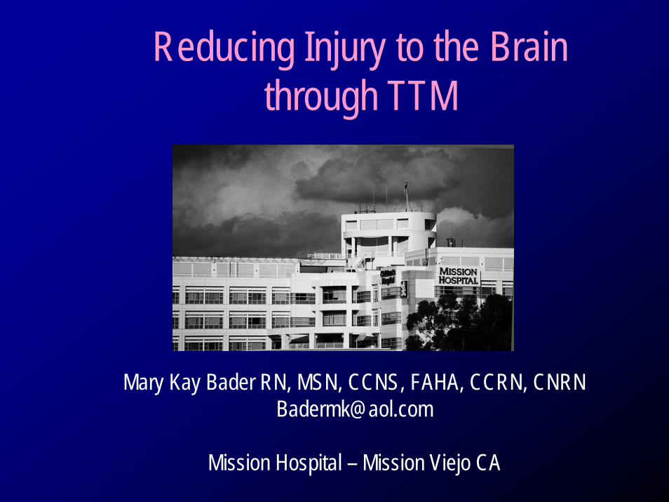
Reducing Injury to the Brain through TTM
Mary Kay Bader RN, MSN, CCNS, FAHA, CCRN, [email protected]
Mission Hospital – Mission Viejo CA
Disclosure• Mary Kay Bader
– Speaker’s Bureau: Integra Neuroscience
Therapeutic Temperature Management
• Temperature Management–Normothermia–Hypothermia
Neurologic Patient
• Temperature control – Avoid hyperthermia– Neuro Populations ++
• Stroke• Traumatic Brain Injury• Spinal Cord Injury
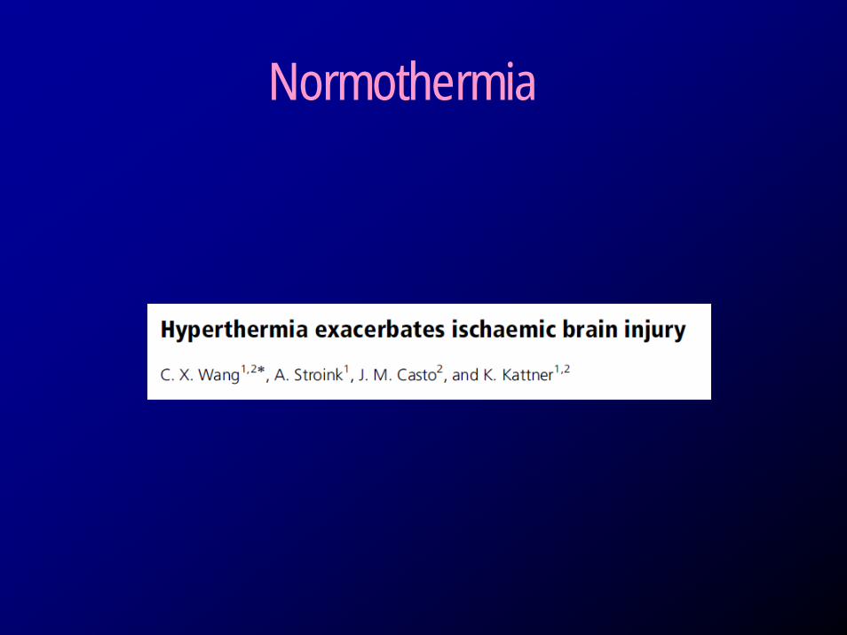
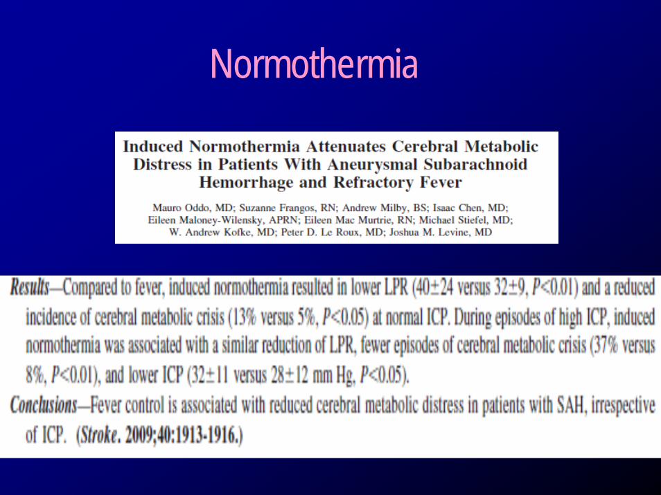
Normothermia
Normothermia
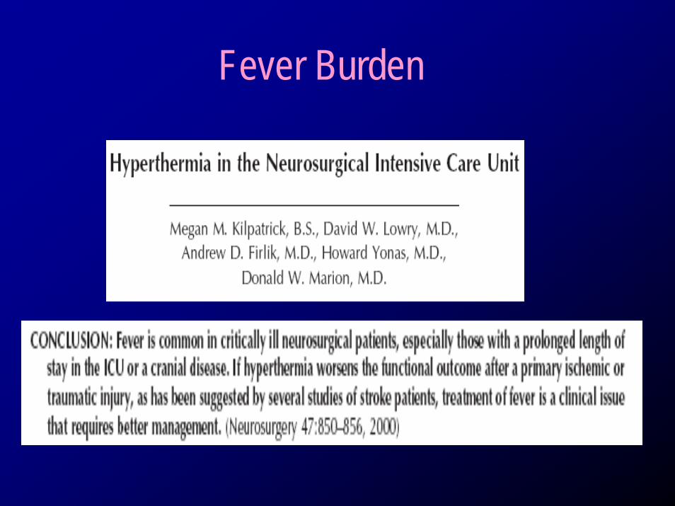
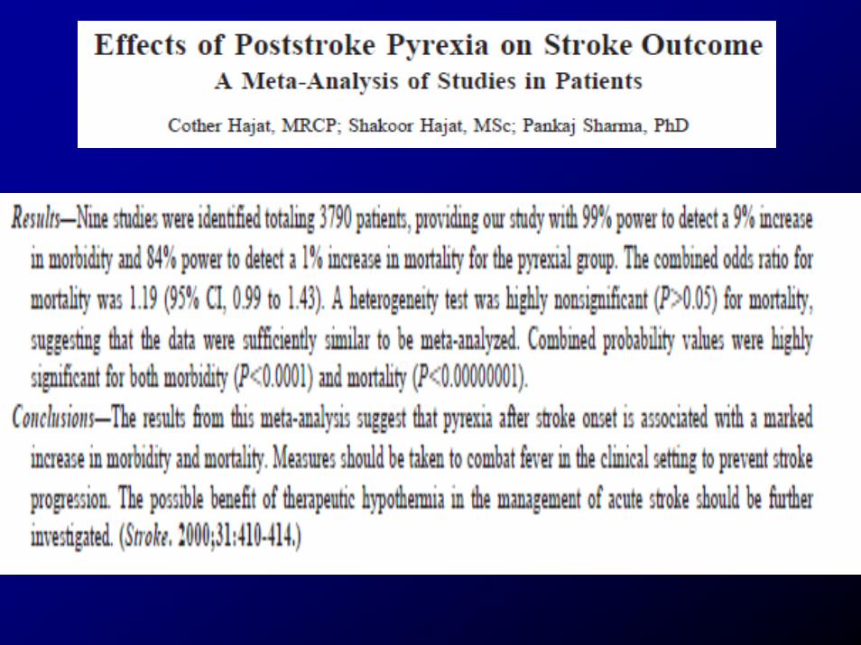
Fever Burden
Normothermia
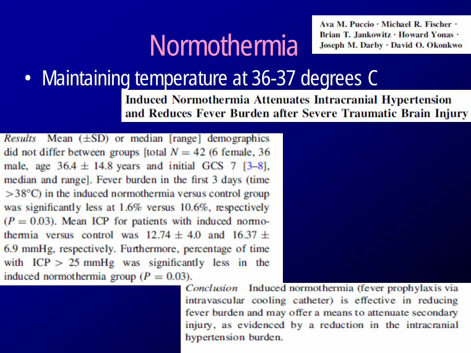
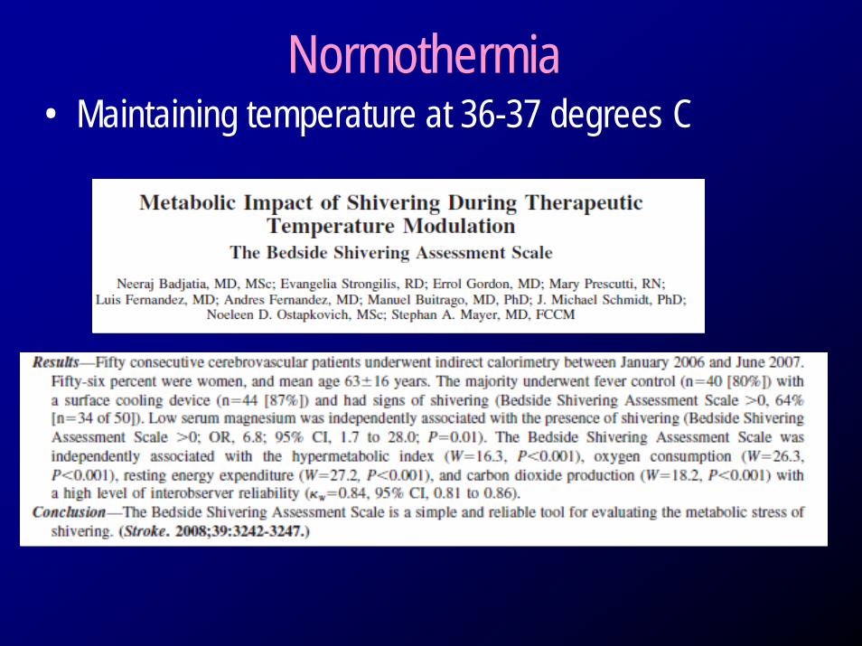
Normothermia• Maintaining temperature at 36-37 degrees C
Normothermia• Maintaining temperature at 36-37 degrees C
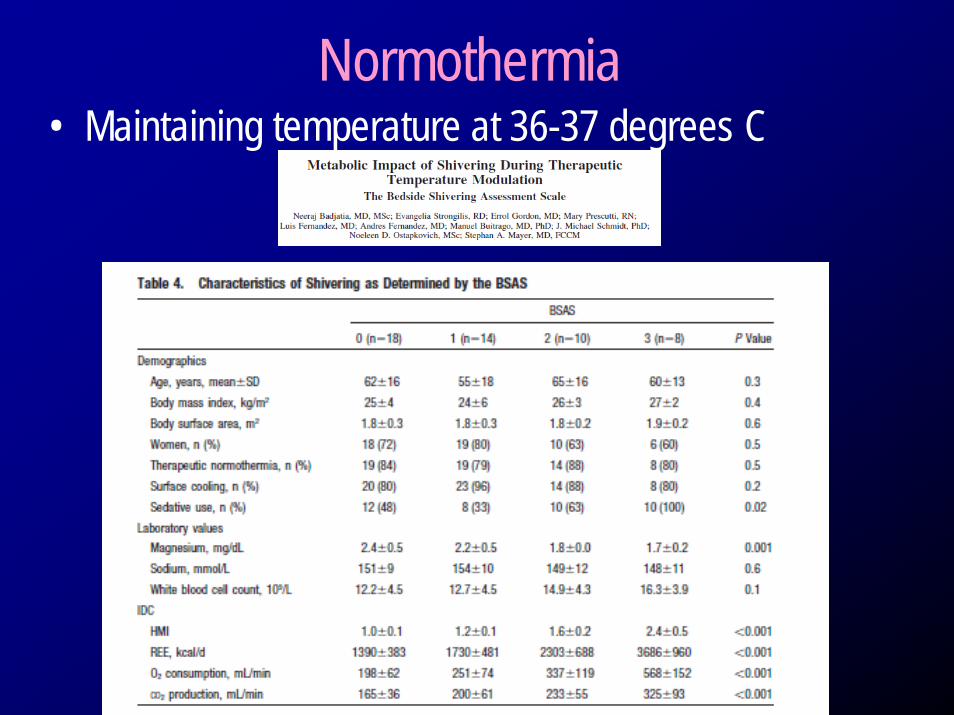
Normothermia• Maintaining temperature at 36-37 degrees C

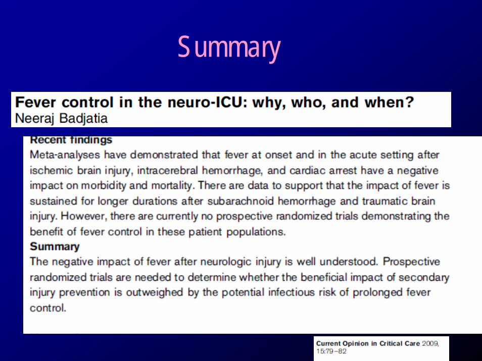
Summary
Hypothermia• What is the evidence in the neuro population?
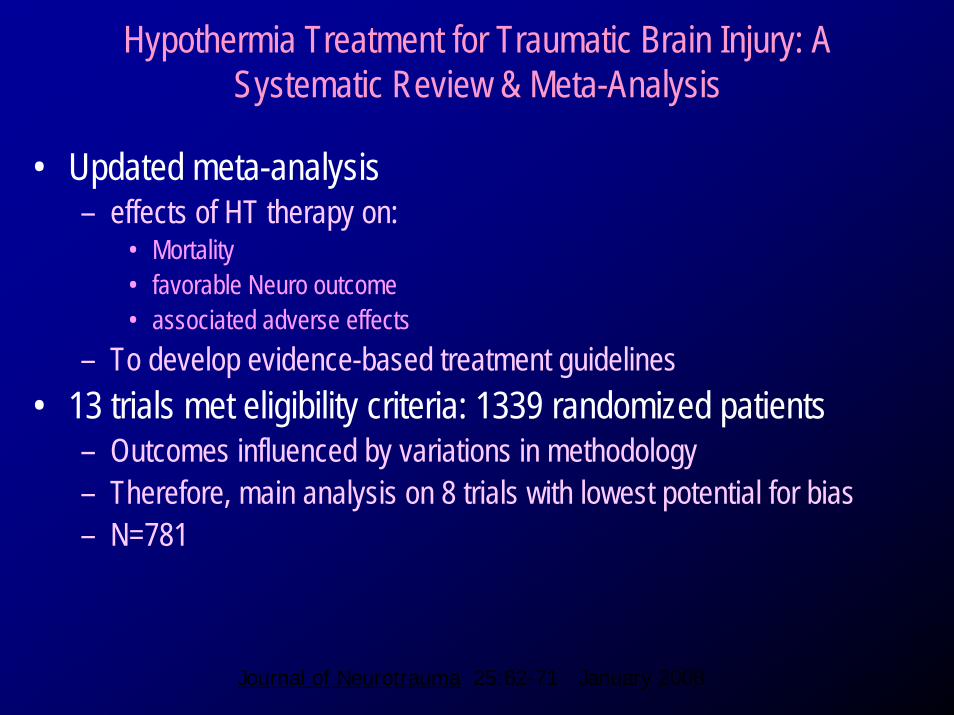
Hypothermia Treatment for Traumatic Brain Injury: A Systematic Review & Meta-Analysis
• Updated meta-analysis – effects of HT therapy on:
• Mortality• favorable Neuro outcome• associated adverse effects
– To develop evidence-based treatment guidelines • 13 trials met eligibility criteria: 1339 randomized patients
– Outcomes influenced by variations in methodology– Therefore, main analysis on 8 trials with lowest potential for bias– N=781
Journal of Neurotrauma 25:62-71 January 2008
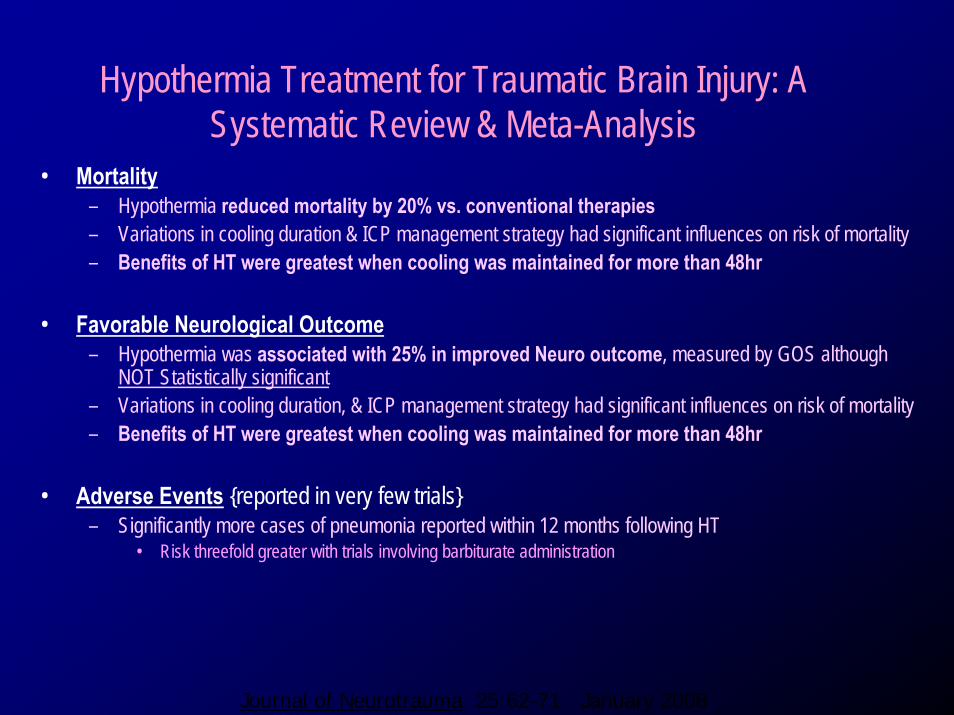
Hypothermia Treatment for Traumatic Brain Injury: A Systematic Review & Meta-Analysis
• Mortality– Hypothermia reduced mortality by 20% vs. conventional therapies– Variations in cooling duration & ICP management strategy had significant influences on risk of mortality– Benefits of HT were greatest when cooling was maintained for more than 48hr
• Favorable Neurological Outcome– Hypothermia was associated with 25% in improved Neuro outcome, measured by GOS although
NOT Statistically significant– Variations in cooling duration, & ICP management strategy had significant influences on risk of mortality– Benefits of HT were greatest when cooling was maintained for more than 48hr
• Adverse Events {reported in very few trials}– Significantly more cases of pneumonia reported within 12 months following HT
• Risk threefold greater with trials involving barbiturate administration
Journal of Neurotrauma 25:62-71 January 2008
Hypothermia in Stroke• Limited studies
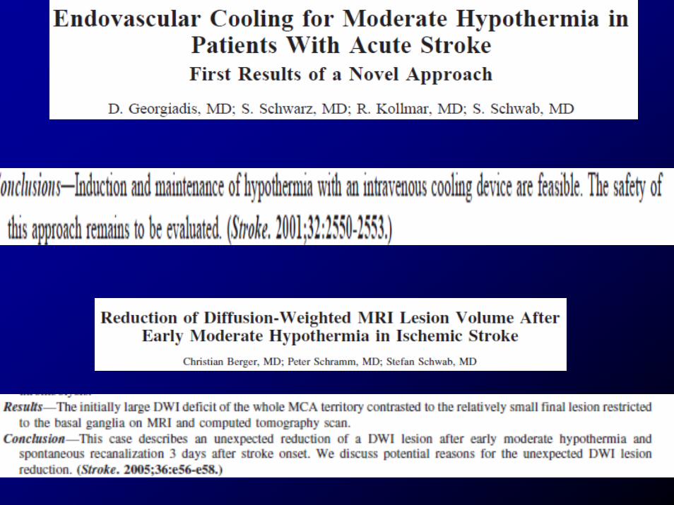
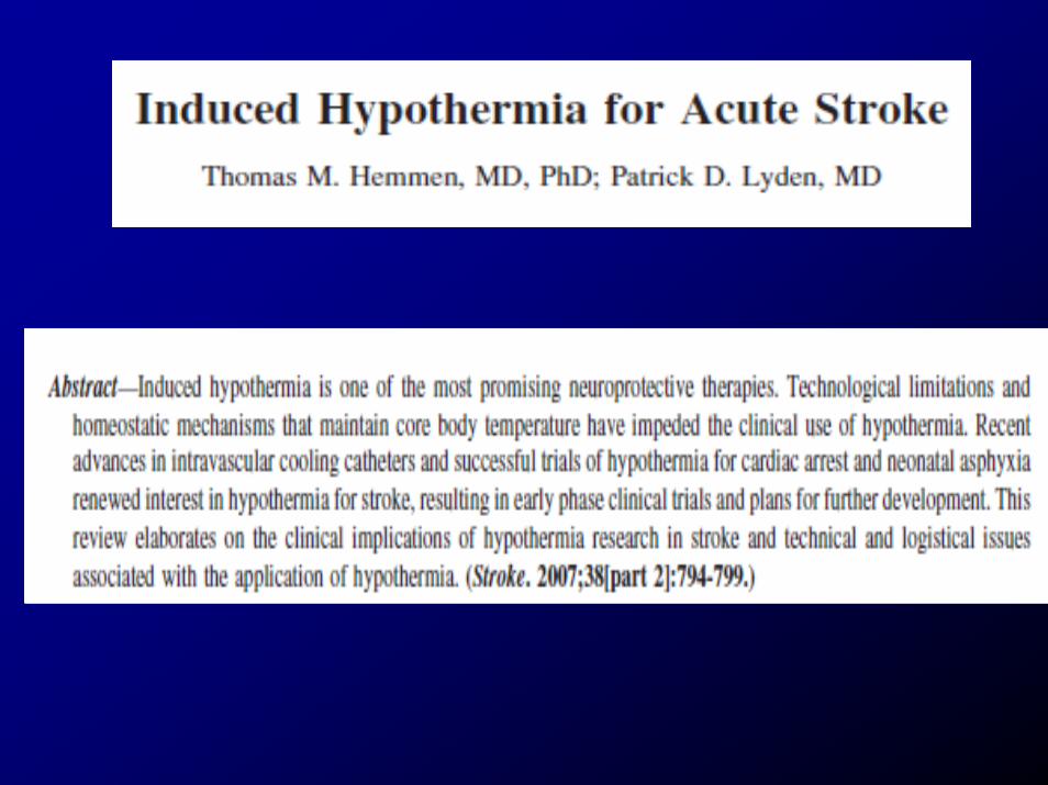
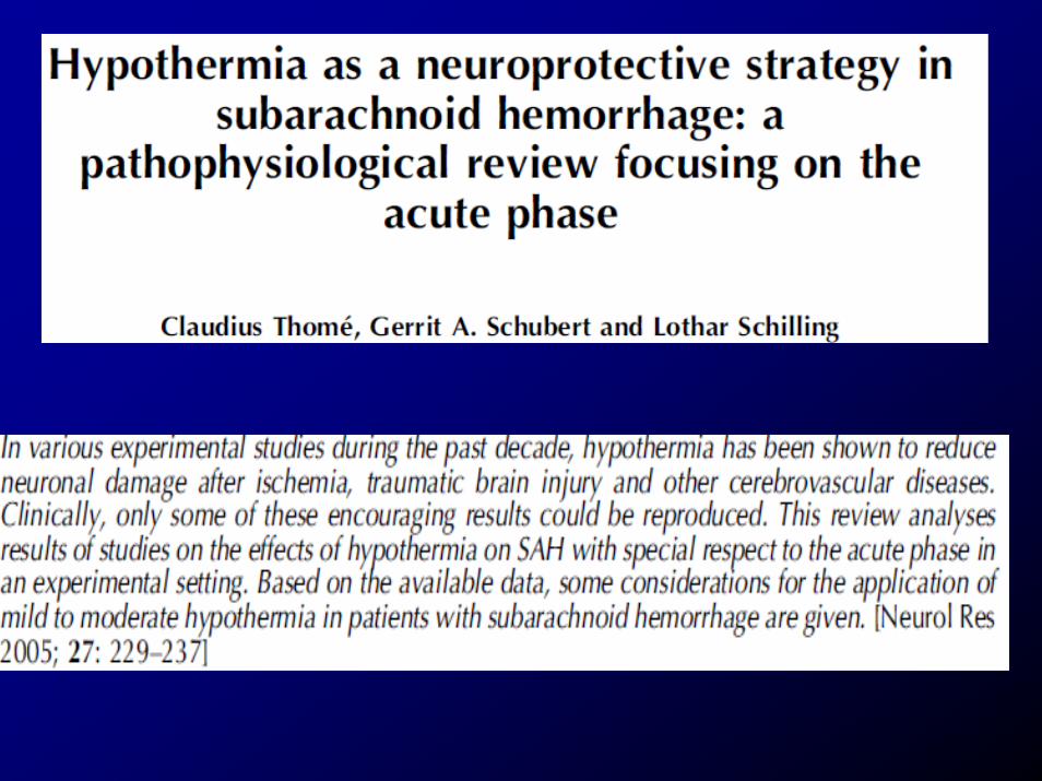

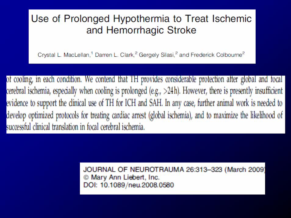
Evidence Based Literature• Very few published studies on efficacy of
hypothermia in the stroke population but…– Experimental animal studies ++– What do you do with the
• impending stroke pt with impending herniation…refractory increased ICP
• Refractory vasospasm from aneurysmal SAH
The Brain at Risk: Initial Anoxia event and reperfusion injury
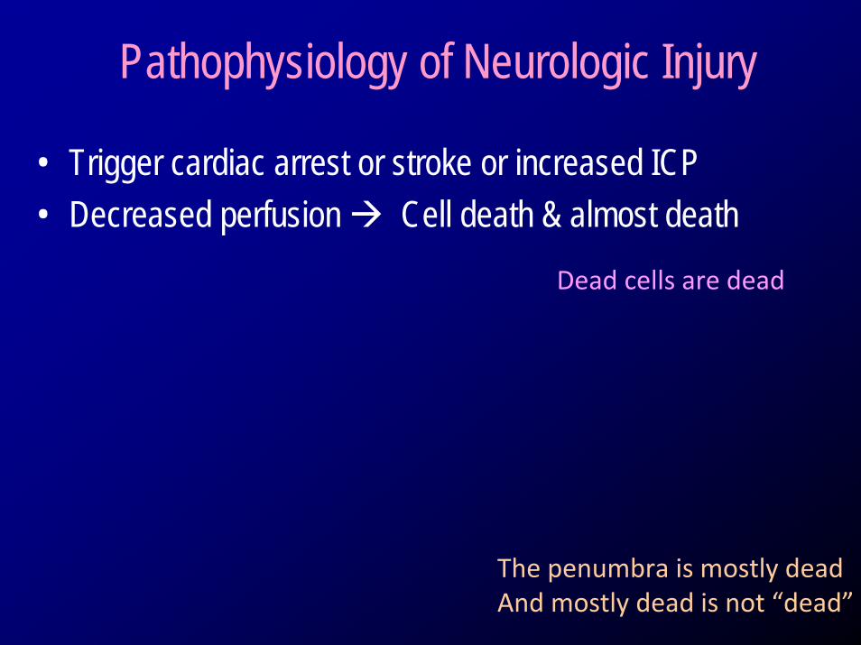
Pathophysiology of Neurologic Injury
• Trigger cardiac arrest or stroke or increased ICP• Decreased perfusion Cell death & almost death
Dead cells are dead
The penumbra is mostly deadAnd mostly dead is not “dead”
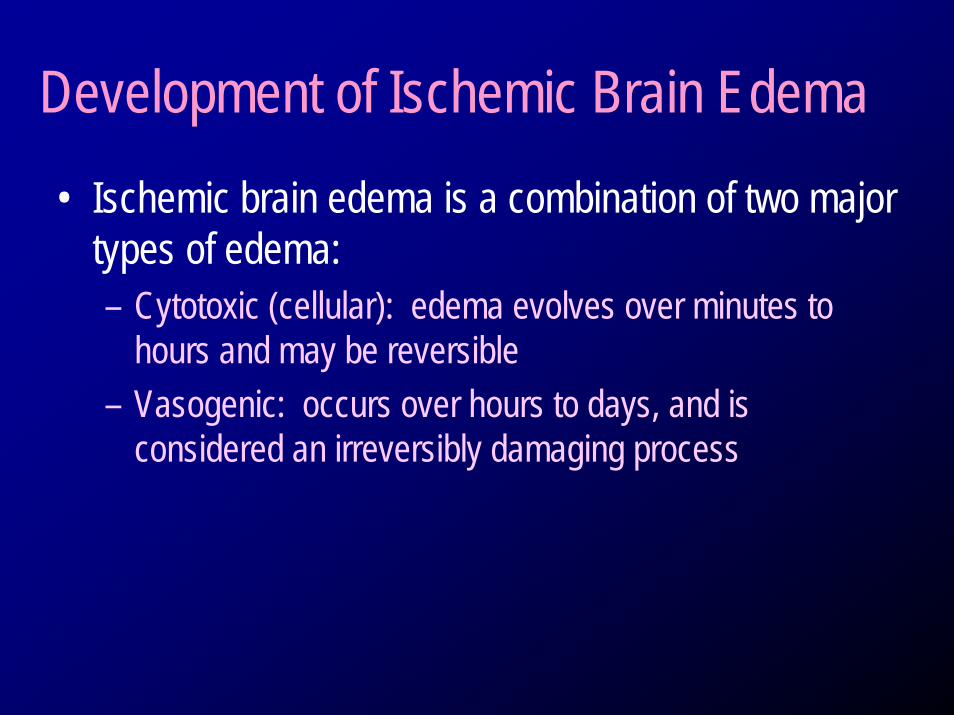
Development of Ischemic Brain Edema • Ischemic brain edema is a combination of two major
types of edema: – Cytotoxic (cellular): edema evolves over minutes to
hours and may be reversible– Vasogenic: occurs over hours to days, and is
considered an irreversibly damaging process
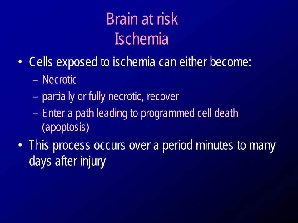
Brain at riskIschemia
• Cells exposed to ischemia can either become:– Necrotic– partially or fully necrotic, recover – Enter a path leading to programmed cell death
(apoptosis)• This process occurs over a period minutes to many
days after injury
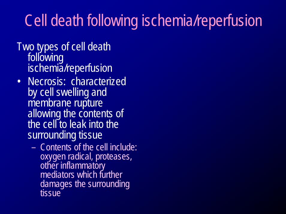
Cell death following ischemia/reperfusionTwo types of cell death
following ischemia/reperfusion
• Necrosis: characterized by cell swelling and membrane rupture allowing the contents of the cell to leak into the surrounding tissue – Contents of the cell include:
oxygen radical, proteases, other inflammatory mediators which further damages the surrounding tissue
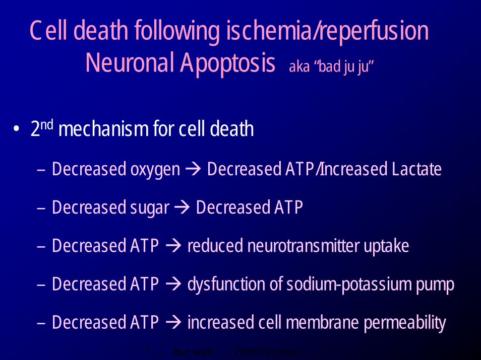
Cell death following ischemia/reperfusion Neuronal Apoptosis aka “bad ju ju”
• 2nd mechanism for cell death
– Decreased oxygen Decreased ATP/Increased Lactate
– Decreased sugar Decreased ATP
– Decreased ATP reduced neurotransmitter uptake
– Decreased ATP dysfunction of sodium-potassium pump
– Decreased ATP increased cell membrane permeability“ . . . But wait . . . There’s more . . . “
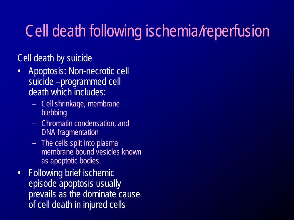
Cell death following ischemia/reperfusionCell death by suicide • Apoptosis: Non-necrotic cell
suicide –programmed cell death which includes:– Cell shrinkage, membrane
blebbing– Chromatin condensation, and
DNA fragmentation– The cells split into plasma
membrane bound vesicles known as apoptotic bodies.
• Following brief ischemic episode apoptosis usually prevails as the dominate cause of cell death in injured cells
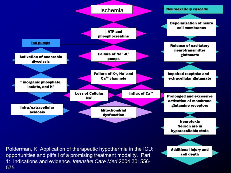
Ischemia
Activation of anaerobic glycolysis
↑ inorganic phosphate, lactate, and H+
Intra/extracellular acidosis
↓ ATP and phosphocreatine
Failure of Na+ -K+
pumps
Failure of K+, Na+ and Ca2+ channels
Loss of Cellular Na+
Influx of Ca2+
Mitochondrial dysfunction
Ion pumps
Neuroexcitory cascade
Depolarization of neuro cell membranes
Release of excitatory neurotransmitter
glutamate
Impaired reuptake and ↑extracellular glutamate
Prolonged and excessive activation of membrane
glutamine receptors
NeurotoxicNeuron are in
hyperexcitable state
Additional injury and cell death
Polderman, K Application of therapeutic hypothermia in the ICU: opportunities and pitfall of a promising treatment modality. Part 1: Indications and evidence. Intensive Care Med 2004 30: 556-575
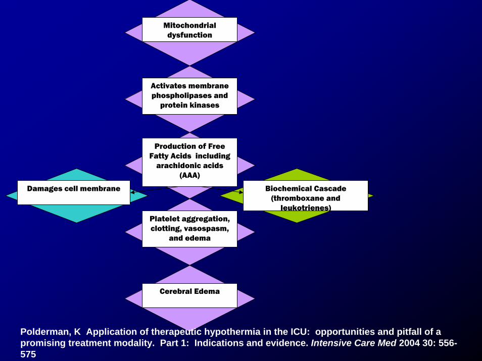
Mitochondrial dysfunction
Activates membrane phospholipases and
protein kinases
Production of Free Fatty Acids including
arachidonic acids (AAA)
Damages cell membrane Biochemical Cascade (thromboxane and
leukotrienes)
Cerebral Edema
Platelet aggregation, clotting, vasospasm,
and edema
Polderman, K Application of therapeutic hypothermia in the ICU: opportunities and pitfall of a promising treatment modality. Part 1: Indications and evidence. Intensive Care Med 2004 30: 556-575
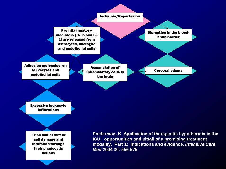
Proinflammatory-mediators (TNFa and IL-
1) are released from astrocytes, microglia and endothelial cells
Adhesion molecules on leukocytes and
endothelial cells
Accumulation of inflammatory cells in
the brain
Excessive leukocyte infiltrations
Ischemia/Reperfusion
↑ risk and extent of cell damage and
infarction through their phagocytic
actions
Disruption in the blood-brain barrier
Cerebral edema
Polderman, K Application of therapeutic hypothermia in the ICU: opportunities and pitfall of a promising treatment modality. Part 1: Indications and evidence. Intensive Care Med 2004 30: 556-575
The Brain at Risk: Mechanisms of Heat Destruction in CNS
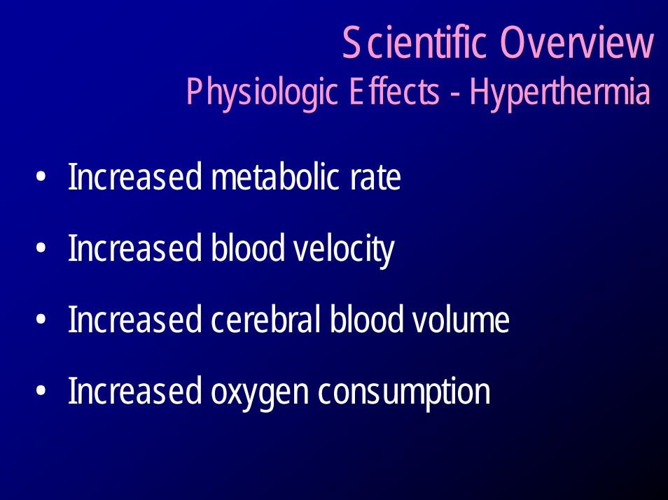
Scientific Overview Physiologic Effects - Hyperthermia
• Increased metabolic rate
• Increased blood velocity
• Increased cerebral blood volume
• Increased oxygen consumption
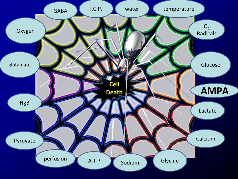
Oxygen
glutamate Glucose
Glycine
water temperatureI.C.P.GABA
Sodium
O2Radicals
Lactate
A T Pperfusion
Calcium
HgB
Pyruvate
CellDeath AMPA
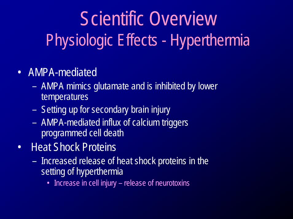
Scientific Overview Physiologic Effects - Hyperthermia
• AMPA-mediated– AMPA mimics glutamate and is inhibited by lower
temperatures– Setting up for secondary brain injury– AMPA-mediated influx of calcium triggers
programmed cell death• Heat Shock Proteins
– Increased release of heat shock proteins in the setting of hyperthermia
• Increase in cell injury – release of neurotoxins
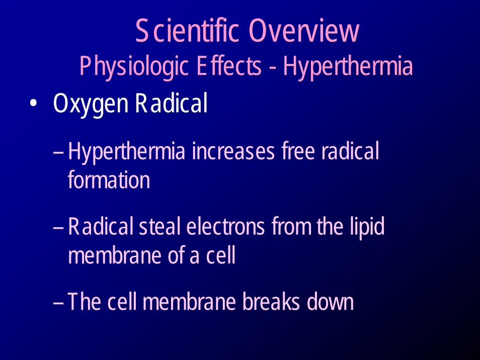
Scientific Overview Physiologic Effects - Hyperthermia
• Oxygen Radical
– Hyperthermia increases free radical formation
– Radical steal electrons from the lipid membrane of a cell
– The cell membrane breaks down
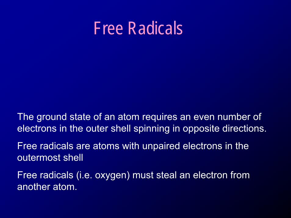
Free Radicals
The ground state of an atom requires an even number of electrons in the outer shell spinning in opposite directions.
Free radicals are atoms with unpaired electrons in the outermost shell
Free radicals (i.e. oxygen) must steal an electron from another atom.
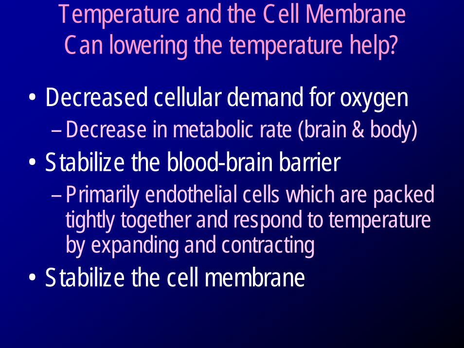
Temperature and the Cell MembraneCan lowering the temperature help?
• Decreased cellular demand for oxygen– Decrease in metabolic rate (brain & body)
• Stabilize the blood-brain barrier– Primarily endothelial cells which are packed
tightly together and respond to temperature by expanding and contracting
• Stabilize the cell membrane
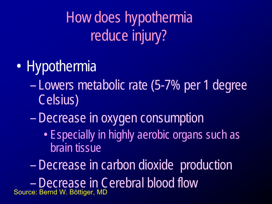
How does hypothermia reduce injury?
• Hypothermia – Lowers metabolic rate (5-7% per 1 degree
Celsius)– Decrease in oxygen consumption
• Especially in highly aerobic organs such as brain tissue
– Decrease in carbon dioxide production– Decrease in Cerebral blood flow
Source: Bernd W. Böttiger, MD
48
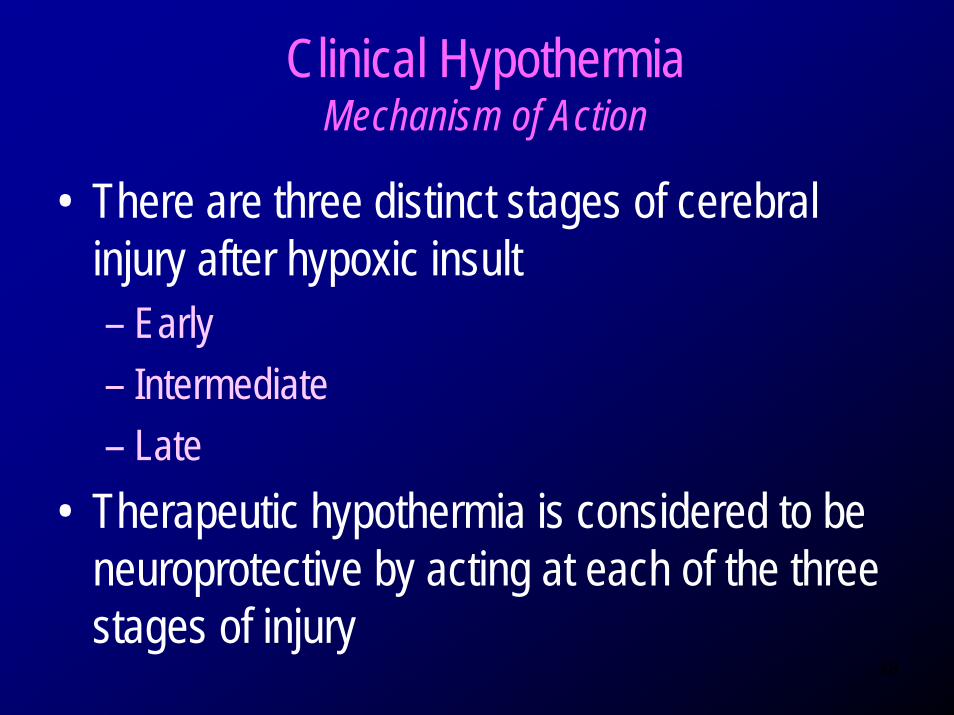
Clinical Hypothermia Mechanism of Action
• There are three distinct stages of cerebral injury after hypoxic insult– Early– Intermediate – Late
• Therapeutic hypothermia is considered to be neuroprotective by acting at each of the three stages of injury
49
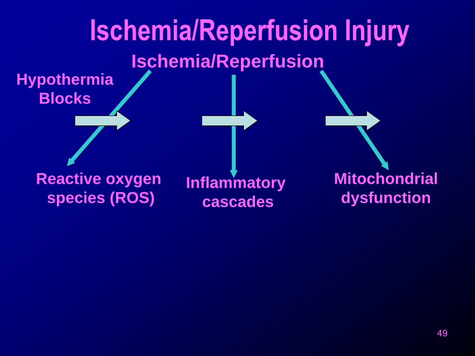
Ischemia/ReperfusionIschemia/Reperfusion Injury
Reactive oxygen species (ROS)
Inflammatory cascades
Mitochondrialdysfunction
Hypothermia Blocks
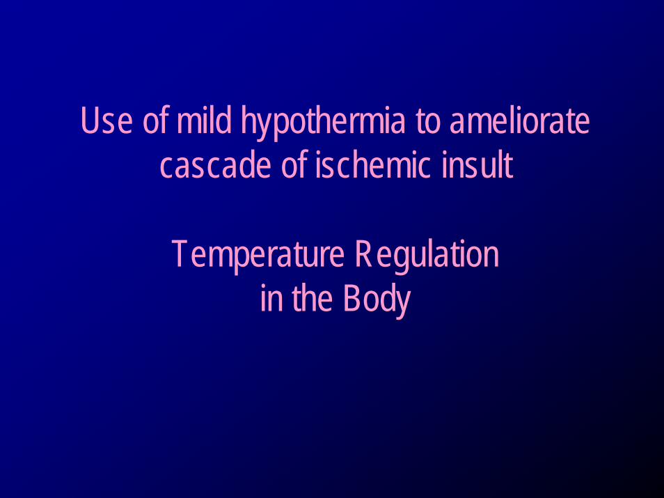
Use of mild hypothermia to ameliorate cascade of ischemic insult
Temperature Regulation in the Body
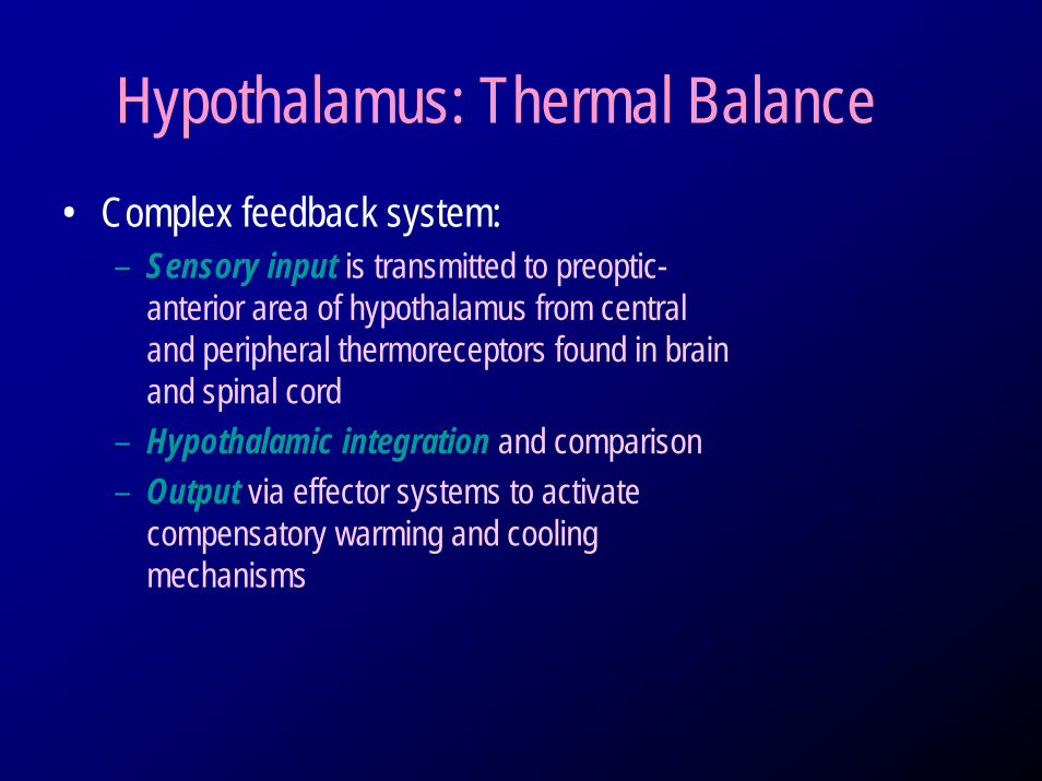
Hypothalamus: Thermal Balance• Complex feedback system:
– Sensory input is transmitted to preoptic-anterior area of hypothalamus from central and peripheral thermoreceptors found in brain and spinal cord
– Hypothalamic integration and comparison– Output via effector systems to activate
compensatory warming and cooling mechanisms
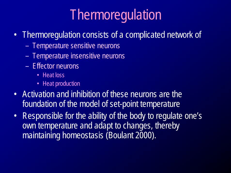
Thermoregulation• Thermoregulation consists of a complicated network of
– Temperature sensitive neurons– Temperature insensitive neurons– Effector neurons
• Heat loss • Heat production
• Activation and inhibition of these neurons are the foundation of the model of set-point temperature
• Responsible for the ability of the body to regulate one’s own temperature and adapt to changes, thereby maintaining homeostasis (Boulant 2000).
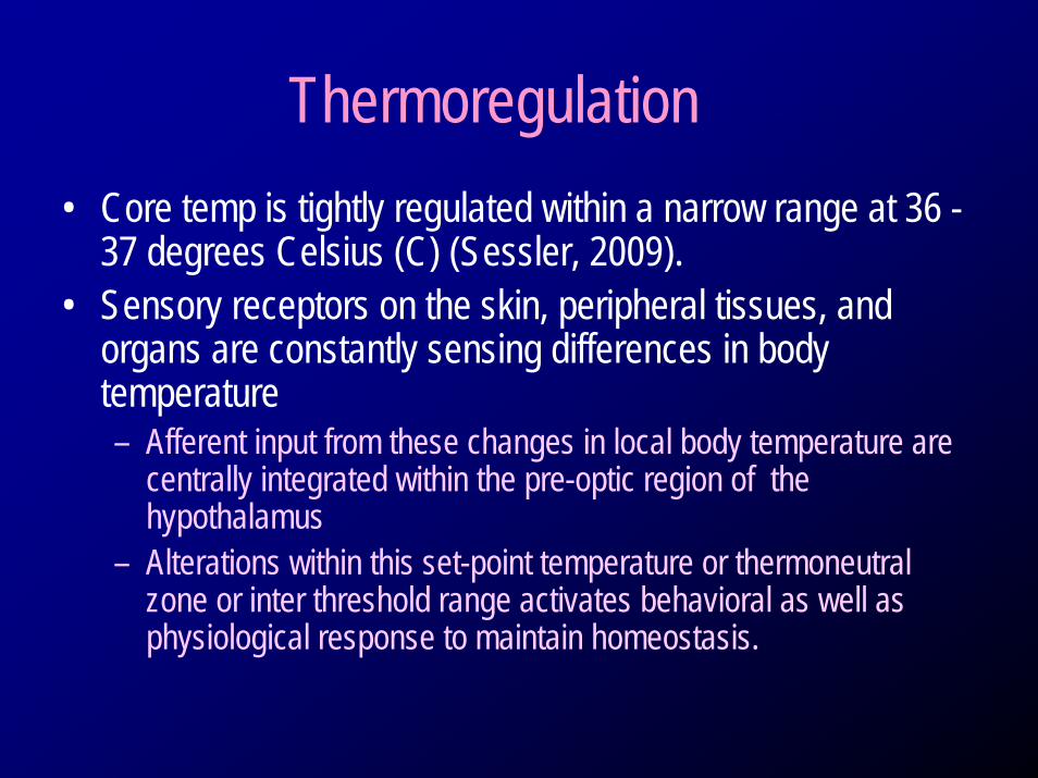
Thermoregulation• Core temp is tightly regulated within a narrow range at 36 -
37 degrees Celsius (C) (Sessler, 2009). • Sensory receptors on the skin, peripheral tissues, and
organs are constantly sensing differences in body temperature– Afferent input from these changes in local body temperature are
centrally integrated within the pre-optic region of the hypothalamus
– Alterations within this set-point temperature or thermoneutralzone or inter threshold range activates behavioral as well as physiological response to maintain homeostasis.
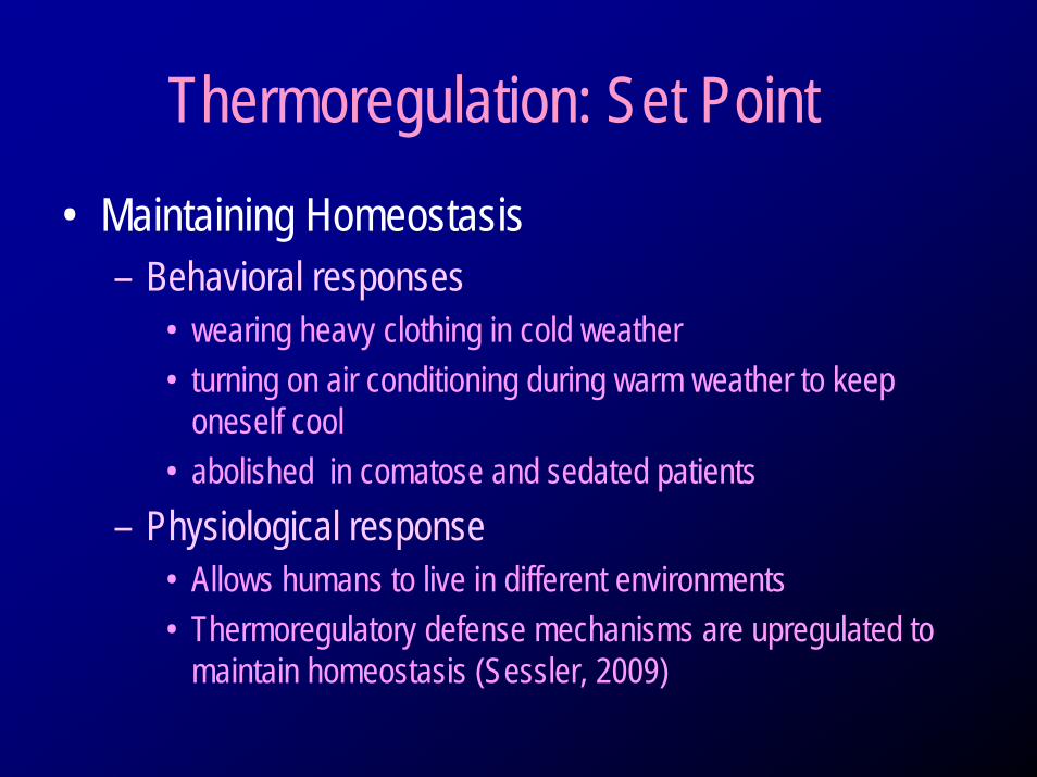
Thermoregulation: Set Point• Maintaining Homeostasis
– Behavioral responses• wearing heavy clothing in cold weather• turning on air conditioning during warm weather to keep
oneself cool• abolished in comatose and sedated patients
– Physiological response• Allows humans to live in different environments• Thermoregulatory defense mechanisms are upregulated to
maintain homeostasis (Sessler, 2009)
Thermoregulation: Set Point
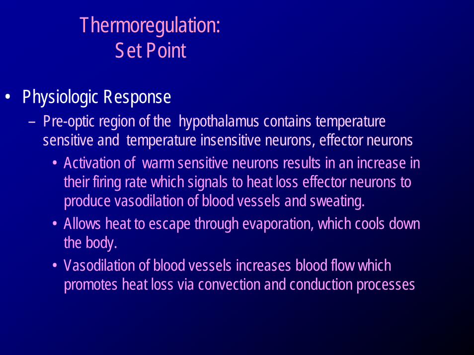
• Physiologic Response – Pre-optic region of the hypothalamus contains temperature
sensitive and temperature insensitive neurons, effector neurons• Activation of warm sensitive neurons results in an increase in
their firing rate which signals to heat loss effector neurons to produce vasodilation of blood vessels and sweating.
• Allows heat to escape through evaporation, which cools down the body.
• Vasodilation of blood vessels increases blood flow which promotes heat loss via convection and conduction processes
Thermoregulation: Set Point
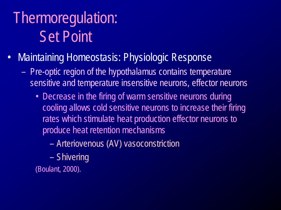
• Maintaining Homeostasis: Physiologic Response – Pre-optic region of the hypothalamus contains temperature
sensitive and temperature insensitive neurons, effector neurons• Decrease in the firing of warm sensitive neurons during
cooling allows cold sensitive neurons to increase their firing rates which stimulate heat production effector neurons to produce heat retention mechanisms
– Arteriovenous (AV) vasoconstriction– Shivering
(Boulant, 2000).
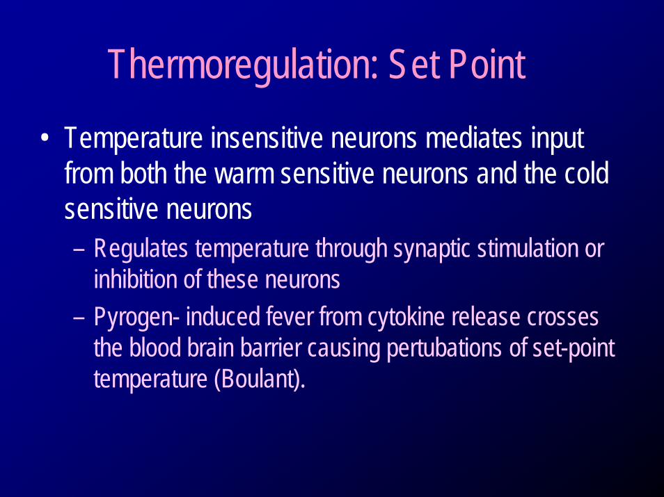
Thermoregulation: Set Point• Temperature insensitive neurons mediates input
from both the warm sensitive neurons and the cold sensitive neurons– Regulates temperature through synaptic stimulation or
inhibition of these neurons– Pyrogen- induced fever from cytokine release crosses
the blood brain barrier causing pertubations of set-point temperature (Boulant).
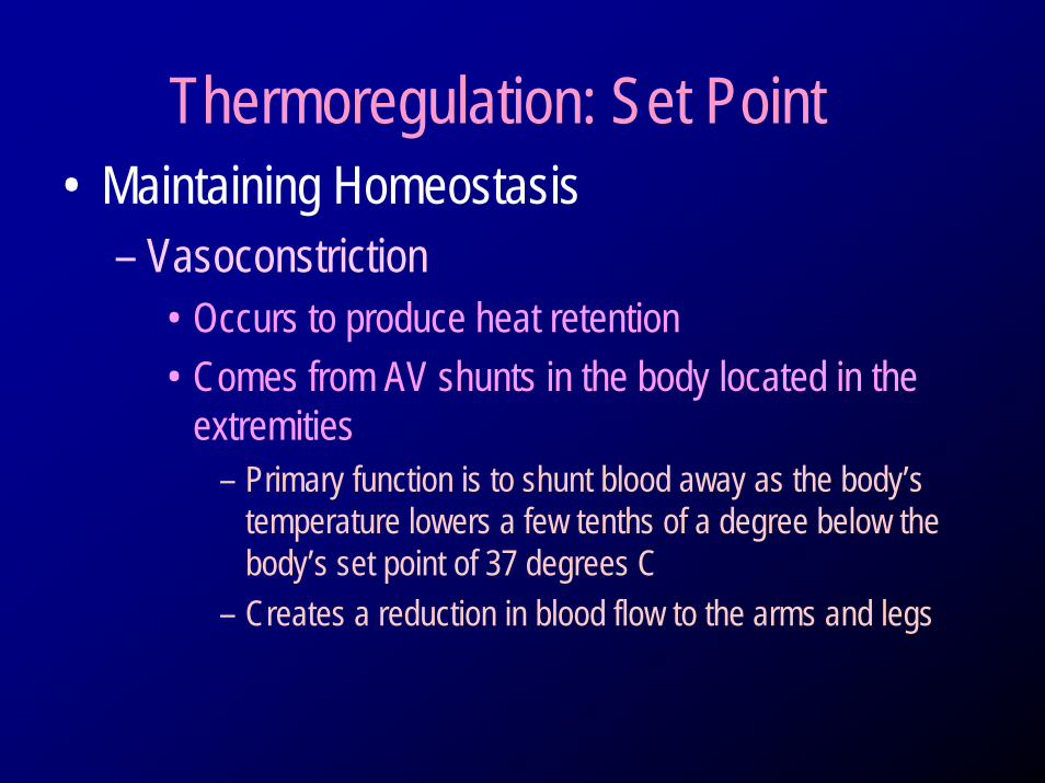
Thermoregulation: Set Point• Maintaining Homeostasis
– Vasoconstriction• Occurs to produce heat retention • Comes from AV shunts in the body located in the
extremities– Primary function is to shunt blood away as the body’s
temperature lowers a few tenths of a degree below the body’s set point of 37 degrees C
– Creates a reduction in blood flow to the arms and legs
Thermoregulation: Set Point• Maintaining Homeostasis
– Vasoconstriction• Piloerection, or goosebumps, are seen first as the
body attempts to shunt blood from the peripheral compartment in an attempt to stop heat loss and conserve heat (Landsberg L, Saville ME, and Young JB 1984).
• Lowers the temperature in the extremities
Thermoregulation: Set Point• Maintaining Homeostasis
– NOTE: Heat created by deep organs located in the trunk and cranium is kept inside this area and is usually 2-4 degrees higher than the peripheral compartment (Sessler 2009).
– Heat flows toward the lower temperature creating a thermoregulatory vasomotion phenomena allowing for the an efficient transfer of heat from the core when needed (Sessler 2009).
Thermoregulation: Set Point• Maintaining Homeostasis
– Shivering – How?• If skin receives continuous sensation of cold, motor neurons
are stimulated creating a shiver response in the muscles of the body
• Motor response begins in the trunk and spreads to the extremities in an attempt to generate heat
• Shivering mechanism occurs when the body temperature falls approximately one degree C below the vasoconstriction threshold
(Sessler 2009)
• an involuntary, rhythmic tremor of skeletal muscle groups which consists of oscillatory involuntary movement (Sessler 2009)– An emergency mechanism – Natural physiological response to an altered hypothalamic set-point
• As temperature descends to below the set-point of 36 degrees Celsius – efferent signals crossing the median forebrain bundle terminating in the
hypothalamus communicates down to the reticulo spinal neurons in the lower brainstem which activates shivering
– Lesions within this “efferent pathway” are associated with absence shivering (Hemingway; Gilbert and Benarroch, 2008).
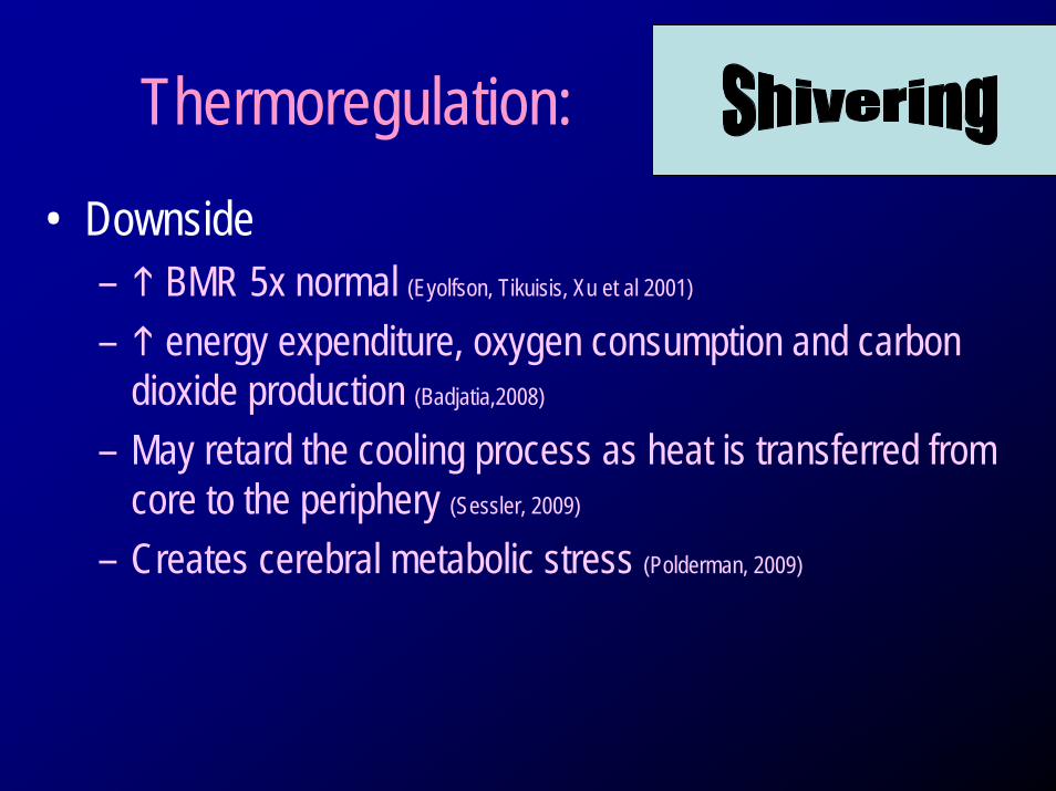
Thermoregulation:• Downside
– BMR 5x normal (Eyolfson, Tikuisis, Xu et al 2001)
– energy expenditure, oxygen consumption and carbon dioxide production (Badjatia,2008)
– May retard the cooling process as heat is transferred from core to the periphery (Sessler, 2009)
– Creates cerebral metabolic stress (Polderman, 2009)
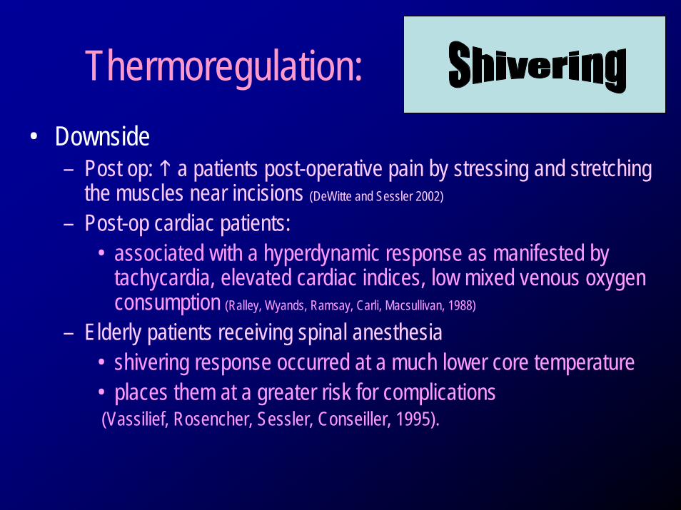
Thermoregulation:• Downside
– Post op: a patients post-operative pain by stressing and stretching the muscles near incisions (DeWitte and Sessler 2002)
– Post-op cardiac patients: • associated with a hyperdynamic response as manifested by
tachycardia, elevated cardiac indices, low mixed venous oxygen consumption (Ralley, Wyands, Ramsay, Carli, Macsullivan, 1988)
– Elderly patients receiving spinal anesthesia• shivering response occurred at a much lower core temperature• places them at a greater risk for complications(Vassilief, Rosencher, Sessler, Conseiller, 1995).
© Medivance 2008
Temperature Regulation
Scientific Overview Systemic Effects of Lowering Temperature
Courtesy of Daiwai Olson
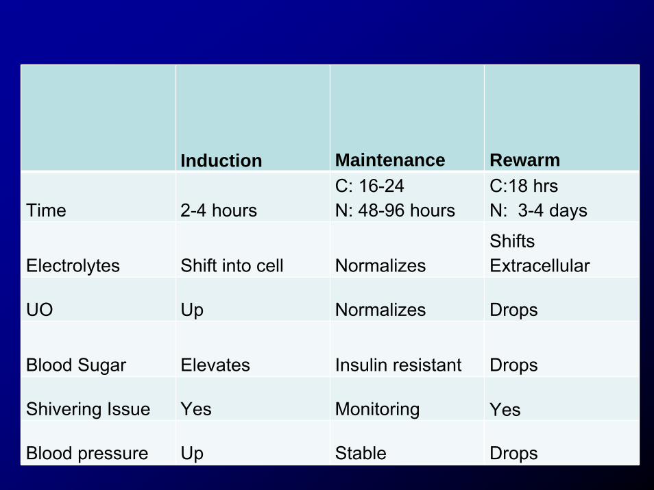
Induction Maintenance Rewarm
Time 2-4 hoursC: 16-24N: 48-96 hours
C:18 hrsN: 3-4 days
Electrolytes Shift into cell NormalizesShifts Extracellular
UO Up Normalizes Drops
Blood Sugar Elevates Insulin resistant Drops
Shivering Issue Yes Monitoring Yes
Blood pressure Up Stable Drops
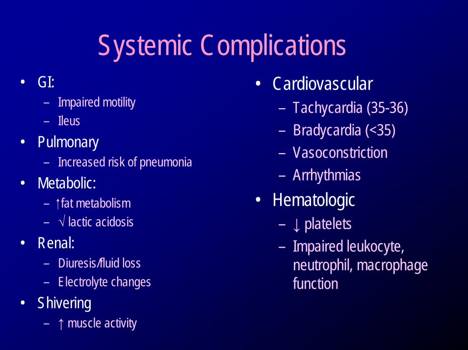
Systemic Complications• GI:
– Impaired motility– Ileus
• Pulmonary– Increased risk of pneumonia
• Metabolic: – fat metabolism– √ lactic acidosis
• Renal:– Diuresis/fluid loss– Electrolyte changes
• Shivering– ↑ muscle activity
• Cardiovascular– Tachycardia (35-36)– Bradycardia (<35)– Vasoconstriction– Arrhythmias
• Hematologic– ↓ platelets– Impaired leukocyte,
neutrophil, macrophage function
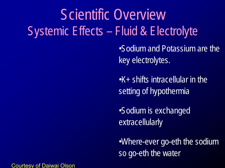
Scientific Overview Systemic Effects – Fluid & Electrolyte
•Sodium and Potassium are the key electrolytes.
•K+ shifts intracellular in the setting of hypothermia
•Sodium is exchanged extracellularly
•Where-ever go-eth the sodium so go-eth the water
Courtesy of Daiwai Olson

Monitoring Labs• Induction: every 1 hour• Maintenance: every 4-6 hours• Re-warming: every 2 hours
Cool
into cell
Warmto plasma
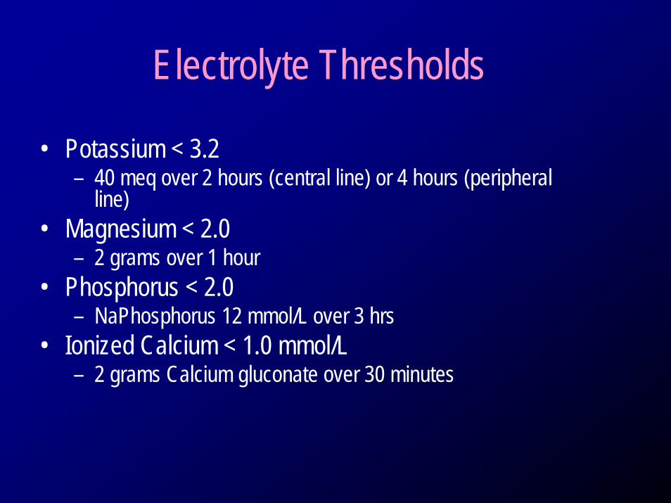
Electrolyte Thresholds
• Potassium < 3.2– 40 meq over 2 hours (central line) or 4 hours (peripheral
line)• Magnesium < 2.0
– 2 grams over 1 hour• Phosphorus < 2.0
– NaPhosphorus 12 mmol/L over 3 hrs• Ionized Calcium < 1.0 mmol/L
– 2 grams Calcium gluconate over 30 minutes
Induction Hypothermia K and insulin
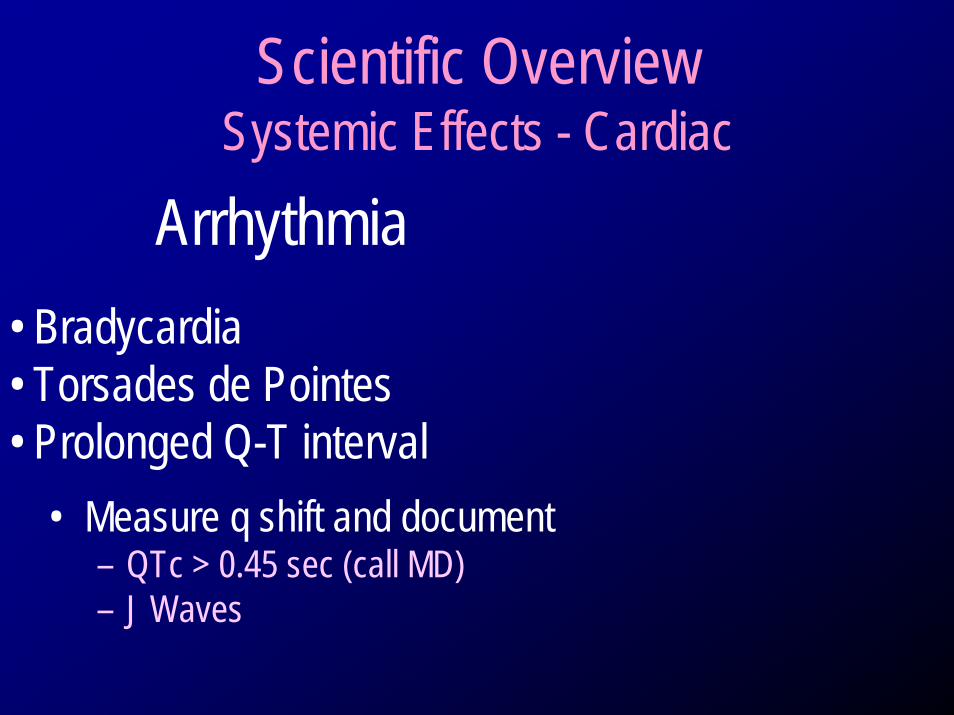
Scientific Overview Systemic Effects - Cardiac
Arrhythmia• Bradycardia• Torsades de Pointes• Prolonged Q-T interval
• Measure q shift and document– QTc > 0.45 sec (call MD)– J Waves
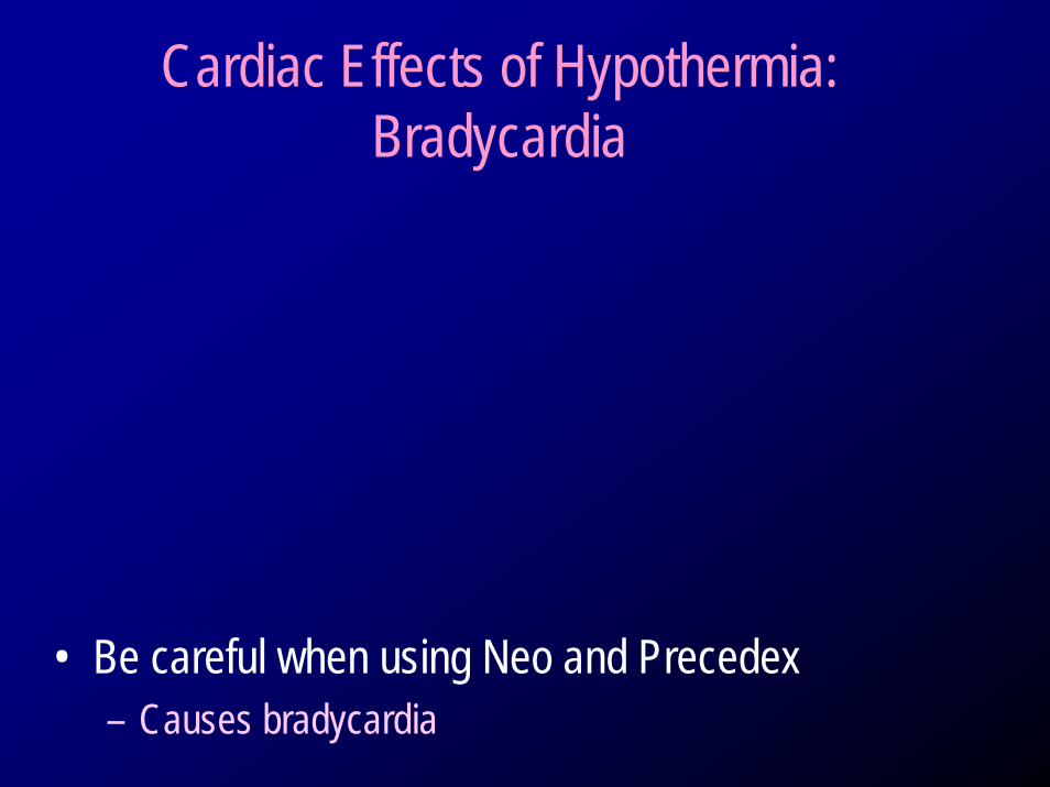
Cardiac Effects of Hypothermia:Bradycardia
• Be careful when using Neo and Precedex– Causes bradycardia
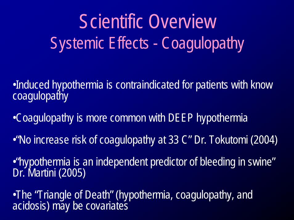
Scientific Overview Systemic Effects - Coagulopathy
•Induced hypothermia is contraindicated for patients with know coagulopathy
•Coagulopathy is more common with DEEP hypothermia
•“No increase risk of coagulopathy at 33 C” Dr. Tokutomi (2004)
•“hypothermia is an independent predictor of bleeding in swine”Dr. Martini (2005)
•The “Triangle of Death” (hypothermia, coagulopathy, and acidosis) may be covariates
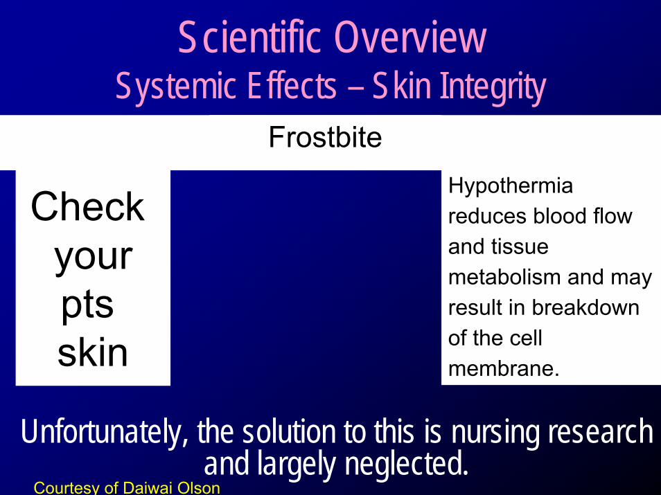
Scientific Overview Systemic Effects – Skin Integrity
Hypothermia reduces blood flow and tissue metabolism and may result in breakdown of the cell membrane.
Unfortunately, the solution to this is nursing research and largely neglected.
Check yourpts skin
Frostbite
Courtesy of Daiwai Olson
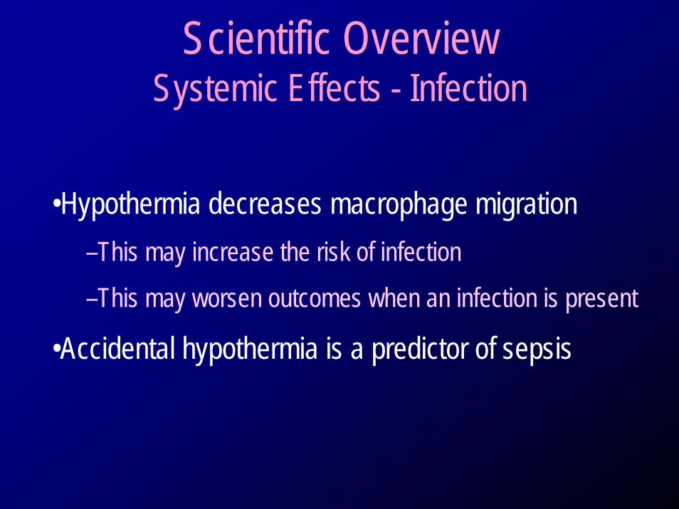
Scientific Overview Systemic Effects - Infection
•Hypothermia decreases macrophage migration–This may increase the risk of infection–This may worsen outcomes when an infection is present
•Accidental hypothermia is a predictor of sepsis
Risk: Increased ICP in Neuro Patients
• ICP increases with attempted rewarm
Management of Temperature
• HACA and Neuro Population– Protocols with specific temp management
directives post HACA and for Neurodisorders• Maintenance of temperature 36-37 C• Automated interventions
– Pre printed physician orders– Meds/cooling strategies

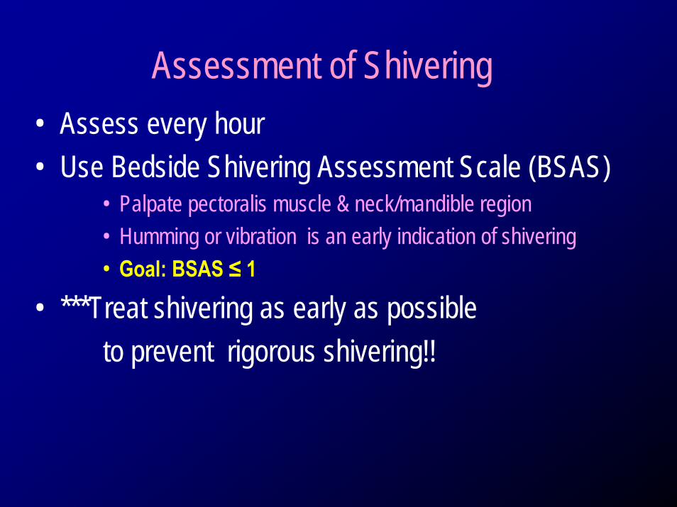
Assessment of Shivering• Assess every hour• Use Bedside Shivering Assessment Scale (BSAS)
• Palpate pectoralis muscle & neck/mandible region• Humming or vibration is an early indication of shivering• Goal: BSAS ≤ 1
• ***Treat shivering as early as possibleto prevent rigorous shivering!!
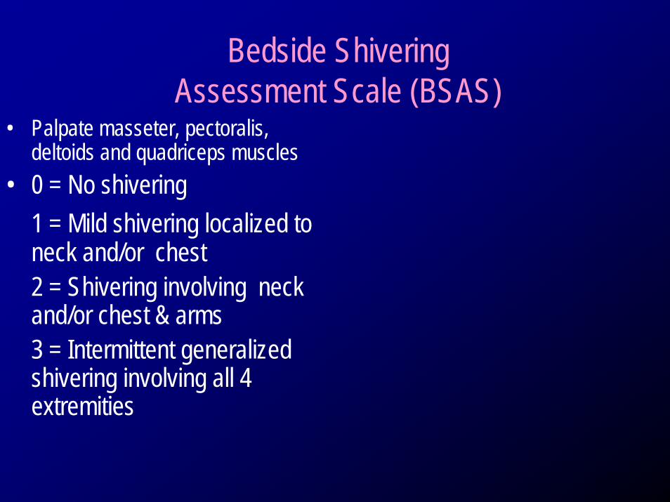
Bedside Shivering Assessment Scale (BSAS)
• Palpate masseter, pectoralis, deltoids and quadriceps muscles
• 0 = No shivering1 = Mild shivering localized to neck and/or chest2 = Shivering involving neck and/or chest & arms3 = Intermittent generalized shivering involving all 4 extremities
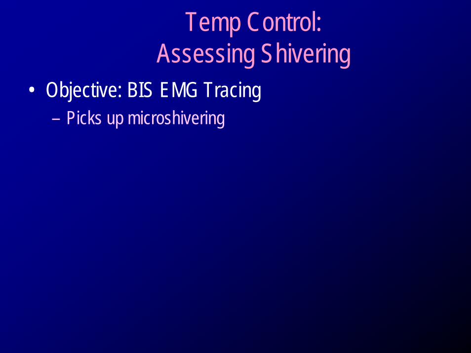
Temp Control:Assessing Shivering
• Objective: BIS EMG Tracing– Picks up microshivering
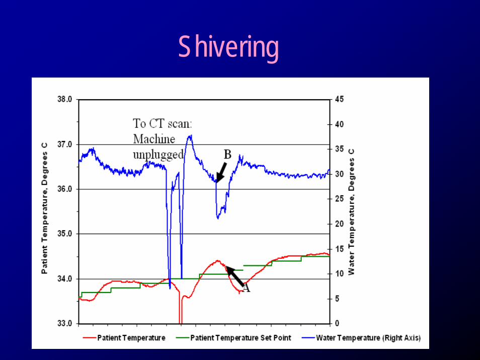
Shivering
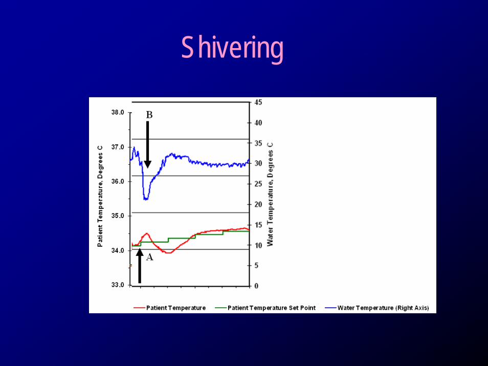
Shivering
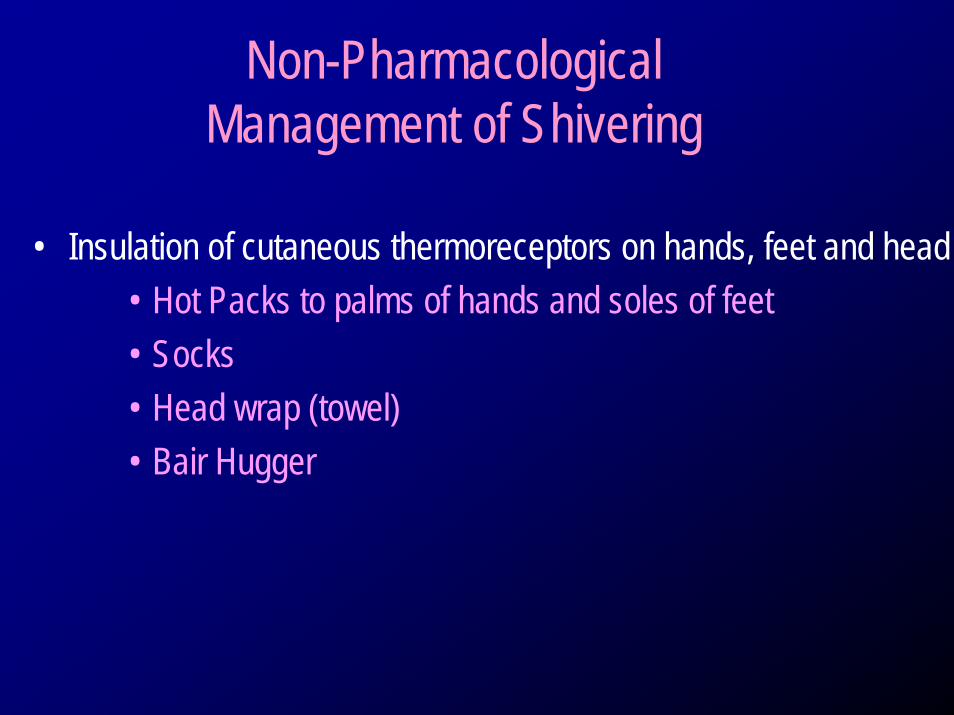
Non-Pharmacological Management of Shivering
• Insulation of cutaneous thermoreceptors on hands, feet and head• Hot Packs to palms of hands and soles of feet• Socks• Head wrap (towel)• Bair Hugger
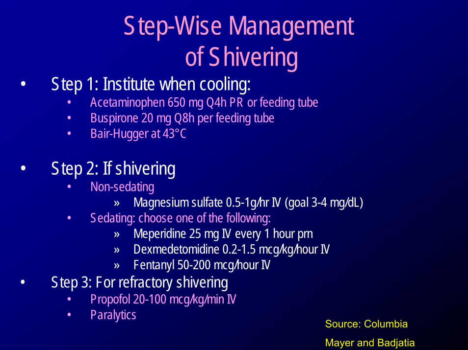
Step-Wise Managementof Shivering
• Step 1: Institute when cooling:• Acetaminophen 650 mg Q4h PR or feeding tube• Buspirone 20 mg Q8h per feeding tube• Bair-Hugger at 43°C
• Step 2: If shivering• Non-sedating
» Magnesium sulfate 0.5-1g/hr IV (goal 3-4 mg/dL)• Sedating: choose one of the following:
» Meperidine 25 mg IV every 1 hour prn» Dexmedetomidine 0.2-1.5 mcg/kg/hour IV » Fentanyl 50-200 mcg/hour IV
• Step 3: For refractory shivering• Propofol 20-100 mcg/kg/min IV • Paralytics
Source: Columbia
Mayer and Badjatia
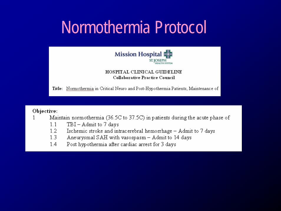
Normothermia Protocol
Normothermia Order Set
Methods to Induce Hypothermia
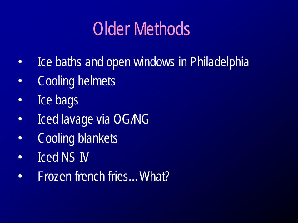
Older Methods• Ice baths and open windows in Philadelphia• Cooling helmets• Ice bags• Iced lavage via OG/NG• Cooling blankets• Iced NS IV• Frozen french fries…What?
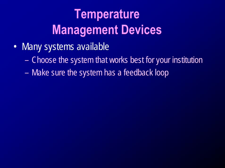
TemperatureManagement Devices
• Many systems available– Choose the system that works best for your institution– Make sure the system has a feedback loop
Methods to Monitor Body Temperature
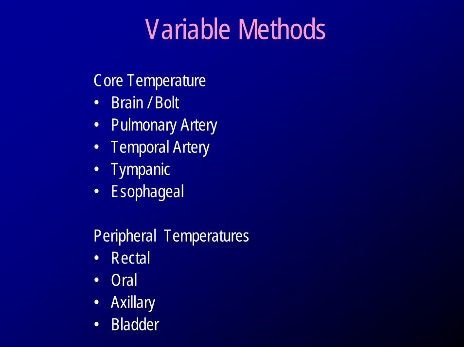
Variable MethodsCore Temperature• Brain / Bolt• Pulmonary Artery• Temporal Artery• Tympanic• Esophageal
Peripheral Temperatures• Rectal• Oral• Axillary• Bladder
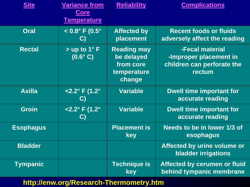
Site Variance from Core
Temperature
Reliability Complications
Oral < 0.8° F (0.5°C)
Affected by placement
Recent foods or fluids adversely affect the reading
Rectal > up to 1° F (0.6° C)
Reading may be delayed from core
temperature change
-Fecal material-Improper placement in
children can perforate the rectum
Axilla <2.2° F (1.2°C)
Variable Dwell time important for accurate reading
Groin <2.2° F (1.2°C)
Variable Dwell time important for accurate reading
Esophagus Placement is key
Needs to be in lower 1/3 of esophagus
Bladder Affected by urine volume or bladder irrigations
Tympanic Technique is key
Affected by cerumen or fluid behind tympanic membrane
http://enw.org/Research-Thermometry.htm
Hypothermia In Neurologic Disorders
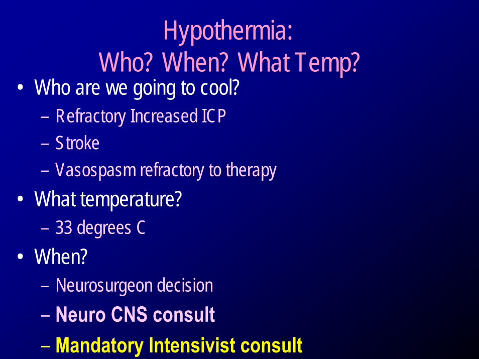
Hypothermia:Who? When? What Temp?
• Who are we going to cool?– Refractory Increased ICP– Stroke– Vasospasm refractory to therapy
• What temperature?– 33 degrees C
• When?– Neurosurgeon decision– Neuro CNS consult– Mandatory Intensivist consult
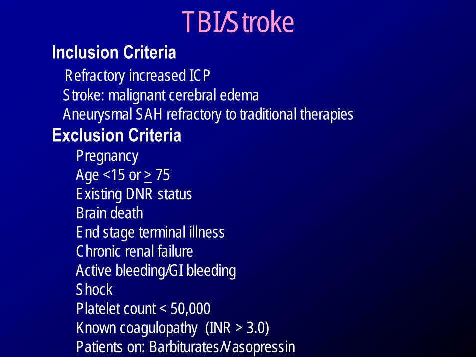
TBI/StrokeInclusion Criteria
Refractory increased ICP Stroke: malignant cerebral edemaAneurysmal SAH refractory to traditional therapies
Exclusion CriteriaPregnancyAge <15 or > 75Existing DNR statusBrain death End stage terminal illnessChronic renal failureActive bleeding/GI bleedingShockPlatelet count < 50,000Known coagulopathy (INR > 3.0)Patients on: Barbiturates/Vasopressin
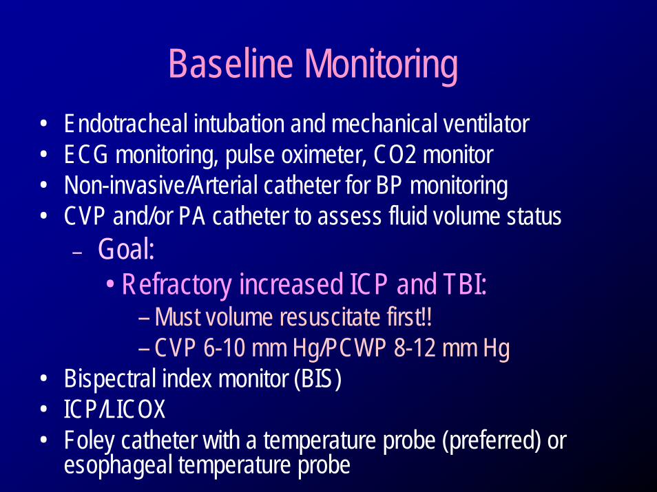
Baseline Monitoring• Endotracheal intubation and mechanical ventilator• ECG monitoring, pulse oximeter, CO2 monitor• Non-invasive/Arterial catheter for BP monitoring• CVP and/or PA catheter to assess fluid volume status
– Goal: • Refractory increased ICP and TBI:
– Must volume resuscitate first!!– CVP 6-10 mm Hg/PCWP 8-12 mm Hg
• Bispectral index monitor (BIS)• ICP/LICOX • Foley catheter with a temperature probe (preferred) or
esophageal temperature probe
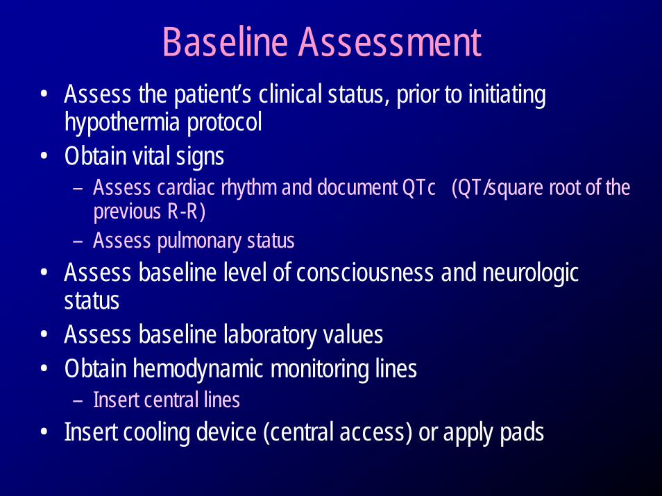
Baseline Assessment• Assess the patient’s clinical status, prior to initiating
hypothermia protocol• Obtain vital signs
– Assess cardiac rhythm and document QTc (QT/square root of the previous R-R)
– Assess pulmonary status• Assess baseline level of consciousness and neurologic
status• Assess baseline laboratory values • Obtain hemodynamic monitoring lines
– Insert central lines • Insert cooling device (central access) or apply pads
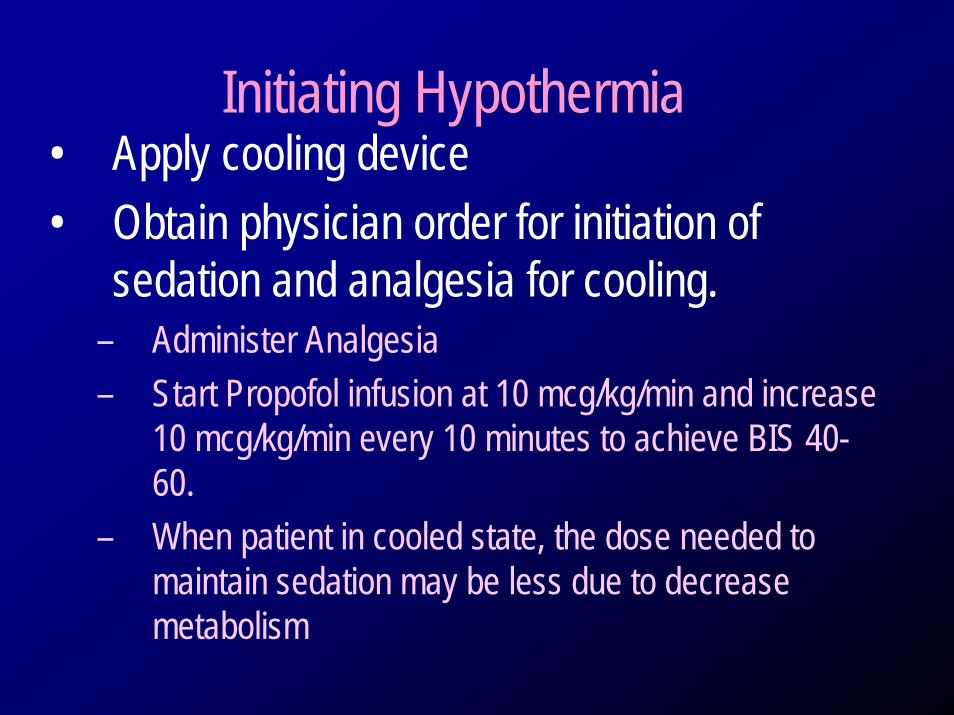
Initiating Hypothermia• Apply cooling device• Obtain physician order for initiation of
sedation and analgesia for cooling. – Administer Analgesia– Start Propofol infusion at 10 mcg/kg/min and increase
10 mcg/kg/min every 10 minutes to achieve BIS 40-60.
– When patient in cooled state, the dose needed to maintain sedation may be less due to decrease metabolism
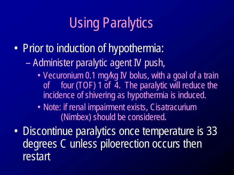
Using Paralytics• Prior to induction of hypothermia:
– Administer paralytic agent IV push, • Vecuronium 0.1 mg/kg IV bolus, with a goal of a train
of four (TOF) 1 of 4. The paralytic will reduce the incidence of shivering as hypothermia is induced.
• Note: if renal impairment exists, Cisatracurium(Nimbex) should be considered.
• Discontinue paralytics once temperature is 33 degrees C unless piloerection occurs then restart

Instituting Cooling
• Induction of hypothermia– 30 cc/kg IV bolus iced saline (4 degrees C) over
30 minutes– Usually drops temperature 2 degrees C– Once complete, begin device cooling
• Helpful to counter the cold diuresis
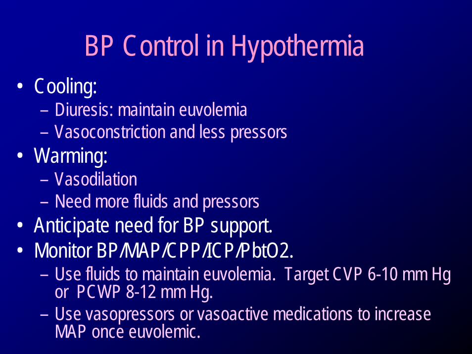
BP Control in Hypothermia• Cooling:
– Diuresis: maintain euvolemia– Vasoconstriction and less pressors
• Warming:– Vasodilation– Need more fluids and pressors
• Anticipate need for BP support.• Monitor BP/MAP/CPP/ICP/PbtO2.
– Use fluids to maintain euvolemia. Target CVP 6-10 mm Hg or PCWP 8-12 mm Hg.
– Use vasopressors or vasoactive medications to increase MAP once euvolemic.
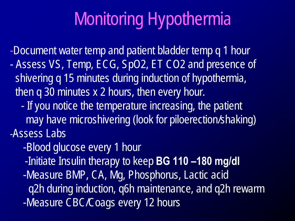
Monitoring Hypothermia-Document water temp and patient bladder temp q 1 hour- Assess VS, Temp, ECG, SpO2, ET CO2 and presence of shivering q 15 minutes during induction of hypothermia, then q 30 minutes x 2 hours, then every hour.
- If you notice the temperature increasing, the patient may have microshivering (look for piloerection/shaking)
-Assess Labs-Blood glucose every 1 hour-Initiate Insulin therapy to keep BG 110 –180 mg/dl-Measure BMP, CA, Mg, Phosphorus, Lactic acid
q2h during induction, q6h maintenance, and q2h rewarm-Measure CBC/Coags every 12 hours
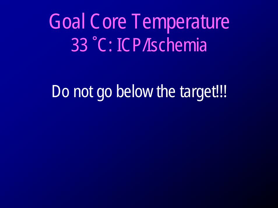
Goal Core Temperature 33 ˚C: ICP/Ischemia
Do not go below the target!!!
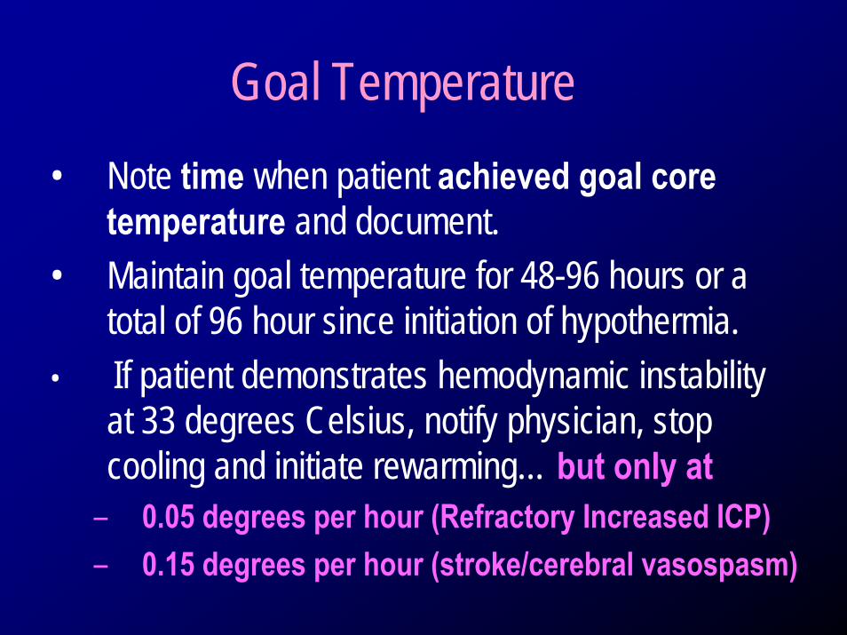
Goal Temperature• Note time when patient achieved goal core
temperature and document. • Maintain goal temperature for 48-96 hours or a
total of 96 hour since initiation of hypothermia.• If patient demonstrates hemodynamic instability
at 33 degrees Celsius, notify physician, stop cooling and initiate rewarming… but only at
– 0.05 degrees per hour (Refractory Increased ICP)– 0.15 degrees per hour (stroke/cerebral vasospasm)
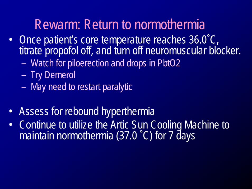
Rewarm: Return to normothermia• Once patient’s core temperature reaches 36.0˚C,
titrate propofol off, and turn off neuromuscular blocker.– Watch for piloerection and drops in PbtO2– Try Demerol– May need to restart paralytic
• Assess for rebound hyperthermia • Continue to utilize the Artic Sun Cooling Machine to
maintain normothermia (37.0 ˚C) for 7 days
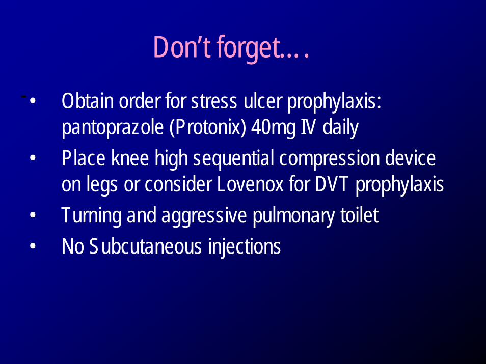
Don’t forget….• Obtain order for stress ulcer prophylaxis:
pantoprazole (Protonix) 40mg IV daily• Place knee high sequential compression device
on legs or consider Lovenox for DVT prophylaxis• Turning and aggressive pulmonary toilet• No Subcutaneous injections
-
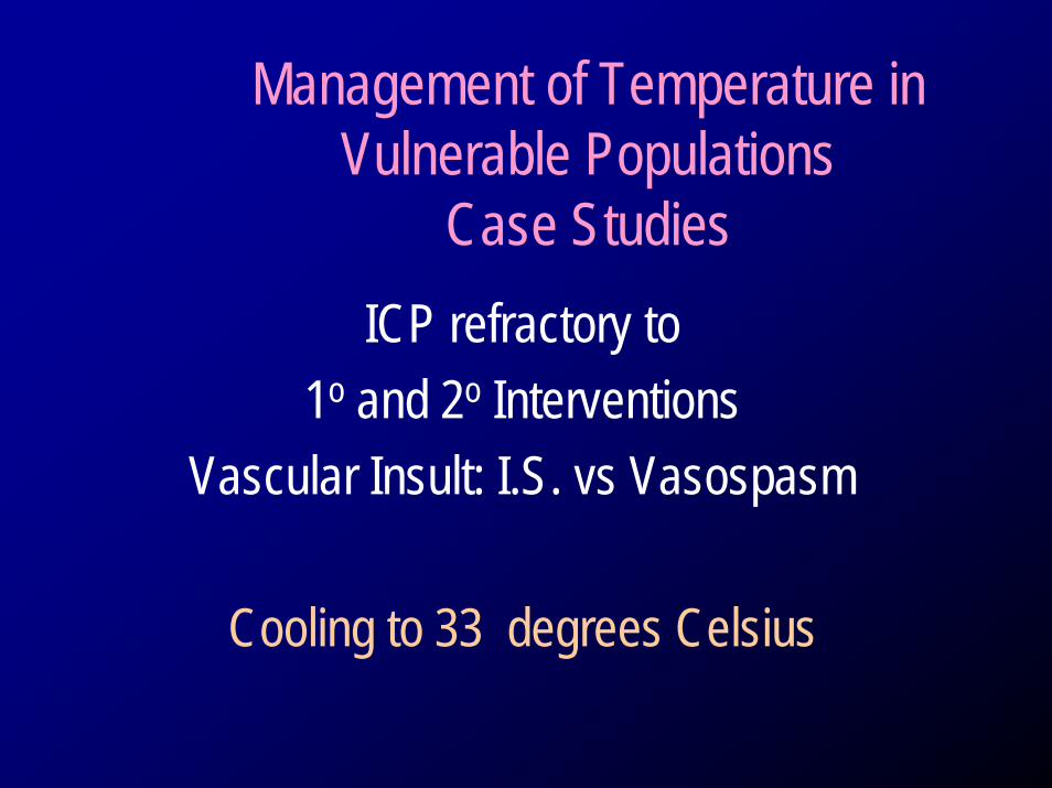
Management of Temperature in Vulnerable Populations
Case StudiesICP refractory to
1o and 2o InterventionsVascular Insult: I.S. vs Vasospasm
Cooling to 33 degrees Celsius
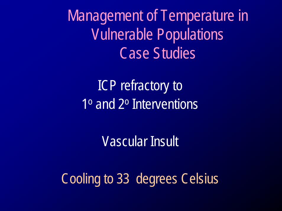
Management of Temperature in Vulnerable Populations
Case Studies
ICP refractory to 1o and 2o Interventions
Vascular Insult
Cooling to 33 degrees Celsius
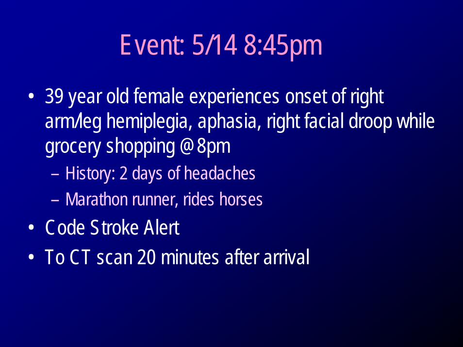
Event: 5/14 8:45pm• 39 year old female experiences onset of right
arm/leg hemiplegia, aphasia, right facial droop while grocery shopping @8pm– History: 2 days of headaches– Marathon runner, rides horses
• Code Stroke Alert• To CT scan 20 minutes after arrival