Embed Size (px)
Citation preview
DOI 10.1212/WNL.53.5.1042 1999;53;1042Neurology
G. Meola, V. Sansone, D. Perani, et al.in proximal myotonic myopathy
spatial function−Reduced cerebral blood flow and impaired visual
November 4, 2012This information is current as of
http://www.neurology.org/content/53/5/1042.full.html
located on the World Wide Web at: The online version of this article, along with updated information and services, is
rights reserved. Print ISSN: 0028-3878. Online ISSN: 1526-632X.Allsince 1951, it is now a weekly with 48 issues per year. Copyright © 1999 by AAN Enterprises, Inc.
® is the official journal of the American Academy of Neurology. Published continuouslyNeurology
brought to you by COREView metadata, citation and similar papers at core.ac.uk
provided by AIR Universita degli studi di Milano
Reduced cerebral blood flow andimpaired visual–spatial function in
proximal myotonic myopathyG. Meola, MD; V. Sansone, MD; D. Perani, MD; A. Colleluori, MD; S. Cappa, MD; M. Cotelli, PhD;
F. Fazio, MD; C.A. Thornton, MD; and R.T. Moxley, MD
Article abstract—Objective: To compare brain involvement in myotonic dystrophy (DM) with that of proximal myotonicmyopathy (PROMM). Background: PROMM is a multisystem disease with many features in common with DM. Methods:Twenty patients with DM (CTG@500–700#), 20 patients with PROMM, and 20 normal control subjects were studied.Neuropsychological testing was performed in 12 patients with PROMM and in 18 patients with DM; brain MRI wasperformed in 17 of 20 PROMM patients and 15 of 20 DM patients. Ten patients with PROMM and 11 patients with DMwere subjected to H2
15O PET. Results: Two-thirds of the patients with PROMM and one-half of those with DM wereimpaired on visual–spatial recall, whereas one-third of the patients with PROMM and less than half of those with DMshowed an impairment in visual–spatial construction. Brain MRI was normal, or showed only nonspecific white matterabnormalities in both PROMM and DM patients. PET studies in PROMM patients showed a bilateral decrease in regionalcerebral blood flow (rCBF) of the orbitofrontal and medial frontal cortex, whereas DM patients had more widespreadhypoperfusion that extended to the dorsolateral frontal cortex and subcortical regions. Conclusions: Impaired visual–spatial function may be present in proximal myotonic myopathy. This correlates best with a reduction in regional cerebralblood flow observed in H2
15O PET brain scans rather than with specific structural abnormalities observed on brain MRI.Key words: Proximal myotonic myopathy—Myotonic dystrophy—PET—MRI—Neuropsychological tests.
NEUROLOGY 1999;53:1042–1050
Proximal myotonic myopathy (PROMM) and myo-tonic dystrophy (DM) both involve multiple organsystems, including the brain.1-9 Hypersomnia(10%),1,3,10 apathy (5%),3,10 grand mal seizures(5%),3,10 and mental retardation (5%)3 have all beennoted in PROMM. Hund et al.10 observed diffuse,confluent lesions in white matter on brain MRI in sixpatients with PROMM in whom no symptoms orsigns of mental disorder were present. Three of six ofthese patients had strokelike episodes. The changeson MRI resemble those described in cerebral autoso-mal dominant arteriopathy with subcortical infarctsand leukoencephalopathy,11 and the authors sug-gested that these abnormalities are a feature ofPROMM. In contrast to these findings, Meola et al.4
reported normal brain MRI findings in four patientswith PROMM. Only one patient, a 70-year-oldwoman with hypertension, had an abnormal MRI,which showed nonspecific white matter lesions.
Common symptoms of mental aberration in DMinclude apathy, indifference, and lack of motivation
even in patients with mild weakness. A suspicious,paranoid, or frankly hostile personality is common.Neuropsychological test results have demonstratedpersonality disorders,12 dementia,13-15 mental re-tardation,13-15 and depression13 in patients withadult-onset DM. Prominent structural changes inthe brain are infrequent, but some patients with DMhave white matter hyperintense lesions in the ante-rior portions of the temporal lobes.16,17 Generalizedatrophy has also been observed.14 The relatively fewneuropathologic studies available have shown a dif-fuse loss of myelin in the deep white matter of thebrain corresponding to the white matter hyperintenselesions observed on the brain scans.15,18 Pathologic tauproteins have been found in the hippocampus, the en-torhinal cortex, and in most of the temporal areas, andthe amount of tau protein was higher in a more se-verely affected patient.19 A possible relationship be-tween the decrease of catecholaminergic neurons in themedullary reticular formation and the presence of alve-olar hypoventilation has also been suggested.20
From the Department of Neurology (Drs. Meola and Sansone) and Institute of Neuroscience and Bioimaging National Research Council (Drs. Perani,Colleluori, and Fazio), H. San Raffaele, University of Milan, Italy; the Neurological Department, University of Brescia, and Instituto di Ricovero E Cura ACarattere Scientifico, S. Giovanni di Dio (Drs. Cappa and Cotelli), Brescia, Italy; and Department of Neurology (Drs. Thornton and Moxley), University ofRochester, NY.Supported by the University of Milan, Italy, MURST 40% and 60% (G.M.); the National Council of Research, Italy (D.P.); Muscular Dystrophy Association(C.T., R.T.M.); General Clinical Research Center–NIH 5M01RR0044 (C.T., R.T.M.), R03 AG14502-01 (C.T., R.T.M.), and R01 AR44069-01A1 (C.T., R.T.M.);Paul Beeson Physician, Faculty, Scholars in Aging Research Program (C.T.); the Saunders Foundation (C.T., R.T.M.); and the Wayne C. Gorrell Jr. MolecularBiology Laboratory (C.T., R.T.M.).Presented in part at the 50th annual meeting of the American Academy of Neurology; Minneapolis, MN; April 1998.Received July 22, 1998. Accepted in final form April 17, 1999.Address correspondence and reprint requests to Dr. Giovanni Meola, Department of Neurology, University of Milan, San Donato Hospital, V. Morandi, 30,20097–San Donato Milanese, Milan, Italy.
1042 Copyright © 1999 by the American Academy of Neurology
To determine whether PROMM patients havebrain alterations similar to those found in patientswith DM we performed neuropsychological testing,brain MRI, and H2
15O PET.
Methods. Selection of patients. We studied 20 patientswith PROMM from 6 unrelated families (2 from Italy and 4from the United States; age range, 18 to 73 years; meanage, 50 years; 11 women and 9 men). In all families atleast one affected person was tested for and did not showan expanded CTG repeat in the DM gene on chromosome19. In family 1, which has been described previously,4 link-age to chromosomes 7 and 17 in addition to chromosome 19was excluded. In this family and in families 3 and 6, link-age to chromosome 3q was also excluded.21,22 The remain-ing families were uninformative for linkage, but CTGrepeat size in the DM gene was normal in the affectedfamily members. Patient 2 was adopted, so there is noavailable information on the family pedigree. However,given the presence of proximal weakness, electromyo-graphic myotonia, and early-onset cataracts in the pres-ence of a normal-size CTG repeat in the DM gene, adiagnosis of PROMM was made. Additional supportivefindings were the presence of preserved tendon reflexes in
the affected limbs and calf hypertrophy. Twenty DM pa-tients (CTG@500–700#) (age range, 24 to 68 years; mean age,43 years; 10 women and 10 men) from 13 unrelated fami-lies from northern Italy were also studied. Comparableneuropsychological tests and neuroimaging procedureswere performed in both the Italian and the US patients.
Seventeen of the 20 patients with PROMM and 18 ofthe 20 DM patients were ambulatory. It was possible toperform neuropsychological testing in 12 of 20 patientswith PROMM and in 18 of 20 patients with DM. MRIevaluation was possible in 17 of 20 PROMM patients andin 15 of 20 DM patients. H2
15O PET scanning of the brainoccurred in 10 of 20 PROMM patients and 11 of 20 DMpatients. Tables 1 and 2 summarize the clinical and labo-ratory findings in the patients with PROMM and DM.
Neuropsychological procedures. Neuropsychologicaltesting was administered by an experienced examiner in aquiet environment in the hospital or at the patient’s home.Approximately 60 to 90 minutes were needed to adminis-ter the battery of tests required. It included a screeningtest for dementia (Mini-Mental State Examination@MMSE#) and tests of nonverbal reasoning (Raven’s coloredmatrices), conceptual abilities (Weigl’s sorting test), audi-tory language comprehension (Token test), verbal fluency
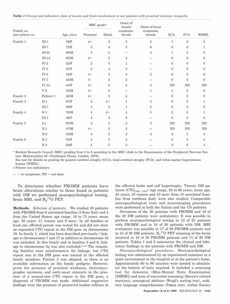
Table 1 Clinical and laboratory data of muscle and brain involvement in our patients with proximal myotonic myopathy
Family no.and patient no. Age, y/sex
MRC grade*Onset ofmuscle
symptoms,decade
Onset of brainsymptoms,
decade GCA† FCA† WMHL†Proximal Distal
Family 1 III-1 59/F 41 5 3 5 1 0 0
III-7† 73/F 3 4 3 6 0 0 1
III-8‡ 68/M 3 5 — 2 1 2 0
III-14 65/M 41 5 4 — 0 0 0
IV-2 22/F 5 5 1 — 0 0 0
IV-3 37/F 2 4 2 — 0 0 0
IV-5 34/F 41 5 3 2 0 0 0
IV-7 43/M 52 5 2 — 0 0 0
IV-14 44/F 41 5 3 3 ND ND ND
V-2 18/M 41 5 — 1 1 0 0
Family 2 Patient 2 45/M 41 5 4 — 0 0 0
Family 3 II-1 67/F 4 41 1 3 0 0 1
III-7 39/F 4 5 1 2 0 0 0
Family 4 II-1 70/M 4 41 4 5 2 0 1
III-1 36/F 4 5 2 — 0 0 0
Family 5 I-1 67/M 4 5 2 2 ND ND ND
II-1 47/M 41 5 2 — ND ND ND
II-2 52/M 4 5 2 4 0 1 0
Family 6 II-1 55/F 4 5 3 — 0 0 0
II-3 50/F 4 5 3 — 0 0 1
* Medical Research Council (MRC) grading from 0 to 5 according to the MRC (Aids to the Examination of the Peripheral Nervous Sys-tem. Memorandum 45—Pendragon House, London, 1976).
† See text for details on grading for general cerebral atrophy (GCA), focal cerebral atrophy (FCA), and white matter hyperintenselesions (WMHL).
‡ Patient not ambulatory.
— 5 no symptoms; ND 5 not done.
September (2 of 2) 1999 NEUROLOGY 53 1043
with phonemic and semantic cues, verbal and spatialshort-term memory (digit span and Corsi block tappingspan), memory for prose (logical memory test), spatiallearning (Corsi supraspan learning), constructional abili-ties and visual spatial recall (Rey’s complex figure), and aself-administered depression rating scale (Cognitive Be-havioral Assessment 2.0 depression scale [Bertolotti G,Michielin P, Sanavio E, Simonetti G, Vidotto G, Zotti AM;Organizzazioni Speciali, Firenze, 1986]). The tests wereadministered and scored according to published proce-dures.23,24 Data were compared with 20 age-, gender-, andeducation-matched control subjects. Not all patients wereable to perform the full battery of tests. A few refused thetesting and a few had severe distal weakness that madeholding a pen impossible. The results of the neuropsycho-logical testing in Patient III-8, from family 1 were notincluded in the statistical analysis because of his history ofpsychiatric disorders and the long-standing use of neuro-leptic drugs. A review of his medical records suggested adiagnosis of schizophrenia, superimposed on delayed men-tal development, which was further complicated by a prob-able vascular dementia. The data from Patient V-2 fromfamily 1 was also not included in the overall analysis be-cause of the long-standing use of antiepileptic drugs and
the patient’s history of delayed mental development. Hisfather refused to permit PET scanning and also refusedthe procedure for himself.
Neuropsychological performance was analyzed by directcomparison of the raw test scores among the three groupsusing one-way analysis of variance. Because of the multi-ple comparisons, the Bonferroni-corrected significancethreshold was set at 0.0042. Furthermore, to identify thenumber of patients who could be considered cognitivelyimpaired, the scores were corrected for age, education, andgender using normative data from a large sample of con-trol subjects (.300). A score in the pathologic range in twoor more tests was considered a criterion for cognitive im-pairment. The number of patients fulfilling this criterionin each group was compared by means of a contingencytable (chi-square statistics).
To assess the possible effect of age on neuropsychologi-cal performance, the age of the cognitively impaired DMpatients was compared with the unimpaired group (thepresence of only two impaired subjects among the PROMMpatients prevented the possibility of a statistical compari-son) using the Mann–Whitney U test.
Magnetic resonance imaging. MRI was performed witha scanner operating at a field strength of 1.5 T, and in-
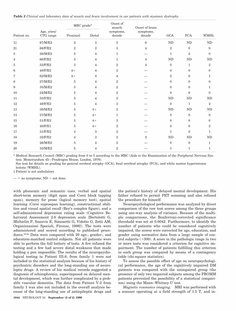
Table 2 Clinical and laboratory data of muscle and brain involvement in our patients with myotonic dystrophy
Patient no.Age, y/sex/CTG range
MRC grade*Onset ofmuscle
symptoms,decade
Onset of brainsymptoms,
decade GCA† FCA† WMHL†Proximal Distal
1‡ 67/M/E2 2 3 5 6 ND ND ND
2‡ 68/F/E2 2 2 3 4 2 0 0
3 28/M/E2 5 4 1 — 1 2 0
4 60/F/E2 3 4 1 4 ND ND ND
5 53/F/E2 3 4 2 4 0 1 2
6 48/F/E2 41 4 2 — 0 0 0
7 62/M/E2 41 4 4 — 2 0 1
8 27/M/E2 5 4 2 — 0 0 0
9 35/M/E2 5 4 2 — 0 0 1
10 24/M/E2 5 4 2 — 0 0 0
11 55/F/E2 5 4 2 — ND ND ND
12 49/F/E2 5 4 3 — 0 1 2
13 38/M/E2 5 41 2 — ND ND ND
14 27/M/E2 5 41 1 — 0 0 0
15 51/F/E2 5 41 3 — 0 0 0
16 40/F/E1 5 41 2 — 0 0 2
17 31/F/E2 4 3 2 — 1 0 1
18 32/F/E2 4 3 2 2 ND ND ND
19 26/M/E2 5 4 2 — 0 0 1
20 33/M/E2 5 4 2 — 1 1 1
* Medical Research Council (MRC) grading from 0 to 5 according to the MRC (Aids to the Examination of the Peripheral Nervous Sys-tem. Memorandum 45—Pendragon House, London, 1976).
† See text for details on grading for general cerebral atrophy (GCA), focal cerebral atrophy (FCA), and white matter hyperintenselesions (WMHL).
‡ Patient is not ambulatory.
— 5 no symptoms; ND 5 not done.
1044 NEUROLOGY 53 September (2 of 2) 1999
cluded spin-echo pulse sequences in T1-, proton density-,and T2-weighted imaging. For all scans, section thicknesswas 5 mm. All MRI studies were evaluated by a neuroradi-ologist without knowledge of the clinical status of the pa-tient. Seventeen patients with PROMM underwent MRI(three patients refused the procedure). MR images wereobtained for 15 of 20 patients with DM. The remaining fiveDM patients were unable to undergo this procedure be-cause of cardiac pacemakers. By mutual agreement be-tween the neuroradiologist and neurologist, our evaluationof the findings on MRI followed the methods of Huber etal.13 with slight modification. The grading is described inthe following paragraphs.
General cerebral atrophy (GCA). GCA was evaluatedrelative to age rather than on an absolute scale. Atrophywas rated on a scale of 0 to 3, with higher numbers indic-ative of increasing severity. A score of 0 representednormal-appearing ventricles and cisterns relative to age,and no parenchymal atrophy with widening of sulci.Higher numbers indicated varying degrees of increasingventricular and cisternal spaces.
Focal cerebral atrophy (FCA). FCA was rated usingthe same scale as that used for GCA, but the rating wasmade with respect to a specific region of the brain, such aswhen a particular lobe (e.g., temporal) was judged to besmaller or more atrophic than expected for the patient’sage (sulci more widened and increased in number).
White matter hyperintense lesions (WMHL). Focalwhite matter disease was judged by both the size andconfluency of these lesions: 0, no lesions; 1, a few bilateralhyperintense lacunae; 2, lesions . 2 cm; and 3, bilaterallesions . 2 cm, usually confluent.
MRI results were compared with MR images obtainedby members of the neuroradiology department in Italyfrom 20 individuals who undertook the examination fordifferent diagnostic purposes: 11 for transient tingling inthe hands; 4 for persistent headache; 2 for transient, non-specific speech problems; and 3 for occasional dizzinessand gait disturbances. No GCA, FCA, or WMHL werepresent in these subjects.
Data for each individual patient are presented in table1 for PROMM and in table 2 for DM.
PET acquisition procedures. Eight patients withPROMM and 11 patients with DM from Italy were studiedby H2
15O PET in a resting state. Two additional patientswith PROMM from the United States had SPECT regionalcerebral blood flow (rCBF) studies of the brain. The PETtomograph used was GE-Advance (General Electric Medi-cal System, Milwaukee, WI). The system consists of 18rings of bismuth germanate, which allows 35 transaxialimages with a slice thickness of 4.25 mm covering an axialfield of view of 15.2 cm. Transmission data were acquiredusing a pair of rotating pin sources filled with 68Ge (10mCi per pin). Image reconstruction was performed with afiltered back-projection algorithm on a 128 3 128 matrixwith a pixel size of 1.9 mm. rCBF was measured by record-ing the distribution of radioactivity following an IV bolusinjection of 7 to 8 mCi H2
15O through a forearm cannula. Theintegrated counts collected for 70 seconds, starting 20 sec-onds after injection time, were used as an index of rCBF.
PET data analysis. Reconstructed images of H215O
PET were transferred to a dedicated workstation for imageprocessing (SUN-SPARC 2, Sun Microsystems, Palo Alto,
CA). The data were analyzed with Statistical ParametricMapping 1996 (using software from the Wellcome Depart-ment of Cognitive Neurology, London, UK) implemented inMatlab version 4.2 (Mathworks Inc., Sherborn, MA).25
PET images were transformed into a stereotacticspace.26 This normalizing spatial conformation matcheseach scan to a reference or template image that alreadyconforms to the standard space.
After specifying the appropriate design matrix, group,subject, and covariate effects were estimated according tothe general linear model at each and every voxel.25 Weapplied analysis of covariance,26 in which the design ma-trix included global activity and age as confounding covari-ates. To test the hypothesis about regionally specificgroup-versus-group effects, the estimates were comparedusing linear contrasts. The resulting set of voxel values foreach contrast constitute a statistical parametric map(SPM) of the t statistic SPM. In this particular implemen-tation of the general linear model, the t statistic is, effec-tively, the rCBF difference between the mean rCBF of thecontrol group and the rCBF of the patient group underinvestigation, divided by the error estimated with the con-trol group data (after correcting for age and global effect).The SPMs were transformed to the unit normal distribu-tion (SPM Z) and thresholded at 3.09 (or corrected at p 50.001). The resulting foci were then characterized in termsof spatial extent (k) and peak heights (u).27
Results. Mental status. The presence of cognitive andbehavioral disorders was determined on the basis of a clin-ical interview with patients and relatives. A diagnosis ofdepression (family 3, Patients II-1 and III-7) and of demen-tia (family 1, Patients III-8 and V-2; family 4, Patient II-1;and family 5, Patient II-2) according to criteria in theDiagnostic and Statistical Manual of Mental Disorders,4th edition (DSM-IV)28 was made in 6 of 20 patients withPROMM. In Patient III-8 from family 1, clinical signs andsymptoms of mental retardation began at 14 years of age.His first admission to a psychiatric hospital was at 27years of age. He was diagnosed at the time as havingparanoid schizophrenia and oligophrenia. At 65 he had astroke with residual mild left hemiparesis. Within 2months he was diagnosed as having dementia of uncertaincause. He is currently in a psychiatric hospital. Patient V-2from family 1 had generalized seizures at 3 years of age,after normal delivery and birth. At 4 years of age, mentaldelay became apparent and seemed to be related to uncon-trolled seizures. Performance at school was poor. At thetime of this report, the patient is unemployed. A diagnosisof dementia was made in two other patients. Memory diffi-culty was the initial symptom in Patient II-1 from family 4who complained of depression and cognitive impairment.His cognitive deficits had progressed to the point that adiagnosis of an Alzheimer’s type of dementia was made.His 36-year-old daughter complains of mood swings andoften feels depressed, but she is able to continue her every-day activity. Patient II-2 from family 5 is currently unem-ployed and is on disability for his disorder. He is currentlycomplaining of problems with memory, language, atten-tion, concentration, and executive functioning. He also re-ports intermittent difficulty with comprehension of bothwritten and oral material, important word-finding prob-lems. He has difficulty with elevated levels of anxiety,depression, and anger. The patient is currently seeing a
September (2 of 2) 1999 NEUROLOGY 53 1045
psychiatrist. His 47-year-old brother (Patient II-1) has pro-found weakness and has much difficulty rising from thefloor and he has trouble rising from a chair. He has bilat-eral cataracts. He complains of attention deficits and mem-ory problems. He is currently taking medication forgeneralized seizures that began at the age of 30. Neuroim-aging scans are unavailable.
The same clinical criteria used to asses the presence ofcognitive and behavioral complaints in PROMM patientswere used for DM patients. Three patients (15%) had adefinite attitude of distrust and suspiciousness (Patients 9,11, and 19) that fitted the criteria of a paranoid personal-ity disorder, and two patients (Patients 14 and 10) had aborderline personality disorder according to the DSM-IVcriteria.28 Recurrent, brief depressive disorders accordingto DSM-IV28 have been found in two patients (Patients 6and 18). In three patients (Patients 1, 2, and 4) there weremultiple cognitive deficits, including memory impairmentand difficulty in executive functioning, that were severeenough to cause impairment in occupational and socialfunctioning so that a diagnosis of dementia was made ac-cording to the DSM-IV criteria.28 Excessive daytime sleep-iness was a subjective complaint in 16 of 20 patients, butonly in threee of these patients was it severe enough tointerfere with everyday activity.
Neuropsychological tests. The intergroup comparison(table 3) indicates that patients with DM have generallylower scores than age- and education-matched control sub-jects. Patients with PROMM performed, in general, at anintermediate level between DM and control subjects. Sig-nificant differences were found for four tests: MMSE, storyrecall, and copy and recall of Rey’s figure. A post hoc anal-ysis using Scheffe’s method showed that patients with DMperformed at a significantly lower level than control sub-jects ( p , 0.05) on MMSE, story recall, and Rey copy. Inthe case of Rey recall, both patients with PROMM and DMwere impaired compared with control subjects.
In regard to normative data, approximately two-thirds(64%) of the patients with PROMM and one-half (53%) ofthose with DM showed an impairment of visual–spatialrecall (Rey recall), whereas only one-third of the patientswith PROMM (36%) and less than half of the DM patients(47%) showed an impairment in visual–spatial construc-tion (Rey copy). An impairment of verbal short-term andlong-term memory was significantly more common in pa-tients with DM than in patients with PROMM (61% ofpatients with DM versus 18% of patients with PROMM: x2
5 5.09; p . 0.005 in the story recall test; and 28% ofpatients with DM failed in the digit span test whereas allpatients with PROMM performed correctly on this test: x2
5 4.0; p . 0.005). Considering the number of scores belowthe age- and education-corrected cutoff value for eachpathologic group (PROMM and DM) on our battery of neu-ropsychological tests, the overall neuropsychological per-formance of impaired tests was 16 of 125 in the PROMMgroup versus 55 of 214 impaired tests in the DM group(x2 5 7.93; p . 0.005). The incidence of more pronouncedneuropsychological impairment, as measured by abnormalperformance in more than two tests, was higher in DMpatients than in PROMM patients (2 of 12 PROMM pa-tients and 9 of 18 DM patients; approaching statisticalsignificance, x2 5 3.44, p 5 0.06). In the DM group, the
average age of the patients showing impairment in testingwas significantly higher (Mann–Whitney U test, z 522.65, p 5 0.008).
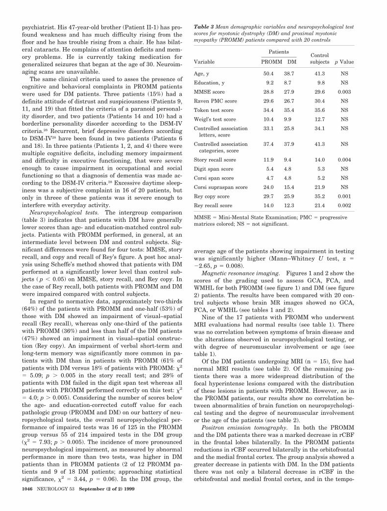
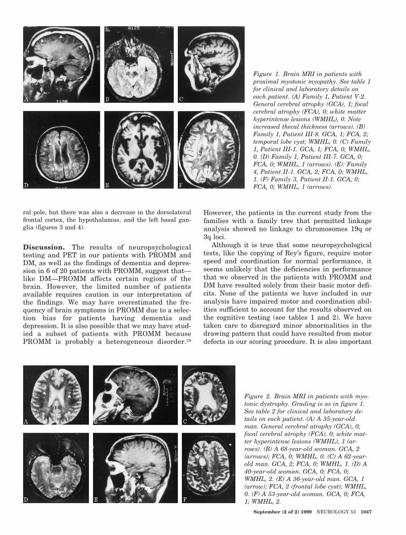
Magnetic resonance imaging. Figures 1 and 2 show thescores of the grading used to assess GCA, FCA, andWMHL for both PROMM (see figure 1) and DM (see figure2) patients. The results have been compared with 20 con-trol subjects whose brain MR images showed no GCA,FCA, or WMHL (see tables 1 and 2).
Nine of the 17 patients with PROMM who underwentMRI evaluations had normal results (see table 1). Therewas no correlation between symptoms of brain disease andthe alterations observed in neuropsychological testing, orwith degree of neuromuscular involvement or age (seetable 1).
Of the DM patients undergoing MRI (n 5 15), five hadnormal MRI results (see table 2). Of the remaining pa-tients there was a more widespread distribution of thefocal hyperintense lesions compared with the distributionof these lesions in patients with PROMM. However, as inthe PROMM patients, our results show no correlation be-tween abnormalities of brain function on neuropsychologi-cal testing and the degree of neuromuscular involvementor the age of the patients (see table 2).
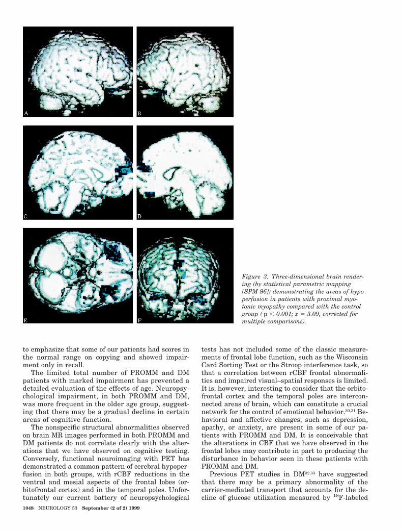
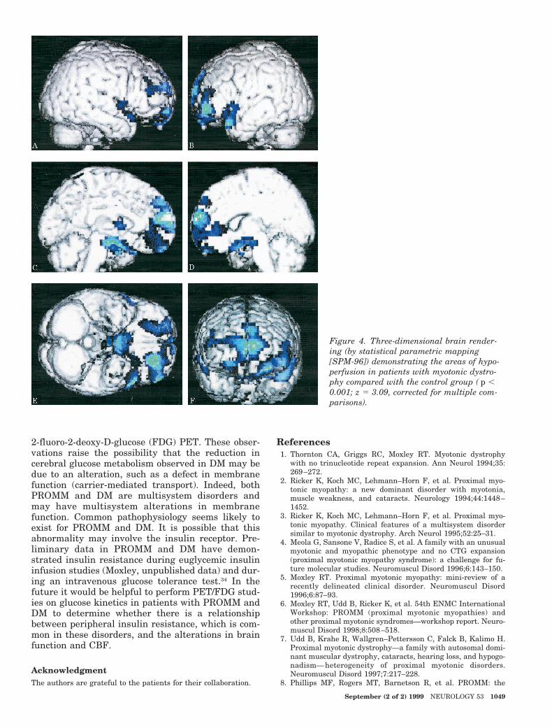
Positron emission tomography. In both the PROMMand the DM patients there was a marked decrease in rCBFin the frontal lobes bilaterally. In the PROMM patientsreductions in rCBF occurred bilaterally in the orbitofrontaland the medial frontal cortex. The group analysis showed agreater decrease in patients with DM. In the DM patientsthere was not only a bilateral decrease in rCBF in theorbitofrontal and medial frontal cortex, and in the tempo-
Table 3 Mean demographic variables and neuropsychological testscores for myotonic dystrophy (DM) and proximal myotonicmyopathy (PROMM) patients compared with 20 controls
Variable
Patients
PROMM DMControlsubjects p Value
Age, y 50.4 38.7 41.3 NS
Education, y 9.2 8.7 9.8 NS
MMSE score 28.8 27.9 29.6 0.003
Raven PMC score 29.6 26.7 30.4 NS
Token test score 34.4 35.4 35.6 NS
Weigl’s test score 10.4 9.9 12.7 NS
Controlled associationletters, score
33.1 25.8 34.1 NS
Controlled associationcategories, score
37.4 37.9 41.3 NS
Story recall score 11.9 9.4 14.0 0.004
Digit span score 5.4 4.8 5.3 NS
Corsi span score 4.7 4.8 5.2 NS
Corsi supraspan score 24.0 15.4 21.9 NS
Rey copy score 29.7 25.9 35.2 0.001
Rey recall score 14.0 12.3 21.4 0.002
MMSE 5 Mini-Mental State Examination; PMC 5 progressivematrices colored; NS 5 not significant.
1046 NEUROLOGY 53 September (2 of 2) 1999
ral pole, but there was also a decrease in the dorsolateralfrontal cortex, the hypothalamus, and the left basal gan-glia (figures 3 and 4).
Discussion. The results of neuropsychologicaltesting and PET in our patients with PROMM andDM, as well as the findings of dementia and depres-sion in 6 of 20 patients with PROMM, suggest that—like DM—PROMM affects certain regions of thebrain. However, the limited number of patientsavailable requires caution in our interpretation ofthe findings. We may have overestimated the fre-quency of brain symptoms in PROMM due to a selec-tion bias for patients having dementia anddepression. It is also possible that we may have stud-ied a subset of patients with PROMM becausePROMM is probably a heterogeneous disorder.29
However, the patients in the current study from thefamilies with a family tree that permitted linkageanalysis showed no linkage to chromosomes 19q or3q loci.
Although it is true that some neuropsychologicaltests, like the copying of Rey’s figure, require motorspeed and coordination for normal performance, itseems unlikely that the deficiencies in performancethat we observed in the patients with PROMM andDM have resulted solely from their basic motor defi-cits. None of the patients we have included in ouranalysis have impaired motor and coordination abil-ities sufficient to account for the results observed onthe cognitive testing (see tables 1 and 2). We havetaken care to disregard minor abnormalities in thedrawing pattern that could have resulted from motordefects in our scoring procedure. It is also important
Figure 1. Brain MRI in patients withproximal myotonic myopathy. See table 1for clinical and laboratory details oneach patient. (A) Family 1, Patient V-2.General cerebral atrophy (GCA), 1; focalcerebral atrophy (FCA), 0; white matterhyperintense lesions (WMHL), 0. Noteincreased thecal thickness (arrows). (B)Family 1, Patient III-8. GCA, 1; FCA, 2;temporal lobe cyst; WMHL, 0. (C) Family1, Patient III-1. GCA, 1; FCA, 0; WMHL,0. (D) Family 1, Patient III-7. GCA, 0;FCA, 0; WMHL, 1 (arrows). (E): Family4, Patient II-1. GCA, 2; FCA, 0; WMHL,1. (F) Family 3, Patient II-1. GCA, 0;FCA, 0; WMHL, 1 (arrows).
Figure 2. Brain MRI in patients with myo-tonic dystrophy. Grading is as in figure 1.See table 2 for clinical and laboratory de-tails on each patient. (A) A 35-year-oldman. General cerebral atrophy (GCA), 0;focal cerebral atrophy (FCA), 0; white mat-ter hyperintense lesions (WMHL), 1 (ar-rows). (B) A 68-year-old woman. GCA, 2(arrows); FCA, 0; WMHL, 0. (C) A 62-year-old man. GCA, 2; FCA, 0; WMHL, 1. (D) A40-year-old woman. GCA, 0; FCA, 0;WMHL, 2. (E) A 36-year-old man. GCA, 1(arrow); FCA, 2 (frontal lobe cyst); WMHL,0. (F) A 53-year-old woman. GCA, 0; FCA,1; WMHL, 2.
September (2 of 2) 1999 NEUROLOGY 53 1047
to emphasize that some of our patients had scores inthe normal range on copying and showed impair-ment only in recall.
The limited total number of PROMM and DMpatients with marked impairment has prevented adetailed evaluation of the effects of age. Neuropsy-chological impairment, in both PROMM and DM,was more frequent in the older age group, suggest-ing that there may be a gradual decline in certainareas of cognitive function.
The nonspecific structural abnormalities observedon brain MR images performed in both PROMM andDM patients do not correlate clearly with the alter-ations that we have observed on cognitive testing.Conversely, functional neuroimaging with PET hasdemonstrated a common pattern of cerebral hypoper-fusion in both groups, with rCBF reductions in theventral and mesial aspects of the frontal lobes (or-bitofrontal cortex) and in the temporal poles. Unfor-tunately our current battery of neuropsychological
tests has not included some of the classic measure-ments of frontal lobe function, such as the WisconsinCard Sorting Test or the Stroop interference task, sothat a correlation between rCBF frontal abnormali-ties and impaired visual–spatial responses is limited.It is, however, interesting to consider that the orbito-frontal cortex and the temporal poles are intercon-nected areas of brain, which can constitute a crucialnetwork for the control of emotional behavior.30,31 Be-havioral and affective changes, such as depression,apathy, or anxiety, are present in some of our pa-tients with PROMM and DM. It is conceivable thatthe alterations in CBF that we have observed in thefrontal lobes may contribute in part to producing thedisturbance in behavior seen in these patients withPROMM and DM.
Previous PET studies in DM32,33 have suggestedthat there may be a primary abnormality of thecarrier-mediated transport that accounts for the de-cline of glucose utilization measured by 18F-labeled
Figure 3. Three-dimensional brain render-ing (by statistical parametric mapping[SPM-96]) demonstrating the areas of hypo-perfusion in patients with proximal myo-tonic myopathy compared with the controlgroup ( p , 0.001; z 5 3.09, corrected formultiple comparisons).
1048 NEUROLOGY 53 September (2 of 2) 1999
2-fluoro-2-deoxy-D-glucose (FDG) PET. These obser-vations raise the possibility that the reduction incerebral glucose metabolism observed in DM may bedue to an alteration, such as a defect in membranefunction (carrier-mediated transport). Indeed, bothPROMM and DM are multisystem disorders andmay have multisystem alterations in membranefunction. Common pathophysiology seems likely toexist for PROMM and DM. It is possible that thisabnormality may involve the insulin receptor. Pre-liminary data in PROMM and DM have demon-strated insulin resistance during euglycemic insulininfusion studies (Moxley, unpublished data) and dur-ing an intravenous glucose tolerance test.34 In thefuture it would be helpful to perform PET/FDG stud-ies on glucose kinetics in patients with PROMM andDM to determine whether there is a relationshipbetween peripheral insulin resistance, which is com-mon in these disorders, and the alterations in brainfunction and CBF.
AcknowledgmentThe authors are grateful to the patients for their collaboration.
References1. Thornton CA, Griggs RC, Moxley RT. Myotonic dystrophy
with no trinucleotide repeat expansion. Ann Neurol 1994;35:269–272.
2. Ricker K, Koch MC, Lehmann–Horn F, et al. Proximal myo-tonic myopathy: a new dominant disorder with myotonia,muscle weakness, and cataracts. Neurology 1994;44:1448–1452.
3. Ricker K, Koch MC, Lehmann–Horn F, et al. Proximal myo-tonic myopathy. Clinical features of a multisystem disordersimilar to myotonic dystrophy. Arch Neurol 1995;52:25–31.
4. Meola G, Sansone V, Radice S, et al. A family with an unusualmyotonic and myopathic phenotype and no CTG expansion(proximal myotonic myopathy syndrome): a challenge for fu-ture molecular studies. Neuromuscul Disord 1996;6:143–150.
5. Moxley RT. Proximal myotonic myopathy: mini-review of arecently delineated clinical disorder. Neuromuscul Disord1996;6:87–93.
6. Moxley RT, Udd B, Ricker K, et al. 54th ENMC InternationalWorkshop: PROMM (proximal myotonic myopathies) andother proximal myotonic syndromes—workshop report. Neuro-muscul Disord 1998;8:508–518.
7. Udd B, Krahe R, Wallgren–Pettersson C, Falck B, Kalimo H.Proximal myotonic dystrophy—a family with autosomal domi-nant muscular dystrophy, cataracts, hearing loss, and hypogo-nadism— heterogeneity of proximal myotonic disorders.Neuromuscul Disord 1997;7:217–228.
8. Phillips MF, Rogers MT, Barnetson R, et al. PROMM: the
Figure 4. Three-dimensional brain render-ing (by statistical parametric mapping[SPM-96]) demonstrating the areas of hypo-perfusion in patients with myotonic dystro-phy compared with the control group ( p ,0.001; z 5 3.09, corrected for multiple com-parisons).
September (2 of 2) 1999 NEUROLOGY 53 1049

expanding phenotype. A family with proximal myopathy, myo-tonia and deafness. Neuromuscul Disord 1998;8:439–446.
9. Harper PS. Myotonic dystrophy. 2nd ed. London: WB Saun-ders, 1989.
10. Hund E, Jansen O, Koch MC, et al. Proximal myotonic myop-athy with MRI white matter abnormalities of the brain. Neu-rology 1997;48:33–37.
11. Ruchous MM, Guerouaou D, Vandenhaute B, et al. Systemicvascular smooth muscle cell impairment in cerebral autoso-mal dominant arteriopathy with subcortical infarcts and leu-koencephalopathy. Acta Neuropathol 1995;89:500–512.
12. Delaporte C. Personality patterns in patients with myotonicdystrophy. Arch Neurol 1998;55:635–640.
13. Huber SJ, Kissel JT, Shuttleworth EC, et al. Magnetic reso-nance imaging and clinical correlates of intellectual impair-ment in myotonic dystrophy. Arch Neurol 1989;46:536–540.
14. Damian MS, Schilling G, Bachmann G, Simon C, Stoppler S,Dorndorf W. White matter lesions and cognitive deficits: rele-vance of lesion pattern? Acta Neurol Scand 1994;90:430–436.
15. Abe K, Fujimura H, Toyooka K, et al. Involvement of thecentral nervous system in myotonic dystrophy. J Neurol Sci1994;127:179–185.
16. Miaux Y, Chiras J, Eymard B, et al. Cranial MRI findings inmyotonic dystrophy. Neuroradiology 1997;39:166–170.
17. Chang L, Ernst T, Osborn D, Seltzer W, Leonido–Yee M,Poland RE. Proton spectroscopy in myotonic dystrophy. Corre-lations with CTG repeats. Arch Neurol 1998;55:305–311.
18. Ogata A, Terae S, Fujita M, Tashiro K. Anterior temporalwhite matter lesions in myotonic dystrophy with intellectualimpairment: an MRI and neuropathological study. Neuroradi-ology 1998;40:411–415.
19. Vermersch P, Sergeant N, Ruchoux MM, et al. Specific tauvariants in the brains of patients with myotonic dystrophy.Neurology 1996;47:711–717.
20. Ono S, Takahashi K, Jinnai K, et al. Loss of catecholaminergicneurons in the medullary reticular formation in myotonic dys-trophy. Neurology 1998;51:1121–1124.
21. Ranum LPW, Rasmussen PF, Benzow KA, Koob MD, Day JW.Genetic mapping of a second myotonic dystrophy locus. NatGenet 1998;19:196–198.
22. Ricker K, Grimm T, Koch MC, et al. Linkage of proximal
myotonic myopathy to chromosome 3q. Neurology 1999;52:170–171.
23. Mariani C, Farina E, Cappa SF, et al. Neuropsychologicalassessment in multiple sclerosis: a follow-up study with mag-netic resonance imaging. J Neurol 1991;238:395–400.
24. Lezak MD. Neuropsychological assessment. 3rd ed. New York:Oxford University Press, 1995.
25. Friston KJ, Holmes AP, Worsley KJ, Poline JP, Frith CD,Frackowiak RSJ. Statistical parametric maps in functionalimaging: a general linear approach. Hum Brain Mapp 1995;2:189–210.
26. Talairach J, Tournoux P. Coplanar stereotactic atlas of thehuman brain. Dimensional proportional system: an approachto cerebral imaging. Stuttgart: Thieme, 1988.
27. Signorini M, Paulesu E, Friston K, et al. Rapid assessment ofregional cerebral metabolic abnormalities in single subjectswith quantitative and nonquantitative @18F#FDG PET: A clin-ical validation of statistical parametric mapping. Neuroimage1999;9:63–80.
28. American Psychiatric Association. Diagnostic and statisticalmanual of mental disorders. 4th ed. Washington, DC: Ameri-can Psychiatric Association, 1994.
29. Thornton CA, Ashizawa T. Getting a grip on the myotonicdystrophies. Neurology 1999;52:12–13.
30. Kupferman I. Localization of higher cognitive and affectivefunctions: the association cortices. In: Kandel ER, SchwartzJH, Jessel TM, eds. Principles of neural science. 3rd ed. NewYork: Elsevier Publishers, 1991:823–838.
31. Stuss DT, Benson DF. The frontal lobes. New York: RavenPress, 1986.
32. Fiorelli M, Duboc D, Mazoyer BM, et al. Decreased cerebralglucose utilization in myotonic dystrophy. Neurology 1992;42:91–94.
33. Annane D, Fiorelli M, Mazoyer B, et al. Impaired cerebralglucose metabolism in myotonic dystrophy: a triplet-sizedependent phenomenon. Neuromuscul Disord 1998;8:39 – 45.
34. Moxley RT, Sansone V, Lifton A, Thornton CA. Insulin resis-tance in proximal myotonic myopathy (PROMM). Neurology1997;48(suppl 3):A229. Abstract.
1050 NEUROLOGY 53 September (2 of 2) 1999
DOI 10.1212/WNL.53.5.1042 1999;53;1042Neurology
G. Meola, V. Sansone, D. Perani, et al.myotonic myopathy
spatial function in proximal−Reduced cerebral blood flow and impaired visual
November 4, 2012This information is current as of
ServicesUpdated Information &
http://www.neurology.org/content/53/5/1042.full.htmlincluding high resolution figures, can be found at:
Referenceshttp://www.neurology.org/content/53/5/1042.full.html#ref-list-1This article cites 28 articles, 7 of which can be accessed free at:
Citations
rlshttp://www.neurology.org/content/53/5/1042.full.html#related-uThis article has been cited by 8 HighWire-hosted articles:
Permissions & Licensing
http://www.neurology.org/misc/about.xhtml#permissionstables) or in its entirety can be found online at: Information about reproducing this article in parts (figures,
Reprints http://www.neurology.org/misc/addir.xhtml#reprintsus
Information about ordering reprints can be found online: