Embed Size (px)
Citation preview

1
Redox Regulation of Plant Homeodomain Transcription Factors*
Adriana E. Tron, Carlos W. Bertoncini, Raquel L. Chan, and Daniel H. Gonzalez
Cátedra de Biología Celular y Molecular, Facultad de Bioquímica y Ciencias
Biológicas, Universidad Nacional del Litoral, CC 242 Paraje El Pozo, 3000 Santa Fe,
Argentina
Running Title: Redox Regulation of Plant Homeodomain Transcription Factors
Correspondence to:
Daniel H. Gonzalez
Cátedra de Biología Celular y Molecular
Fac. de Bioq. y Cs. Biol. (UNL)
CC 242 Paraje El Pozo
3000 Santa Fe
Argentina
Tel/Fax: 54-342-4575219
E-mail: [email protected]
Copyright 2002 by The American Society for Biochemistry and Molecular Biology, Inc.
JBC Papers in Press. Published on July 1, 2002 as Manuscript M203297200 by guest on Septem
ber 25, 2018http://w
ww
.jbc.org/D
ownloaded from

2
Summary
Several families of plant transcription factors contain a conserved DNA binding
motif known as the homeodomain. In two of these families, named Hd-Zip and glabra2,
the homeodomain is associated with a leucine zipper-like dimerization motif. A group
of Hd-Zip proteins, namely Hd-ZipII, contain a set of conserved cysteines within the
dimerization motif and adjacent to it. Incubation of one of these proteins, Hahb-10, in
the presence of thiol-reducing agents such as dithiothreitol or reduced glutathione
produced a significant increase in DNA binding. Under such conditions, the protein
migrated as a monomer in non-reducing SDS-polyacrylamide gels. Under oxidizing
conditions, a significant proportion of the protein migrated as dimers, suggesting the
formation of intermolecular disulfide bonds. A similar behaviour was observed for the
glabra2 protein HAHR1, which also contains two conserved cysteines within its
dimerization domain. Site-directed mutagenesis of the cysteines to serines indicated that
each of them has different roles in the activation of the proteins. Purified thioredoxin
was able to direct the NADPH-dependent activation of Hahb-10 and HAHR1 in the
presence of thioredoxin reductase. The results suggest that redox conditions may
operate to regulate the activity of these groups of plant transcription factors within plant
cells.
by guest on September 25, 2018
http://ww
w.jbc.org/
Dow
nloaded from
3
INTRODUCTION
The homeodomain (HD)1 is a 61-amino-acid protein motif found in eukaryotic
transcription factors generally involved in the regulation of developmental processes (1-
3). It folds into a characteristic three-helix structure that interacts specifically with DNA
(1,4-6). Helices II and III form a structure that resembles the helix-turn-helix motif
found in many prokaryotic transcription factors. Helix III (the recognition helix) fits
into the major groove of DNA making extensive contacts with specific bases and the
sugar-phosphate backbone (7-10). In spite of the resemblance in structure between the
HD and the helix-turn-helix motif, a striking difference is that HDs usually bind DNA
as monomers with high affinity (11,12). This fact has been explained by the presence of
extended contacts along the recognition helix and the N-terminal arm of the HD.
HDs are present in almost every eukaryotic organism that has been investigated.
In plants, several families of HD proteins have been described (13). One of these
families, named Hd-Zip, comprises proteins with a typical leucine zipper motif adjacent
to the C-terminal end of the HD (14,15). As expected, these proteins bind DNA as
dimers (16). The removal of the leucine zipper, or the introduction of extra amino acids
between the HD and the zipper, significantly reduces binding affinity, indicating that
the leucine zipper is responsible for the correct positioning of the HD for efficient
binding (16). The analysis of binding at different protein concentrations suggests that
dimer formation is a pre-requisite for DNA binding (17). It has been suggested that Hd-
Zip proteins may be involved in regulating developmental processes associated with the
response of plants to environmental conditions (13,18,19).
A different family of plant HD proteins, named glabra2, consists of larger
proteins with an N-terminal HD. These proteins also bind DNA as dimers, and possess a
by guest on September 25, 2018
http://ww
w.jbc.org/
Dow
nloaded from

4
dimerization motif that resembles a leucine zipper truncated by a loop (20,21). Most
members of the glabra2 family are expressed specifically in epidermal cells and the first
member to be identified (glabra2) is involved in the development of trichomes, root
hairs and the seed coat mucilage (22-26).
The HD and dimerization motif constitute the most conserved part of the
different members of each family. Sequence analysis also revealed the presence of
conserved cysteine-containing motifs within variable regions (i.e.: within the variable
loop in glabra2 proteins and near the C-terminus of the leucine zipper in Hd-ZipII
proteins). Since conserved cysteines have been reported to be involved in the redox
regulation of the properties of several transcription factors (27-33), we have tested the
effect of oxidants and reductants on DNA binding and quaternary structure of two
proteins from these families. Our results indicate that redox conditions are a key factor
determining the binding of these proteins to DNA and the formation of covalent
oligomeric structures. We propose that a redox-dependent mechanism may operate in
vivo to modulate the activity of these transcription factors in response to metabolic
and/or environmental signals.
EXPERIMENTAL PROCEDURES
Cloning, Expression, and Purification of Recombinant Proteins--An NheI/XbaI
fragment containing sequences coding for the entire HD and dimerization motif plus
additional amino acids (positions 86 to 325) from the sunflower glabra2 protein
HAHR1 (34), was excised from pHX1 (which contains this fragment in vector pUC119)
with HincII and EcoRI, and cloned into the SmaI and EcoRI sites of pGEX-3X as
described (35). For the production of a protein with only the first half of the
dimerization motif (amino acids 86 to 184), a KpnI deletion of this clone was used.
by guest on September 25, 2018
http://ww
w.jbc.org/
Dow
nloaded from
5
Fragments encoding proteins with either Cys or Ser at positions 185 and 188, were
obtained by PCR on native HAHR1 and oligonucleotide 5’-
GGCGAATTCTTGGTGATGCTCCCTGTG-3’ in combination with either 5’-
GGGCTGGCAAGCCACGTTTGGTG-3’ (for CC-HAHR1), 5’-
AAGGGTACCAGTACCAAC-3’ (for SC HAHR1), or 5’-
GCGGGTACCAGTACCAACAGTGGTTTC-3’ (for SS-HAHR1). For CS HAHR1,
oligonucleotides 5’-GGCGAATTCTTGGTGATGCTCCCTGTG-3’ and 5’-
AAGGGTACCTGTACCAAC-3’ were used with DNA from SS-HAHR1 as template.
Amplified products were digested with KpnI and EcoRI and cloned into similar sites in
the HAHR1 expressing plasmid. These constructs express proteins bearing the entire
HD and dimerization motif (amino acids 86 to 234).
An SpeI fragment encoding amino acids 81 to 231 from the sunflower Hd-ZipII
protein Hahb-10 (36), previously cloned into the XbaI site of pMAL-c2, was used as
source for expression in pGEX4T-3, using BamHI and SalI sites for excision and
cloning. Fragments encoding the entire Hd-Zip domain plus the CPSCE motif (amino
acids 81 to 208) were amplified and cloned in frame into the BamHI and EcoRI sites of
pGEX4T-3. Amplifications were performed using oligonucleotides pGEX1 (5’-
GGGCTGGCAAGCCACGTTTGGTG-3’) and CC10 (5’-
CCGAATTCCCGATCTGTTCACACGAC-3’, for CCCCC-Hahb-10), SC10 (5’-
CCGAATTCCCGATCTGTTCACACGACGGAGACACG-3’, for CCCSC-Hahb-10),
or CS10 (5’-CCGAATTCCCGATCTGTTCAGACGAC-3’, for CCCCS-Hahb-10 and
CCCSS-Hahb-10), using either native or CCCSC-Hahb-10 sequences as templates.
Mutants with Cys to Ser changes within the leucine zipper were constructed using
complementary oligonucleotides 5’-TAAATACTCAGAGTCCACCT-3’ and 5’-
AGGTGGACTCTGAGTATTTA-3’ (for SCCCC-Hahb-10) or 5’-
by guest on September 25, 2018
http://ww
w.jbc.org/
Dow
nloaded from
6
TAATGTGTTGGAGGATCTCTTTAA-3’ and 5’-
TTAAAGAGATCCTCCAACACATTA-3’ (for CSSCC-Hahb-10) together with
primers pGEX1 and CC10, to amplify partially overlapping N-terminal and C-terminal
Hahb-10 fragments. The resulting products were mixed in buffer containing 50 mM
Tris-HCl (pH 7.2), 10 mM MgSO4, and 0.1 mM dithiothreitol (DTT), incubated at 95ºC
during 5 min, and annealed by allowing the solution to cool to 24ºC in approximately 1
h. After this, 0.5 mM of each dNTP and 5 units of the Klenow fragment of E. coli DNA
polymerase I were added, and incubation was followed for 1 h at 37ºC. A portion of this
reaction was directly used to amplify the annealed fragments using primers pGEX1 and
CC10. Mutants SCCSS-Hahb-10 and CSSSS-Hahb-10 were obtained in a similar way
but using primer CS10 instead of CC10 and DNA from CCCSC-Hahb-10 as template.
Mutant SSSSS-Hahb-10 was obtained by mutagenizing CSSSS-Hahb-10. All
constructions were checked by DNA sequence analysis.
For expression, E. coli JM109 cells bearing the corresponding plasmids were
grown and induced as described previously (35). Purification and cleavage of the fusion
products were carried out essentially as described by Smith and Johnson (37), with
modifications described by Palena et al. (35). Purified proteins (>95% as judged by
Coomassie Brilliant Blue staining of denaturing polyacrylamide gels) were used for the
assays. Protein amounts were measured as described by Sedmak and Grossberg (38).
His-tagged Escherichia coli thioredoxin encoded in plasmid pET-32a(+)
(Novagen, Inc.) was expresseed from this plasmid and purified by nickel affinity
chromatography. E. coli thioredoxin reductase was expressed from plasmid pTrR301
and purified as described by Mulrooney (39).
Treatment of Proteins with Redox Agents--Purified proteins were dialyzed
overnight at 4ºC in 50 mM Tris-HCl (pH 8.0). Treatments with redox agents were
by guest on September 25, 2018
http://ww
w.jbc.org/
Dow
nloaded from
7
performed in this buffer during 1 h at room temperature. Reagents were dissolved in the
same buffer.
DNA Binding Assays--For electrophoretic mobility shift assays (EMSA),
aliquots of purified proteins were incubated with double stranded DNA generated by
hybridization of the complementary oligonucleotides 5’-
AATTCAGATCTCAATGATTGAGAG-3’ and 5’-
GATCCTCTCAATCATTGAGATCTG-3’ (for Hahb-10), or 5’-
AATTCAGATCTCATTAAATGAGAG-3’ and 5’-
GATCCTCTCATTTAATGAGATCTG-3’ (for HAHR1), and labeled with [α-
32P]dATP by filling-in the 3’-ends using the Klenow fragment of E. coli DNA
polymerase I. Binding reactions (20 µl) containing 20 mM HEPES (pH 7.5), 50 mM
KCl, 2 mM MgCl2, 0.5 mM EDTA, 0.5% Triton X-100, 1µg poly(dI-dC), 10%
glycerol, 0.6 ng (30000 c.p.m.) of labelled oligonucleotide and 50 ng protein were
incubated for 20 min at room temperature, supplemented with 2.5% Ficoll and
immediately loaded onto a running gel (5% acrylamide, 0.08% bis-acrylamide in 0.5 ×
TBE plus 2.5% glycerol; 1 × TBE is 90 mM Tris-borate, pH 8.3, 2 mM EDTA). The gel
was run in 0.5 × TBE at 20 mA for 2 h and dried prior to autoradiography. Control
experiments indicated that after 20 min of incubation binding equilibrium was attained.
For quantitative analyses of binding affinity, poly(dI-dC) was omitted, and radioactive
bands were cut from exposed gels and measured by scintillation counting. Data
handling and curve fitting were performed using Sigma plot software. Overall
dissociation constants (K12) of the dimer-DNA complexes into monomers and free DNA
were calculated using the equation K12 = P2 × D/P2D, according to the binding
sequence: 2 P → P2, and P2 + D → P2D, were P, P2, D, and P2D represent protein
monomers and dimers, and free and bound DNA, respectively (17).
by guest on September 25, 2018
http://ww
w.jbc.org/
Dow
nloaded from
8
Electrophoresis of Proteins--Non-reducing SDS-polyacrylamide gels were
performed essentially as described by Laemmli (40), except that ß-mercaptoethanol was
omitted from the loading buffer. Samples (1 µg protein) were preincubated at room
temperature in 50 mM Tris-HCl (pH 8.0) plus the indicated additions, mixed with
loading buffer, boiled during 5 min and loaded onto a 12% (w/v) polyacrylamide gel.
After electrophoresis, gels were stained with Coomassie Brilliant Blue.
Preparation of Nuclei and Western Blots--Sunflower nuclei and nuclear extracts
were prepared from 4-day-old seedlings according to the technique described in Maliga
et al. (41). Protein patterns were analysed by SDS-polyacrylamide gel electrophoresis
and total protein concentration was measured as described (38). For Western blots,
aliquots of extracts (5 µg protein) previously dialyzed in buffer without reductants were
incubated under different conditions and loaded onto non-reducing SDS-polyacrylamide
gels. After electrophoresis, proteins were transferred to nitrocellulose and developed
using anti-HAHR1 polyclonal antibodies (35) and chemiluminiscent peroxidase
reagents (Amersham Pharmacia Biotech) using standard protocols.
RESULTS
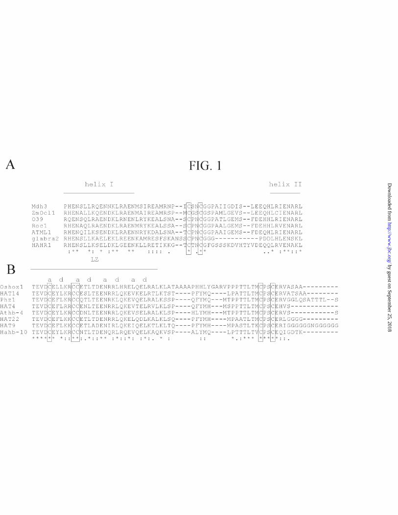
Redox Agents Modulate HARH1 Binding to DNA --We have previously
demonstrated that the sunflower HD protein HAHR1 binds a pseudopalindromic 9-bp
DNA sequence as a dimer (20,42). Amino acid sequence comparisons of HAHR1 with
other proteins from the glabra2 family showed a high conservation along the HD and
dimerization domain, with the sole exception of the disordered loop that separates helix
I and II of the dimerization domain (Fig. 1A). This loop is characterized by the presence
of Gly, Pro and Ser, but is variable in length and defined positions are only poorly
conserved, suggesting that its main role is as a flexible linker between the two helices.
by guest on September 25, 2018
http://ww
w.jbc.org/
Dow
nloaded from

9
Within this context, it is noteworthy that a block that contains two cysteines (CXXCG)
is present within the loop of all glabra2-like proteins described up to now (Fig. 1A).
This conservation is likely an indicator of an essential function of this segment and,
particularly, of the conserved cysteines within it.
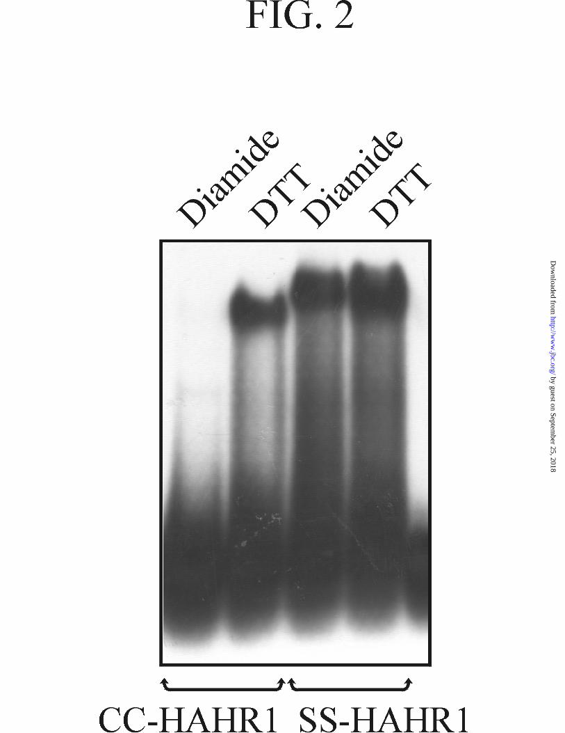
To test the role of the oxidation state of the conserved cysteines on the properties
of HAHR1, we have incubated the recombinant protein in the presence of the oxidant
diamide and the sulfhydryl reductant DTT and analyzed the extent of DNA binding
using EMSA (Fig. 2). Incubation in the presence of DTT produced a marked increase in
DNA binding with respect to samples incubated with diamide, suggesting that cysteines
in the reduced state are required for efficient binding. A similar observation was made
when the binding reactions were subjected to UV photocrosslinking and analyzed by
denaturing polyacrylamide gels (not shown). This indicates that the observed changes in
DNA binding are not due to differential dissociation of the protein-DNA complex
during electrophoresis. Similar differences in binding activity were obtained in the
absence and presence of the non-specific competitor poly(dI-dC). Determination of the
overall dissociation constant of bound dimers into free monomers (K12) yielded values
of 3.10(±0.01) × 10-14 M2 and 1.88(±0.09) × 10-13 M2 for the proteins incubated in the
presence of DTT or diamide, respectively, indicating that the reducing agent produces
an increase in the affinity of the protein for DNA. Kinetic analysis showed that the rate
of association of the complex is increased by incubation with DTT, while the rate of
complex dissociation is not significantly affected (not shown).
When both cysteines present within the dimerization motif were mutated to
serine a protein active under both redox conditions was obtained (Fig. 2). To further
define the requirements for the redox conversion of HAHR1, we have tested the effect
of the redox pair oxidized glutathione (GSSG)/reduced glutathione (GSH). As shown in
by guest on September 25, 2018
http://ww
w.jbc.org/
Dow
nloaded from
10
Fig. 3 (left panel), a significant activation was obtained with GSH indicating that this
compound, which has a lower reduction potential than DTT, is able to reduce HAHR1
cysteines, although less efficiently than DTT. Interestingly, a sample incubated without
any oxidant or reductant showed similar binding activity as those incubated with
oxidants (Fig. 3, left panel), suggesting that the protein is spontaneously oxidized during
dialysis or incubation without reducing agents.
The individual role of each of the two cysteines present in the loop of the
dimerization motif was analyzed by producing proteins with single mutations to serine.
Both proteins were sensitive to oxidation, although to a different extent. CS-HAHR1
was partially active under oxidizing conditions and was activated by GSH, even if DTT
was required for full activation (Fig. 3, right panel). SC-HAHR1, on the contrary,
showed significant binding only in the presence of DTT. The different behavior of the
single mutants may reflect differences in redox potential of the respective cysteines.
Oxidized HAHR1 Forms Intermolecular Disulfide Bonds--The fact that HAHR1
forms dimers in solution suggests that the cysteines present within the loop of the
dimerization motif may form intermolecular disulfide bonds when oxidized. This
possibility was analyzed by performing denaturing polyacrylamide gels under non-
reducing conditions of proteins subjected to different treatments. As shown in Fig. 4
(upper panel), proteins with either one or both cysteines formed species with
significantly reduced mobility, corresponding to dimers, in the absence of reducing
agents. Proteins treated with DTT behaved as monomers, suggesting that they are non-
covalent dimers. Incubation in the presence of oxidizing agents did not enhance the
amount of species with reduced mobility, while GSH was as efficient as DTT as
reducing agent (Fig. 4, middle and lower panel). It should be noted that there is not an
exact correlation of the amount of monomers and DNA binding activity. This may be
by guest on September 25, 2018
http://ww
w.jbc.org/
Dow
nloaded from
11
due to the different pre-treatment of samples (i.e.: samples to be analyzed in SDS-
polyacrylamide gels were boiled before loading to completely disrupt non-covalent
interactions, while samples to be analyzed by mobility shift assays were incubated at
room temperature). Nevertheless, the formation of interchain disulfide bonds between
adjacent monomers seems to be related with the decrease in DNA binding.
The native protein present in sunflower nuclei showed a similar behavior.
Western blots of nuclear extracts incubated in the presence of reductants developed with
anti-HAHR1 antibodies showed a distinct band of about 70 kDa, corresponding to the
monomer (Fig. 5, upper panel). This band was not observed when extracts were
incubated under oxidizing conditions most likely because of the formation of larger
species which barely entered the gel. In fact, reactive bands in the region corresponding
to the stacking gel were evident under these conditions (not shown). The response of
HAHR1 differed from that of other proteins from the nuclear extract, as judged from
Coomassie Blue stained gels (Fig. 5, lower panel). While a major group of proteins
changed their mobility only in the presence of DTT, several minor bands remained
unchanged under all conditions.
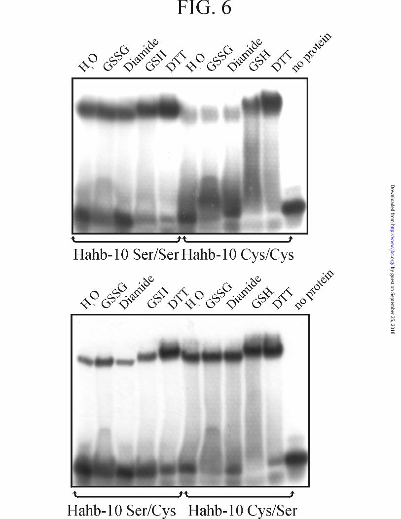
Hahb-10 is Activated by Treatment with DTT--Analysis of conserved cysteines
in other plant HD proteins revealed that in all proteins from the Hd-ZipII subclass
described up to now there is a CPSCE motif adjacent to the leucine zipper (Fig. 1B).
This motif contains two conserved cysteines and resembles the CXXCG motif present
in glabra2 proteins. To determine if Hd-ZipII proteins also undergo redox-dependent
changes, we have expressed the sunflower Hd-ZipII protein Hahb-10 (36) in E. coli and
performed similar studies as those described above for HAHR1. Just as HAHR1, Hahb-
10 binds DNA poorly under oxidizing conditions (Fig. 6, upper panel). Treatment with
DTT produced a significant increase in DNA binding, indicating that the reduction of
by guest on September 25, 2018
http://ww
w.jbc.org/
Dow
nloaded from
12
cysteines is required for its activation. GSH also produced an increase in binding,
although lower than that produced by DTT. Measurement of the dissociation constants
under oxidizing [K12 = 2.1(± 0.1) × 10-13 M2] and reducing [K12 = 1.3(±0.1) × 10-14 M2]
conditions indicated that DTT produces an increase in binding affinity. Kinetic analysis
showed that both the association and dissociation rates of the complex were changed
(i.e.: the association rate was increased and the dissociation rate was decreased in the
presence of DTT). Mutation of both cysteines to serine significantly reduced the
oxidation-dependent decrease in DNA binding (Fig. 6, upper panel). However, the
mutated protein still retained partial sensitivity to oxidizing agents, which is more
evident when DNA binding is analyzed at lower protein concentrations (see below).
This may be caused by the presence of additional cysteines within the leucine zipper of
Hahb-10.
Role of Individual Cysteines in Hahb-10 Redox-mediated Activation--Single
mutations of cysteines in the CPSCE motif of Hahb-10 produced proteins with
intermediate DNA binding activity under oxidizing conditions (Fig. 6, lower panel).
SC-Hahb-10 was less active that CS-Hahb-10 in the presence of oxidants, and was
significantly activated only in the presence of DTT. This indicates that the behavior of
the single cysteine mutants within the CXXCX motif is very similar for HAHR1 and
Hahb-10.
Non-reducing gels indicated the presence of covalently bound species under
oxidizing conditions for all the proteins under study (Fig. 7). Native Hahb-10 formed
species that migrated considerably slower than dimers, which in turn where
predominant in cysteine mutants under oxidizing conditions. Full conversion into
monomers was only attained in the presence of DTT, while GSH was only partially
active (Fig. 7). This shows that there is a close correlation between the formation of
by guest on September 25, 2018
http://ww
w.jbc.org/
Dow
nloaded from

13
intermolecular covalent bonds and the decrease in DNA binding. On comparing these
parameters for the different proteins under oxidizing conditions, however, it becomes
evident that at least part of the proteins that participate in covalent bond formation are
active in DNA binding. This suggests that the formation of certain disulfide bonds
influences binding activity in a different way than the formation of others. Accordingly,
the role of cysteines present in the leucine zipper of Hahb-10 was also investigated.
The portion of Hahb-10 used for these studies has three additional cysteines
within the leucine zipper (Fig. 1B). Two of them are present at a positions (that is,
facing the dimer interface) of the second and third heptads (a2 and a3, respectively),
while the other is at the g position of the second heptad (g2), adjacent to a3 and also near
the interface according to known leucine zipper structures (43). Cysteines a2 and a3 are
conserved in all Hd-ZipII proteins, while cysteine g2 is present in most of them. The
role of these cysteines was studied by the analysis of the properties of proteins with
different combinations of mutated cysteines. The nomenclature used for the different
proteins is presented in Fig. 8A. From the analysis of the DNA binding properties of
mutants (Fig. 8B), the following observations were made: 1) proteins in which cysteines
g2 and a3 are mutated to serine are largely insensitive to redox conditions in terms of
binding activity (Fig. 8B, upper panel); 2) proteins with cysteine at positions g2 and a3
are highly sensitive to oxidation (Fig. 8B, lower panel); 3) proteins in which cysteine a2
is mutated to serine show a significant decrease in binding under all conditions. These
results indicate that adjacent cysteines g2 and a3 are main determinants of the sensitivity
of DNA binding to oxidation, and that cysteines within the CPSCE motif also influence
this behavior. Oxidation of cysteine a2 does not influence DNA binding.
The formation of intermolecular disulfide bonds was analyzed by non-reducing
polyacrylamide gel electrophoresis. As expected, all proteins, with the sole exception of
by guest on September 25, 2018
http://ww
w.jbc.org/
Dow
nloaded from

14
the all-serine mutant, produced cross-linked species under oxidizing conditions (Fig. 9).
SSSCC-Hahb-10 also showed a significant proportion of monomers (lower panel),
which were not observed for the other proteins. Cysteines within the leucine zipper are
then probably responsible for the formation of cross-linked species. CSSSS-Hahb-10
forms dimers under oxidizing conditions (Fig. 9, middle panel) suggesting that cysteines
a2 from two adjacent monomers form cross-linked active dimers. Conversely, covalent
dimers formed by SCCSS-Hahb-10 (upper panel), seem to be inactive. This confirms
that disulfide bond formation may lead to either active or inactive proteins depending on
the nature of the cysteines that are involved.
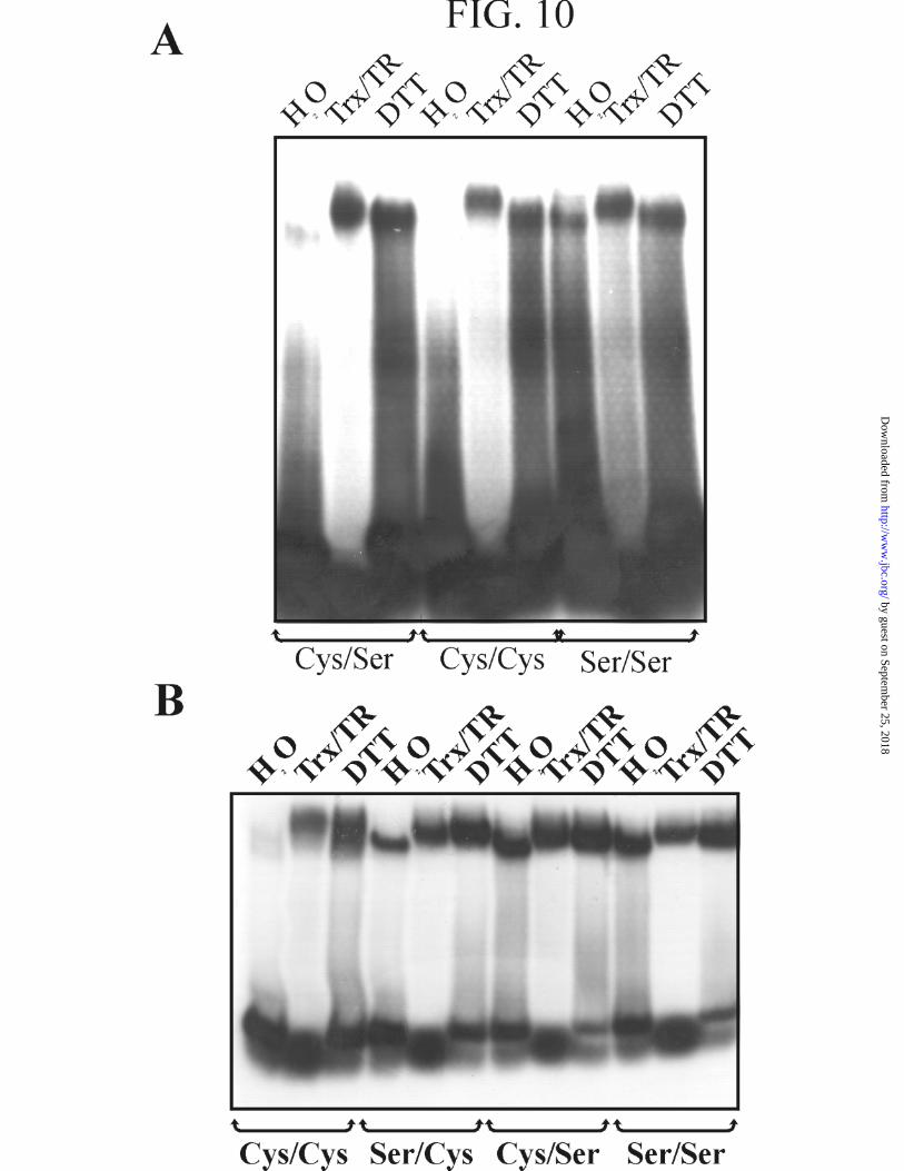
Reduced Thioredoxin Catalyzes the Activation of HAHR1 and Hahb-10--The
fact that the reduction of disulfide bonds promotes the activation of HAHR1 and Hahb-
10 raises the possibility that a similar reaction is used under physiological conditions to
regulate the properties of these transcription factors. Redox changes in protein cysteines
are usually catalyzed in vivo by thioredoxin, a small protein that is in turn reduced by
NADPH in the presence of thioredoxin reductase (28,44). We have then analyzed the
thioredoxin-dependent activation of HAHR1 and Hahb-10, and their respective mutants,
using recombinant thioredoxin and thioredoxin reductase purified from E. coli. As
shown in Fig. 10, this system was as efficient as DTT in the activation of HAHR1 and
Hahb-10. This is consistent with a role of thioredoxin in catalyzing thiol/disulfide
exchanges in these transcription factors in vivo.
DISCUSSION
Post-translational modifications are extensively used by cells to modulate the
activity of proteins. Among these modifications, dithiol/disulfide exchanges are key
components of regulatory systems that respond to changes in redox conditions (28,44-
by guest on September 25, 2018
http://ww
w.jbc.org/
Dow
nloaded from
15
46). In the present work, we show that two plant HD transcription factors undergo
dithiol/disulfide exchanges that produce changes in their affinity for their DNA target
sequences. One of these factors, HAHR1, belongs to the glabra2 family which members
are thought to participate in epidermal cell development both at the embryo and adult
plant level (22-26). Since the cysteines involved in redox modulation (i.e.: those present
in the loop of the dimerization motif) are conserved in all members of the glabra2
family, we propose that all glabra2-like plant HD proteins undergo similar changes.
This also applies to Hahb-10 and Hd-ZipII proteins, which possess a set of conserved
cysteines in and outside the leucine zipper motif.
Although both proteins tested are activated in the presence of reductants, they
show a different response to the cellular redox agent GSH than to DTT. The first of
these compounds produces only a partial activation of HAHR1 and Hahb-10, probably
because only part of the disulfide bonds are reduced. Full activation requires the action
of more potent reductants. In vivo, GSH itself participates in the reduction of proteins,
sometimes with the aid of a protein called glutaredoxin (45). A different system, with
higher reducing potential, is composed by thioredoxin, a protein that is in turn reduced
by NADPH in a reaction catalyzed by NADPH-thioredoxin reductase (28,44-46). Our
results using purified proteins in vitro indicate that this system is able to promote full
activation of HAHR1 and Hahb-10. We propose, then, that the intracellular levels of
GSH and reduced thioredoxin operate in concert to influence the activation state of the
proteins under study.
Regarding the physiological significance of our observations, it should be
mentioned that redox agents are known to influence several aspects of development in
plants. Root growth, root hair number and root hair length respond to the inclusion of
redox agents in the growth medium (47,48), and mutants in the enzyme γ-
by guest on September 25, 2018
http://ww
w.jbc.org/
Dow
nloaded from
16
glutamylcysteine synthetase, involved in the systhesis of GSH, show altered root
development and reduced cell division rates (49). Root hair development establishes a
link between GSH and glabra2 proteins. Mutations in glabra2 produce an increase in
root hair number, suggesting that the encoded protein is involved in repressing root hair
formation (21,23). If GSH acts through activation of glabra2, then the inclusion of this
redox agent should promote a decrease in root hair number. Previous studies indicate,
however, that GSH has the opposite effect (48). While these results are difficult to
concile, it should be mentioned that a recent report indicates that the overexpression of
glabra2 produces plants similar in phenotype than glabra2 mutants (50). An increase in
glabra2 activity over a certain threshold may then produce an increase in root hair
number.
Considering Hd-ZipII proteins, current knowledge indicates that some of these
proteins are involved in certain developmental responses of plants to illumination. The
genes for two Arabidopsis Hd-ZipII proteins, Athb-2 and -4, are induced by far-red-rich
light (18), and changes in the expression levels of HAT4 or Athb-2 (which are the same
protein) influence hypocotyl elongation and leaf expansion, mimicking certain effects of
illumination (19,51). Then, also in this case there is a link between environmental
conditions and the modification of plant development. We propose that part of these
modifications are brought about by environmentally-induced changes in cellular redox
agents that in turn modulate the activity of transcription factors of the type reported in
this study.
Regarding the nature of the structural modifications that render these proteins
inactive under oxidation conditions, it is noteworthy that the residues that participate in
this process are located outside the DNA binding motif. For Hahb-10, it can be
envisaged that intermolecular cross-linking of residues within the leucine zipper may
by guest on September 25, 2018
http://ww
w.jbc.org/
Dow
nloaded from

17
affect the correct positioning of monomers for DNA binding. In the crystal structure of
the GCN4 leucine zipper, the side chains of residues at position a lie in close proximity
(Fig. 11). In addition, contacts between residue g from one chain and residue a from the
following heptad of the other chain have been shown to occur (43). From our results, it
is evident that a2 residues form intermolecular cross-links that do not affect DNA
binding by Hahb-10. Cross-links involving g2 and a3, on the contrary, seem to produce
inactive dimers, probably because they cause a distortion in the leucine zipper structure.
Our results also suggest that cysteines located within more disordered regions of
HAHR1 and Hahb-10 also influence DNA binding. Knowledge of the changes brought
about by redox transitions within these regions will require detailed structural studies of
these proteins.
Acknowledgments--We gratefully acknowledge Drs. Scott B. Mulrooney
(Michigan State University) and Charles H. Williams, Jr. (Michigan University) for the
generous gift of plasmids expressing thioredoxin and thioredoxin reductase.
by guest on September 25, 2018
http://ww
w.jbc.org/
Dow
nloaded from
18
REFERENCES
1. Gehring, W. J., Affolter, M., and Bürglin, T. (1994) Annu. Rev. Biochem. 63,
487-526
2. Levine, M., and Hoey, T. (1988) Cell 55, 537-540
3. Gehring, W. J. (1987) Science 236, 1245-1252
4. Qian, Y. Q., Billeter, M., Otting, G., Müller, M., Gehring, W. J., and Wüthrich,
K. (1989) Cell 59, 573-580
5. Qian, Y. K., Furukubo-Tokunaga, K., Müller, M., Resendez-Perez, D., Gehring,
W. J., and Wüthrich, K. (1994) J. Mol. Biol. 238, 333-345
6. Tsao, D. H. H., Gruschus, J. M., Wang, L., Nirenberg, M., and Ferretti, J. A.
(1995) J. Mol. Biol. 251, 297-307
7. Kissinger, C. R., Liu, B., Martin-Blanco, E., Kornberg, T. B., and Pabo, C. O.
(1990) Cell 63, 579-590
8. Otting, G., Qian, Y. Q., Billeter, M., Müller, M., Affolter, M., Gehring, W. J.,
and Wüthrich, K. (1990) EMBO J. 9, 3085-3092
9. Gehring, W. J., Qian, Y. Q., Billeter, M., Furukubo-Tokunaga, K., Schier, A. F.,
Resendez-Perez, D., Affolter, M., Otting, G., and Wüthrich, K. (1994) Cell 78,
211-223
10. Wolberger, C., Vershon, A. K., Liu, B., Johnson, A. D., and Pabo, C. O. (1991)
Cell 67, 517-528
11. Otting, G., Qian, Y. Q., Müller, M., Affolter, M., Gehring, W. J., and Wüthrich,
K. (1988) EMBO J. 7, 4305-4309
12. Affolter, M., Percival-Smith, A., Müller, M., Leupin, W., and Gehring, W. J.
(1990) Proc. Natl. Acad. Sci. USA 87, 4093-4097
by guest on September 25, 2018
http://ww
w.jbc.org/
Dow
nloaded from

19
13. Chan, R. L., Gago, G. M., Palena, C. M., and Gonzalez, D. H. (1998) Biochim.
Biophys. Acta 1442, 1-19
14. Ruberti, I., Sessa, G., Lucchetti, S., and Morelli, G. (1991) EMBO J. 10, 1787-
1791
15. Schena, M., and Davis, R. W. (1992) Proc. Natl. Acad. Sci. USA 89, 3894-3898
16. Sessa, G., Morelli, G., and Ruberti, I. (1993) EMBO J. 12, 3507-3517
17. Palena, C. M., Gonzalez, D. H., and Chan, R. L. (1999) Biochem. J. 341, 81-87
18. Carabelli, M., Sessa, G., Baima, S., Morelli, G., and Ruberti, I. (1993) Plant J. 4,
469-479
19. Schena, M., Lloyd, A. M., and Davis, R. W. (1993) Genes Dev. 7, 367-379
20. Palena, C. M., Chan, R. L., and Gonzalez, D. H. (1997) Biochim. Biophys. Acta
1352, 203-212
21. Di Cristina, M., Sessa, G., Dolan, L., Linstead, P., Baima, S., Ruberti, I., and
Morelli, G. (1996) Plant J. 10, 393-402
22. Lu, P., Porat, R., Nadeau, J. A., and O'Neill, S. D. (1996) Plant Cell 8, 2155-
2168
23. Masucci, J. D., Rerie, W. G., Foreman, D. R., Zhang, M., Galway, M. E., Marks,
M. D., and Schiefelbein, J. W. (1996) Development 122, 1253-1260
24. Hung, C.-Y., Lin, Y., Zhang, M., Pollock, S., Marks, M. D., and Schiefelbein, J.
(1998) Plant Physiol. 117, 73-84
25. Ingram, G. C., Magnard, J.-L., Vergne, P., Dumas, C., and Rogowsky, P. M.
(1999) Plant Mol. Biol. 40, 343-354
26. Rerie, W. G., Feldmann, K. A., and Marks, M. D. (1994) Genes Dev. 8, 1388-
1399
by guest on September 25, 2018
http://ww
w.jbc.org/
Dow
nloaded from
20
27. Akamatsu, Y., Ohno, T., Hirota, K., Kagoshima, H., Yodoi, J., and Shigesada,
K. (1997) J. Biol. Chem. 272, 14497-14500
28. Arnér, E. S. J., and Holmgren, A. (2000) Eur. J. Biochem. 267, 6102-6109
29. Hayashi, T., Ueno, Y., and Okamoto, T. (1993) J. Biol. Chem. 268, 11380-
11388
30. Hirota, K., Matsui, M., Iwata, S., Nishiyama, A., Mori, K., and Yodoi, J. (1997)
Proc. Natl. Acad. Sci. USA 94, 3633-3638
31. Makino, Y., Yoshikawa, N., Okamoto, K., Hirota, K., Yodoi, J., Makino, I., and
Tanaka, H. (1999) J. Biol. Chem. 274, 3182-3188
32. Nakshatri, H., Bhat-Nakshatri, P., and Currie, R. A. (1996) J. Biol. Chem. 271,
28784-28791
33. Schenk, H., Klein, M., Erdbrugger, W., Droge, W., and Schulze-Osthoff, K.
(1994) Proc. Natl. Acad. Sci. USA 91, 1672-1676
34. Valle, E. M., Gonzalez, D. H., Gago, G., and Chan, R. L. (1997) Gene 196, 61-
68
35. Palena, C. M., Gonzalez, D. H., Guelman, S., and Chan, R. L. (1998) Protein
Expr. Purif. 13, 97-103
36. Gonzalez, D. H., Valle, E. M., Gago, G. M., and Chan, R. L. (1997) Biochim.
Biophys. Acta 1351, 137-149
37. Smith, D. B., and Johnson, K. S. (1988) Gene 67, 31-40
38. Sedmak, J., and Grossberg, S. (1977) Anal. Biochem. 79, 544-552
39. Mulrooney, S. B. (1997) Protein Expr. Purif. 9, 372-378
40. Laemmli, U. K. (1970) Nature 227, 680-685
by guest on September 25, 2018
http://ww
w.jbc.org/
Dow
nloaded from

21
41. Maliga, P., Klessig, D. F., Cashmore, A. R., Gruissem, W., and Varner, J. E.
(1995) in Methods in Plant Molecular Biology. A Laboratory Course Manual,
pp. 233-260, Cold Spring Harbor Laboratory Press, New York
42. Tron, A. E., Bertoncini, C. W., Palena, C. M., Chan, R. L., and Gonzalez, D. H.
(2001) Nucleic Acids Res. 29, 4866-4872
43. O'Shea, E. K., Klemm, J. D., Kim, P. S., and Alber, T. (1991) Science 254, 539-
544
44. Buchanan, B. B., Schurmann, P., Decottignies, P., and Lozano, R. M. (1994)
Arch. Biochem. Biophys. 314, 257-260
45. Holmgren, A. (1989) J. Biol. Chem. 264, 13963-13966
46. Holmgren, A. (1985) Annu. Rev. Biochem. 54, 237-271
47. May, M. J., Vernoux, T., Leaver, C. J., Van Montagu, M., and Inzé, D. (1998) J.
Exp. Bot. 49, 649-667
48. Sánchez-Fernández, R., Fricker, M., Corben, L. B., White, N. S., Sheard, N.,
Leaver, C. J., Van Montagu, M., Inzé, D., and May, M. J. (1997) Proc. Natl.
Acad. Sci. USA 94, 2745-2750
49. Vernoux, T., Wilson, R. C., Seeley, K. A., Reichheld, J.-P., Muroy, S., Brown,
S., Maughan, S. C., Cobbett, C. S., Van Montagu, M., Inzé, D., May, M. J., and
Sung, Z. R. (2000) Plant Cell 12, 97-109
50. Ohashi, Y., Oka, A., Ruberti, I., Morelli, G., and Aoyama, T. (2002) Plant J. 29,
359-369
51. Steindler, C., Matteucci, A., Sessa, G., Weimar, T., Ohgishi, M., Aoyama, T.,
Morelli, G., and Ruberti, I. (1999) Development 126, 4235-4245
52. Thompson, J. D., Higgins, D. G., and Gibson, T. J. (1994) Nucleic Acids Res. 22,
4673-4680
by guest on September 25, 2018
http://ww
w.jbc.org/
Dow
nloaded from

22
Footnotes:
* This work was supported by grants from CONICET, ANPCyT, Universidad Nacional
del Litoral and Fundación Antorchas (Argentina). RLC and DHG are members of
CONICET; AET is a fellow of the same Institution. CWB was an undergraduate fellow
of Universidad Nacional del Litoral.
1 The abbreviations used are: HD, homeodomain; DTT, dithiothreitol; EMSA,
electrophoretic mobility shift assay; GSSG, oxidized glutathione; GSH, reduced
glutathione.
by guest on September 25, 2018
http://ww
w.jbc.org/
Dow
nloaded from

23
Figure Legends
FIG. 1. Conserved cysteines at or near the dimerization domain of plant HD
proteins. Full-length protein sequences were aligned using Clustal W (52). Conserved
cysteines are boxed. Identical amino acids at a defined position are denoted by asterisks.
Double-dots indicate similar amino acids. A, Amino acid sequence alignment of a
region of the dimerization motif of seven glabra2-like proteins: Malus x domestica
Mdh3 (acc. no. AAC79430), Zea mays ZmOcl1 (acc. no. CAB51059), Phalaenopsis sp.
O39 (acc. no. AAB37230), Oryza sativa Roc1 (acc. no. BAB85750), Arabidopsis
thaliana ATML1 and glabra2 (acc. no. T05850 and P46607), and Helianthus annuus
HAHR1 (acc. no. AAC37514). A detail of the alignment, comprising helix I, the loop,
and helix II, is shown. B, The leucine zipper (LZ) and adjacent regions of eight Hd-ZipII
proteins: Oryza sativa Oshox1 (acc. no. AAF19980), Arabidopsis thaliana HAT14,
HAT4, Athb-4, HAT22 and HAT9 (acc. no. CAD24012, CAA79670, AAC31833,
T06026 and P46603), Pimpinella brachycarpa Phz1 (acc. no. CAA64491), and
Helianthus annuus Hahb-10 (acc. no. AAA79778); a and d indicate the first and fourth
residue of each heptad, respectively.
FIG. 2. Redox changes modulate HAHR1 DNA binding activity. EMSA of
wild-type and its double Cys-to-Ser mutant (CC-HAHR1 and SS-HAHR1, respectively).
Proteins were incubated in the presence of either 10 mM diamide or 25 mM DTT during
1 h at room temperature before performing the DNA binding assay.
FIG. 3. Effect of different redox agents on the DNA binding activity of
HAHR1 and its cysteine substitution mutants. EMSA of wild-type HAHR1
by guest on September 25, 2018
http://ww
w.jbc.org/
Dow
nloaded from

24
(Cys/Cys) and its single and double Cys-to-Ser mutants (Cys/Ser, Cys-188→Ser;
Ser/Cys, Cys-185→Ser; Ser/Ser, double mutant) previously incubated in the presence of
25 mM GSSG, 10 mM diamide, 25 mM GSH, 25 mM DTT, or in the absence of redox
agents (H2O) as indicated in Fig. 2.
FIG. 4. Non-reducing polyacrylamide gel of HAHR1 and its cysteine
mutants incubated in the presence of various redox agents. The different proteins
were incubated under the conditions described in Fig. 3 before the addition of sample
buffer and electrophoretic separation. Arrows indicate the position of monomers, dimers
and higher order structures according to standard molecular weight markers shown in
lane M of the upper panel.
FIG. 5. Native HAHR1 in nuclear extracts undergoes redox-dependent
changes in electrophoretic migration. Aliquots of a sunflower nuclear protein extract
were incubated in the presence of redox agents as described in Fig. 3, subjected to non-
reducing polyacrylamide gel electrophoresis, and either analyzed with anti-HAHR1
antibodies by Western blot (upper panel) or stained with Coomassie Brilliant Blue
(lower panel). A sample of recombinant HAHR1 (GST-HAHR1), incubated in the
presence of DTT, was included as control.
FIG. 6. Effect of different redox agents on the DNA binding activity of
Hahb-10 and its cysteine mutants within the CPSCE motif. EMSA of wild-type
Hahb-10 (Cys/Cys) and single and double Cys-to-Ser mutants (Ser/Cys, Cys-201→Ser;
Cys/Ser, Cys-204→Ser; Ser/Ser, double mutant) previously incubated in the presence of
redox agents as indicated in Fig. 3.
by guest on September 25, 2018
http://ww
w.jbc.org/
Dow
nloaded from

25
FIG. 7. Non-reducing polyacrylamide gel of Hahb-10 and its cysteine
mutants incubated in the presence of various redox agents. The different proteins
were incubated under the conditions described in Fig. 3 before the addition of sample
buffer and electrophoretic separation. Arrows indicate the position of monomers, dimers
and higher order structures according to standard molecular weight markers shown in
lane M of the upper panel. See the legend of Fig. 6 for the designation of the mutants.
FIG. 8. Role of cysteines at different positions in the redox-dependent
activation of Hahb-10. A, Scheme showing the relative positions of the cysteines in
Hahb-10 and the nomenclature of the different mutants analyzed in this study. The
amino acid position of each cysteine, relative to the initial methionine, and the position
within the leucine zipper (a2, g2, or a3), are shown. Regions corresponding to the HD
and the leucine zipper (LZ) are boxed. Below, the name and amino acids present at each
position are shown. B, Effect of incubation with DTT on the DNA binding activity of
Hahb-10 and its mutants. The different proteins were incubated in the presence or
absence of 25 mM DTT as described before analyzing DNA binding by EMSA. See
part A for the nomenclature used for the different proteins.
FIG. 9. Formation of intermolecular disulfide bonds by Hahb-10 cysteine
mutants under different redox conditions. The different proteins were incubated
under the conditions described in Fig. 3 before the addition of sample buffer and
electrophoretic separation on a non-reducing polyacrylamide gel. Arrows indicate the
position of monomers, dimers and higher order structures according to standard
molecular weight markers. See Fig. 8A for nomenclature of the proteins.
by guest on September 25, 2018
http://ww
w.jbc.org/
Dow
nloaded from

26
FIG. 10. Thioredoxin can replace DTT in the redox activation of HAHR1
and Hahb-10. A, EMSA of wild-type HAHR1 (Cys/Cys) and its single and double Cys-
to-Ser mutants (Cys/Ser, Cys-188→Ser; Ser/Ser, Cys-185/188→Ser double mutant)
previously incubated in the presence of 0.013 µg/µl each thioredoxin and thioredoxin
reductase plus 0.55 mM NADPH (Trx/TR), 25 mM DTT, or in the absence of redox
agents (H2O) as indicated in Fig. 2. B, Same as in A for wild-type (Cys/Cys) and
mutants of Hahb-10 within the CPSCE motif (Ser/Cys, Cys-201→Ser; Cys/Ser, Cys-
204→Ser; Ser/Ser, double mutant).
FIG. 11. Proximity of amino acids at a and g positions in the GCN4 leucine
zipper. The picture, based on the crystal structure of the GCN4 leucine zipper (43) and
generated using the program RasMol, shows the backbone of the two strands of the
leucine zipper coiled coil together with the side chains of residues at positions a2, a3 and
g2. These positions are occupied by cysteines in the Hahb-10 leucine zipper.
by guest on September 25, 2018
http://ww
w.jbc.org/
Dow
nloaded from
Adriana E. Tron, Carlos W. Bertoncini, Raquel L. Chan and Daniel H. GonzalezRedox regulation of plant homeodomain transcription factors
published online July 1, 2002J. Biol. Chem.
10.1074/jbc.M203297200Access the most updated version of this article at doi:
Alerts:
When a correction for this article is posted•
When this article is cited•
to choose from all of JBC's e-mail alertsClick here
by guest on September 25, 2018
http://ww
w.jbc.org/
Dow
nloaded from


![Programa de Interpretación Ambiental en el Paraje Natural Municipal Los Calderones. Chulilla[1]](https://img.pdfslide.us/doc/110x75/568c4c931a28ab4916a0b2bf/programa-de-interpretacion-ambiental-en-el-paraje-natural-municipal-los-calderones.jpg)