Embed Size (px)
Citation preview
Tattoo-Associated Uveitis
TRUCIAN A. OSTHEIMER, BRYN M. BURKHOLDER, THERESA G. LEUNG, NICHOLAS J. BUTLER,JAMES P. DUNN, AND JENNIFER E. THORNE
� PURPOSE: To describe the clinical presentation of uve-itis with coincident onset of raised and indurated tattooedskin.� DESIGN: Case series.� METHODS: Seven consecutive patients were evaluatedat a tertiary ophthalmologic facility with coincident uve-itis and cutaneous tattoo induration over an 18-monthperiod. All subjects underwent complete ophthalmicexamination and a focused systemic medical evaluationincluding serologic testing and imaging studies. Two par-ticipants underwent biopsy of their tattoos. The patients’clinical courses and responses to treatment over a follow-up period of 1–20 months are reported (mean follow-up[ 9months).Main outcomemeasures included degreeof intraocular inflammation, ocular complications, visualacuity, clinically observable tattooed skin changes, andbiopsy results.� RESULTS: Five of 7 patients had bilateral nongranulom-atous anterior uveitis: 4 with chronic and 1 with recurrentdisease. The remaining 2 patients had bilateral chronicgranulomatous panuveitis. Biopsies of raised and induratedtattoos were performed in 2 patients and demonstratednoncaseating granulomatous inflammation surroundingtattoo ink in the dermis. The skin changes resolved in allpatients, with a faster response noted in those treatedwith high-dose oral prednisone for intraocular inflamma-tion. Five patients subsequently experienced recurrentflares of intraocular inflammation in conjunction withthe recurrence of raised and indurated tattoos.� CONCLUSIONS: These cases represent a subset ofpatients in whom skin tattooing may have incited animmune response leading to simultaneous inflammationof the eyes and tattooed skin. (Am J Ophthalmol2014;158:637–643. � 2014 by Elsevier Inc. All rightsreserved.)
IN 1952, LUBECK AND EPSTEIN PUBLISHED THE FIRST
report of a patient with bilateral intraocular inflamma-tion and simultaneous tattoo granulomas in the setting
Accepted for publication May 16, 2014.From Wilmer Eye Institute, Johns Hopkins University School of
Medicine (T.A.O., B.M.B., T.G.L., N.J.B., J.P.D., J.E.T.); andDepartment of Epidemiology, Bloomberg School of Public Health, JohnsHopkins University (J.E.T.), Baltimore, Maryland.
James P. Dunn is currently employed at the Wills Eye Institute, Phila-delphia, Pennsylvania.
Inquiries to Trucian A. Ostheimer, Wilmer Eye Institute, 600 NWolfeSt, Woods 476, Baltimore, MD 21287; e-mail: [email protected]
0002-9394/$36.00http://dx.doi.org/10.1016/j.ajo.2014.05.019
� 2014 BY ELSEVIER INC.
of systemic sarcoidosis.1 This was followed, in 1969, by thefirst case series to describe bilateral intraocular inflamma-tion with the simultaneous development of tattoo granu-lomas in 3 patients felt to have no evidence of systemicsarcoidosis at the time of presentation.2 The pathologichallmark of sarcoidosis is the noncaseating granuloma;however, it remains a diagnosis of exclusion because of itslack of pathognomonic histopathology, imaging, or sero-logic studies.3 Among patients with sarcoidosis, anywherefrom 25% to 80% may suffer from ocular or adnexalinvolvement,4 and approximately 25%–35% of patientsdevelop cutaneous findings.3 Anterior uveitis is the mostcommon ocular manifestation of sarcoidosis, occurring in65% of patients with ophthalmic involvement.3
We present 7 patients with no prior diagnosis of sarcoid-osis who developed bilateral uveitis in temporal associationwith inflammation of tattooed skin.
METHODS
A RETROSPECTIVE REVIEW OF 7 CONSECUTIVE PATIENTS
with bilateral uveitis and associated cutaneous changes sug-gestive of tattoo inflammation evaluated over a 20-monthperiod was conducted at the Division of Ocular Immu-nology, Wilmer Eye Institute. The study was approved bythe Johns Hopkins School of Medicine InstitutionalReview Board and adhered to all tenets of the Declarationof Helsinki. All patient data were handled in accordancewith the Health Information Portability and Account-ability Act.All patients underwent a complete ophthalmologic
examination and received a medical evaluation (Table)in an attempt to rule out syphilis (fluorescent treponemalantibody-absorption [FTA-ABS] and rapid plasma reagintesting [RPR]) and sarcoidosis (chest x-ray and/orcomputed tomography [CT] chest, serum angiotensin-converting enzyme [ACE] and/or serum lysozyme). Testingfor HLA-B27 positivity and infectious etiologies such asMycobacterium tuberculosis, Toxoplasma gondii, Bartonellahenselae, and Borrelia burdorferi was performed in selectedpatients. Two of the 7 patients underwent biopsy of theirinflamed tattoos. The patients’ clinical courses andresponses to treatment were reviewed over a follow-upperiod of 1–20 months.
� SELECTEDCASEREPORT: PATIENT 1: Patient 1 was a 20-year-old African-American man who initially presented for
637ALL RIGHTS RESERVED.
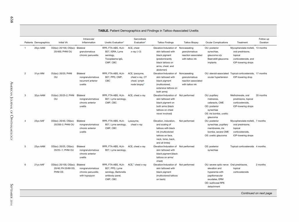
TABLE. Patient Demographics and Findings in Tattoo-Associated Uveitis
Patients Demographics Initial VA
Intraocular
Inflammation Uveitic EvaluationaSarcoidosis
Evaluationa Tattoo Findings Tattoo Biopsy Ocular Complications Treatment
Follow-up
Duration
1 20yo AAM OD(sc): 20/100, OS(sc):
20/400; PHNI OU
Bilateral
granulomatous
chronic panuveitis
RPR, FTA-ABS, HLA-
B27, IGRA, Lyme
serology,
Toxoplasma IgG,
CMP, CBC
ACE, chest
x-ray (32)
Elevation/induration of
skin tattooed with
black pigment
(predominantly
black tattoos on
arms, chest, and
abdomen)
Noncaseating
granulomatous
reaction associated
with tattoo ink
OU: posterior
synechiae,
glaucoma s/p
Baerveldt glaucoma
implants
Mycophenolate mofetil,
oral prednisone,
topical
corticosteroids, and
IOP-lowering drops
13 months
2 31yo WM OU(sc): 20/25; PHNI
OU
Bilateral
nongranulomatous
recurrent anterior
uveitis
RPR, FTA-ABS, HLA-
B27, PPD, CMP,
CBC
ACE, lysozyme,
chest x-ray, CT
chest, lymph
node biopsyb
Elevation/induration of
skin tattooed with
black pigment
(multicolored,
extensive tattoos on
both arms)
Noncaseating
granulomatous
reaction associated
with tattoo ink
OU: steroid-associated
ocular hypertension
Topical corticosteroids,
IOP-lowering drop
17 months
3 32yo AAM OU(sc): 20/25-2; PHNI
OU
Bilateral
nongranulomatous
chronic anterior
uveitis
RPR, FTA-ABS, HLA-
B27, Lyme serology,
CMP, CBC
ACE, chest x-ray Elevation/induration of
skin tattooed with
black pigment on
both arms (black
tattoos on chest
never involved)
Not performed OU: pupillary
mebranes,
cataracts, CME
OD: posterior
synechiae
OS: iris bombe, uveitic
glaucoma
Methotrexate, oral
prednisone, topical
corticosteroids,
IOP-lowering drops
20 months
4 23yo AAF OD(sc): 20/40, OS(sc):
20/200-2; PHNI OU
Bilateral
nongranulomatous
chronic anterior
uveitis
RPR, FTA-ABS, HLA-
B27, Lyme serology,
CMP, CBC
Lysozyme,
chest x-ray
Elevation, induration,
and scaling of
tattoos with black
ink (multicolored
tattoos on face,
neck, torso, back,
and all limbs)
Not performed OU: posterior
synechiae, pupillary
membranes, iris
bombe, severe CME
OS: uveitic glaucoma
Mycophenolate mofetil,
oral prednisone,
topical
corticosteroids,
IOP-lowering drops
7 months
5 23yo AAM OD(sc): 20/25, OS(sc):
20/20þ1; PHNI OU
Bilateral
nongranulomatous
chronic anterior
uveitis
RPR, FTA-ABS, HLA-
B27, Lyme serology,
ACE, chest x-ray Elevation/induration of
skin tattooed with
black pigment (black
tattoos on arms/
chest)
Not performed OS: posterior
synechiae
Topical corticosteroids 4 months
6 21yo AAF OD(sc): 20/100, OS(sc):
20/40; PH 20/80 OD,
PHNI OS
Bilateral
nongranulomatous
chronic panuveitis
with hypopyon
RPR, FTA-ABS, HLA-
B27, PPD, Lyme
serology, Bartonella
antibody panel,
CMP, CBC
ACE,b chest x-ray Elevation/induration of
skin tattooed with
black pigment
(multicolored tattoos
on back)
Not performed OU: severe optic nerve
elevation and
hyperemia with
papillomacular
exudates, ERM
OD: subfoveal RPE
detachment
Oral prednisone,
topical
corticosteroids
2 months
Continued on next page
638
SEP
TEMBER
2014
AMER
ICANJO
URNALOFO
PHTHALM
OLO
GY
TABLE.PatientDemographicsandFindingsin
Tattoo-A
ssociatedUveitis(Continued)
Patients
Demographics
InitialV
A
Intraocular
Inflammation
Uveitic
Evaluationa
Sarcoidosis
Evaluationa
TattooFindings
TattooBiopsy
OcularComplications
Treatm
ent
Follow-up
Duration
742yoAAM
OD(sc):20/20,OS(sc):
20/40;PHNIOD,PH
OS20/32
Bilateral
nongranulomatous
chronic
anterior
uveitis
FTA-A
BS,HLA-B
27,
CMP,CBC
ACE,lysozyme,b
chestx-ray,CT
chest
Elevation/indurationof
skin
tattooedwith
blackpigment(arm
)
Notperform
ed
OU:posterior
synechiae
OD:neurosensory
retinaldetachment
OS:severe
CME
Oralprednisone;
systemic
immunosuppression
recommended
1month
(lostto
follo
w-up)
AAF¼African-A
mericanfemale;AAM
¼African-A
mericanmale;ACE¼serum
angiotensin-convertingenzyme;CBC¼complete
bloodcount;CME¼cystoid
macularedema;CMP¼
complete
metabolic
panelincludingliverfunctiontesting;C
T¼computedtomography;E
RM
¼epiretinalm
embrane;FTA-A
BS¼syphilisfluorescenttreponemalantibody-absorption;IgG¼im
munoglobulin
G;IG
RA¼
interferongammareleaseassay(Q
uantiFERON–TBGold);IO
P¼
intraocularpressure;PH¼
visuala
cuitymeasuredwithpinhole
occluder;PHNI¼
visuala
cuitymeasuredwithpinhole
occluderofferednoim
provementinvision;P
PD¼tuberculosispurifiedproteinderivativeskintesting;R
PE¼retinalp
igmentepithelium;R
PR¼syphilisrapid
plasmaregain;sc¼withoutcorrection;
VA¼
visualacuity;WM
¼whitemale;yo¼
yearold.
aAllresultsunremarkable/negativeunlessotherw
iseindicated.
bPatient2underw
entbiopsyofanenlargedaxillary
lymphnode,whichdisplayedanoncaseatinggranulomatousreaction.Patient6hadanelevatedACEva
lueof85(referencerange:9–67U/L).
Patient7hadanelevatedlysozymevalueof32(referencerange:9–17mg/m
L)andanorm
alserum
ACEvalueof47.
VOL. 158, NO. 3 TATTOO-ASSOCIA
evaluation of a 1-week history of blurred vision, photo-phobia, and pain in both eyes. He had experienced similarsymptoms 6 months earlier, which lasted approximately1 month before spontaneously resolving. Review of systemswas notable for elevation and swelling of 8 tattoos on hisarms and chest that occurred in conjunction with his ocularsymptoms on both occasions (Figure 1). All of his tattooswere performed during a 1-year period, approximately6 months prior to his initial ocular complaints.Upon presentation, best-corrected visual acuity
(BCVA) was 20/100 in the right eye and 20/400 in theleft eye, with an intraocular pressure (IOP) of 11 mm Hgin the right eye and 10 mm Hg in the left eye. Slit-lampexamination revealed diffuse conjunctival injection, densemutton-fat keratic precipitates with overlying cornealmicrocystic edema, and posterior synechiae bilaterally.Both eyes displayed anterior chamber inflammation of 3þcells and 1þ flare, with 2þ anterior vitreous cells. Bothoptic nerves appeared hyperemic and edematous, but amore detailed posterior segment examination, includingan assessment of vitreous haze, was limited by bilateralcorneal edema and posterior synechiae.An initial evaluation, consisting of FTA-ABS, RPR,
QuantiFERON-TB Gold, chest x-ray, and Lyme antibody,was unrevealing. He was initially treated with intensivetopical corticosteroids and cycloplegic drops. Three daysafter presentation, BCVA improved to 20/50 in the righteye and 20/60 in the left eye, with an IOP of 20 mm Hgin the right eye and 22 mm Hg in the left eye. However,his intraocular inflammation persisted and high-dose(1 mg/kg/day) oral prednisone therapy was initiated. Thebilateral anterior chamber inflammation and elevated tat-toos resolved over the course of 3 weeks; however, hisIOP became elevated at this time (33 mm Hg in the righteye and 27 mm Hg in the left eye). Topical IOP-loweringtherapy was initiated in a stepwise manner, which escalatedto maximal topical therapy. Topical prednisolone acetate1% was changed to loteprednol etabonate 0.5% approxi-mately 8 weeks after presentation in an attempt to mini-mize any steroid-response component; however, oralacetazolamide was necessary. A prolonged attempt to taperhis oral prednisone led to a recurrence of intraocularinflammation and tattoo elevation, and 3 months afterhis initial presentation he was referred for dermatologicevaluation. His oral prednisone dose was tapered from40 mg/day to 30 mg/day 6 days prior to this dermatologyappointment, and a biopsy was taken from an area of previ-ously affected skin on his chest. Histologic sections of thisbiopsy displayed macrophages in clusters around the dermalsuperficial vascular plexus that contained tattoo pigmentwith surrounding granulomatous inflammation. A repeatchest x-ray, comprehensive chemistry panel, and serumACE were ordered by dermatology, with all results inter-preted as normal. He then returned for routine follow-upnearly 3 weeks later on 20 mg/day of prednisone, at whichtime he was noted to have diffuse elevation of all tattoos
639TED UVEITIS
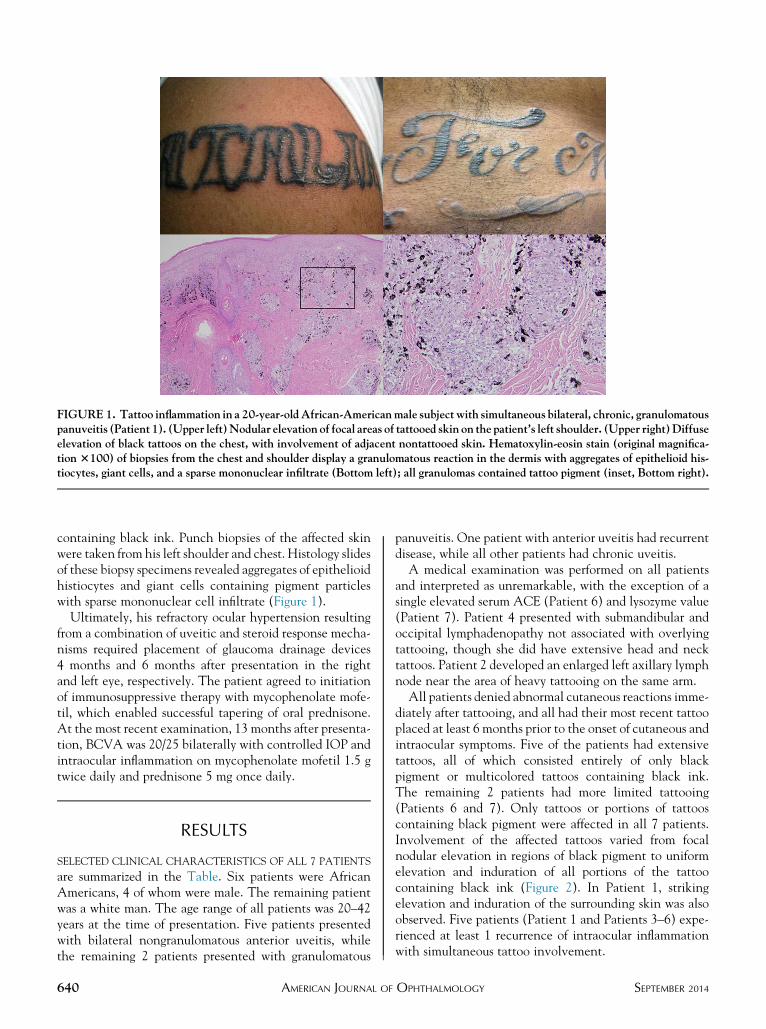
FIGURE 1. Tattoo inflammation in a 20-year-oldAfrican-Americanmale subject with simultaneous bilateral, chronic, granulomatouspanuveitis (Patient 1). (Upper left)Nodular elevation of focal areas of tattooed skin on the patient’s left shoulder. (Upper right)Diffuseelevation of black tattoos on the chest, with involvement of adjacent nontattooed skin. Hematoxylin-eosin stain (original magnifica-tion 3100) of biopsies from the chest and shoulder display a granulomatous reaction in the dermis with aggregates of epithelioid his-tiocytes, giant cells, and a sparse mononuclear infiltrate (Bottom left); all granulomas contained tattoo pigment (inset, Bottom right).
containing black ink. Punch biopsies of the affected skinwere taken from his left shoulder and chest. Histology slidesof these biopsy specimens revealed aggregates of epithelioidhistiocytes and giant cells containing pigment particleswith sparse mononuclear cell infiltrate (Figure 1).
Ultimately, his refractory ocular hypertension resultingfrom a combination of uveitic and steroid response mecha-nisms required placement of glaucoma drainage devices4 months and 6 months after presentation in the rightand left eye, respectively. The patient agreed to initiationof immunosuppressive therapy with mycophenolate mofe-til, which enabled successful tapering of oral prednisone.At the most recent examination, 13 months after presenta-tion, BCVA was 20/25 bilaterally with controlled IOP andintraocular inflammation on mycophenolate mofetil 1.5 gtwice daily and prednisone 5 mg once daily.
RESULTS
SELECTED CLINICAL CHARACTERISTICS OF ALL 7 PATIENTS
are summarized in the Table. Six patients were AfricanAmericans, 4 of whom were male. The remaining patientwas a white man. The age range of all patients was 20–42years at the time of presentation. Five patients presentedwith bilateral nongranulomatous anterior uveitis, whilethe remaining 2 patients presented with granulomatous
640 AMERICAN JOURNAL OF
panuveitis. One patient with anterior uveitis had recurrentdisease, while all other patients had chronic uveitis.A medical examination was performed on all patients
and interpreted as unremarkable, with the exception of asingle elevated serum ACE (Patient 6) and lysozyme value(Patient 7). Patient 4 presented with submandibular andoccipital lymphadenopathy not associated with overlyingtattooing, though she did have extensive head and necktattoos. Patient 2 developed an enlarged left axillary lymphnode near the area of heavy tattooing on the same arm.All patients denied abnormal cutaneous reactions imme-
diately after tattooing, and all had their most recent tattooplaced at least 6 months prior to the onset of cutaneous andintraocular symptoms. Five of the patients had extensivetattoos, all of which consisted entirely of only blackpigment or multicolored tattoos containing black ink.The remaining 2 patients had more limited tattooing(Patients 6 and 7). Only tattoos or portions of tattooscontaining black pigment were affected in all 7 patients.Involvement of the affected tattoos varied from focalnodular elevation in regions of black pigment to uniformelevation and induration of all portions of the tattoocontaining black ink (Figure 2). In Patient 1, strikingelevation and induration of the surrounding skin was alsoobserved. Five patients (Patient 1 and Patients 3–6) expe-rienced at least 1 recurrence of intraocular inflammationwith simultaneous tattoo involvement.
SEPTEMBER 2014OPHTHALMOLOGY
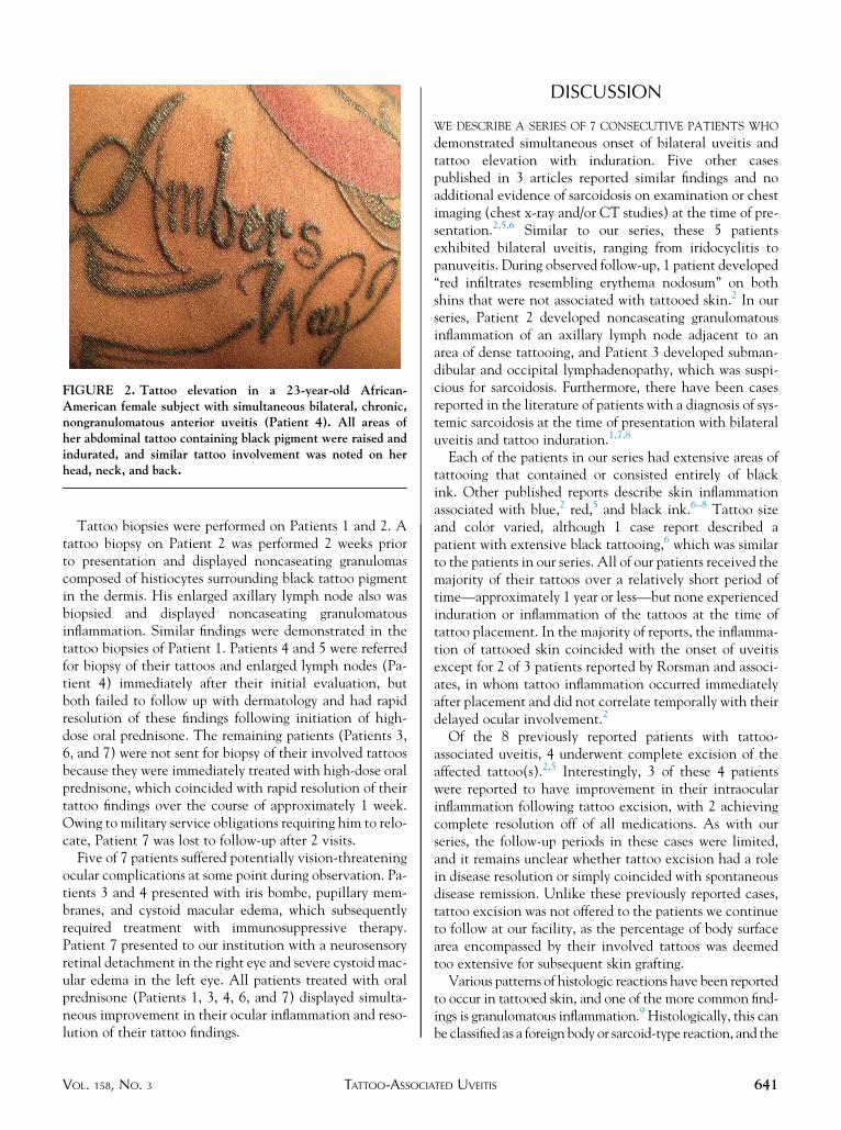
FIGURE 2. Tattoo elevation in a 23-year-old African-American female subject with simultaneous bilateral, chronic,nongranulomatous anterior uveitis (Patient 4). All areas ofher abdominal tattoo containing black pigment were raised andindurated, and similar tattoo involvement was noted on herhead, neck, and back.
Tattoo biopsies were performed on Patients 1 and 2. Atattoo biopsy on Patient 2 was performed 2 weeks priorto presentation and displayed noncaseating granulomascomposed of histiocytes surrounding black tattoo pigmentin the dermis. His enlarged axillary lymph node also wasbiopsied and displayed noncaseating granulomatousinflammation. Similar findings were demonstrated in thetattoo biopsies of Patient 1. Patients 4 and 5 were referredfor biopsy of their tattoos and enlarged lymph nodes (Pa-tient 4) immediately after their initial evaluation, butboth failed to follow up with dermatology and had rapidresolution of these findings following initiation of high-dose oral prednisone. The remaining patients (Patients 3,6, and 7) were not sent for biopsy of their involved tattoosbecause they were immediately treated with high-dose oralprednisone, which coincided with rapid resolution of theirtattoo findings over the course of approximately 1 week.Owing to military service obligations requiring him to relo-cate, Patient 7 was lost to follow-up after 2 visits.
Five of 7 patients suffered potentially vision-threateningocular complications at some point during observation. Pa-tients 3 and 4 presented with iris bombe, pupillary mem-branes, and cystoid macular edema, which subsequentlyrequired treatment with immunosuppressive therapy.Patient 7 presented to our institution with a neurosensoryretinal detachment in the right eye and severe cystoid mac-ular edema in the left eye. All patients treated with oralprednisone (Patients 1, 3, 4, 6, and 7) displayed simulta-neous improvement in their ocular inflammation and reso-lution of their tattoo findings.
VOL. 158, NO. 3 TATTOO-ASSOCIA
DISCUSSION
WE DESCRIBE A SERIES OF 7 CONSECUTIVE PATIENTS WHO
demonstrated simultaneous onset of bilateral uveitis andtattoo elevation with induration. Five other casespublished in 3 articles reported similar findings and noadditional evidence of sarcoidosis on examination or chestimaging (chest x-ray and/or CT studies) at the time of pre-sentation.2,5,6 Similar to our series, these 5 patientsexhibited bilateral uveitis, ranging from iridocyclitis topanuveitis. During observed follow-up, 1 patient developed‘‘red infiltrates resembling erythema nodosum’’ on bothshins that were not associated with tattooed skin.2 In ourseries, Patient 2 developed noncaseating granulomatousinflammation of an axillary lymph node adjacent to anarea of dense tattooing, and Patient 3 developed subman-dibular and occipital lymphadenopathy, which was suspi-cious for sarcoidosis. Furthermore, there have been casesreported in the literature of patients with a diagnosis of sys-temic sarcoidosis at the time of presentation with bilateraluveitis and tattoo induration.1,7,8
Each of the patients in our series had extensive areas oftattooing that contained or consisted entirely of blackink. Other published reports describe skin inflammationassociated with blue,2 red,5 and black ink.6–8 Tattoo sizeand color varied, although 1 case report described apatient with extensive black tattooing,6 which was similarto the patients in our series. All of our patients received themajority of their tattoos over a relatively short period oftime—approximately 1 year or less—but none experiencedinduration or inflammation of the tattoos at the time oftattoo placement. In the majority of reports, the inflamma-tion of tattooed skin coincided with the onset of uveitisexcept for 2 of 3 patients reported by Rorsman and associ-ates, in whom tattoo inflammation occurred immediatelyafter placement and did not correlate temporally with theirdelayed ocular involvement.2
Of the 8 previously reported patients with tattoo-associated uveitis, 4 underwent complete excision of theaffected tattoo(s).2,5 Interestingly, 3 of these 4 patientswere reported to have improvement in their intraocularinflammation following tattoo excision, with 2 achievingcomplete resolution off of all medications. As with ourseries, the follow-up periods in these cases were limited,and it remains unclear whether tattoo excision had a rolein disease resolution or simply coincided with spontaneousdisease remission. Unlike these previously reported cases,tattoo excision was not offered to the patients we continueto follow at our facility, as the percentage of body surfacearea encompassed by their involved tattoos was deemedtoo extensive for subsequent skin grafting.Various patterns of histologic reactions have been reported
to occur in tattooed skin, and one of the more common find-ings is granulomatous inflammation.9 Histologically, this canbe classified as a foreign body or sarcoid-type reaction, and the
641TED UVEITIS
differentiation of these 2 types of granulomas may be bothchallenging and open to controversy.9,10 Allergic reactionsto tattoo pigment can also occur, which may exhibit avariety of histologic forms, some of which are alsoconsistent with sarcoidosis.9 Granulomatous reactionsconfined to single tattoo colors typically represent a localhypersensitivity reaction to specific components of tattoopigment, but they may also represent a manifestation ofsystemic sarcoidosis.9 Interestingly, reports of allergic reac-tions to black tattoo pigment are very rare.11 The histologicappearance of tattoo biopsy specimens obtained from 2 ofthe 7 patients in our series were interpreted as noncaseatinggranulomatous inflammation in association with dermaltattoo pigment, which is consistent with but not specific forsarcoidosis.
Although the etiology of sarcoidosis remains unclear, it ishypothesized that the disease process is initiatedwhena genet-ically susceptible host is exposed to an inciting environmentalantigen(s).3,10 In such an event, an exaggerated immuneresponse characterized by the activation of macrophagesand CD4þ T lymphocytes occurs, resulting in cytokineproduction consistent with a TH1-type immune response,ultimately leading to granuloma formation.3,10 Consideringthe multiple environmental risk factors reported to date, itseems reasonable to conclude that the development ofsarcoidosis is likely the end result of immune responses to apotentially large variety of environmental triggers.3,4
The production of black tattoo ink is based on soot,which may ‘‘contain toxic, mutagenic or carcinogeniccompounds such as carbon black and polycyclic aromatichydrocarbons or phenol.’’12 Carbon black nanoparticle
642 AMERICAN JOURNAL OF
exposure in lung cell lines13 and mice14 have shownthat these particles ‘‘induce inflammation, oxidizeDNA, cause DNA strand breaks and increase the mutantfrequency following long-term exposure at a subcytotoxicconcentration.’’15 All of our patients received the major-ity of their tattoos over a relatively short period oftime—approximately 1 year or less—which may haveconferred an increased risk of disease development as aresult of the relatively large antigenic and/or toxicload. Interestingly, the US Food and Drug Administra-tion has not approved any tattoo pigments for injectioninto the skin, and many pigments used in tattoo inks areindustrial-grade colors suitable for printers’ ink or auto-mobile paint.16 Among adults 18–50 years of age inthis country, the prevalence of tattoos may be as highas 24%.17
Ultimately, the patients in our series seem to represent asubset of patients in whom some component of tattoopigment initiated a localized cutaneous response that bysome means also played a role in the simultaneous develop-ment of ocular inflammation. Whether the pathophysi-ology of this process is similar to systemic sarcoidosis, isthe result of a hypersensitivity response, or is attributbleto some other mechanism is not yet known. Continuedobservation for the development of additional organ systeminvolvement consistent with sarcoidosis, and the potentialbenefit of tattoo removal, if performed, may be usefulknowledge. Altogether, the clinical presentation of thepatients collected for this series nearly equals the cumula-tive number of previously reported cases, suggesting thatthis association is likely underappreciated.
ALL AUTHORSHAVE COMPLETED AND SUBMITTED THE ICMJE FORM FOR DISCLOSUREOF POTENTIAL CONFLICTS OF INTEREST.Jennifer E. Thorne discloses the following: grant funding from theNational Institutes of Health (Bethesda,Maryland,USA), Research to Prevent BlindnessSybil B. Harrington Special Scholars Award (New York, New York, USA), Allergan (Irvine, California, USA); consultant for AbbVie (North Chicago,Illinois, USA), Gilead (Foster City, California, USA), Navigant (Chicago, Illinois, USA), Xoma (Berkeley, California, USA). The authors indicate nofunding support. Contributions of authors: all listed authorsmade substantial contributions regarding (1) data acquisition (T.O., B.B., T.L., N.B., J.D., J.T.);(2) drafting (T.O.) or revising the article (T.O., B.B., T.L., N.B., J.D., J.T.); and (3) approval of submitted version (T.O., B.B., T.L., N.B., J.D., J.T.).
Pathology consultation was provided by Gulsun Erdag, MD, Johns Hopkins School of Medicine, Baltimore, Maryland, USA.
REFERENCES
1. Lubeck G, Epstein E. Complications of tattooing. Calif Med1952;76(2):83–85.
2. Rorsman H, Brehmer-Andersson E, Dahlquist I, et al. Tattoogranuloma and uveitis. Lancet 1969;294(7610):27–28.
3. Iannuzzi MC, Rybicki BA, Teirstein AS. Sarcoidosis.NEngl JMed 2007;357(21):2153–2165.
4. Bonfioli AA, Orefice F. Sarcoidosis. Semin Ophthalmol 2005;20(3):177–182.
5. Barbarasi Z, Kiss E, Balaton G, Vajo Z. Cutaneous granulomaand uveitis caused by a tattoo. Wien Klin Wochenschr 2008;120(1-2):18.
6. Saliba N, Owen ME, Beare N. Tattoo-associated uveitis. Eye2010;24(8):1406.
7. Anolik R, Mandal R, Franks A. Sarcoidal tattoo granu-loma. Dermatol Online J 2010;16(11):19. Available at,http://escholarship.org/uc/item/1fm1d840. Accessed May6, 2014.
8. Post J, Hull P. Tattoo reactions as a sign of sarcoidosis. CMAJ2012;184(4):432.
9. Morales-Callaghan AM, Aquilar-Bernier M, Martinez-Garcia G, Miranda-Romero A. Sarcoid granuloma on blacktattoo. J Am Acad Dermatol 2006;55(5 Suppl):S71–S73.
10. Marcoval J, Mana J, Moreno A, Gallego I, Fortuno Y, Peyri J.Foreign bodies in granulomatous cutaneous lesions of patientswith systemic sarcoidosis. Arch Dermatol 2001;137(4):427–430.
11. Kaur RR, Kirby W, Maibach H. Cutaneous allergic reactionsto tattoo ink. J Cosmet Dermatol 2009;8(4):295–300.
SEPTEMBER 2014OPHTHALMOLOGY

12. Wenzel SM, Rittmann I, Landthaler M, BaumlerW. Adversereactions after tattooing: review of the literature and com-parison to results of a survey. Dermatology 2013;226(2):138–147.
13. Jacobsen NR, Saber AT,White P, et al. Increased mutant fre-quency by carbon black, but not quartz, in the lacZ and clltransgenes of muta mouse lung epithelial cells. Environ MolMutagen 2007;48(6):451–461.
14. Jacobsen NR, Moller P, Jensen KA, et al. Lung inflamma-tion and genotoxicity following pulmonary exposureto nanoparticles in ApoE-/- mice. Part Fibre Toxicol2009;6:2.
VOL. 158, NO. 3 TATTOO-ASSOCIA
15. Hogsberg T, Jacobsen NR, Clausen PA, Serup J. Black tattooinks induce reactive oxygen species production correlatingwith aggregation of pigment nanoparticles and product brandbut not with the polycyclic aromatic hydrocarbon content.Exp Dermatol 2013;22(7):464–469.
16. U.S. Food andDrugAdministration.ConsumerHealth Informa-tion. Think before you ink: are tattoos safe? Available at, http://www.fda.gov/forconsumers/consumerupdates/ucm048919.htm.Accessed May 6, 2014.
17. Laumann AE, Derick AJ. Tattoos and body piercings in theUnited States: a national data set. J Am Acad Dermatol2006;55(3):413–421.
643TED UVEITIS

Biosketch
Trucian Ostheimer, MD, graduated from the Ohio State University College of Medicine in 2008, and completed his
ophthalmology residency at the Illinois Eye & Ear Infirmary in 2012. He is currently completing a two-year ocular
immunology fellowship at the Wilmer Eye Institute, and has a special interest in birdshot chorioretinopathy. Dr
Ostheimer will begin a fellowship in vitreoretinal surgery at the University of Washington in July, 2014.
643.e1 SEPTEMBER 2014AMERICAN JOURNAL OF OPHTHALMOLOGY