Embed Size (px)
Citation preview
Red Blood Cells Disorders
Ali M. JawadProfessor of HaematologyDepartment of Medicine
College of Medicine -
University of Baghdad2011
Objectives1.
Quick revision of the physiology of normal haematopoiesis.
2.
Define the term anaemia, its pathophysiology
and diagnostic approach..
3.
List the common causes and symptoms of iron-deficiency anaemia. State how iron deficiency anaemia
may be treated.
4.
Define the terms megaloblastic
anaemia
and anaemia
of chronic diseases. In each case discuss their causes, consequences and management.
5.
Define the terms aplastic
anaemia
and myelodysplastic syndrome. In each case an over view of clinical features,
diagnosis and treatment is outlined.6.
Define the term haemolytic
anaemia
including thalassaemia
and
sickle cell anaemia, and discuss the clinical features, diagnosis and treatment.
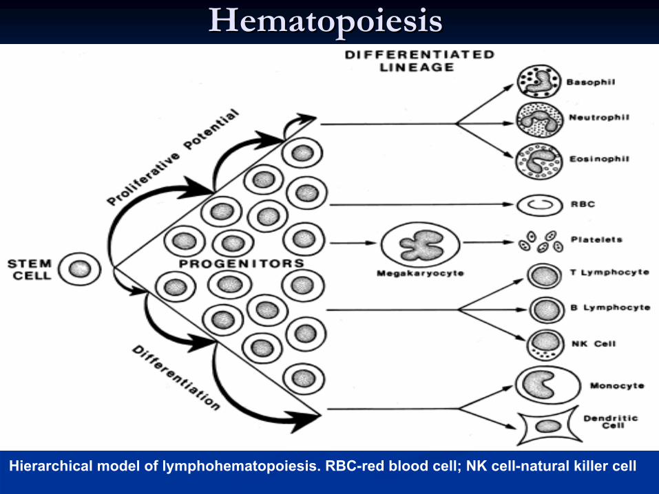
HematopoiesisHematopoiesis
Hierarchical model of lymphohematopoiesis. RBC-red blood cell; NK cell-natural killer cell
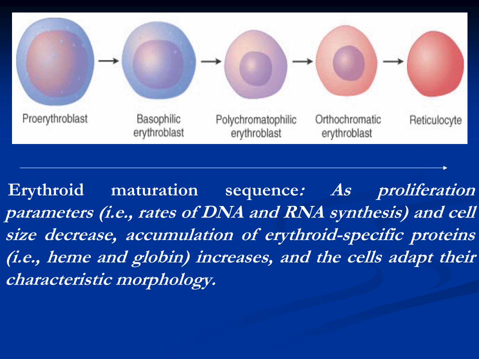
Erythroid
maturation sequence: As proliferation parameters (i.e., rates of DNA and RNA synthesis) and cell size decrease, accumulation of erythroid-specific proteins (i.e., heme and globin) increases, and the cells adapt their characteristic morphology.
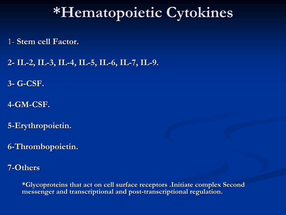
**HematopoieticHematopoietic CytokinesCytokines
11--
Stem cell Factor.Stem cell Factor.
22--
ILIL--2, IL2, IL--3, IL3, IL--4, IL4, IL--5, IL5, IL--6, IL6, IL--7, IL7, IL--9.9.
33--
GG--CSF.CSF.
44--GMGM--CSF.CSF.
55--Erythropoietin.Erythropoietin.
66--Thrombopoietin.Thrombopoietin.
77--OthersOthers
**GlycoproteinsGlycoproteins
that act on cell surface receptors .Initiate complex Second that act on cell surface receptors .Initiate complex Second
messenger and transcriptional and postmessenger and transcriptional and post--transcriptional regulation.transcriptional regulation.
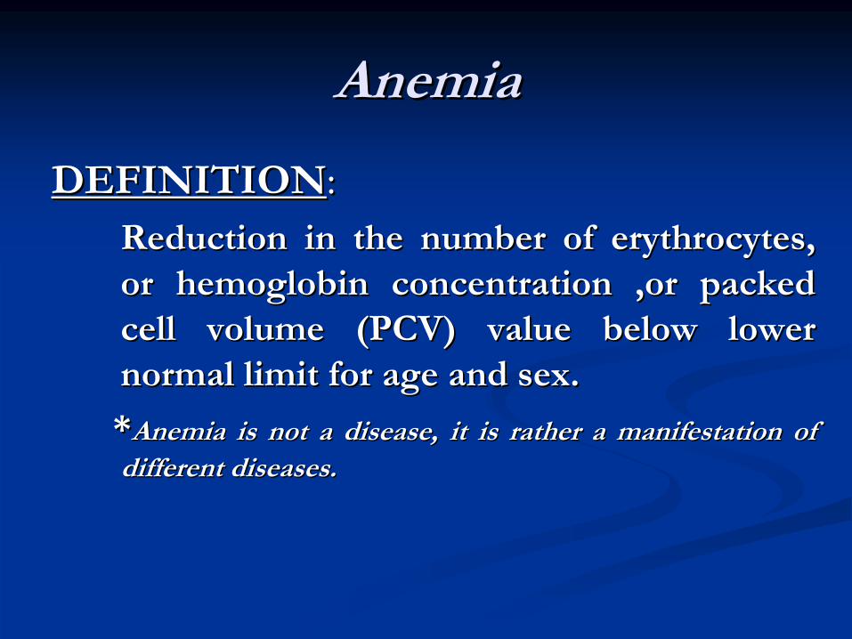
AnemiaAnemia
DEFINITIONDEFINITION::Reduction in the number of erythrocytes, Reduction in the number of erythrocytes, or hemoglobin concentration ,or packed or hemoglobin concentration ,or packed cell volume (PCV) value below lower cell volume (PCV) value below lower normal limit for age and sex.normal limit for age and sex.
**Anemia is not a disease, it is rather a manifestation of Anemia is not a disease, it is rather a manifestation of different diseases.different diseases.
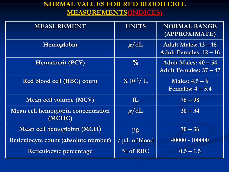
NORMAL VALUES FOR RED BLOOD CELL NORMAL VALUES FOR RED BLOOD CELL (INDICES)(INDICES)MEASUREMENTSMEASUREMENTS
NORMAL RANGE NORMAL RANGE (APPROXIMATE)(APPROXIMATE)
UNITSUNITSMEASUREMENTMEASUREMENT
Adult Males: 13 Adult Males: 13 ––
1818
Adult Females: 12 Adult Females: 12 ––
1616
g/dLg/dLHemoglobinHemoglobin
Adult Males: 40 Adult Males: 40 ––
5454
Adult Females: 37 Adult Females: 37 ––
4747
%%HematocritHematocrit
(PCV)(PCV)
Males: 4.5 Males: 4.5 ––
66
Females: 4 Females: 4 ––
5.45.4X 10X 101212/ L/ LRed blood cell (RBC) countRed blood cell (RBC) count
78 78 ––
9898fLfLMean cell volume (MCV)Mean cell volume (MCV)
30 30 ––
3434g/dLg/dLMean cell hemoglobin concentration Mean cell hemoglobin concentration (MCHC(MCHC))
30 30 ––
3636pgpgMean cell hemoglobin (MCHMean cell hemoglobin (MCH))
40000 40000 --
100000100000/ / LL
of bloodof bloodReticulocyteReticulocyte
count (absolute number)count (absolute number)
0.5 0.5 ––
1.51.5% of RBC% of RBCReticulocyteReticulocyte
percentagepercentage

Normal Red Blood CellNormal Red Blood Cell
The circulating erythrocyte under normal The circulating erythrocyte under normal conditions has an average lifespan of approximately conditions has an average lifespan of approximately 120 days120 days. It is a . It is a nonnon--nucleatednucleated, non, non--dividing cell in dividing cell in which more than 90% of the protein content is the which more than 90% of the protein content is the oxygenoxygen--carrying molecule, carrying molecule, hemoglobinhemoglobin. The . The erythrocyte's sole responsibility is to erythrocyte's sole responsibility is to deliver oxygen todeliver oxygen to
the tissues of the bodythe tissues of the body. Thus, the primary . Thus, the primary consequence of anemia is consequence of anemia is tissue hypoxiatissue hypoxia. . ErythropoiesisErythropoiesis
is driven by a feedback loop. Oxygenis driven by a feedback loop. Oxygen--
sensing cells in the area of the sensing cells in the area of the juxtaglomerularjuxtaglomerular apparatus of the apparatus of the kidneykidney respond to local tissue hypoxia respond to local tissue hypoxia
by increasing production of by increasing production of erythropoietinerythropoietin
(EPO), (EPO), which is the primary regulatory hormone for which is the primary regulatory hormone for erythropoiesiserythropoiesis..

Etiology Of AnemiaEtiology Of Anemia
11--
Decreased Production of RBC/Hemoglobin Decreased Production of RBC/Hemoglobin ((HypoproliferativeHypoproliferative
AnemiaAnemia).).
22--
Increased Destruction of Increased Destruction of RBC(RBC(HemolysisHemolysis).).
33--
Blood Loss.Blood Loss.
44--
Sequestration.Sequestration.

Effects of AnemiaEffects of Anemia
Physiological: Physiological:
due to tissue hypoxia.due to tissue hypoxia.11--Increased 2,3 DPG in Increased 2,3 DPG in RBC(helpsRBC(helps
to deliver more oxygen to to deliver more oxygen to
tissues).tissues).22--Increased Cardiac Output (heart rate and stroke volume).Increased Cardiac Output (heart rate and stroke volume).33--Redistribution of Blood Flow to vital organs.Redistribution of Blood Flow to vital organs.
Clinical:Clinical:
11--Pallor of the skin and mucous membranes.Pallor of the skin and mucous membranes.22--Easy Fatigability due to muscle hypoxia.Easy Fatigability due to muscle hypoxia.33--Cardiovascular: dizziness, SOB, worsening of IHD and HF.Cardiovascular: dizziness, SOB, worsening of IHD and HF.44--Loss of Concentration due to brain hypoxia.Loss of Concentration due to brain hypoxia.55--Other effects.Other effects.

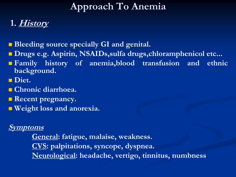
Approach To Anemia
1. History
Bleeding source specially GI and genital.
Drugs e.g. Aspirin, NSAIDs,sulfa
drugs,chloramphenicol
etc...
Family history of anemia,blood
transfusion and ethnic
background.
Diet.
Chronic diarrhoea.
Recent pregnancy.
Weight loss and anorexia.
SymptomsGeneral: fatigue, malaise, weakness. CVS: palpitations, syncope, dyspnea.Neurological: headache, vertigo, tinnitus, numbness

Approach To Anemia2. Physical Examination
CVS: tachycardia, systolic flow murmur, wide pulse pressure,evidence of CHF.
Pallor: mucous membranes, conjunctiva (Hb
< 9g/dl), skin creases (Hb
< 7g/dl).
Splenomegaly; seen in different hematological and non hematological conditions.
Lymphadenopathy,bleeding
spots.
Rectal (occult blood).
Others•
Koilonychia
(spoon-shaped nails)and
Glossitis
as in iron deficiency
anemia,•
Neurological findings as in pernicious anemia and vitamin B6 deficiency.
•
Jaundice as in hemolytic anemia3. Complete Blood Counts and Blood Film
Hemoglobin level, WBC count and differential, platelet count, blood film morphology, reticulocyte
count and percentage, RBC indices.
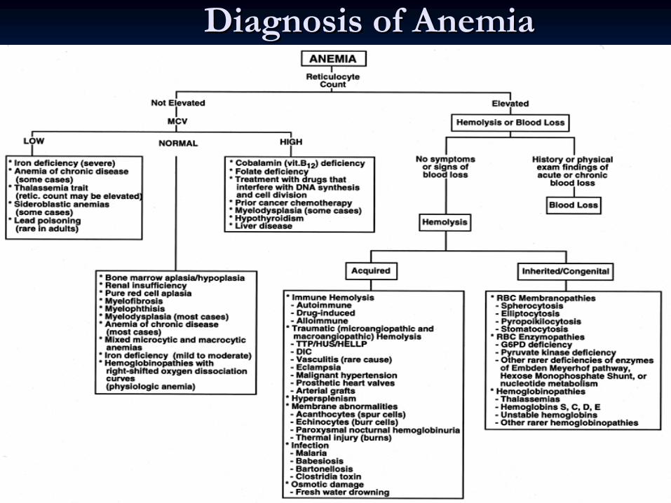
Diagnosis of AnemiaDiagnosis of Anemia
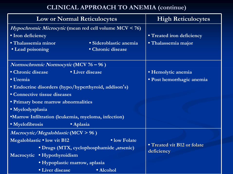
CLINICAL APPROACH TO ANEMIA (continue)
High ReticulocytesLow or Normal Reticulocytes
•
Treated iron deficiency
•
Thalassemia
major
Hypochromic Microcytic (mean red cell volume MCV < 76)
•
Iron deficiency
•
Thalassemia
minor •
Sideroblastic
anemia •
Lead poisoning •
Chronic disease
• Hemolytic anemia
•
Post hemorrhagic anemia
Normochromic Normocytic (MCV 76 –
96 )
•
Chronic disease •
Liver disease
•
Uremia
•
Endocrine disorders (hypo/hyperthyroid, addison’s)
•
Connective tissue diseases
•
Primary bone marrow abnormalities
•
Myelodysplasia
•Marrow Infiltration (leukemia, myeloma, infection)
•
Myelofibrosis
•
Aplasia
•
Treated Treated vitvit
B12 or B12 or folatefolate
deficiencydeficiency
Macrocytic/Megaloblastic (MCV > 96 )
Megaloblastic
•
low vit
B12 •
low Folate
•
Drugs (MTX, cyclophosphamide
,arsenic)
Macrocytic
•
Hypothyroidism
•
Hypoplastic
marrow, aplasia
•
Liver disease •
Alcohol
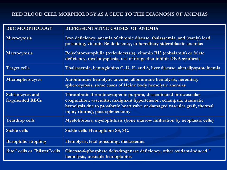
RED BLOOD CELL MORPHOLOGY AS A CLUE TO THE DIAGNOSIS OF ANEMIASRED BLOOD CELL MORPHOLOGY AS A CLUE TO THE DIAGNOSIS OF ANEMIAS
REPRESENTATIVE CAUSES OF ANEMIA REPRESENTATIVE CAUSES OF ANEMIA RBC MORPHOLOGY RBC MORPHOLOGY
Iron deficiency, anemia of chronic disease, Iron deficiency, anemia of chronic disease, thalassemiathalassemia, and (rarely) lead , and (rarely) lead poisoning, vitamin B6 deficiency, or hereditary poisoning, vitamin B6 deficiency, or hereditary sideroblasticsideroblastic
anemiasanemiasMicrocytosisMicrocytosis
PolychromatophiliaPolychromatophilia
((reticulocytsisreticulocytsis), vitamin B12 (), vitamin B12 (cobalamincobalamin) or ) or folatefolate
deficiency, deficiency, myelodysplasiamyelodysplasia, use of drugs that inhibit DNA synthesis, use of drugs that inhibit DNA synthesisMacrocytosisMacrocytosis
ThalassemiaThalassemia, , hemoglobinshemoglobins
C, D, E, and S, liver disease, C, D, E, and S, liver disease, abetalipoproteinemiaabetalipoproteinemiaTarget cellsTarget cells
Autoimmune hemolytic anemia, Autoimmune hemolytic anemia, alloimmunealloimmune
hemolysishemolysis, hereditary , hereditary spherocytosisspherocytosis, some cases of Heinz body hemolytic , some cases of Heinz body hemolytic anemiasanemias
MicrospherocytesMicrospherocytes
ThromboticThrombotic
thrombocytopenic thrombocytopenic purpurapurpura, disseminated intravascular , disseminated intravascular coagulation, coagulation, vasculitisvasculitis, malignant hypertension, , malignant hypertension, eclampsiaeclampsia, traumatic , traumatic hemolysishemolysis
due to prosthetic heart valve or damaged vascular graft, thermadue to prosthetic heart valve or damaged vascular graft, thermal l injury (burns), postinjury (burns), post--splenectomysplenectomy
SchistocytesSchistocytes
and and fragmented fragmented RBCsRBCs
MyelofibrosisMyelofibrosis, , myelophthisismyelophthisis
(bone marrow infiltration by (bone marrow infiltration by neoplasticneoplastic
cells)cells)Teardrop cellsTeardrop cells
Sickle cells Hemoglobin SS, SC. Sickle cells Hemoglobin SS, SC. Sickle cellsSickle cells
HemolysisHemolysis, lead poisoning, , lead poisoning, thalassemiathalassemiaBasophilic stipplingBasophilic stippling
""GlucoseGlucose--66--phosphate phosphate dehydrogenasedehydrogenase
deficiency, other oxidantdeficiency, other oxidant--induced induced hemolysishemolysis, unstable , unstable hemoglobinshemoglobins
Bite" cells or "Bite" cells or "blister"cellsblister"cells

Approach To Anemia(continue)Other Investigations are guided by the clinical impression after the
initial evaluation.
1-Iron Studies.2-Measurment of vitamin B12 and Folate
serum
levels.3-Bone Marrow Aspirate and Biopsy.4-Coomb,s Test.5-Osmotic Fragility test.6-Hemoglobin Electrophoresis.7-RBC enzyme studies.8-Chromosomal studies,Molecular
studies.
9-Other studies.

Normal Blood FilmNormal Blood Film

ReticulocytesReticulocytes

Tear Drop CellsTear Drop Cells

Basophilic StipplingBasophilic Stippling
MacrocytesMacrocytes
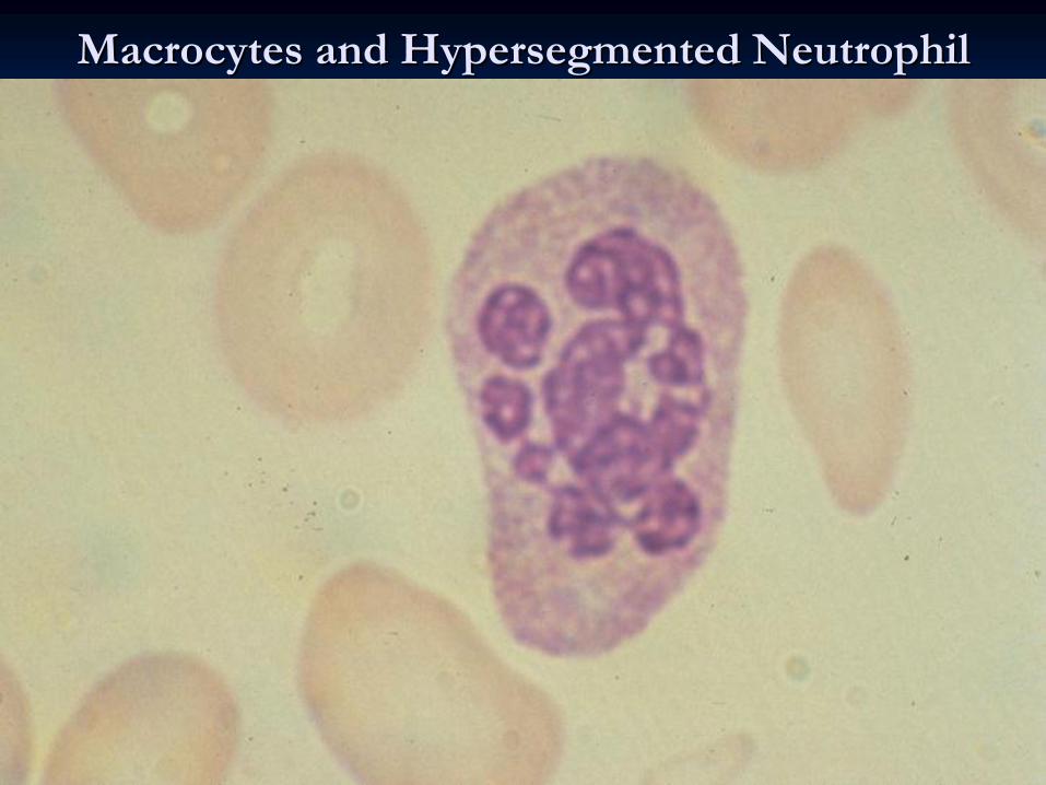
and and HypersegmentedHypersegmented
NeutrophilNeutrophil
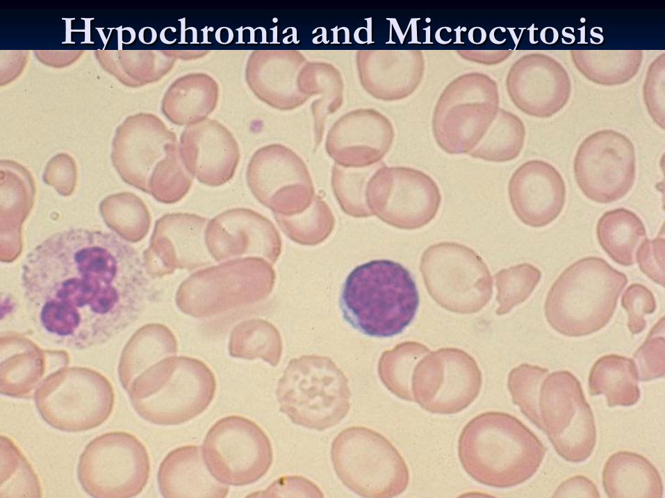
HypochromiaHypochromia and and MicrocytosisMicrocytosis
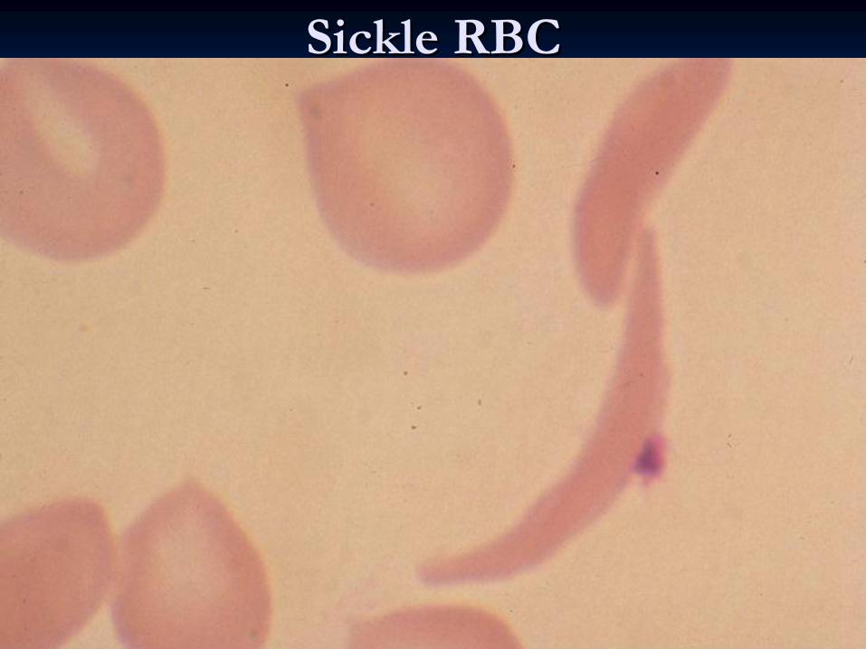
Sickle RBCSickle RBC
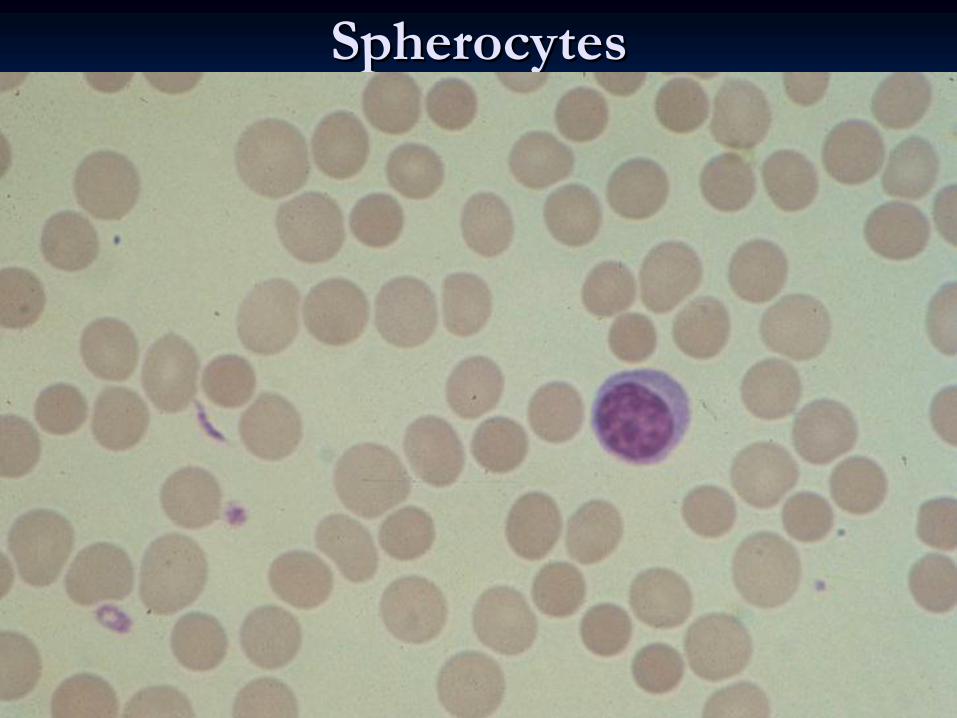
SpherocytesSpherocytes
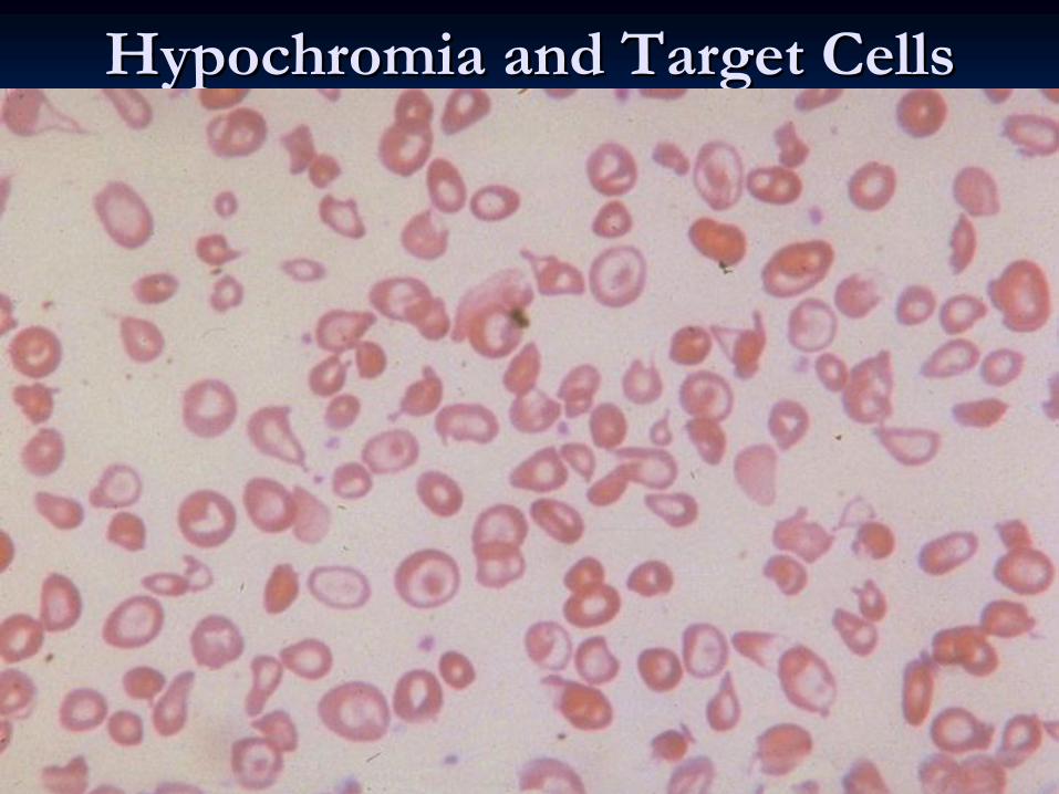
HypochromiaHypochromia and Target Cellsand Target Cells
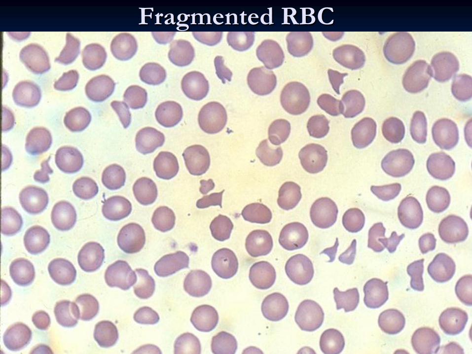
Fragmented RBCFragmented RBC

Blister RBCBlister RBC
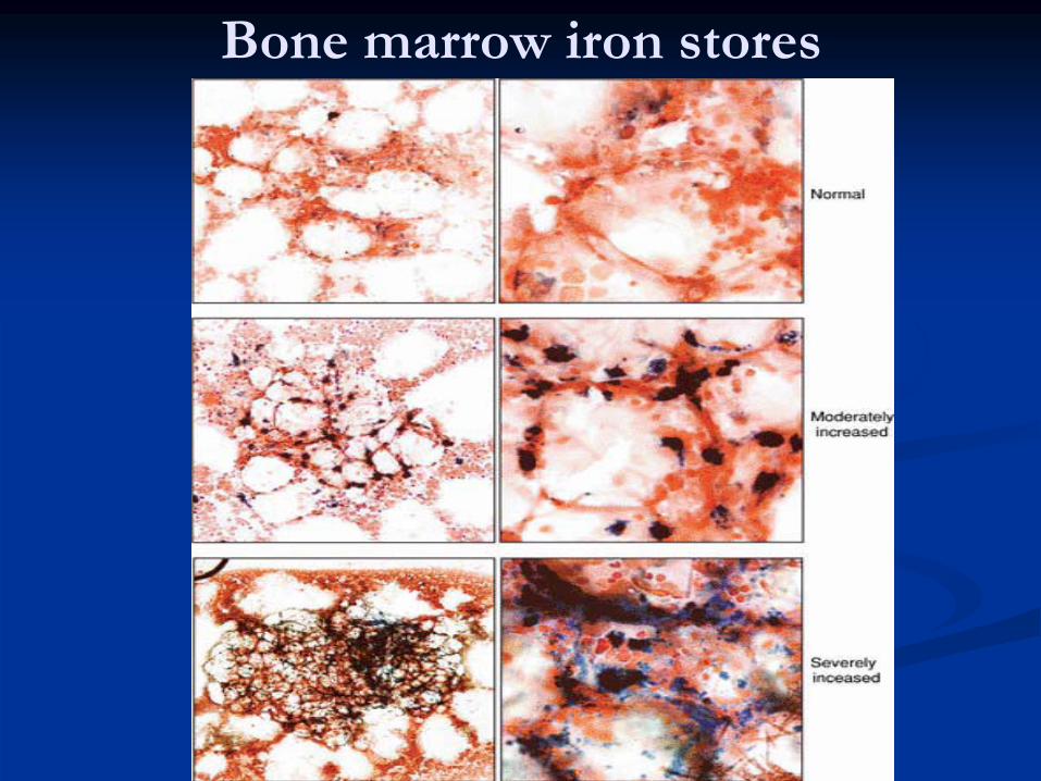
Bone marrow iron stores

Aplastic Bone Marrow Biopsy

Megaloblastic
Bone Marrow
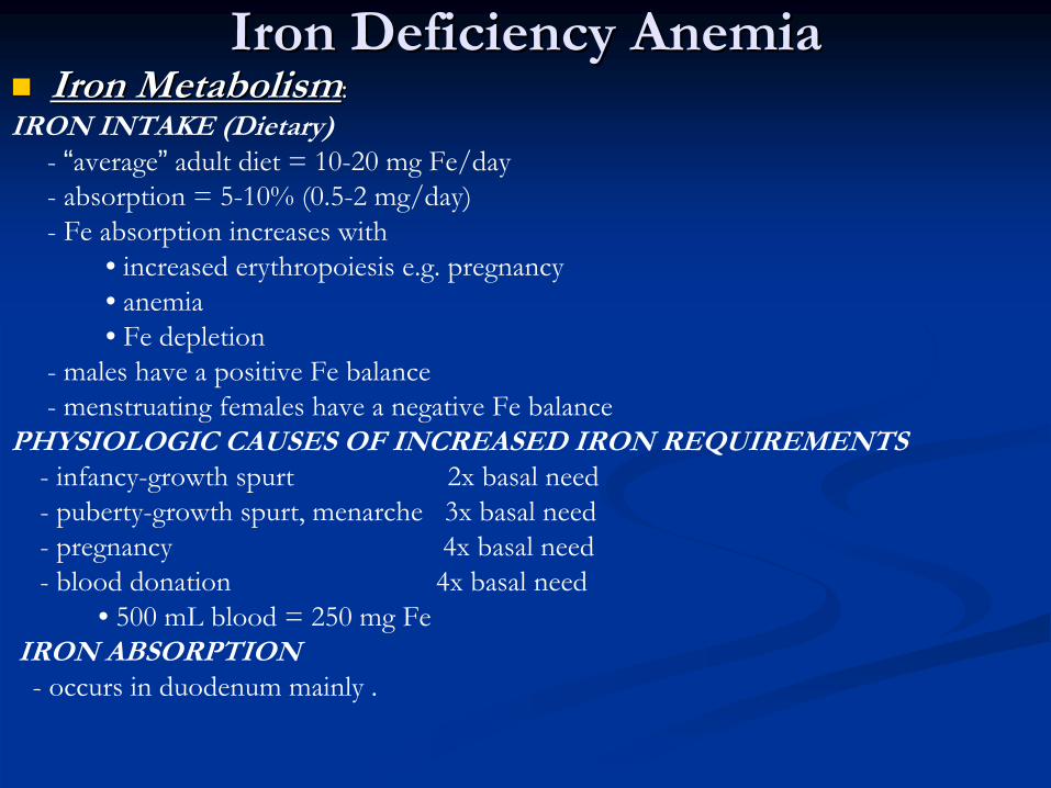
Iron Deficiency AnemiaIron Deficiency Anemia
Iron MetabolismIron Metabolism::
IRON INTAKE (Dietary)-
“average” adult diet = 10-20 mg Fe/day
-
absorption = 5-10% (0.5-2 mg/day)-
Fe absorption increases with
• increased erythropoiesis
e.g. pregnancy• anemia• Fe depletion
-
males have a positive Fe balance-
menstruating females have a negative Fe balance
PHYSIOLOGIC CAUSES OF INCREASED IRON REQUIREMENTS-
infancy-growth spurt 2x basal need
-
puberty-growth spurt, menarche 3x basal need-
pregnancy 4x basal need
-
blood donation 4x basal need• 500 mL
blood = 250 mg Fe
IRON ABSORPTION-
occurs in duodenum mainly .
IRON TRANSPORT
Majority of non-heme
Fe in plasma is bound to a beta-globulin called
transferrin
Transferrin
• carries Fe from mucosal cell to RBC precursors in marrow.• carries Fe from storage pool in hepatocytes
and macrophages to RBC
precursors in marrow.IRON STORAGE
Fe is stored in two forms: ferritin
and hemosiderin.
Ferritin• ferric Fe complexed
to a protein called apoferritin.
• hepatocytes
are the main site of ferritin
storage.• minute quantities are present in plasma in equilibrium with theintracellular ferritin.
Hemosiderin• aggregates or crystals of ferritin
with the apoferritin
partially removed
• macrophage-monocyte
system is the main source of hemosidirin.

IRON METABOLISM
Causes of IDACauses of IDA
PHYSIOLOGIC CAUSES
Increased need for iron in the body.
Infancy.
Adolescence, menstruation.
Pregnancy, lactation.PATHOLOGIC CAUSES
In adult males and post-menopausal females, Fe deficiency is usually related to chronic blood loss mainly from GIT.
Dietary deficiencies (rarely the only etiology)• cow’s milk (infant diet)• poor dietary iron intake
(elderly)
Absorption imbalances• post-gastrectomy• malabsorption
Hemorrhage• obvious causes -
menorrhagia
• occult -
peptic ulcer disease, aspirin, GI tract cancer,ankylostoma.
Intravascular hemolysis;iron
will be lost in the urine.

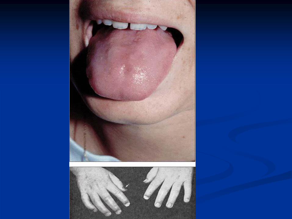
CLINICAL PRESENTATION Of IDA
Iron deficiency may cause fatigue before clinical anemia develops
Brittle hair
Dry skin
Dysphagia
(esophageal web, Plummer-Vinson ring)
Nail changes
• brittle• koilonychia
Glossitis
Angular stomatitis
Pica (appetite for bizarre substances e.g. ice, paint, clay)
IRON INDICES* Bone marrow biopsy is the gold standard test for iron stores.
Serum ferritin
•
Single most important blood test for iron stores.•
Falsely elevated in inflammatory disease, liver disease
(from necrotic hepatocytes), neoplasm and hyperthyroidism.
Serum iron
•
A measure Fe present in blood.•
Virtually all serum iron is bound to transferrin.
•
Only a trace of serum Fe is free or complexed
in ferritin.
Total iron binding capacity (TIBC)
•
measure of total amount of transferrin
present in blood.•
normally, one third of the TIBC is saturated with Fe, the remainder is unsaturated
Transferrin Saturation
•
serum Fe divided by TIBC, expressed as a proportion or a percentage.

Diagnosis of IDADiagnosis of IDALaboratory Investigations1-
Peripheral blood film•
Hypochromic
microcytic
RBC.
•
Pencil forms.•
Target cells (thin).
•
Platelet count may be elevated.2-
Serum Iron Studies•
low s.iron,high
TIBC,low
Fe saturation
•
S. ferritin
< 20 ug/l
is diagnostic of iron deficiency anemia,Iron
deficiency
anemia unlikely if ferritin
> 100 ug/l.
•
Increased plasma level of soluble transferrin
receptors.
3-
Hemoglobin Electrophoresis decreased Hb
A2 percentage.
4-
Bone marrow study
(Needed in difficult cases)
•
Predominence
of intermediate and late erythroblasts.
•
Micronormoblastic
maturation of erythroid
precursors.
•
Fe stain (Prussian blue) shows decreased iron in macrophages.•
Decreased sideroblasts.
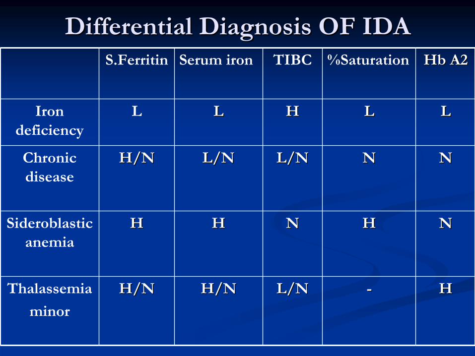
Differential Diagnosis OF IDA Differential Diagnosis OF IDA HbHb
A2A2%SaturationTIBCSerum ironS.Ferritin
LLLLHHLLLIron deficiency
NNNNL/NL/NL/NL/NH/NH/NChronic disease
NNHHNNHHHHSideroblastic anemia
HH--L/NL/NH/NH/NH/NH/NThalassemia
minor

Treatment of IDATreatment of IDA1-
TREAT THE UNDERLYING CAUSE : to control the site of blood loss,correct
malabsorption,improve
oral iron intake etc…
2-
IRON REPLACEMENT
Different preparations available: tablets, syrup, parenteral
Dose: ferrous sulphate
325 mg PO TID or ferrous gluconate
300 mg PO TID until anemia corrects and then for 3 months after to replenish the stores.
Reticulocytes
begin to increase after one week indicating response.
Ensure that the hemoglobin returns completely to normal.
If serum ferritin
returns normal discontinue iron therapy.
Treatment of iron deficiency anemia is made somewhat difficult by the frequent induction of nausea, dyspepsia, constipation, and diarrhea by oral iron preparations.
Blood is given for severely symptomatic anemia.
In the rare patient who cannot tolerate or cannot absorb iron frIn the rare patient who cannot tolerate or cannot absorb iron from the om the gastrointestinal tract and in individuals who require large irongastrointestinal tract and in individuals who require large iron boluses boluses to compensate for chronic blood loss, to compensate for chronic blood loss, parenteralparenteral iron is available as an iron is available as an ironiron--dextrandextran complex (complex (ImferonImferon) and iron ) and iron sorbitolsorbitol; they should be used ; they should be used with caution because of the threat of acute anaphylaxis and with caution because of the threat of acute anaphylaxis and subacutesubacute
((arthralgiasarthralgias, , myalgiasmyalgias, and , and adenopathyadenopathy) side effects.) side effects.
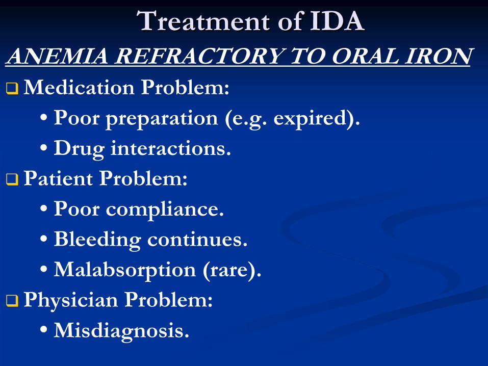
Treatment of IDATreatment of IDAANEMIA REFRACTORY TO ORAL IRONMedication Problem:
•
Poor preparation (e.g. expired).•
Drug interactions.
Patient Problem:•
Poor compliance.
•
Bleeding continues. •
Malabsorption
(rare).
Physician Problem:•
Misdiagnosis.

MegaloblasticMegaloblastic AnemiasAnemias
DEFINITION:DEFINITION:
MegaloblasticMegaloblastic
anemiasanemias
are caused by are caused by various defects in various defects in DNA synthesisDNA synthesis
that that
lead to a common set of hematologic lead to a common set of hematologic abnormalities of abnormalities of bone marrow and bone marrow and peripheral blood cellsperipheral blood cells. The term. The term megaloblasticmegaloblastic refers to a refers to a morphologic morphologic abnormality(mainlyabnormality(mainly
affecting the size affecting the size
and morphology) of the cell and it's and morphology) of the cell and it's nucleusnucleus. The . The erythrocyticerythrocytic, granulocytic, , granulocytic, and megakaryocytic cell lines are all and megakaryocytic cell lines are all involved, and involved, and pancytopeniapancytopenia
maymay result.result.

Causes of Causes of MegaloblasticMegaloblastic
AnemiaAnemiaI. I. Cobalamin(vitCobalamin(vit
B12) deficiencyB12) deficiency
A. Decreased ingestion: vegetarians.A. Decreased ingestion: vegetarians.B. Impaired absorption: small intestinal disease.B. Impaired absorption: small intestinal disease.C.ImpairedC.Impaired
utilization.utilization.
II. II. FolateFolate
deficiencydeficiencyA. Decreased A. Decreased ingestion:prolongedingestion:prolonged
parenteralparenteral
feeding,alcoholismfeeding,alcoholism..B. Impaired B. Impaired absorption:smallabsorption:small
intestinal disease.intestinal disease.
C. Impaired C. Impaired utilization:drugutilization:drug
induced induced egeg; sulfa ; sulfa drugs,methotrexate,phenytoindrugs,methotrexate,phenytoin……
D. Increased D. Increased requirement:pregnancy,hemolysisrequirement:pregnancy,hemolysis..E. Increased E. Increased loss:throughloss:through
urine.urine.
III. Drugs III. Drugs —— metabolic inhibitorsmetabolic inhibitorsIV. MiscellaneousIV. Miscellaneous
A. Inborn errorsA. Inborn errorsB. Unexplained disordersB. Unexplained disorders

CobalamineCobalamine MetabolismMetabolism
CobalamineCobalamine
is an animal product. Daily need=1microgramis an animal product. Daily need=1microgram
Under normal conditions of gastric acidity, dietary Under normal conditions of gastric acidity, dietary cobalamincobalamin
enters the duodenum bound to R protein. enters the duodenum bound to R protein.
Additional Additional cobalamincobalamin
bound to R protein enters the bound to R protein enters the duodenum after secretion into bile by the liver (the only duodenum after secretion into bile by the liver (the only significant route by which significant route by which cobalamincobalamin
is lost from the body). is lost from the body).
Pancreatic proteases partially degrade salivary and Pancreatic proteases partially degrade salivary and biliarybiliary
R R proteinprotein––cobalamincobalamin
complexes in the jejunum; complexes in the jejunum; cobalamincobalamin
is is
bound to intrinsic factor only after this process occurs. The bound to intrinsic factor only after this process occurs. The intrinsic factorintrinsic factor––cobalamincobalamin
complex remains intact until it complex remains intact until it
reaches the distal end of the ileum, where it binds with high reaches the distal end of the ileum, where it binds with high affinity to specific receptors located on affinity to specific receptors located on ilealileal
mucosal cells. mucosal cells.
CobalaminCobalamin
then enters these cells and reaches the portal then enters these cells and reaches the portal plasma, which contains three plasma, which contains three cobalamincobalamin
binding proteins binding proteins
known as known as transcobalamintranscobalamin
I, II, and III).I, II, and III).
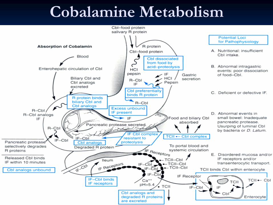
Cobalamine Metabolism

FolateFolate MetabolismMetabolism
FolateFolate
is widely distributed in plants and in products of animal origiis widely distributed in plants and in products of animal origin; n;
green vegetables are particularly rich sources of green vegetables are particularly rich sources of folatefolate
.Daily .Daily need=50microgram.need=50microgram.
FolatesFolates
in natural foods are conjugated to chains of in natural foods are conjugated to chains of polyglutamicpolyglutamic
acid. Enzymes in the lumen of the small intestine convert the acid. Enzymes in the lumen of the small intestine convert the polyglutamatepolyglutamate
forms of forms of folatefolate
to the to the monoglutamatemonoglutamate
and and diglutamatediglutamate
forms, which are readily absorbed in the proximal portion of theforms, which are readily absorbed in the proximal portion of the jejunum. Most of the jejunum. Most of the folatefolate
in plasma is present as 5in plasma is present as 5--
methyltetrahydrofolate in the methyltetrahydrofolate in the monoglutamatemonoglutamate
form. The majority is form. The majority is loosely bound to albumin, from which it is readily taken up by tloosely bound to albumin, from which it is readily taken up by the he highhigh--affinity affinity folatefolate
receptors present on cells throughout the body. receptors present on cells throughout the body.
Once it enters the cell, 5Once it enters the cell, 5--methyltetrahydrofolate must be converted methyltetrahydrofolate must be converted to to tetrahydrofolatetetrahydrofolate
by the by the cobalamincobalamin--dependent enzyme dependent enzyme methioninemethionine
synthasesynthase
before it can be converted to the before it can be converted to the polyglutamatepolyglutamate
form and form and take part in the other take part in the other folatefolate--dependent enzymatic reactions. In dependent enzymatic reactions. In addition to being secreted into bile and reabsorbed in the smalladdition to being secreted into bile and reabsorbed in the small
intestine, intestine, folatesfolates
are also degraded and excreted in the urineare also degraded and excreted in the urine

FolateFolate
and and CobalamineCobalamine
metabolismmetabolism
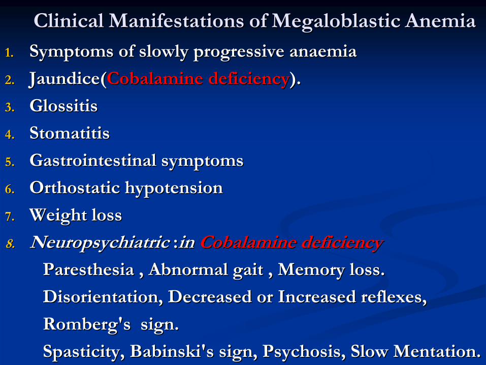
Clinical Manifestations of Clinical Manifestations of MegaloblasticMegaloblastic
AnemiaAnemia
1.1.
Symptoms of slowly progressive Symptoms of slowly progressive anaemiaanaemia
2.2.
Jaundice(Jaundice(CobalamineCobalamine
deficiencydeficiency).).
3.3.
GlossitisGlossitis
4.4.
StomatitisStomatitis
5.5.
Gastrointestinal symptomsGastrointestinal symptoms
6.6.
Orthostatic hypotensionOrthostatic hypotension
7.7.
Weight lossWeight loss
8.8.
NeuropsychiatricNeuropsychiatric ::in in CobalamineCobalamine deficiencydeficiency
ParesthesiaParesthesia
, Abnormal gait , Memory loss., Abnormal gait , Memory loss.
Disorientation, Decreased or Increased reflexes, Disorientation, Decreased or Increased reflexes,
Romberg's sign.Romberg's sign.
SpasticitySpasticity, , Babinski'sBabinski's
sign, Psychosis, Slow sign, Psychosis, Slow MentationMentation..
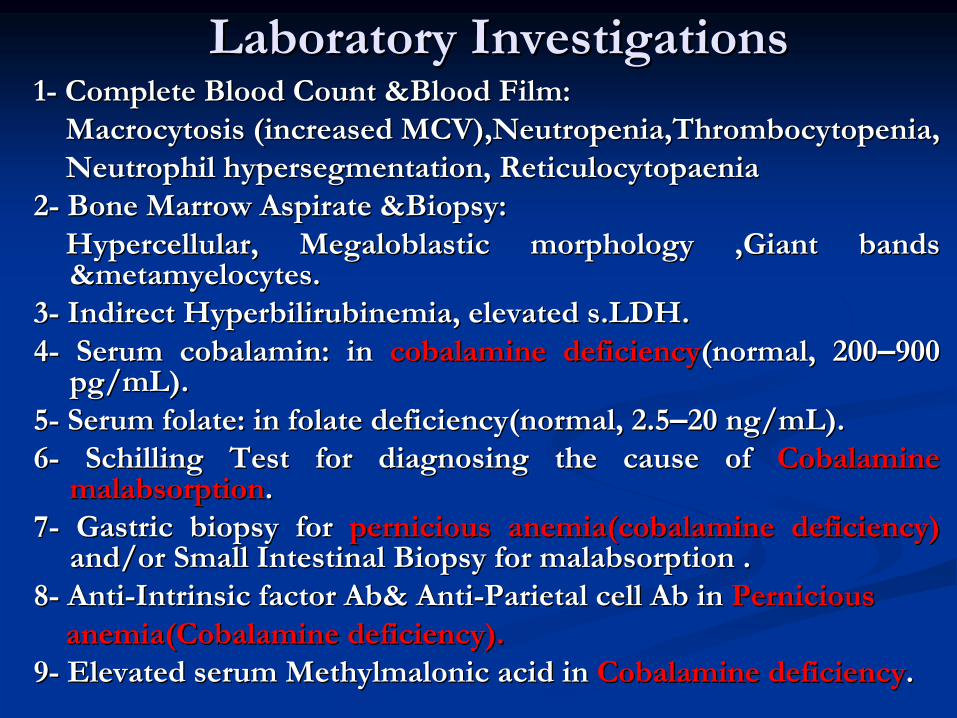
Laboratory InvestigationsLaboratory Investigations11--
Complete Blood Count &Blood Film:Complete Blood Count &Blood Film:MacrocytosisMacrocytosis
(increased (increased MCV),Neutropenia,ThrombocytopeniaMCV),Neutropenia,Thrombocytopenia,,
NeutrophilNeutrophil
hypersegmentationhypersegmentation, , ReticulocytopaeniaReticulocytopaenia22--
Bone Marrow Aspirate &Biopsy:Bone Marrow Aspirate &Biopsy:HypercellularHypercellular, , MegaloblasticMegaloblastic
morphologymorphology
,Giant bands ,Giant bands
&&metamyelocytesmetamyelocytes..33--
Indirect Indirect HyperbilirubinemiaHyperbilirubinemia, elevated , elevated s.LDHs.LDH..
44--
Serum Serum cobalamincobalamin: in : in cobalaminecobalamine
deficiencydeficiency(normal(normal, 200, 200––900 900 pg/pg/mLmL).).
55--
Serum Serum folatefolate: in : in folatefolate
deficiency(normaldeficiency(normal, 2.5, 2.5––20 20 ng/mLng/mL).).66--
Schilling Test for diagnosing the cause of Schilling Test for diagnosing the cause of CobalamineCobalamine
malabsorptionmalabsorption..77--
Gastric biopsy for Gastric biopsy for pernicious pernicious anemia(cobalamineanemia(cobalamine
deficiency)deficiency)
and/or Small Intestinal Biopsy for and/or Small Intestinal Biopsy for malabsorptionmalabsorption
..88--
AntiAnti--Intrinsic factor Intrinsic factor AbAb& Anti& Anti--Parietal cell Parietal cell AbAb
in in Pernicious Pernicious
anemia(Cobalamineanemia(Cobalamine
deficiency).deficiency).99--
Elevated serum Elevated serum MethylmalonicMethylmalonic
acid in acid in CobalamineCobalamine
deficiencydeficiency..
PERNICIOUS ANEMIAThe most common cause of cobalamin
malabsorption
is
pernicious anemia, a disease of unknown origin in which the fundamental defect is atrophy of the gastric (parietal cell) oxyntic
mucosa eventually leading to the absence of IF and
HCl
secretion. Because cobalamin
is only absorbed by binding to IF and uptake by ileal
IF-cobalamin
receptors, the
net consequence is severe cobalamin
malabsorption
leading to cobalamin
deficiency.
There is a significant association of pernicious anemia with other autoimmune diseases. There is a positive family history for about 30% of patients, among whom the risk of familial pernicious anemia is 20 times as high as in the general populationThe
histologic
appearance of the gastric mucosa
(infiltration with plasma cells and lymphocytes) is also strongly reminiscent of autoimmune-type lesions. There is also a high incidence of anti-parietal cell IgG
antibodies in
the serum of 90% of patients with pernicious anemia.

Treatment of Treatment of MegaloblasticMegaloblastic AnemiaAnemia
CobalamineCobalamine Deficiency:Deficiency:
HydroxycobalamineHydroxycobalamine
1 mg 1 mg i.mi.m. daily for 10 days . daily for 10 days ,then once every one month for ,then once every one month for life.Ironlife.Iron
is added is added
for slow response. for slow response. HypokalemiaHypokalemia
may develop may develop during therapy. during therapy. ReticulocytosisReticulocytosis
at day 7 will indicate at day 7 will indicate
response.response.Blood is given for Blood is given for severlyseverly
symptomatic patients.symptomatic patients.
FolateFolate Deficiency:Deficiency:
Oral Folic acid 5 mg/day for 3 weeks then weekly Oral Folic acid 5 mg/day for 3 weeks then weekly as maintenance.as maintenance.

Anemia of Chronic DiseaseAnemia of Chronic DiseaseEtiology
Infections, cancer, endocrine disorders (e.g. thyroid).
Inflammatory and rheumatologic disease.
Renal disease.
Pathophysiology
A mild hemolytic component is often present, red blood cell survival is moderately decreased.
Erythropoietin levels are normal or slightly elevated but are inappropriately low for the degree of anemia, erythropoietin level is low in renal failure
Iron cannot be removed from its storage pool in hepatocytes
and RES cells.

Anemia of Chronic DiseaseAnemia of Chronic DiseaseDiagnosis
RBC are usually normocytic
and normochromic
if the
anemia is mild, may be microcytic
and normochromic
if the anemia is moderate, may be microcytic
and
hypochromic
if the anemia is severe ,Hb
rarely < 9 g/dL except in renal failure.
Serum iron, TIBC, and % saturation all normal or slightly reduced, serum ferritin
is normal or increased.
Normal or increased iron stores in bone marrow, decreased “normal”
sideroblasts.
Management
Resolves if underlying disease is treated.
Erythropoietin may normalize the hemoglobin value especially in chronic renal failure. Dose of erythropoietin required is lower for patients with renal disease

AplasticAplastic AnemiaAnemia
Etiology
Radiation
Drugs
•
anticipated (chemotherapy)•
idiosyncratic (chloramphenicol, phenylbutazone)
Chemicals
•
benzene and other organic solvents•
DDT and insecticides
Post viral e.g. hepatitis B, parvovirus,HIV.
Idiopathic
•
often immune (cell mediated)
Paroxysmal nocturnal hemoglobinuria
Marrow replacement
Congenital: Fanconi
anemia,associated
with dysmorphic
features.

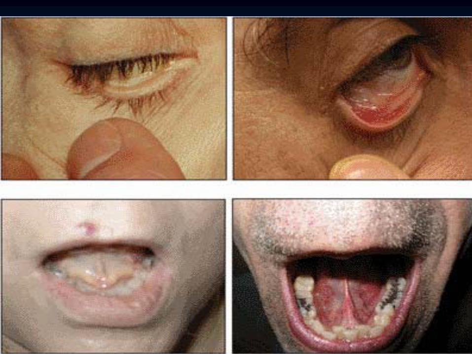
Abnormal Thumbs in Fanconi
Anemia
Clinical Presentation of Aplastic
Anemia
Occurs at any age
Slightly more common in males.
Can present acutely or insidiously.
Features of anemia or neutropenia
o
r thrombocytopenia (any combination).
1.
Thrombocytopenia as bruising, bleeding gums, epistaxis.
2.
Anemia as SOB, pallor and fatigue.
3.
Presentation of neutropenia
ranges from infection in the mouth to septicemia.

AplasticAplastic AnemiaAnemia
Diagnosis1-
CBC: Pancytopenia•
normochromic
normocytic
anemia.
•
neutrophil
count < 1.5 x 10⁹/L.
•
platelet count < 20 x 109/L.•
corrected reticulocyte
count < 1%.
2-
Bone marrow aspirate and biopsy•
aplasia
or hypoplasia
of marrow cells with fat replacement.
Management
Removal of offending agents.
Supportive care (red cell and platelet transfusions, antibiotics).
Antithymocyte
globulin (50-60% of patients respond) for patients who are >45
years of age and those who have no donor for bone marrow transplant
Cyclosporin
A,mainly
useful for mild cases.
Allogeneic
bone marrow transplantation for patients<45 y
•
minimize blood products on presentation.•
only irradiated, leuko-depleted blood products should be used to minimize
CMV transmission.•
CMV negative blood for CMV negative patients.
MYELODYSPLASTIC SYNDROMES (MDS)PathophysiologyPathophysiology
A group of clonal
bone marrow stem cell disorders
characterized by one or more cytopenias
with anemia present.
Ineffective hematopoiesis
despite presence of adequate numbers of progenitor cells (bone marrow is usually hyper-cellular).
Dysplastic changes affect all the hematopoietic cell lines due to abnormal maturation and differentiation which include abnormal size , nuclear shape and cytoplasmic
granules
The blood elements are dysfunctional.
There is increased liability for transformation to AML.
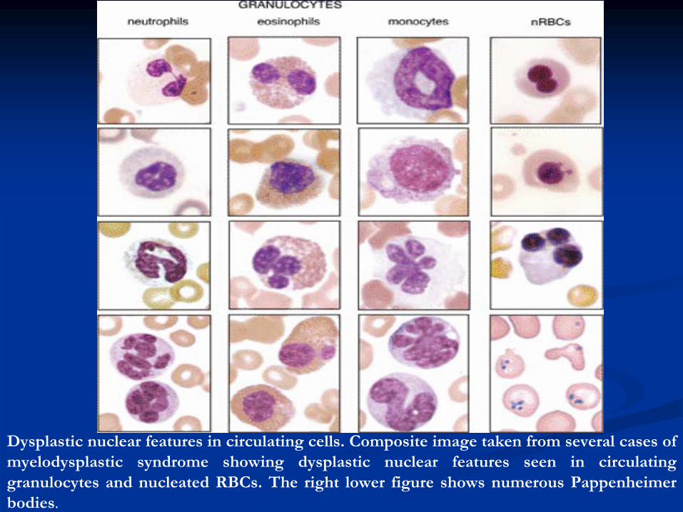
Dysplastic nuclear features in circulating cells. Composite image taken from several cases of myelodysplastic
syndrome showing dysplastic nuclear features seen in circulating
granulocytes and nucleated RBCs. The right lower figure shows numerous Pappenheimer
bodies.

MDSMDSTypes
Refractory anemia (RA).Refractory anemia with ring sideroblasts
(RARS).Refractory anemia with excess blasts
(RAEB).Refractory anemia with excess blasts in
transformation (RAEB-T).Chronic myelomonocytic
leukemia
(CMML).
MDSMDSClinical Presentation
Related to bone marrow failure, most common in elderly, usually > 70 and post-chemotherapy or radiation
Usually insidious in onset: fatigue, weakness, pallor, infections, bruising and rarely weight loss, and hepatosplenomegaly
Diagnosis1-
Anemia ±
thrombocytopenia ±
neutropenia
RBC: variable morphology with decreased reticulocyte count,
WBC: decrease in granulocytes and abnormal function, Platelet: either too large or too small and thrombocytopenia
2-
Bone marrow : dysmyelopoiesis
in bone marrow precursors
3-
Chromosomal Abnormalities:5,7,8 ,others
MDSMDSManagement1-
Symptomatic (transfusion, antibiotics)
2-
Growth factors: Erythropoietin,G-CSF.
3-
Cytotoxics
for RAEB & RAEB- T&CMML
4-
Bone marrow transplant for young patients with advanced disease.
5-
Immune modulating and differentiating agents .

Hemolytic anemias Definition:Anemias
that result from shortening of RBC life span,RBC
destruction could be extravascular
or intravascular
Etiology Etiology
Congenital1-
Membrane abnormalities•
Hereditary spherocytosis
•
Hereditary elliptocytosis2-Haemoglobinopathies
•
Lack of haemoglobin
chain synthesis Thalassaemias
•
Amino acid substitution on the haemoglobin
chain
Haemoglobin
S. C, D3-
Red cell enzyme detects
•
Glucose-B-phosphate dehydrogenase
deficiency
Acquired1-
Immune
•Isoimmune •Autoimmune Warm antibody
Cold antibody •Alloimmune
2-
Non-immune
• Mechanical-
Artificial cardiac valves
-
Burns
-
Microangiopattlic
-
March haemoglobinuria
•Infections •Clostridium perfringens,
malaria •Drugs, chemicals3-
Paroxysmal nocturnal haemoglobinuria
(PNH).
Approach To Hemolytic anemiaApproach To Hemolytic anemiaA- Prove The Presence of Hemolysis (Evidence of hemolysis):
Clinical Features: anemia +jaundice.
Laboratory Tests1-
Low Hb, Increased reticulocyte count and percentage.
2-
Indirect
hyperbilirubinemia,raised
s.LDH,increased
urinary urobilinogen.
3-
Low serum haptoglobin.4-
Hemosiderinurea,hemoglobinurea
in cases of intravascular
hemolysis.B-Find The Cause Of Hemolysis:
1-
Blood Film Morphology: Target Cells,Sickle
Cells,Heinz
Bodies, Blister Cells, Fragmented RBC, Spherocytes.
2-
Hemoglobin Electrophoresis: for Hemoglobinopathies.3-
Osmotic Fragility test for Spherocytosis
4-
Enzyme assay for Enzymopathies.5-
Coomb,s
test for immune hemolysis.
6-
Ham,s test for PNH.

Hereditary Hereditary spherocytosisspherocytosisThis is an This is an autosornalautosornal
dominantdominant
disorder in which disorder in which
the principal abnormality appears to be a deficiency of the principal abnormality appears to be a deficiency of spectrinspectrin,a,a
red cell membrane protein. Approximately red cell membrane protein. Approximately
25% of patients have no family history. The erythrocyte 25% of patients have no family history. The erythrocyte envelope is abnormally permeable and the sodium envelope is abnormally permeable and the sodium pumps are overworked. The exact nature of the red pumps are overworked. The exact nature of the red blood cell defect may vary from family to family. The blood cell defect may vary from family to family. The erythrocytes lose their biconcave shape, become erythrocytes lose their biconcave shape, become sphericalspherical
and are more susceptible to osmotic and are more susceptible to osmotic lysislysis. .
These These spherocytesspherocytes
are destroyed almost exclusively by are destroyed almost exclusively by the the spleenspleen. The severity of the disorder is very variable . The severity of the disorder is very variable even within an affected family. even within an affected family. HaemolysisHaemolysis
is mainly is mainly
extravascularextravascular..
Diagnosis &TreatmentDiagnosis &TreatmentClinical Features:
The severity of anemia
is variable from asymptomatic to
transfusion dependent anemia.Jaundice
is also variable,as
well as splenomegaly.
Complications include
1-
Crises (hemolytic, megaloblastic, and aplastic). 2-
Pigment Gall stones.
Lab Tests:
Evidence of Hemolysis: Anemia, Reticulocytosis, raised S.LDH…
Spherocytes
on blood film.
+ve
Osmotic Fragility Test.
-ve
Coombs Test.
Treatment :1-Blood Transfusion.2-Folic acid.3-Splenectomy for moderate to severe cases.
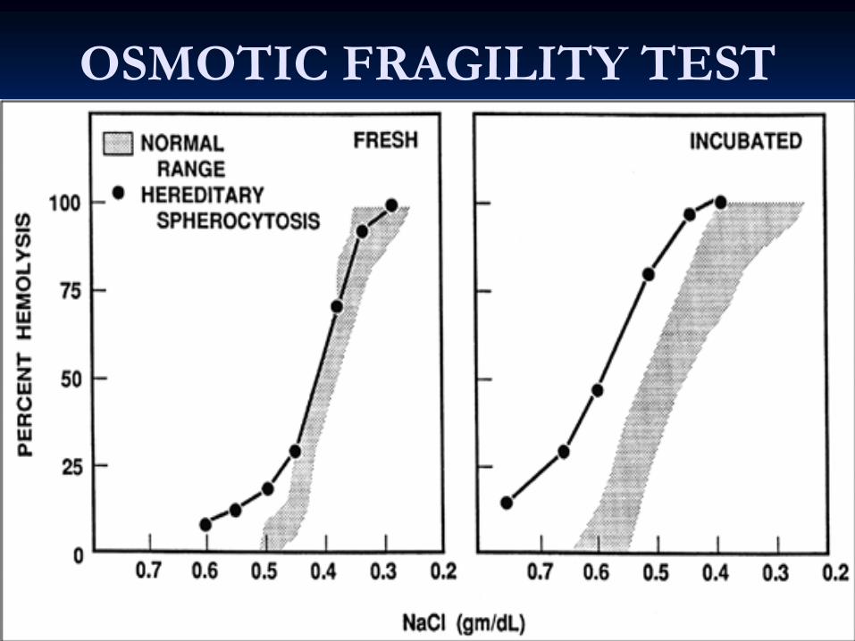
OSMOTIC FRAGILITY TEST

Glucose 6 Phosphate Glucose 6 Phosphate DehydrogenaseDehydrogenase
DeficiencyDeficiency
G6PD Enzyme is the first one in the hexosmonophosphate
shunt, the function of this
shunt is to service the enzymes glutathione reductase
and glutathione peroxidase, which protect
the red cells against damage due to oxidation.
The deficiency is inherited as an X-linked disorder
with a high frequency among Black Africans who possess an electrophoretic
enzyme polymorphism
with A and B type enzymes. The enzyme is A type (A-)
in deficient Black Africans. In Caucasians only
the normal B type enzyme is found and the deficient type is also B (B-).

G6PD deficiencyG6PD deficiency
Oxidant damage of RBC followed by intavascular
hemolysis
is induced by:1-
Infections .
2-
Ingestion of Fava
Beans.3-
Oxidant drugs like: sulfa, dapsone, antimalarial,
chloramphenicol….., 4-
Surgery.
Clinical manifestations:1-
Most cases present
with episodic intravascular hemolysiswith fever, rapid anemia, jaundice and deep colored urine.
2-
Rarely the hemolysis
is chronic.3-
Neonatal jaundice.

G6PD deficiencyG6PD deficiency
Lab Tests:1-
Anemia, reticulocytosis,
indirect hyperbilirubinemia…2-
Blister RBC on blood film,
and Heinz bodies.3-
Hemoglobinurea
and later
hemosideriurea.4-
Enzyme assay is useful
after recovery.Treatment:1-
Avoid fava
beans and oxidant drugs.
2-
For the hemolytic episode: stop the offending factor, blood transfusion, folic acid.
HAEMOGLOBINOPATHIESHAEMOGLOBINOPATHIES
The haemoglobinopathies
can be classified into
two subgroups:
1-
Where there is an alteration in the amino acid structure of the polypeptide chains of the glohin
fraction of haemoglohin, commonly called the abnormal haemoglohins: the best-known example is haemoglobin
S found in sickle-cell
anaemia.
2- Where the amino acid sequence is normal but polypeptide chain production is impaired or absent for a variety of reasons: these are the Thalassemias.

Hemoglobin Structure and Production
Fetal hemoglobin, HbF
(α2γ2) switches to adult forms HbA
(α2ß2) and HbA2 (α2δ2) at
3-6 months of life.
HbA
constitutes 97% of adult hemoglobin.
HbA2 constitutes 3% of adult hemoglobin.
4 α
genes are located on chromosome 16.
2 ß
genes are located on chromosome 11.
There is the possibility of mixed defects
e.g. ß-thalassemia
minor and sickle cell (HbS) trait.

Hemoglobin Structure

THALASSEMIASHETEROZYGOU ß
THALASSEMIA
: ß-Thalassemia
Minor
Common condition in Mediterranean Basin ,Africa,Asia
Clinical Presentation
Mild or no anemia.
Spleen sometimes is palpable.
May be masked by Fe deficiency and sometimes confused with iron deficiency anemia.
Diagnosis1-
Hb
9-12 g/dL, MCV < 70
2-
Microcytosis
+/– hypochromia,target
cells present,basophilic stippling usually present.
3-
Hb
electrophoresis: Hb
A2 increased to 3.5-5% (normal 1.5 -
3.5%), 50% of individuals have slight increase in HbF.
Management
Add folic acid.
Patient and family should receive genetic counseling.

HOMOZYGOUS ß
THALASSEMIA (ß-Thalassemia
Major)Pathophysiology
Ineffective beta chain synthesis due to point mutation in the beta gene promoter or enhancer on chromosome 11, excess alpha chains relative to beta chain leading to ineffective erythropoiesis
and
hemolysis
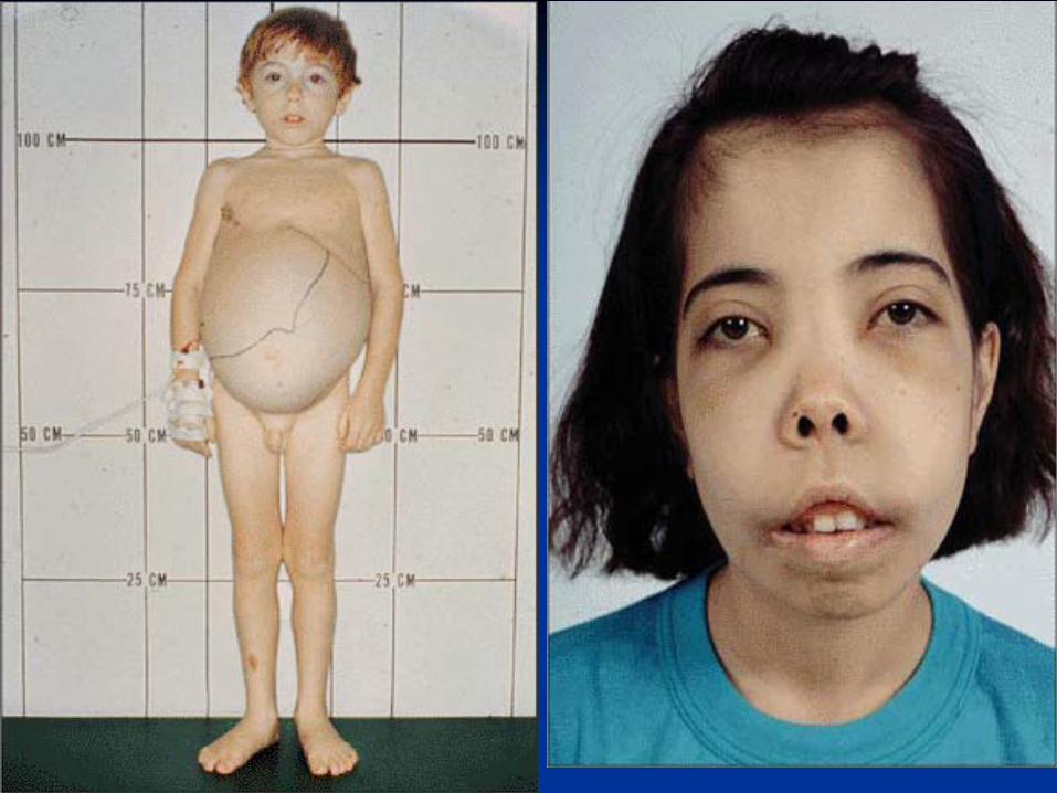
of RBC, compensatory increase in HbFClinical Presentation
Start presenting at 3-6 months
because of replacement of HbF
by HbA
Severe anemia developing in the first year of life
Jaundice
Stunted growth and development (hypogonadal
dwarf)
Gross hepatosplenomegaly
(extramedullary
hematopoiesis)
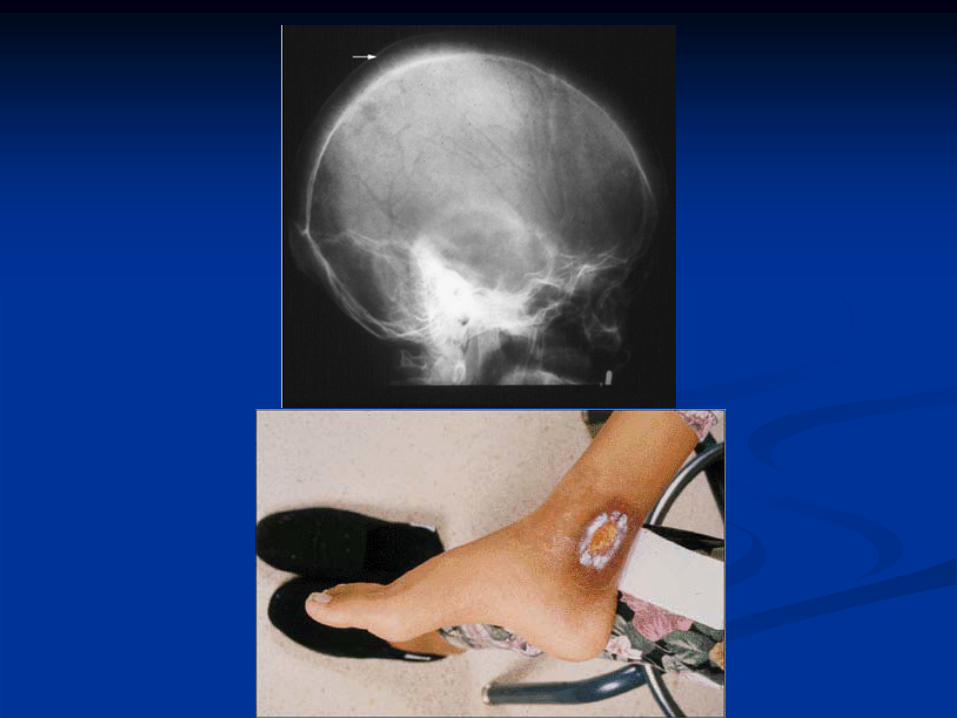
Skeletal changes (expanded marrow cavity)•
Skull x-ray has “hair-on-end”
appearance
•
Pathological fractures common
Evidence of increased Hb
catabolism (e.g. gallstones)
Death from•
Untreated anemia .
•
Infection (early).•
Iron overload (late, secondary to transfusions), usually20-30 years old.
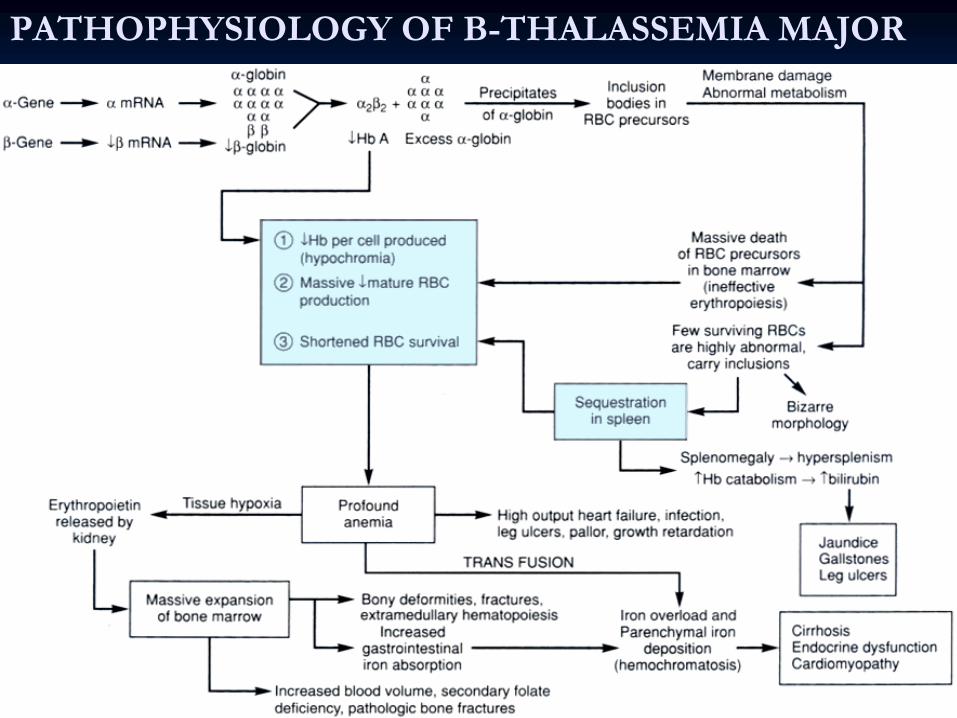
PATHOPHYSIOLOGY OF B-THALASSEMIA MAJOR
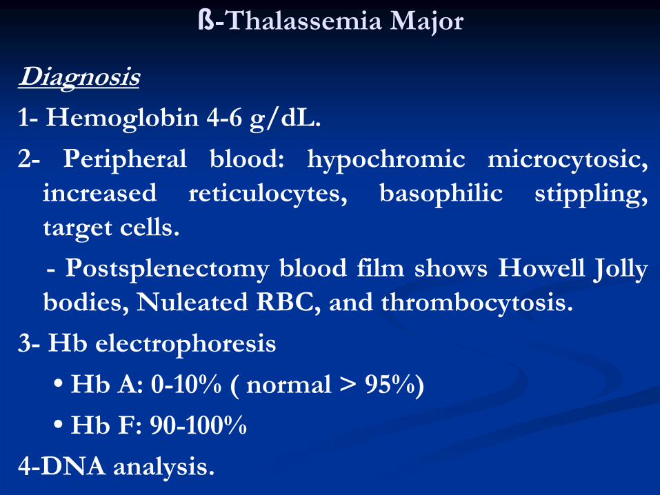
ß-Thalassemia
Major
Diagnosis1-
Hemoglobin 4-6 g/dL.
2-
Peripheral blood: hypochromic
microcytosic, increased reticulocytes, basophilic stippling, target cells.
-
Postsplenectomy
blood film shows Howell Jolly bodies, Nuleated
RBC, and thrombocytosis.
3-
Hb
electrophoresis
•
Hb
A: 0-10% ( normal > 95%)
•
Hb
F: 90-100%
4-DNA analysis.
ß-Thalassemia
Major
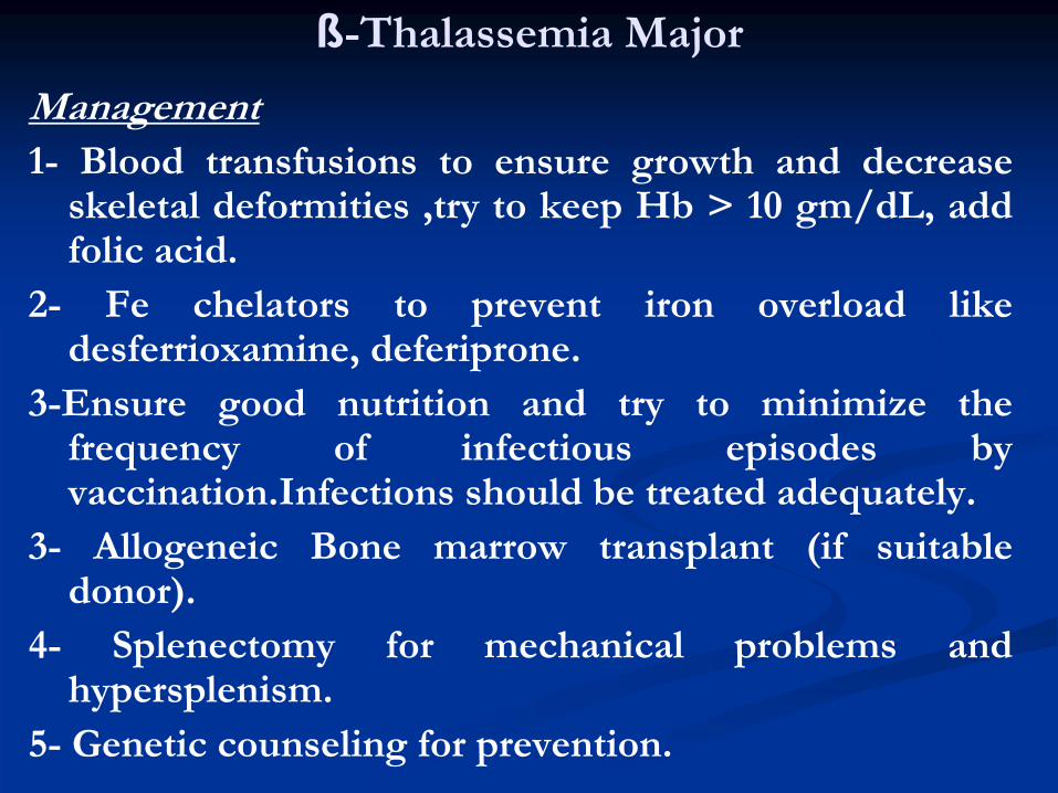
Management1-
Blood transfusions to ensure growth and decrease
skeletal deformities ,try to keep Hb
> 10 gm/dL, add folic acid.
2-
Fe chelators
to prevent iron overload like desferrioxamine, deferiprone.
3-Ensure good nutrition and try to minimize the frequency of infectious episodes by vaccination.Infections
should be treated adequately.
3-
Allogeneic
Bone marrow transplant (if suitable donor).
4-
Splenectomy
for mechanical problems and hypersplenism.
5-
Genetic counseling for prevention.
ALPHA THALASSEMIASPathophysiology
Autosomal
recessive
Deficit of alpha chains
4 grades of severity depending on the number of defective alpha genes
1 -
silent: αα/ α-2 -
trait: αα/--
or α-/ α-
3 -
Hb
H Disease (presents in adults) : α-/--4 -
Hb
Bart’s (hydrops
fetalis): --/--
Hb
Bart’s made of 4 gamma chains; not compatible with life
Hb
H made of 4 beta chains, is unstable, and leads to inclusion
bodiesDiagnosis
1-
Peripheral blood film: microcytes, hypochromia, occasional target cells, screen for Hb
H inclusion bodies.
2-
Hb
electrophoresis not diagnostic.3-
DNA analysis using alpha gene probe.
Management: same as beta thalassemia.

PATHOPHYSIOLOGY OF ALPHA THALASSEMIAS
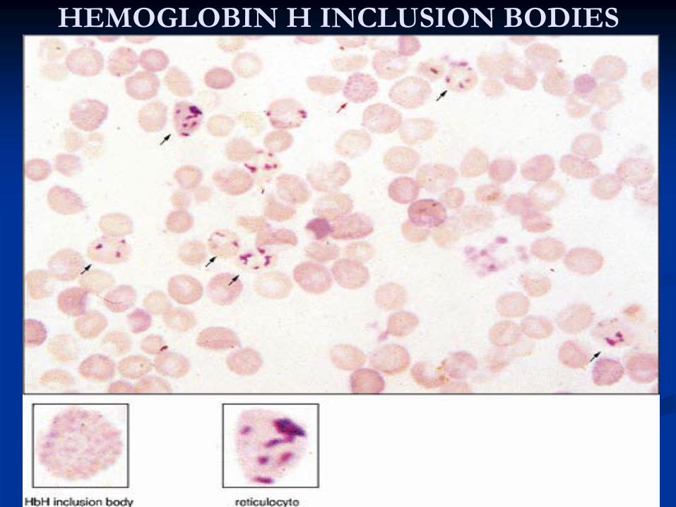
HEMOGLOBIN H INCLUSION BODIES

Sickle Cell anemiaSickle Cell anemia
Autosomal
recessive
Amino acid substitution of valine
for glutamate in
position 6 of betaglobin
chain.
*It has a wide geographical distribution.
Sickle Cell anemiaSickle Cell anemiaMechanisms of Sickling
At low PO2, deoxy
Hb
S polymerizes, leading to rigid
crystal-like rods that distort membrane = SICKLES
The PO2 level at which sickling
occurs is related to the
% of Hb
S present-
If the patient is heterozygous (Hb
AS), the sickling
phenomenon occurs at a PO2 of 40 mmHg-
If the patient is homozygous (Hb
SS), sickling
occurs
at 80 mmHg
Sickling
is also aggravated by
•
Acidosis.•
Increased CO2.
•
Increased 2,3-DPG.•
Increased temperature and osmolality.
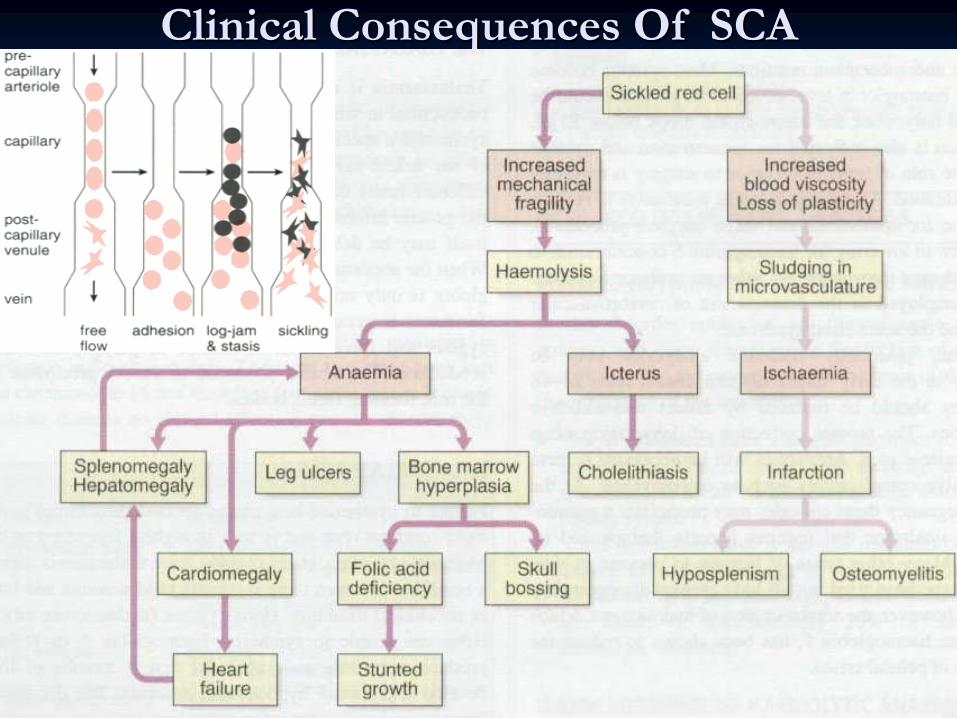
Clinical Consequences Of SCAClinical Consequences Of SCA

Heterozygous SCA: Hb
S TraitClinical presentation:• the patient will appear normal except at times of extreme hypoxia and infection, elderly patients may suffer from loss of renal concentration ability. Diagnosis:1-
Hb
level is normal
2-
Peripheral blood: normal except for a few target cells3-
Hb
electrophoresis (confirmatory test): Hb
A fraction
of 65% ; Hb
S fraction of 35%Treatment:1-
Avoid hypoxia during flying and surgery.
2-
Folic acid for pregnant.3-
Genetic counseling.
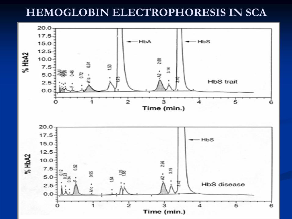
HEMOGLOBIN ELECTROPHORESIS IN SCA
Homozygous SCA: Hb
SS DiseaseClinical presentation1-
Chronic hemolytic anemia with jaundice in the first year of life.
2-
Retarded growth and development +/–
skeletal changes.3-
Susceptibility to infections by encapsulated organisms due to
hyposplenism.4-
Spleen enlarged in children and atrophic in adults.
5-
Crises:
Vaso
-
occlusive crises (infarction) leading to pain, fever , leukocytosis, acidosis& dehydration. Any organ or tissue can be involved.
Hyperhemolytic
crises associated malaria.
Sequestration crises presenting with anemia and rapidly enlarging spleen or liver.
Aplastic
crisis due to parvovirus infection or folate
deficiency, leading to rapid anemia and reticulocytopenia.
Acute Chest Syndrome presenting as fever, chest pain, cough and hypoxia.
6-
Iron overload due to repeated blood transfusion (less likely compared to Thalassemia).
7-Gall stones,leg
ulcers.

Homozygous SCA: Hb
SS Disease
Diagnosis
1-
Peripheral blood: sickled
cells,target
cells, reticulocytosis.
2-
Indirect hyperbilirubinemia, raised s.LDH3-
Screening test: sickle cell preparation searching for sickling
phenomenon.
4-
Hb
electrophoresis (confirmatory test): Hb
S fraction > 80%,the rest is Hb
F.

Homozygous SCA: Hb
SS DiseaseManagement1-
Prevention Of Sickling
Attacks:
•
Avoid conditions that favor sickling
(hypoxia, acidosis, dehydration, fever).•
Vaccination in childhood e.g. pneumococcus,
meningococcus.•
Good hygiene and nutrition.
2-
Genetic counseling.3-
Blood transfusion to keep Hb>8 g/dl + Iron chelation
for
frequent transfusions.4-
Folic acid to avoid folate
deficiency.
5-
Hydroxyurea
to enhance production of Hb
F, presence of Hb F in the SS cells decreases polymerization and precipitation
of Hb
S.Note: Hydroxyurea
is cytotoxic
and may cause bone marrow
suppression it is indicated in severe cases.6-
Experimental anti-sickling
agents.
7-
Allogeneic
Bone marrow transplant for selected patients.
Homozygous SCA: Hb
SS DiseaseTreatment of Vaso-Occlusive Crisis
Oxygen.
Good Hydration (reduces viscosity).
Antimicrobials.
Correct acidosis if severe.
Analgesics/narcotics (give enough to relieve pain).
Exchange transfusion for CNS crisis.

AUTOIMMUNE HEMOLYTIC ANEMIATypes:Types:
11--
Warm Antibody type :usually Warm Antibody type :usually IgGIgG..22--
Cold Antibody type:Cold Antibody type: usually usually IgMIgM..
Warm Antibody type)Warm Antibody type)(AUTOIMMUNE HEMOLYTIC ANEMIAPathophysiology: RBC are coated with IgG
or complement (C3d)
or both leading to extravascular
hemolysis
in RES (mainly spleen).
Classification:1-
idiopathic
2-
secondary to•
Lymphoproliferative
disorders (CLL, Hodgkin’s disease,
Non -
Hodgkin’s lymphoma)•
Autoimmune (SLE)
3-
Drug induced (penicillin, quinine/quinidine, alpha methyl dopa)Clinical Features:Usually insidious :anemia, jaundice and splenomegaly.

Mechanism of extravascular
hemolysis
in autoimmune hemolytic anemia. (A) Macrophage
encounters an lgG-coated erythrocyte and binds to it via its Fc
receptors. Thus entrapped,
the RBC loses bits of its membrane as a result of digestion by the macrophage. The discoid erythrocyte transforms into a sphere. (B) RBC lightly coated with lgG
(and therefore
incapable of activating the complement cascade) is preferentially removed in the sluggish circulation of the spleen. (C) RBC with a heavy coat of lgG; thus, C3b (black circles) can be removed both by the spleen and the liver.

AUTOIMMUNE HEMOLYTIC ANEMIA (Warm Antibody type)Warm Antibody type)
Diagnosis1-
Spherocytes
in blood film, reticulocytosis.
2-
Indirect hyperbilirubinemia, raised s.LDH.3-
Positive direct antiglobulin
test (direct Coombs’) best detected at
37ºC (hence “warm-reacting antibodies”)4-
Exclude delayed transfusion reaction.
Management
Treat underlying cause
Corticosteroids: prednisolone
1mg/Kg until response then taper
over 2-3 months.
Splenectomy
for relapsed and corticosteroid resistant cases .
Immunosuppressives
like azathioprine
for relapses after
splenectomy
Blood transfusion is used with caution.
Add folic acid.

Autoimmune Hemolytic Anemia with Cold-Reacting Antibodies
Pathophysiology Either monoclonal or polyclonal IgM
Antibodies attach to RBC
surface antigens in peripheral circulation where T < 37ºC. Antibodies will detach from the surface antigen if T > thermal amplitude Thermal amplitude is the temperature at which IgM
is attached
to RBC surface Associated with intravascular hemolysisClassification Idiopathic Secondary to
•
Lymphoproliferative
disorders (CLL, Hodgkin’s disease, non-Hodgkin’s lymphoma)•
Infections (Mycoplasma
pneumoniae, EBV).
Clinical Features:Anemia, acrocyanosis, joint pain, vasculitic
rash, Raynaud
phenomena, and rarely splenomegaly.
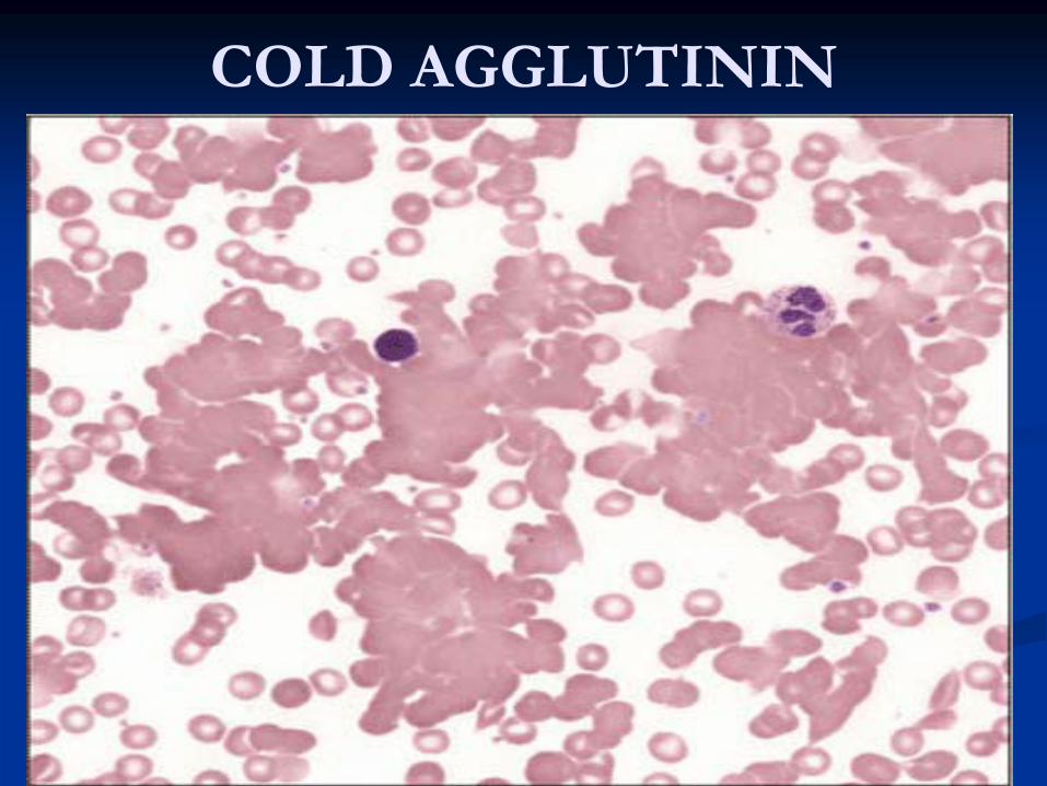
COLD AGGLUTININ
Autoimmune Hemolytic Anemia with Cold-Reacting Antibodies (IgM)
Diagnosis1-
Anemia, mild reticulocytosis, RBC agglutination in
blood film .
2-
Positive cold agglutinin test best at 4ºC.
3-
Positive direct Coombs’
for complement at any temperature.
Management
Treat underlying cause
Warm the patient above the thermal amplitude of the antibody
Plasmapheresis.
Immunosuppressives
like chlorambucil.

Non Immune Non Immune HemolysisHemolysis11--
Infections:Infections:
Bacterial, Malaria, Bacterial, Malaria, BabesiaBabesia..
22--
Mechanical:Mechanical:
MicroangiopathicMicroangiopathic
HemolysisHemolysis
(MAHA): TTP, HUS, (MAHA): TTP, HUS,
DIC.DIC.
March March HemoglobinureaHemoglobinurea..
Mechanical Cardiac Valves.Mechanical Cardiac Valves.
33--
Snake bite.Snake bite.
44--
Burns.Burns.
Drug Induced Drug Induced HemolysisHemolysis
11--
HaptenHapten
Mechanism: high dose PenicillinMechanism: high dose Penicillin
22--
Complement Fixation : Complement Fixation : quinidinequinidine
, , phenacetinphenacetin..
33--
Autoantibody production: LAutoantibody production: L--dopa, methyldopa.dopa, methyldopa.
44--
Nonspecific: Nonspecific: cephalothincephalothin..
55--
Metabolic: sulfa drugs.Metabolic: sulfa drugs.

HypersplenismHypersplenismIt is a state of sequestration of one or more of
blood elements in an enlarged spleen.
Causes1-
Portal hypertension
2-
Myeloproliferative
disorders.3-
Thalassemia
major.
4-
Others.
Diagnosis1-
Reduction of one or more of blood elements.
2-
Normal cellularity
of bone marrow.
TreatmentTreat the underlying condition, splenectomy
may
be indicated for increased transfusion requirements..