Embed Size (px)
Citation preview
Letter to the Editor
Vol. 26, No. 6, 2014 779
Received September 2, 2013, Revised November 20, 2013, Accepted for publication December 11, 2013
Corresponding author: Kwang Hyun Cho, Department of Dermatology, Seoul National University College of Medicine, 101 Daehak-ro, Jongno-gu, Seoul 110-744, Korea. Tel: 82-2-2072-2412, Fax: 82-2- 742-7344, E-mail: [email protected]
This is an Open Access article distributed under the terms of the Creative Commons Attribution Non-Commercial License (http:// creativecommons.org/licenses/by-nc/3.0) which permits unrestrictednon-commercial use, distribution, and reproduction in any medium, provided the original work is properly cited.
http://dx.doi.org/10.5021/ad.2014.26.6.779
Recurrent Acral Lentiginous Melanoma In Situ Suggesting the Field Cell Theory
Jin Yong Kim1,2,3, Eun Jung Hwang1, Mira Choi1,2,3, Seong Jin Jo1,2,3, Kwang Hyun Cho1,2,3
1Department of Dermatology, Seoul National University College of Medicine, 2Institute of Human-Environment Interface Biology, Seoul National University, 3Laboratory of Cutaneous Aging and Hair Research, Biomedical Research Institute, Seoul National University Hospital, Seoul, Korea
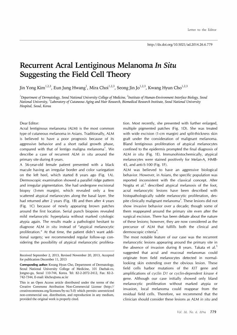
Dear Editor:Acral lentiginous melanoma (ALM) is the most common type of cutaneous melanoma in Asians. Traditionally, ALM is believed to have a poor prognosis because of its aggressive behavior and a short radial growth phase, compared with that of lentigo maligna melanoma1. We describe a case of recurrent ALM in situ around the primary site during 8 years.A 56-year-old female patient presented with a black macule having an irregular border and color variegation on the left heel, which started 8 years ago (Fig. 1A). Dermoscopic examination showed a parallel ridge pattern and irregular pigmentation. She had undergone excisional biopsy (3-mm margin), which revealed only a few scattered atypical melanocytes along the basal layer. She had returned after 2 years (Fig. 1B) and then after 4 years (Fig. 1C) because of newly appearing brown patches around the first location. Serial punch biopsies revealed mild melanocytic hyperplasia without marked cytologic atypia again. The results made a pathologist hesitant to diagnose ALM in situ instead of “atypical melanocytic proliferation.” At that time, the patient didn't want addi-tional surgery; we recommended regular follow-up con-sidering the possibility of atypical melanocytic prolifera-
tion. Most recently, she presented with further enlarged, multiple pigmented patches (Fig. 1D). She was treated with wide excision (1-cm margin) and split-thickness skin graft under the consideration of malignant melanoma. Bland lentiginous proliferation of atypical melanocytes confined to the epidermis prompted the final diagnosis of ALM in situ (Fig. 1E). Immunohistochemically, atypical melanocytes were stained positively for Melan-A, HMB- 45, and anti-S-100 (Fig. 1F).ALM was believed to have an aggressive biological behavior. However, in Asians, the specific population was reported inconsistent with the classical concept. After Nogita et al.2 described atypical melanosis of the foot, acral melanocytic lesions have been described with histopathologically subtle melanocytic proliferation, des-pite clinically malignant melanoma3. These lesions did not show invasive behavior over a decade; though some of them reappeared around the primary site even after the surgical excision. There has been debate about the nature of these lesions; however, they are now considered as the precursor of ALM that fulfills both the clinical and dermoscopic criteria4.The most notable feature of our case was the recurrent melanocytic lesions appearing around the primary site in the absence of invasion during 8 years. Takata et al.5 suggested that acral and mucosal melanomas could originate from field melanocytes detected in normal- looking skin extending over the obvious lesion. These field cells harbor mutations of the KIT gene and amplifications of cyclin D1 or cyclin-dependent kinase 4 gene. Although our case initially showed only bland melanocytic proliferation without marked atypia or invasion, local melanoma could reappear from the residual field cells. Therefore, we recommend that the clinician should consider these lesions as ALM in situ and
Letter to the Editor
780 Ann Dermatol
Fig. 1. Acral lentiginous melanoma in situ. Clinical and histopatholo-gical feature. (A) Initial presenta-tion 8 years ago. (B) Second pre-sentation 6 years ago. (C) Third presentation 4 years ago. (D) Last presentation. Left, gross picture of the lesion; center, close-up picture of the lesion showing multiple dark brown to black pigmented enlargedpatches with irregular border and variegated color on the left heel; right, histopathologic feature de-monstrating bland proliferationof scattered melanocytes without marked atypia or dermal invasion (A∼D: H&E, ×200). (E) Lentiginous hyperplasia of melanocytes was prominent only in the epidermis (H&E, ×200). (F) Melan-A staining showed atypical melanocytes confi-ned to the epidermis (immunoper-oxidase, ×200).
treat with early complete excision. Also, it is better to perform an excisional biopsy for a review of the entire lesion in the suspicious acral melanocytic lesions. Finally, regular follow-up for several years is important for the detection of reappearing melanoma around the primary site, even after the surgical excision.
REFERENCES
1. Arrington JH 3rd, Reed RJ, Ichinose H, Krementz ET. Plantar lentiginous melanoma: a distinctive variant of human cutaneous malignant melanoma. Am J Surg Pathol 1977;1:131-143.
2. Nogita T, Wong TY, Ohara K, Mizushima J, Mihm MC Jr, Kawashima M. Atypical melanosis of the foot. A report of
Letter to the Editor
Vol. 26, No. 6, 2014 781
Received September 30, 2013, Revised January 4, 2014, Accepted for publication February 1, 2014
Corresponding author: Jeong Deuk Lee, Department of Dermatology, Incheon St. Mary's Hospital, College of Medicine, The Catholic University of Korea, 56 Dongsu-ro, Bupyeong-gu, Incheon 403-720, Korea. Tel: 82-32-280-5700, Fax: 82-32-506-9514, E-mail: [email protected]
This is an Open Access article distributed under the terms of the Creative Commons Attribution Non-Commercial License (http:// creativecommons.org/licenses/by-nc/3.0) which permits unrestricted non-commercial use, distribution, and reproduction in any medium, provided the original work is properly cited.
three cases in Japanese populations. Arch Dermatol 1994; 130:1042-1045.
3. Kilinc Karaarslan I, Akalin T, Unal I, Ozdemir F. Atypical melanosis of the foot showing a dermoscopic feature of the parallel ridge pattern. J Dermatol 2007;34:56-59.
4. Chiu HH, Hu SC, Ke CL, Cheng ST. Dermoscopy identifies histopathologically indiscernible malignant lesion of atypical
melanosis of the foot, an early lesion of acral lentiginous melanoma in situ. Dermatol Surg 2008;34:979-983.
5. Takata M, Murata H, Saida T. Molecular pathogenesis of malignant melanoma: a different perspective from the studies of melanocytic nevus and acral melanoma. Pigment Cell Melanoma Res 2010;23:64-71.
http://dx.doi.org/10.5021/ad.2014.26.6.781
Foreign Body Reaction due to a Retained Cuff from a Central Venous Catheter
So Min Kim, Hee Jin Jun, Hei Sung Kim, Sang Hyun Cho, Jeong Deuk Lee
Department of Dermatology, Incheon St. Mary’s Hospital, College of Medicine, The Catholic University of Korea, Incheon, Korea
Dear Editor:Foreign body reaction is a tissue response to extraneous materials such as injected materials or implanted medical devices1. Here, we report a unique foreign body reaction caused by a retained cuff from a central venous catheter. A 63-year-old male patient with a history of end-stage renal disease presented with an asymptomatic, firm mass on the right chest for several months. One year ago, because of swelling and tenderness on the continuous ambulatory peritoneal dialysis (CAPD) catheter site, his CAPD catheter was removed, and a hemodialysis (HD) catheter was inserted through the right internal jugular vein. The CAPD catheter was reinserted after 2 weeks, and the HD catheter was removed by manual traction after 2 months. The patient visited our clinic with a 2 cm, skin-colored, subcutaneous mass on the right chest (Fig. 1). On incisional biopsy, there was an odorous, pus-like drainage and pieces of foreign material (Fig. 1). Histo-logical examination showed groups of fibers with adjacent
granulation tissue (Fig. 2). He was referred to the Depar-tment of General Surgery, and the catheter remains were completely removed. He had no complications.Venous access catheters are used for treatments such as HD and chemotherapy. Many catheters have polyester cuffs at the end for anchorage to the subcutaneous tissue. The catheters can be removed by traction or with a cutdown procedure2,3. When catheters are removed by traction, parts of the cuffs can break off and be retained in the subcutaneous tissue in 10%∼50% of cases2. The reported complications of retained cuffs include infection, abscess, discharge, and delayed healing2,3. Our patient had an odorous, pus-like drainage; however, we did not perform bacterial culture or Gram stain. Antibiotics were given, and the wound site healed without complications.Retained cuffs are clinically insignificant unless infection occurs2-4. In a study by Kohli et al.3, 428 cuffed central venous catheters were removed by traction, and catheter cuffs were retained in only 41 (10%) of the patients. Of