Embed Size (px)
Citation preview
Chapter 3.4
Recording sleep and wake
A.W. De Weerd* (The Netherlands) and P. Clarenbach (Germany)
Introduction
As well as in all medical disorders, taking the
patient's history is very important in sleep medi-
cine, too. Polygraphic studies, however, are indis-
pensable and recording of sleep and wake gets a
more and more important role in clinical neurophy-
siology.
This chapter is meant to give an overview of
technical aspects, how to perform recording and
to give an estimate of the diagnostic yield of poly(-
somno)graphy. Choices are pointed out instead of
rigid rules. The text aims at providing outlines that
can serve to choose ways of performing polysom-
nography.
To make such choices one should realize what
may be encountered in sleep medicine. Thus,
insight into the classi®cation of sleep and wake
disorders is necessary before registration is started
(Table 1).
Polygraphy should be taken in the strict sense of
the word. That means not only recording of signals
related to clinical neurophysiology as EEG and
EMG but also parameters from other ®elds, for
example pulmonary physiology.
There are several possibilities for recording
sleep. The most extensive but also very expensive
and time consuming way is polysomnography in
the laboratory, in fact the `gold standard'. Another
possibility is polysomnography at home using
portable recorders. Both methods give insight in
many parameters of sleep.
Polysomnography at home can easily be
extended to daytime recording and provides data
not only on sleep but on wakefulness, naps etc. as
well. Finally, there is limited polygraphy with one
to four channels of recording of various parameters,
often respiration or movements.
Standardized methods to measure wakefulness
are the Multiple Sleep Latency (MSLT) and the
Maintenance of Wakefulness (MWT) tests.
As there are many combinations of what can be
recorded (see Table 2), the concept of `modules' is
introduced here. A module is a way of recording
signals related to sleep and wake de®ned by the
method used, for example EEG, EMG, EOG,
ECG, thermistor recording of respiration, etc.
It is the aim of this chapter to describe possibi-
lities and give advise how to perform recording of
sleep and wake. Still, one can make his own `mix of
modules' to get the ideal recording for that parti-
cular patient and during given circumstances. Each
module will be described in technical details
(Section 2) and most used combination of modules
(Section 3). Rules for assessment and the relative
159
Recommendations for the Practice of Clinical Neurophysiology:
Guidelines of the International Federation of Clinical Physiology (EEG Suppl. 52)
Editors: G. Deuschl and A. Eisen
q 1999 International Federation of Clinical Neurophysiology. All rights reserved.
Published by Elsevier Science B.V.
* Correspondence to: Dr. A.W. De Weerd, Center for
Sleep and Wake Disorders and Department of Clinical
Neurophysiology, Medical Center Haaglanden, Westeinde
Hospital, P.O. Box 432, 2501 CK The Hague (The Nether-
lands).
value of the various ways of recording disorders
will be given in Sections 4 and 5.
Technical aspects
For insight into a disorder in sleep and wake and
its intensity it is necessary to record: (1) sleep, (2)
respiration, and (3) other factors.
Sleep and sleep depth
EEG
Recording is done with normal scalp electrodes
very well ®xed to provide many hours of recording
without artifacts. The recording can be limited to
two (in some cases even one) channels. According
to the classical guidelines given by Rechtschaffen
and Kales, EEG is recorded from the channels C3-
A2 and C4-A1. Good results can be obtained with
other derivations, for example, Fpz-Cz and Pz-Oz.
As many sleep-related EEG phenomena are located
over central regions, recording from at least these
areas is necessary.
It is recommended to use paper velocity of 10
mm/s; a time constant of at least 0.3 s, preferably
at 1.2 s; low-pass ®lter of 70 Hz and ampli®cation
of 70 mV/cm. Even with these settings dif®culties
may arise. On standard EEG paper or computer
screen such recordings are displayed in epochs of
30 s. This allows easy scoring in the Rechtschaffen
and Kales system which has 30 s as standard epoch,
but for example for the evaluation of apneas that
may last up to two min this time-base is too short.
Thus, there should be a possibility to change paper
velocity or enlarge the time-base of the computer
display.
EOG
Movements of both eyes together are recorded or
160
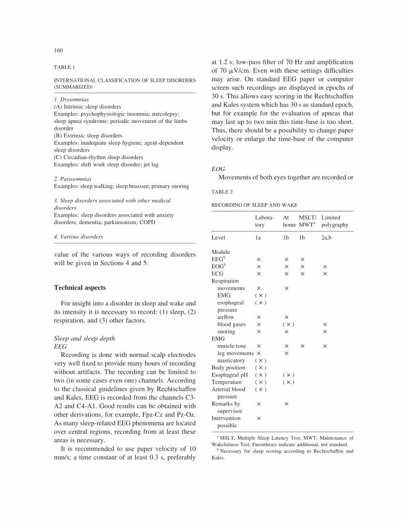
TABLE 1
INTERNATIONAL CLASSIFICATION OF SLEEP DISORDERS
(SUMMARIZED)
1. Dyssomnias
(A) Intrinsic sleep disorders
Examples: psychophysiologic insomnia; narcolepsy;
sleep apnea syndrome; periodic movement of the limbs
disorder
(B) Extrinsic sleep disorders
Examples: inadequate sleep hygiene; agent-dependent
sleep disorders
(C) Circadian-rhythm sleep disorders
Examples: shift work sleep disorder; jet lag
2. Parasomnias
Examples: sleep walking; sleep bruxism; primary snoring
3. Sleep disorders associated with other medical
disorders
Examples: sleep disorders associated with anxiety
disorders; dementia; parkinsonism; COPD
4. Various disorders
TABLE 2
RECORDING OF SLEEP AND WAKE
Labora-
tory
At
home
MSLT/
MWTa
Limited
polygraphy
Level 1a 1b 1b 2a,b
Module
EEGb£ £ £
EOGb£ £ £ £
ECG £ £ £ £
Respiration
movements £ £
EMG ( £ )
esophageal
pressure
( £ )
air¯ow £ £
blood gases £ ( £ ) £
snoring £ £ £
EMG
muscle tone £ £ £ £
leg movements £ £
masticatory ( £ )
Body position ( £ )
Esophageal pH ( £ ) ( £ )
Temperature ( £ ) ( £ )
Arterial blood
pressure
( £ )
Remarks by
supervisor
£ £
Intervention
possible
£
a MSLT, Multiple Sleep Latency Test; MWT, Maintenance of
Wakefulness Test. Parentheses indicate additional, not standard.b Necessary for sleep scoring according to Rechtschaffen and
Kales.
alternatively, movements of each eye. Surface elec-
trodes are ®xed at the outer corners of the eyes.
Filter settings should allow analysis of slow and
rapid eye movements (recommended time constant:
1.2 s; low-pass ®lter: 30 Hz, ampli®cation 200 mV/
cm).
Muscle tone
Surface electrodes are used for this recording as
well, mostly from the mental or submental
muscles (recommended settings: time constant:
0.03 s, low-pass ®lter 70±120 Hz, ampli®cation
30 mV/cm).
Respiration
Respiratory movements (� effort)
Measurement of excursions of breast and
abdomen. It is possible to limit the measurements
to movements of the abdominal wall only. Methods
are induction-plethysmography, elastic bands and
measurement of thoracic impedance.
EMG of respiratory muscles. Qualitative EMG
from respiratory muscles is possible, using surface
electrodes. Only in very adipose people this method
is not feasible. EMG of the intercostal muscles can
be recorded in the second and third intercostal
space next to the sternum. The EMG from the
diaphragma can be recorded in the eighth to tenth
intercostal space in the ®rst axillar line (recom-
mended settings similar to those for the evaluation
of muscle tone).
Esophageal pressure. Changes in pleural pres-
sure are related to inspiratory movements. They
can be measured directly from changes in esopha-
geal pressure measured by a balloon or cathetertip-
manometer. Changes in pressure in a respiratory
rhythm are a qualitative and quantitative parameter
for inspiratory movements. Measurement of
esophageal pressure is up to now the only reliable
method to diagnose the so-called Upper Airway
Resistance Syndrome.
Adequacy of respiration.
Air¯ow. Air¯ow can be measured by a ther-
mistor that transduces changes in temperature
(between in- and outgoing air) into electrical
signals. The thermistor is ®xed before nose or
mouth or both. The method is semi-quantitative
and gives only limited information on the volume
of air passed.
Blood gases. For measurement of arterial oxygen
saturation (SaO2) the method of choice is pulse-
oxymetry either from a ®nger or from the earlobe.
One should realize that it takes a period up to 30 s
before hypopnea or apnea is measurable in a lower
SaO2.
In patients without pulmonary disorders the
pCO2 at the end of an expiration is representative
for the arterial pCO2. In patients who do have lung-
disorders end-tidal pCO2 is only a qualitative
measure. Measurement of pCO2 is the only way
to quantify hypoventilation. Unfortunately, end-
tidal pCO2 is practically measurable only by
wearing a whole-face mask or by taking samples
from a catheter in the nasal pharynx. Both methods
are of limited use in sleep studies.
Technically it is possible to quantify hypoventi-
lation with transcutaneous electrodes. Due to the
long-time constant of such systems they can be
used only in patients with chronic hypoventilation
that is more or less stable. Thus, the system is
adequate in patients with COPD, but cannot be
used in patients with fast changes in pCO2, for
example, in sleep apnea syndrome.
Other factors
EMG activity (often resulting in visually detectable
movements)
This activity is measured over the anterior tibial
muscles of both sides, if possible as well over the
extensor carpi muscles of both sides. Surface elec-
trodes are ®xed over the muscles with an inter-elec-
trode distance of 2±4 cm with settings similar to
those used in the detection of muscle tone but
with lower ampli®cation. In case only one channel
is available, one electrode is put over the right ante-
rior tibial muscle and the other over the left side.
Similar techniques can be used in the analysis of
abnormal activity in the masseter muscles or in the
arms.
Body position
In particular for the assessment of snoring and
apneas, it is important to know whether the patient
161
lies on his back or in another position. Gravity
sensors (mercury tilt-switch or accelerometer) or
video registration can be used.
Body temperature
The method of choice is a rectal probe with a
thermal element.
162
ECG
A one-channel derivation is adequate as the
important parameters are cardiac frequency and
the occurrence of (ventricular) extrasystoles. For
the analysis of these parameters a good quality
QRS-complex suf®ces.
Snoring (sounds)
Measurements through a miniature microphone
or other vibration-sensitive device, for example
built into the thermistor, or a microphone ®xed on
the skin over the larynx.
Arterial blood pressure
Measurement through photo-plethysmography.
Spontaneous erections
Measurement using elastic bands.
Esophageal pH
Measured by a thin catheter electrode.
Recording of all these signals can be done in
different ways. Registration on paper is the classical
way; more modern is recording on the hard disc of
computer and ± after assessment ± storage on, for
example, CD-ROM.
Combination of modules
The modules mentioned above can be combined
in various ways. This can lead to recording using a
wide selection with nearly all modules to recording
of only a few parameters and allows for classi®ca-
tion in `levels of registration' (see Table 2).
Level 1
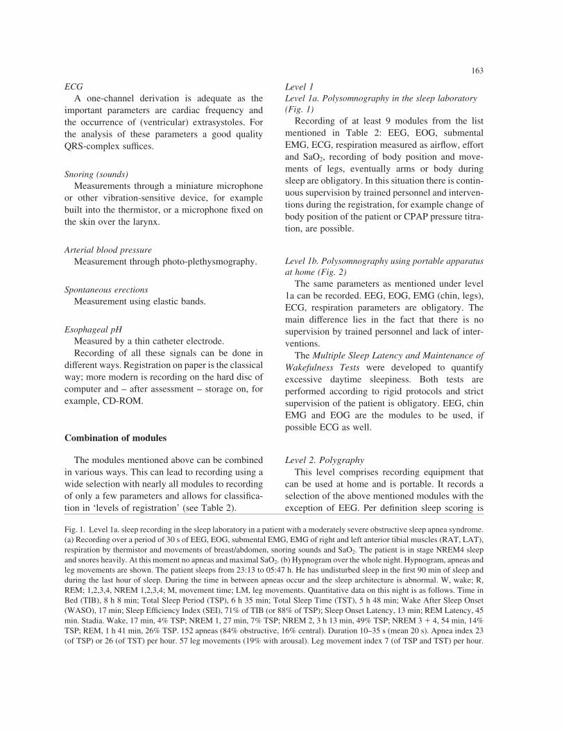
Level 1a. Polysomnography in the sleep laboratory
(Fig. 1)
Recording of at least 9 modules from the list
mentioned in Table 2: EEG, EOG, submental
EMG, ECG, respiration measured as air¯ow, effort
and SaO2, recording of body position and move-
ments of legs, eventually arms or body during
sleep are obligatory. In this situation there is contin-
uous supervision by trained personnel and interven-
tions during the registration, for example change of
body position of the patient or CPAP pressure titra-
tion, are possible.
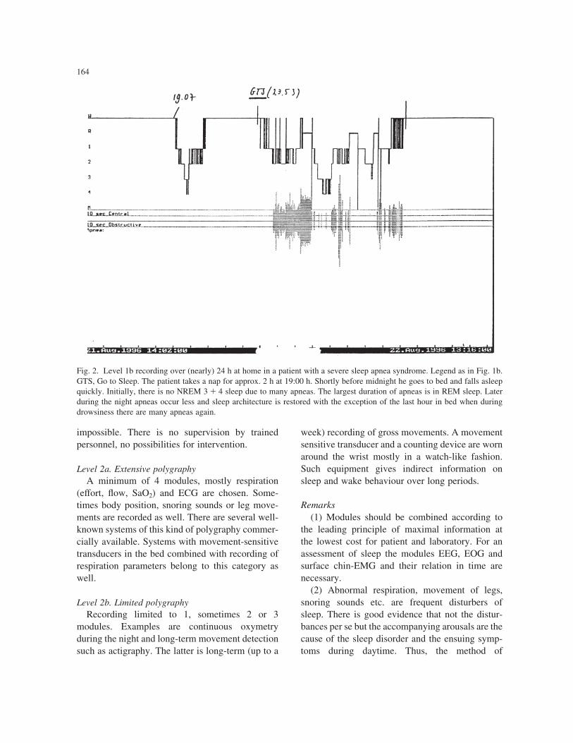
Level 1b. Polysomnography using portable apparatus
at home (Fig. 2)
The same parameters as mentioned under level
1a can be recorded. EEG, EOG, EMG (chin, legs),
ECG, respiration parameters are obligatory. The
main difference lies in the fact that there is no
supervision by trained personnel and lack of inter-
ventions.
The Multiple Sleep Latency and Maintenance of
Wakefulness Tests were developed to quantify
excessive daytime sleepiness. Both tests are
performed according to rigid protocols and strict
supervision of the patient is obligatory. EEG, chin
EMG and EOG are the modules to be used, if
possible ECG as well.
Level 2. Polygraphy
This level comprises recording equipment that
can be used at home and is portable. It records a
selection of the above mentioned modules with the
exception of EEG. Per de®nition sleep scoring is
163
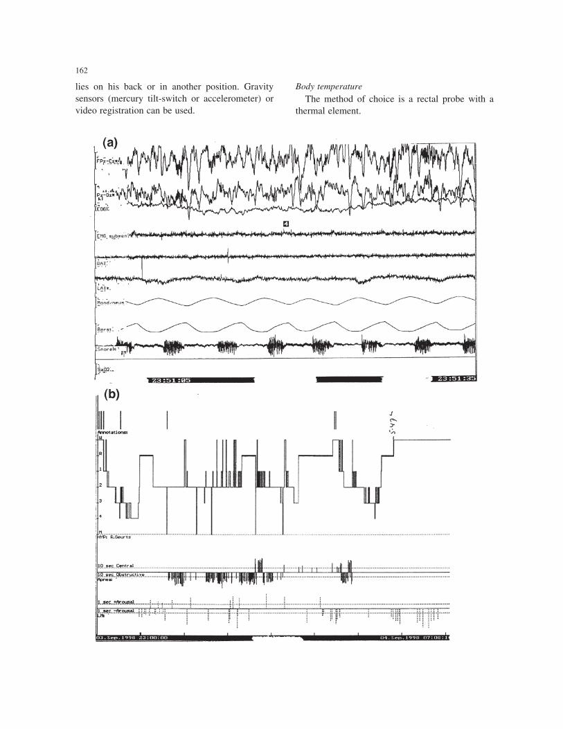
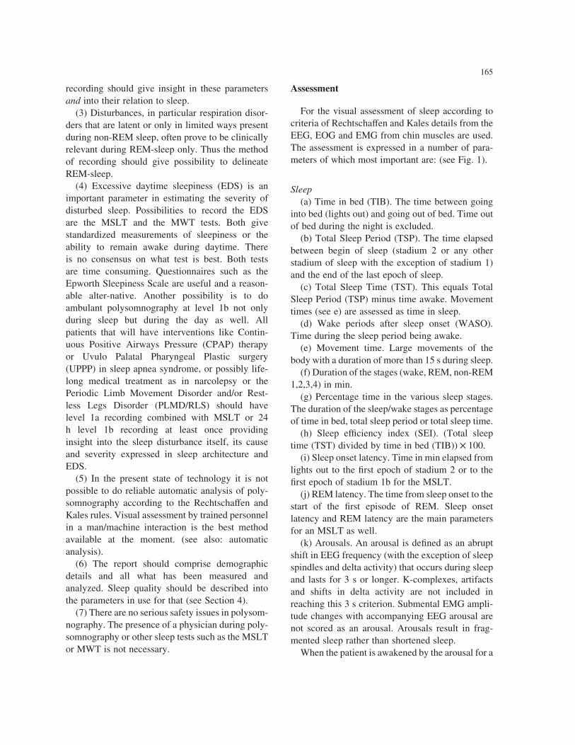
Fig. 1. Level 1a. sleep recording in the sleep laboratory in a patient with a moderately severe obstructive sleep apnea syndrome.
(a) Recording over a period of 30 s of EEG, EOG, submental EMG, EMG of right and left anterior tibial muscles (RAT, LAT),
respiration by thermistor and movements of breast/abdomen, snoring sounds and SaO2. The patient is in stage NREM4 sleep
and snores heavily. At this moment no apneas and maximal SaO2. (b) Hypnogram over the whole night. Hypnogram, apneas and
leg movements are shown. The patient sleeps from 23:13 to 05:47 h. He has undisturbed sleep in the ®rst 90 min of sleep and
during the last hour of sleep. During the time in between apneas occur and the sleep architecture is abnormal. W, wake; R,
REM; 1,2,3,4, NREM 1,2,3,4; M, movement time; LM, leg movements. Quantitative data on this night is as follows. Time in
Bed (TIB), 8 h 8 min; Total Sleep Period (TSP), 6 h 35 min; Total Sleep Time (TST), 5 h 48 min; Wake After Sleep Onset
(WASO), 17 min; Sleep Ef®ciency Index (SEI), 71% of TIB (or 88% of TSP); Sleep Onset Latency, 13 min; REM Latency, 45
min. Stadia. Wake, 17 min, 4% TSP; NREM 1, 27 min, 7% TSP; NREM 2, 3 h 13 min, 49% TSP; NREM 31 4, 54 min, 14%
TSP; REM, 1 h 41 min, 26% TSP. 152 apneas (84% obstructive, 16% central). Duration 10±35 s (mean 20 s). Apnea index 23
(of TSP) or 26 (of TST) per hour. 57 leg movements (19% with arousal). Leg movement index 7 (of TSP and TST) per hour.
impossible. There is no supervision by trained
personnel, no possibilities for intervention.
Level 2a. Extensive polygraphy
A minimum of 4 modules, mostly respiration
(effort, ¯ow, SaO2) and ECG are chosen. Some-
times body position, snoring sounds or leg move-
ments are recorded as well. There are several well-
known systems of this kind of polygraphy commer-
cially available. Systems with movement-sensitive
transducers in the bed combined with recording of
respiration parameters belong to this category as
well.
Level 2b. Limited polygraphy
Recording limited to 1, sometimes 2 or 3
modules. Examples are continuous oxymetry
during the night and long-term movement detection
such as actigraphy. The latter is long-term (up to a
week) recording of gross movements. A movement
sensitive transducer and a counting device are worn
around the wrist mostly in a watch-like fashion.
Such equipment gives indirect information on
sleep and wake behaviour over long periods.
Remarks
(1) Modules should be combined according to
the leading principle of maximal information at
the lowest cost for patient and laboratory. For an
assessment of sleep the modules EEG, EOG and
surface chin-EMG and their relation in time are
necessary.
(2) Abnormal respiration, movement of legs,
snoring sounds etc. are frequent disturbers of
sleep. There is good evidence that not the distur-
bances per se but the accompanying arousals are the
cause of the sleep disorder and the ensuing symp-
toms during daytime. Thus, the method of
164
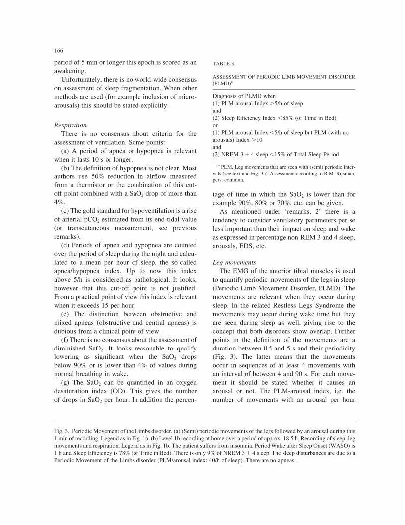
Fig. 2. Level 1b recording over (nearly) 24 h at home in a patient with a severe sleep apnea syndrome. Legend as in Fig. 1b.
GTS, Go to Sleep. The patient takes a nap for approx. 2 h at 19:00 h. Shortly before midnight he goes to bed and falls asleep
quickly. Initially, there is no NREM 31 4 sleep due to many apneas. The largest duration of apneas is in REM sleep. Later
during the night apneas occur less and sleep architecture is restored with the exception of the last hour in bed when during
drowsiness there are many apneas again.
recording should give insight in these parameters
and into their relation to sleep.
(3) Disturbances, in particular respiration disor-
ders that are latent or only in limited ways present
during non-REM sleep, often prove to be clinically
relevant during REM-sleep only. Thus the method
of recording should give possibility to delineate
REM-sleep.
(4) Excessive daytime sleepiness (EDS) is an
important parameter in estimating the severity of
disturbed sleep. Possibilities to record the EDS
are the MSLT and the MWT tests. Both give
standardized measurements of sleepiness or the
ability to remain awake during daytime. There
is no consensus on what test is best. Both tests
are time consuming. Questionnaires such as the
Epworth Sleepiness Scale are useful and a reason-
able alter-native. Another possibility is to do
ambulant polysomnography at level 1b not only
during sleep but during the day as well. All
patients that will have interventions like Contin-
uous Positive Airways Pressure (CPAP) therapy
or Uvulo Palatal Pharyngeal Plastic surgery
(UPPP) in sleep apnea syndrome, or possibly life-
long medical treatment as in narcolepsy or the
Periodic Limb Movement Disorder and/or Rest-
less Legs Disorder (PLMD/RLS) should have
level 1a recording combined with MSLT or 24
h level 1b recording at least once providing
insight into the sleep disturbance itself, its cause
and severity expressed in sleep architecture and
EDS.
(5) In the present state of technology it is not
possible to do reliable automatic analysis of poly-
somnography according to the Rechtschaffen and
Kales rules. Visual assessment by trained personnel
in a man/machine interaction is the best method
available at the moment. (see also: automatic
analysis).
(6) The report should comprise demographic
details and all what has been measured and
analyzed. Sleep quality should be described into
the parameters in use for that (see Section 4).
(7) There are no serious safety issues in polysom-
nography. The presence of a physician during poly-
somnography or other sleep tests such as the MSLT
or MWT is not necessary.
Assessment
For the visual assessment of sleep according to
criteria of Rechtschaffen and Kales details from the
EEG, EOG and EMG from chin muscles are used.
The assessment is expressed in a number of para-
meters of which most important are: (see Fig. 1).
Sleep
(a) Time in bed (TIB). The time between going
into bed (lights out) and going out of bed. Time out
of bed during the night is excluded.
(b) Total Sleep Period (TSP). The time elapsed
between begin of sleep (stadium 2 or any other
stadium of sleep with the exception of stadium 1)
and the end of the last epoch of sleep.
(c) Total Sleep Time (TST). This equals Total
Sleep Period (TSP) minus time awake. Movement
times (see e) are assessed as time in sleep.
(d) Wake periods after sleep onset (WASO).
Time during the sleep period being awake.
(e) Movement time. Large movements of the
body with a duration of more than 15 s during sleep.
(f) Duration of the stages (wake, REM, non-REM
1,2,3,4) in min.
(g) Percentage time in the various sleep stages.
The duration of the sleep/wake stages as percentage
of time in bed, total sleep period or total sleep time.
(h) Sleep ef®ciency index (SEI). (Total sleep
time (TST) divided by time in bed (TIB�� £ 100.
(i) Sleep onset latency. Time in min elapsed from
lights out to the ®rst epoch of stadium 2 or to the
®rst epoch of stadium 1b for the MSLT.
(j) REM latency. The time from sleep onset to the
start of the ®rst episode of REM. Sleep onset
latency and REM latency are the main parameters
for an MSLT as well.
(k) Arousals. An arousal is de®ned as an abrupt
shift in EEG frequency (with the exception of sleep
spindles and delta activity) that occurs during sleep
and lasts for 3 s or longer. K-complexes, artifacts
and shifts in delta activity are not included in
reaching this 3 s criterion. Submental EMG ampli-
tude changes with accompanying EEG arousal are
not scored as an arousal. Arousals result in frag-
mented sleep rather than shortened sleep.
When the patient is awakened by the arousal for a
165
period of 5 min or longer this epoch is scored as an
awakening.
Unfortunately, there is no world-wide consensus
on assessment of sleep fragmentation. When other
methods are used (for example inclusion of micro-
arousals) this should be stated explicitly.
Respiration
There is no consensus about criteria for the
assessment of ventilation. Some points:
(a) A period of apnea or hypopnea is relevant
when it lasts 10 s or longer.
(b) The de®nition of hypopnea is not clear. Most
authors use 50% reduction in air¯ow measured
from a thermistor or the combination of this cut-
off point combined with a SaO2 drop of more than
4%.
(c) The gold standard for hypoventilation is a rise
of arterial pCO2 estimated from its end-tidal value
(or transcutaneous measurement, see previous
remarks).
(d) Periods of apnea and hypopnea are counted
over the period of sleep during the night and calcu-
lated to a mean per hour of sleep, the so-called
apnea/hypopnea index. Up to now this index
above 5/h is considered as pathological. It looks,
however that this cut-off point is not justi®ed.
From a practical point of view this index is relevant
when it exceeds 15 per hour.
(e) The distinction between obstructive and
mixed apneas (obstructive and central apneas) is
dubious from a clinical point of view.
(f) There is no consensus about the assessment of
diminished SaO2. It looks reasonable to qualify
lowering as signi®cant when the SaO2 drops
below 90% or is lower than 4% of values during
normal breathing in wake.
(g) The SaO2 can be quanti®ed in an oxygen
desaturation index (OD). This gives the number
of drops in SaO2 per hour. In addition the percen-
tage of time in which the SaO2 is lower than for
example 90%, 80% or 70%, etc. can be given.
As mentioned under `remarks, 2' there is a
tendency to consider ventilatory parameters per se
less important than their impact on sleep and wake
as expressed in percentage non-REM 3 and 4 sleep,
arousals, EDS, etc.
Leg movements
The EMG of the anterior tibial muscles is used
to quantify periodic movements of the legs in sleep
(Periodic Limb Movement Disorder, PLMD). The
movements are relevant when they occur during
sleep. In the related Restless Legs Syndrome the
movements may occur during wake time but they
are seen during sleep as well, giving rise to the
concept that both disorders show overlap. Further
points in the de®nition of the movements are a
duration between 0.5 and 5 s and their periodicity
(Fig. 3). The latter means that the movements
occur in sequences of at least 4 movements with
an interval of between 4 and 90 s. For each move-
ment it should be stated whether it causes an
arousal or not. The PLM-arousal index, i.e. the
number of movements with an arousal per hour
166
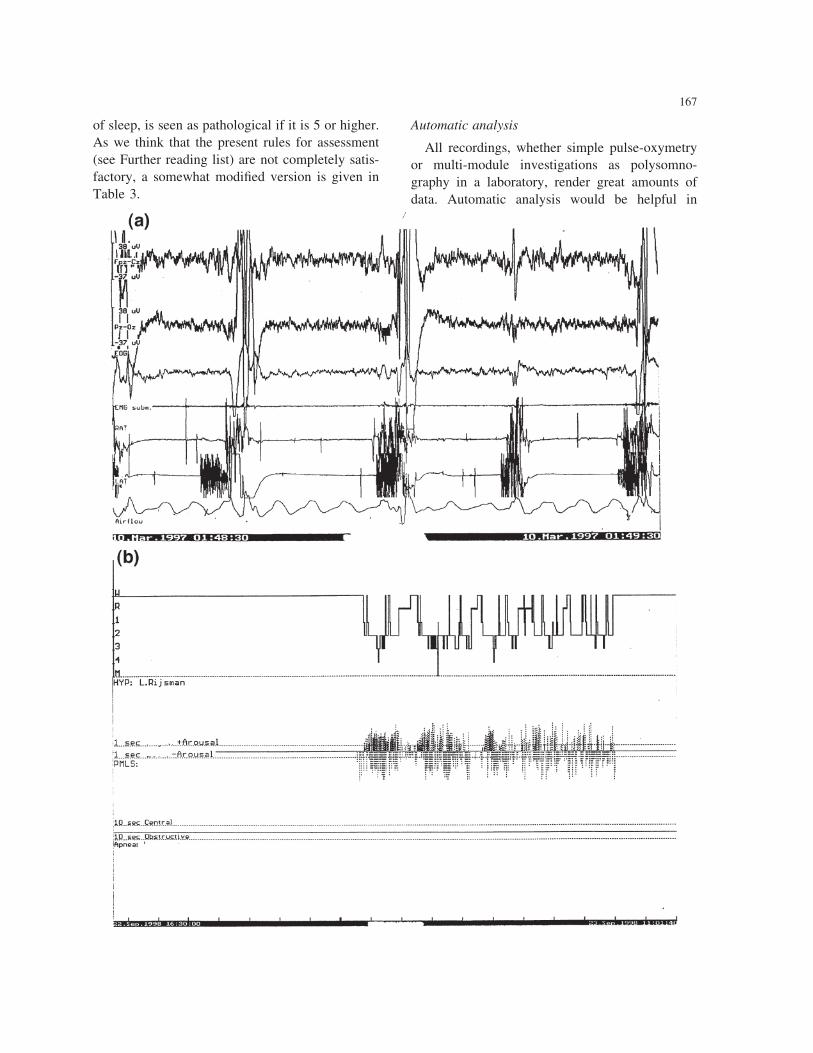
Fig. 3. Periodic Movement of the Limbs disorder. (a) (Semi) periodic movements of the legs followed by an arousal during this
1 min of recording. Legend as in Fig. 1a. (b) Level 1b recording at home over a period of approx. 18.5 h. Recording of sleep, leg
movements and respiration. Legend as in Fig. 1b. The patient suffers from insomnia. Period Wake after Sleep Onset (WASO) is
1 h and Sleep Ef®ciency is 78% (of Time in Bed). There is only 9% of NREM 31 4 sleep. The sleep disturbances are due to a
Periodic Movement of the Limbs disorder (PLM/arousal index: 40/h of sleep). There are no apneas.
TABLE 3
ASSESSMENT OF PERIODIC LIMB MOVEMENT DISORDER
(PLMD)a
Diagnosis of PLMD when
(1) PLM-arousal Index .5/h of sleep
and
(2) Sleep Ef®ciency Index ,85% (of Time in Bed)
or
(1) PLM-arousal Index ,5/h of sleep but PLM (with no
arousals) Index .10
and
(2) NREM 31 4 sleep ,15% of Total Sleep Period
a PLM, Leg movements that are seen with (semi) periodic inter-
vals (see text and Fig. 3a). Assessment according to R.M. Rijsman,
pers. commun.
of sleep, is seen as pathological if it is 5 or higher.
As we think that the present rules for assessment
(see Further reading list) are not completely satis-
factory, a somewhat modi®ed version is given in
Table 3.
Automatic analysis
All recordings, whether simple pulse-oxymetry
or multi-module investigations as polysomno-
graphy in a laboratory, render great amounts of
data. Automatic analysis would be helpful in
167
terms of time, accuracy and costs. Such programs
are available. It should, however, be realized that
each automatic method harbors the danger of
`garbage in, garbage out', meaning that one should
be sure that the signals are of high quality and
should be easily available as raw data for non-
automatic re-analysis. Up to now, an automatic
analysis of parameters related to respiration,
snoring sounds and (leg) movements is technically
feasible. The same holds for the ECG. Large
problems, however, are encountered in the auto-
matic analysis of sleep itself. This is due to arti-
facts and aspects of the prevailing Rechtschaffen
and Kales method of scoring of sleep. Admittedly
there are methods of automatic analysis according
to the Rechtschaffen and Kales rules, but the
performance of such systems is at best 80% accor-
dance to visually scored sleep in normal subjects.
This is not acceptable and means that the visual
assessment of sleep following the criteria of
Rechtschaffen and Kales is as yet the only useful
method.
The main problems are the discontinuity (stages
wake, REM and NREM 1±4) and low-time resolu-
tion (30 s) inherent to the Rechtschaffen and Kales
method. Most important in this respect is that the
Rechtschaffen and Kales scoring for a certain sleep
stage is based on sleep phenomena that are not
concurrent. An automatic system has to `decide'
on data that are dispersed over epochs of sometimes
many minutes of recording. For example, for the
decision to score NREM 2 sleep, sleep spindles,
K-complexes, moderate amplitude background
activity etc. are to be detected. These phenomena
do not occur simultaneously. Hence the automatic
analyzator has to remember what was seen during
an earlier epoch and has to combine this informa-
tion with data from the epoch that is analyzed at that
moment. Fortunately, other methods that differ
signi®cantly from the classical Rechtschaffen and
Kales assessment allow fully automatic and contin-
uous assessment of sleep. Most promising are the
developments in the delta plot analysis. Very
important for research in new methods is the easy
exchange of sleep data between laboratories that is
available now, for example, in the `European Data
Format'.
Diagnostic yield
The value of a method can be given in various
parameters. At each such study in this respect,
comparison has to be made with a gold standard.
In sleep medicine this ideal is not available, but
polysomnography in the sleep laboratory at level
1a approaches this ideal.
As there are serious drawbacks for this `gold
standard' examination such as costs, duration and
limited availability, one should always try to use
more limited ways of registration that are cheaper
and that give the same or even better answers for
the questions asked by the clinician: Which patient
has to have what kind of registrations to get the
optimal results?
Guidelines for the practical use of
poly(somno)graphy
Level 1a. Polysomnography in the sleep laboratory
This method with its comprehensive measure-
ment of physiological parameters is indicated for
patients in whom the diagnosis is problematic.
For example patients suspected of Sleep Apnea
Syndrome in which there are confounding clinical
details or in which other sleep disorders such as
PMLD are possible as well. Furthermore, level 1a
examination should be performed in all patients
who will get interventions for example CPAP or
surgery for Sleep Apnea Syndrome or intensive
medical therapy for narcolepsy, PMLD, etc. This
supervised form of registration in the sleep labora-
tory allows interventions. That is the reason why
this method of registration is indicated for titration
of CPAP pressure. Drawbacks are its cost in time
and the lack of insight in sleepiness during daytime.
The so-called siesta-sleep registration, i.e. the
recording of an afternoon nap, is at the 1a level
from a technical point of view but it has a low
sensitivity and speci®city for disturbances in
sleep. Patients with Sleep Apnea Syndrome often
have no respiratory disturbances during a siesta-
sleep registration but still prove to have the disorder
during night sleep and vice versa. Thus, a level 1a
registration is still necessary and makes a siesta
sleep registration redundant.
168
Level 1b. Polysomnography at home with a
portable system
This way of registration is suitable for the same
categories of patients that would otherwise have
had polysomnography in the sleep laboratory.
There are, however, some differences. Although
technical facilities are large at the moment there
are still some limitations in parameters that can
be recorded by a portable system. Furthermore,
the method of registration with a portable system
is not supervised and as such not useful for inter-
ventions. Registration at home has its own advan-
tages. It can be performed under circumstances that
are normal for the patient. He or she sleeps in his/
her own bed and falls asleep at his/her own sofa! In
case ambulant poly(somno)graphy is extended over
periods of 24 h or longer the method gives insight
into the waking period during daytime and allows
quanti®cation of (excessive) daytime sleepiness or
other phenomena that occur during the day. The
method is much cheaper and more suitable for
follow-up studies than polysomnography in the
sleep laboratory.
The MSLT and MWT tests are performed in
order to answer limited questions: is there excessive
daytime sleepiness or inability to stay awake? They
are meant to give quanti®cation of these questions.
The tests are performed under supervision of a
trained technician but are not intended for interven-
tion. For the questions posed their sensitivity is high
and results have face-value, but speci®city is low.
For example, a pathologic MSLT can be encoun-
tered in Narcolepsy, Sleep Apnea Syndrome,
PLMD, etc.
Polygraphy (levels 2a and 2b)
In this category belong all forms of limited poly-
graphy at home using portable equipment. Combi-
nations of various parameters with the exception of
EEG are recorded. In particular for screening and
follow-up for the Sleep Apnea Syndrome there is a
role for this equipment. The sensitivity and speci®-
city, for this disorder, of level 2a equipment is at
about 80±95% (when compared with polysomno-
graphy at level 1a or 1b). This indicates that there
is a place for such equipment. The indication for
use in other sleep and wake disorders is limited as
the methods give only indirect information on
disturbances of sleep itself.
Equipment that is intended for monitoring of one
or two parameters during sleep (level 2b) can be
used for screening of Sleep Apnea patients. An
example is long-time pulse-oxymetry. For this
method some validation studies were performed
in which comparison was made with polysomno-
graphy at level 1a or 1b. Depending on the severity
of respiratory disorders during sleep sensitivity and
speci®city varied between 40% and 100%. The
more severe the Sleep Apnea Syndrome, the more
exact were the results of pulse oxymetry. This
conforms to the expectation that in clear abnormal-
ities diagnosis can be made with more simple
methods.
Further reading
American Academy of Neurology. Assessment: Techniques asso-
ciated with the diagnosis and management of sleep disorders.
Report of the Therapeutics and Technology Assessment
Subcommittee of the American Academy of Neurology.
Neurology, 1992, 42: 269±275.
American Electroencephalographic Society. Guideline Fifteen:
Guidelines for polygraphic assessment of sleep-related disor-
ders (polysomnography). J. Clin Neurophysiol., 1994, 11:
116±124.
American Sleep Disorders Association. Guilleminault, C.
(chairman). EEG arousals: scoring rules and examples. Sleep,
1992, 15: 173±184.
American Sleep Disorders Association. Guilleminault, C.
(chairman). Recording and scoring leg movements. Sleep,
1993, 16: 749±759.
American Sleep Disorders Association. Standards of Practice
Committee. Practice parameters for the use of portable
recording in the assessment of obstructive sleep apnea. Sleep,
1994,17: 372±327.
American Sleep Disorders Association. Ferber, R. (chairman).
Portable recording in the assessment of obstructive sleep
apnea. Sleep, 1994, 17: 378±392.
American Sleep Disorders Association. Chesson, A.L. (chairman).
Practice parameters for the indications for polysomnography
and related procedures. Sleep, 1997, 20: 432±487.
American Sleep Disorders Association. Chesson, A.L. (chairman).
The indications for polysomnography and related procedures.
Sleep, 1997, 20: 423±487.
American Sleep Disorders Association. The International Classi®-
cation of Sleep Disorders (revised). Diagnostic and Coding
Manual. ASDA, 1997.
American Thoracic Society. Indication and standards for cardiopul-
monary sleep studies. Am. Rev. Respir. Dis., 1989, 139: 559±
568.
Carskadon, M.A., Dement, W.C., Mitler, M.M., Roth, T., West-
brook, P.R. and Keenan, S. Guidelines for the multiple sleep
169
latency test (MSLT): a standard measure of sleepiness. Sleep,
1986, 9: 519±524.
Coleman, R.M. Periodic movements in sleep (nocturnal myoclonus)
and restless legs syndrome. In: Guilleminault, C. (Ed.), Sleeping
and Waking Disorders; Indications and Techniques. Addison-
Wesley, Menlo Park, 1982: 265±295.
Kemp, B. Consensus report. A proposal for computer-based sleep/
wake analysis. J. Sleep Res., 1993, 2: 179±185.
Kemp, B., VaÈrri, A., Rosa, A.C., Nielsen, K.D. and Gade, J.A.
Simple format for exchange of digitized polygraphic recordings.
Electroenceph. clin. Neurophysiol., 1992, 82: 391±393.
Mitler, M.M., Gujavarty, K.S. and Browman, C.P. Maintenance of
wakefulness test: a polysomnographic technique for evaluation
treatment ef®cacy in patients with excessive somnolence. Elec-
troenceph. clin. Neurophysiol., 1982, 53: 658±661.
Penzel, T., Hajak, G., Hoffman, R.M., Lund, R., Podzus, T., Poll-
maÈcher, T., SchaÈfer, T., Schulz, H., Sonnenschein, W. and
Spieweg, I. Empfehlungen zur DurchfuÈhrung und Auswertung
polygraphischer Ableitungen im diagnostischen Schla¯abor.
EEG/EMG, 1993, 24: 65±70.
Rechtschaffen, A. and Kales, A. A Manual of Standardized Termi-
nology, Techniques and Scoring System for Sleep Stages of
Human Subjects. Natl. Inst. Neurol. Dis. Blind. (NIH Publ.
204), Bethesda, MD, 1968.
Stradling, J.R. Consensus report. Sleep studies for sleep-related
breathing disorders. J. Sleep Res., 1992, 1: 265±273.
170